Research Article
Silver Nanoparticles Affect Functional Bioenergetic Traits in
the Invasive Red Sea Mussel
Brachidontes pharaonis
Ilenia Saggese,
1Gianluca Sarà,
2and Francesco Dondero
11Dipartimento di Scienze ed Innovazione Tecnologica, University of Piemonte Orientale, Via Michel 11, 15121 Alessandria, Italy 2Dipartimento di Scienze della Terra e del Mare, University of Palermo, Viale delle Scienze Ed. 16, 90128 Palermo, Italy
Correspondence should be addressed to Francesco Dondero; [email protected] Received 3 May 2016; Accepted 22 June 2016
Academic Editor: Zongming Ren
Copyright © 2016 Ilenia Saggese et al. This is an open access article distributed under the Creative Commons Attribution License, which permits unrestricted use, distribution, and reproduction in any medium, provided the original work is properly cited. We investigated the functional trait responses to 5 nm metallic silver nanoparticle (AgNPs) exposure in the Lessepsian-entry bivalve
B. pharaonis. Respiration rate (oxygen consumption), heartbeat rate, and absorption efficiency were evaluated across an 8-day
exposure period in mesocosmal conditions. Basal reference values from not-exposed specimens were statistically compared with
those obtained from animals treated with three sublethal nanoparticle concentrations (2𝜇g L−1, 20𝜇g L−1, and 40𝜇g L−1). Our data
showed statistically significant effects on the average respiration rate of B. pharaonis. Moreover, complex nonlinear dynamics were observed as a function of the concentration level and time. Heartbeat rates largely increased with no acclimation in animals exposed to the two highest levels with similar temporal dynamics. Eventually, a decreasing trend for absorption efficiency might indicate energetic constraints. In general, these data support the possible impact of engineered nanomaterials in marine environments and support the relevance of functional trait assessment in present and future ecotoxicological studies.
1. Introduction
The quick development of nanotechnology is posing con-cerns on the environmental impact of engineered nanoma-terials (ENMs) into delicate environments such as marine coastal areas. The production and commercial use of ENMs have been linearly increased during the last decade and involve the following: personal care, cosmetics, fabrics and textile, household appliances, electronics and computers, health and medicine, renewable energies, and environmental remediation [1]. According to the Nanotechnology Con-sumer Products Inventory [2], currently, there are 1841 ENM-containing products in the consumer marketplace, of which 438 (24%) contain nanosilver. Silver is a xenobiotic metal widely used in industrial applications (more than 60% of the metal demand) such as chemical catalysis, batteries, photog-raphy, electronics, and brazing and soldering [3]. Due to its antimicrobial properties it has been largely employed as addi-tive in personal care products, coatings, paints, fabrics, food, and beverage. Nowadays, most of the latter applications as well as those targeted to health, fitness, and household use are based on silver ENMs [2]. Indeed, silver at the nanoscale level
shows distinctive physicochemical properties and biological activities like excellent conductivity, chemical stability, and increased catalytic activity [4]. These distinctive properties differ significantly from those of larger particles and have made silver nanoparticles (AgNPs) extremely attractive for the production of consumer materials and, therefore, the investigation of their environmental fate and toxicological properties [4, 5]. Nanosilver predicted environmental con-centrations (PECs) are yet affected by uncertainty since differ-ent approaches in modeling and measuremdiffer-ent methods com-plicate validation. Gottschalk and coworkers [6] reviewed the outcome from different predictive models. For superficial
waters they indicated a wide silver range spanning from 10−4
to 100ppm with a median value around 10−2ppm. Waste water
treatment plants are able to capture most (nano)silver into an insoluble sulfide form [7]; however, ENMs may enter into the environment through different routes such as outdoor urban sources [8] that are not necessarily intercepted by treatment plants. Air transport and in general the hydrogeological cycle have a primary role in ENM run-off and transport, thus, making marine ecosystems the terminal sinks [9]. PECs for seawater environments are practically unknown.
Volume 2016, Article ID 1872351, 7 pages http://dx.doi.org/10.1155/2016/1872351
The antimicrobial as well as toxic effects of silver are
attributed to the Ag+ion that is a strong electrophile reacting
with most macromolecules containing S, O, and N [5, 10, 11]. The toxicity of AgNPs in the environment is certainly a function of the silver metal speciation that highly depends on the environment itself but also intrinsic characteristics such as particle sintering, coating, surface potential, size,
and Ag+ dissolution rate [12–14]. It is widely recognized
that Ag-ENMs can exert toxic effects on aquatic organisms such as algae, mollusks, crustaceans, and fish for which classical ecotoxicological endpoints have been evaluated [1, 15]; however, very little is known about marine species [16].
Marine bivalves are model species to study ENM effects. Since they are filter-feeder organisms, they bioaccumulate toxicants either in dissolved or in particle-adsorbed forms in the water column [17, 18]. However, the main routes of NPs uptake and the level at which they can penetrate into the organism are not completely known [5]. In the bivalve Mytilus edulis Moore [17] showed that the principal process for the translocation of polystyrene NPs across the membrane is endocytosis. Regarding AgNP data on toxicological effects on bivalves, this information is sparse. According to Zuykov et al. [19] AgNPs can be accumulated in the extrapallial fluid of the blue mussel Mytilus edulis and concentrate mostly in ganglia. Gomes et al. [20] reported an oxidative stress syndrome in soft tissues of Mytilus spp. with induction of heavy metal binding proteins in gills. In the oyster C. virginica there were embryo toxicity and effects on lysosomal integrity of hepatopancreas cells [21]. In this context, functional traits such as food assimilation, respiration, and heartbeat rates [22] may represent a straightforward and useful approach to investigate AgNPs metabolic and bioenergetic effects. Here we report the effects of AgNPs on main functional traits of a new proposed model species, the Lessepsian-entry bivalve Brachidontes pharaonis [23, 24]. Indeed, we tested the sensitivity of B. pharaonis to submicromolar amounts (0–
40𝜇g L−1) of a 5 nm commercial AgNP in an 8-day exposure
experiment carried out in laboratory under mesocosmal con-ditions. The response variables observed were respiration rate (RR), heartbeat rate (HBR), and absorption efficiency (AE).
2. Material and Methods
2.1. Animals and Treatments with Silver Nanoparticles. Spec-imens of Brachidontes pharaonis were collected from the Ettore Pond of Stagnone di Marsala (Trapani, Western Sicily,
37∘ 52 north; 12∘ 28 east). Once collected, mussels were
brought back to the laboratory in controlled conditions of
temperature (16∘C) and humidity (100%). They were cleaned
from epibionts and moved into tanks at 0.5 L per animal
in filtered recirculating seawater at 20∘C. Organisms were
acclimated in laboratory conditions for 15 days at 20∘C, 36‰
salinity, and pH8.2 ± 0.1. The total number of animals used
in this experiment was 360, with a size ranging from 22 to 27 mm. Both during the acclimation and during experimental periods, organisms were fed three times per day (ad libitum) with fresh cultures of Isochrysis galbana at an initial titer of
15,000 cells mL−1. Animals were divided into 4 experimental
groups of 90 specimens, with 3 independent tanks per group. Each group corresponded to a different exposure level to silver nanoparticles: 0 (not-exposed, reference control), 2, 20,
and 40𝜇g L−1. This concentration range is compatible with
previous studies on AgNPs [20, 25].
Silver nanoparticles (5 nm average diameter) with an alkane coating were supplied by AMEPOX (Lodz, Poland) in
a stable ultrapure-water solution at 1 g L−1. This material has
been already used in a large ENM toxicity-testing framework within the NanoFATE project (https://wiki.ceh.ac.uk/display/ nanofate/Home) [26–29].
2.2. Respiration Rate. Metabolic respiration rates were evalu-ated as oxygen consumption according to a reliable procedure already tested in companion papers [30, 31]. Measurements were carried out every 2 days on six randomly selected
specimens per treatment (0, 2, 20, and 40𝜇g L−1). Briefly,
single B. pharaonis specimens were placed in glass respiro-metric chambers (0.5 L) containing filtered air-saturated seawater. Magnetic stirring ensured water mixing within the chamber, while oxygen reduction was measured by means of computer-assisted optical oxygen-meter probes (FireSting
O2, PyroScience GmbH, Aachen, Germany) in four different
chambers (one per condition) simultaneously. The rate of oxygen consumption was then calculated according to [32]:
RR (O2𝜇moles/h) = [𝐶(𝑡0) − 𝐶(𝑡1)] ⋅ (𝑉𝑟) ⋅ 60/(𝑡1 − 𝑡0),
where 𝑡0 and 𝑡1 represent start and finish times (min) of
the measurement period;𝐶(𝑡) is concentration of oxygen in
the water (𝜇moles O2L−1) at time 𝑡; and 𝑉𝑟 is volume of
respirometer minus the animal. Respiration rate correction for body mass was carried out using dry weight [32]. 2.3. Heartbeat Rate. Heartbeat rate (HBR, beats/min) was evaluated by means of a noninvasive cardioplethysmographic technique [22, 33]. To record the mussel heartbeat, infrared sensors were glued on the left side of the mussel shell, just below the umbone. To avoid stress to the organism, sensors were positioned the day before the measurement. The heartbeat signals obtained were amplified, filtered, and then detected by means of a portable oscilloscope PicoScope 236 (Pico Technology Ltd., UK) connected to a laptop computer equipped with PicoScope 6.0 software. During the experimental session, HBR was recorded at intervals of 10 minutes per mussel. HBR values for each animal were obtained as the average of 3 randomly selected views of the 10-min measurement. Measurements were made every 2 days on six randomly selected individuals per treatment.
2.4. Absorption Efficiency. Food absorption efficiency was measured by comparing the proportions of organic matter in the algal cells and mussels feces according to the equation
of Conover [34]: AE = (𝐹 − 𝐸)/[(1 − 𝐸)𝐹], where 𝐹 is a
relationship between dry weight and ash-free dry weight
of algal food while𝐸 is a relationship between dry weight
and ash-free dry weight of fecal pellets. Fecal pellets from each treatment were collected daily and placed in separate vials. Collected feces were filtered on preweighted glass fiber
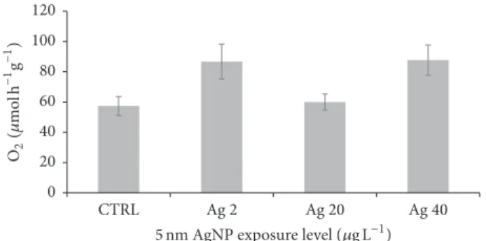
5 nm AgNP exposure level (𝜇g LAg 2 Ag 20 −1) Ag 40 CTRL 0 20 40 60 80 100 120 O2 (𝜇 mo l h −1g −1)
Figure 1: Effects of 5 nm AgNP on average B. pharaonis respiration rates (RR). Shown is the normalized average (±SEM) respiration rate
(𝜇mol h−1g−1) across the 8-day exposure period. The ANOVA post
hoc test did not show statistically significant differences between the control not-exposed samples and the AgNP treated ones (TNK-test 𝑃 > 0.05).
ammonium formate to remove salts [35]. Then, samples
were dried at 100∘C for 48 h and dry weights were recorded
immediately after cooling in a desiccator. Eventually, samples
were ashed in a furnace at 450∘C for 2 h and then reweighted
to obtain the ash-free dry weight. The results are the average of six different measurements per condition.
2.5. Statistical Analysis. An analysis of variance (ANOVA) was performed using R (software version 2.15.1) to test the dependence of RR, HBR, and AE on AgNP concentration (CONC, fixed, 4 levels) and time (TIME, fixed, 5 levels). The assumption of homoscedasticity was tested using Cochran’s 𝐶 test. Post hoc comparisons were made using the Student-Newman-Keuls test (SNK-test). The alpha values are reported on each table or figure.
3. Results and Discussion
The exposure to 5 nm AgNPs significantly influenced the overall B. pharaonis respiration rate (RR) (Table 1, Figure 1). This is the result of complex dynamics especially occurring
in specimens treated with 2 and 40𝜇g L−1AgNP during the
8-day exposure (Figure 2). These samples showed opposite temporal trends and seem to reflect different compensa-tion strategies to discrete nanoparticle amounts. The lowest concentration, in fact, determined a progressive increase of oxygen consumption in time and by contrast the highest Ag level caused an increase of respiration during the first part of the exposure, then, followed by a constant decrease to
an average value of about 49𝜇mol h−1g−1 similar to that of
control (Figure 2). The intermediate Ag level displayed neg-ligible effects with respect to control. The average individual respiration rate of control specimens observed in this study
was around 10𝜇M mol O2h−1. This measure was compatible
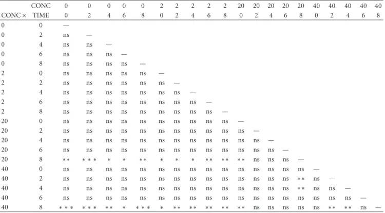
with previous values reported in the same species [24]. 5 nm AgNPs had statistically significant effects on the heartbeat rate (HBR) (Table 2). Figure 3 shows the temporal trends obtained across the 8-day exposure period. These are consistent with an increase of HBR in samples exposed to the
Table 1: ANOVA details for the effects of 5 AgNP exposure on B.
pharaonis normalized respiration rate (RR).
Source DF MS 𝐹 𝑃
CONC 3 8570.4 3.62 ∗
TIME 4 5490.6 2.32 ns
CONC× TIME 12 4336.8 1.83 ns
Residuals 40 2365.7
Factors tested for dependence were concentration (CONC) and exposure time (TIME). Shown are DF, degree of freedom; MS, mean square;𝐹, 𝐹-test result; and𝑃, 𝑃 value [∗ = 𝑃 ≤ 0.05; and ns = no significant difference (𝑃 > 0.05)].
Table 2: ANOVA details for the effects of AgNP exposure on B.
pharaonis heart beat rate (HBR).
Source DF MS 𝐹 𝑃
CONC 4 215.2 5.342 ∗∗
TIME 3 510.8 12.679 ∗ ∗ ∗
CONC× TIME 12 124.8 3.098 ∗∗
Residuals 40 40.3
Factors tested for dependence were concentration (CONC) and exposure time (TIME). Shown are DF, degree of freedom; MS, mean square;𝐹, 𝐹-test result; and𝑃, 𝑃 value [∗∗ = 𝑃 ≤ 0.01; ∗ ∗ ∗ = 𝑃 ≤ 0.001; and ns = no significant difference (𝑃 > 0.05)].
highest silver levels, that is, 20 and 40𝜇g L−1. The ANOVA
output (Table 3) clearly indicates that HBRs are higher in the latter samples than in control animals as well as those exposed
to 2𝜇g L−1AgNPs (Table 3).
Basal HBRs were similar to values previously reported [33].
Finally, no statistically significant effects were found in absorption efficiency (AE) of mussels exposed to AgNPs (𝑃 > 0.05). However, Figure 4 depicts a decreasing AE trend across the silver concentration gradient.
Respiration and heartbeat rates are two important phys-iological traits in ectothermic organisms, such as bivalves. They may reflect the energetic budget availability thus informing on higher organizational levels, that is, growth and fecundity [36–38]. For this reason RR and HBR have been used to predict physical and chemical stress effects for a long time. Numerous researchers have reported a dose dependent decrease of HBR for the chronic effects of heavy metal exposure, such as copper, however, starting
from concentrations higher than 50𝜇g L−1[39–41]. It should
be pointed out that, in the present study, silver was con-tinuously presented to mussel in a nonreactive form, that is, the elemental state. Preliminary data obtained by our research group suggest a sudden precipitation of AgNP in seawater and a relatively low dissolution rate of silver to
Ag+ ions (unpublished observations). This means that the
actual bioavailable as well as active silver fraction in the water column was certainly below the nominal concentration used, shifting the actual tested range within a genuine ppb scale. The results presented in this work indicate that AgNPs can affect two important physiological processes of the marine bivalve B. pharaonis, that is, metabolic respiration rate and
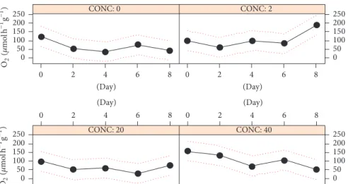
CONC: 0 CONC: 2 CONC: 20 CONC: 40 0 50 100 150 200 250 O2 (𝜇 mo l h −1 g −1 ) 0 50 100 150 200 250 2 4 6 8 0 (Day) 2 4 6 8 0 (Day) 0 2 4 6 8 (Day) 0 2 4 6 8 (Day) 0 50 100 150 200 250 O2 (𝜇 mo l h −1 g −1 ) 0 50 100 150 200 250
Figure 2: B. pharaonis respiration rates (RR) dynamics. The plot shows respiration rates (RR,𝜇mol h−1g−1) versus AgNP concentrations
(𝜇g L−1). Continuous line: average RR; dotted-line, 1-SD confidence interval.
CONC: 0 CONC: 2 CONC: 20 CONC: 40 40 50 60 70 80 2 4 6 8 0 (Day) 40 50 60 70 80 0 2 4 6 8 (Day) 2 4 6 8 0 (Day) 0 2 4 6 8 (Day) 40 50 60 7080 40 50 60 70 80 (B ea t min −1) (B ea t min −1 )
Figure 3: B. pharaonis heartbeat rates (HBRs) dynamics. The plot shows heartbeat rates (HBRs, beat min−1) versus AgNP concentrations
(𝜇g L−1). Continuous line: average HBR; dotted-line, 1-SD confidence interval.
heartbeat rate. HBRs showed a consistent increase in samples
exposed to the two highest AgNP levels (20 and 40𝜇g L−1)
(Figure 3). This finding is only in apparent contrast with the aforementioned previous studies, since our data can be reconciled by hormesis. Hormesis represents a relatively new paradigm in toxicology for which responses to different kinds of toxic agents (or drugs) behave nonlinearly within a threshold level, usually very low and thus disregarded in common toxicological frameworks [42, 43]. Indeed, in specimens of B. pharaonis and Mytilaster minimus subjected to relatively high and recurrent levels of disturbance—such as hypersalinity or hyperthermia—a sharp and continuous increase of HBR has been described before occurring of severe bradycardia [33, 44]. We, therefore, can conclude that the observed HBR changes are likely to represent a compen-sation strategy to counteract the disturbance level escalation, for example, increasing silver burdens accumulating into mussel soft tissues across the exposure.
The outcome of this study showed a more complex response pattern to 5 nm AgNPs concerning the respiration
rate (RR) (Figures 1 and 2). ANOVA pointed out significant effects for the variable concentration (CONC, Table 1) but
nonlinear responses were observed (Figure 1). In 40𝜇g L−1
exposed animals RR dynamics displayed a fair increase fol-lowed by acclimation to control values. This is consistent with the acclimation properties of RR, as reported for moderate temperature shift by Widdows [45]. Instead, no response was observed at the intermediate dose and a delayed RR
intensi-fication was seen at 2𝜇g L−1AgNPs. Similar results were seen
in the bivalve Mytilus galloprovincialis Lam. exposed to ZnO NPs for 12 weeks, that is, low-to-intermediate doses did not render a perfect dose response pattern on some physiological and behavioral traits such as RR and survival [46]. These findings confirm that, in general, ENM toxicity should be a function of factors other than the mere nominal tested concentration since the evolution and fate of NPs in the tested environment can eventually influence the internal dose. To this end, it is worth noting that Mullera et al. [47], as part of the same toxicological framework studied by Hanna et al. [46], reported a good correlation between the internal
Table 3: ANOVA post hoc comparison for HBR. CONC 0 0 0 0 0 2 2 2 2 2 20 20 20 20 20 40 40 40 40 40 CONC× TIME 0 2 4 6 8 0 2 4 6 8 0 2 4 6 8 0 2 4 6 8 0 0 — 0 2 ns — 0 4 ns ns — 0 6 ns ns ns — 0 8 ns ns ns ns — 2 0 ns ns ns ns ns — 2 2 ns ns ns ns ns ns — 2 4 ns ns ns ns ns ns ns — 2 6 ns ns ns ns ns ns ns ns — 2 8 ns ns ns ns ns ns ns ns ns — 20 0 ns ns ns ns ns ns ns ns ns ns — 20 2 ns ns ns ns ns ns ns ns ns ns ns — 20 4 ns ns ns ns ns ns ns ns ns ns ns ns — 20 6 ns ns ns ns ns ns ns ns ns ns ns ns ns — 20 8 ∗∗ ∗ ∗ ∗ ∗ ∗ ∗∗ ∗ ∗ ∗ ∗∗ ∗∗ ∗∗ ns ns ns — 40 0 ns ns ns ns ns ns ns ns ns ns ns ns ns ns ns — 40 2 ns ns ns ns ns ns ns ns ns ns ns ns ns ns ∗∗ ns — 40 4 ns ns ns ns ns ns ns ns ns ns ns ns ns ns ∗∗ ns ns — 40 6 ns ns ns ns ns ns ns ns ns ns ns ns ns ns ns ns ns ns — 40 8 ∗ ∗ ∗ ∗ ∗ ∗ ∗∗ ∗ ∗ ∗ ∗ ∗ ∗∗ ∗∗ ∗∗ ∗∗ ∗∗ ns ns ns ns ns ∗∗ ∗∗ ns —
SNK-test and𝑃 value [∗ = 𝑃 ≤ 0.05; ∗∗ = 𝑃 ≤ 0.01; ∗ ∗ ∗ = 𝑃 ≤ 0.001; and ns = no significant difference (𝑃 > 0.05)].
5 nm AgNP exposure level (𝜇g L−1)
Ag 40 Ag 20 Ag 2 CTRL 0.87 0.88 0.89 0.90 0.91 0.92 0.93 0.94 A bs or ptio n efficienc y (AE)
Figure 4: B. pharaonis absorption efficiency (AE). AE average (±SEM) values for the effects of 5 nm AgNP exposure are depicted. The decreasing trend observed was not statistically significant (1-way
ANOVA,𝑃 = 0.18).
ZnO level and RR. Furthermore, as previously discussed for HBR, nonlinearity (sensu hormesis) represents an intrinsic item of the toxicological response; therefore, RR fluctuations across increasing AgNP doses should be considered a genuine feature of the physiological adaptation process.
Energy flows through the food chain in ecosystems and ecosystems themselves rely on that energy [48]. Our data on AgNP exposure indicated a decreasing trend for food absorption efficiency, although this effect was not statistically
significant (Figure 4). This result is not trivial since, as recently demonstrated by our research group [24], even a small reduction in the efficiency to assimilate food may give rise to important impacts in terms of life history trait’s magnitude (fecundity and growth rate) or energy allocated to byssus quality and quantity [36], eventually increasing the likelihood of subpopulation dislodgment and/or extinction.
While we are aware that mesocosmal conditions can be highly controlled to generate reliable outcomes [24, 49–51], present results, despite their relevance in an ecotoxicolog-ical framework of ENM toxicity assessment, showed that a relatively short exposure period (8 days) is probably not enough to delineate the entire dynamics that characterize Brachidontes sp. response to stress. Therefore, longer expo-sure period should be envisaged in further studies.
4. Conclusions
This work demonstrates that in marine bivalves basic physi-ological functions are impacted by relatively low concentra-tions of AgNP, an emerging contaminant, and supports the use of functional bioenergetics into mechanistic frameworks able to inform on ecological niche changes starting from simple individual responses [52].
Competing Interests
The authors declare that there is no conflict of interests regarding the publication of this paper.
Acknowledgments
This study was supported by a grant to Francesco Dondero (CP-FP 247739 NanoFATE) under the European Commis-sion FP7-Program. Authors are grateful to Valeria Palmeri, Valeria Montalto, and Alessandro Rinaldi for their technical contribution and assistance in laboratory.
References
[1] A. Massarsky, V. L. Trudeau, and T. W. Moon, “Predicting the environmental impact of nanosilver,” Environmental Toxicology
and Pharmacology, vol. 38, no. 3, pp. 861–873, 2014.
[2] M. E. Vance, T. Kuiken, E. P. Vejerano, S. P. McGinnis, M. F. Hochella, and D. R. Hull, “Nanotechnology in the real world: redeveloping the nanomaterial consumer products inventory,”
Beilstein Journal of Nanotechnology, vol. 6, no. 1, pp. 1769–1780,
2015.
[3] The Silver Institute and Thomson Reuters, World Silver Survey
2015. A Summary, 2015.
[4] S. W. P. Wijnhoven, W. J. G. M. Peijnenburg, C. A. Herberts et al., “Nano-silver—a review of available data and knowledge gaps in human and environmental risk assessment,” Nanotoxicology, vol. 3, no. 2, pp. 109–138, 2009.
[5] J. Fabrega, S. N. Luoma, C. R. Tyler, T. S. Galloway, and J. R. Lead, “Silver nanoparticles: Behaviour and effects in the aquatic environment,” Environment International, vol. 37, no. 2, pp. 517– 531, 2011.
[6] F. Gottschalk, T. Sun, and B. Nowack, “Environmental concen-trations of engineered nanomaterials: review of modeling and analytical studies,” Environmental Pollution, vol. 181, pp. 287– 300, 2013.
[7] B. Nowack, “Nanosilver revisited downstream,” Science, vol. 330, no. 6007, pp. 1054–1055, 2010.
[8] M. Baalousha, Y. Yang, M. E. Vance et al., “Outdoor urban nanomaterials: the emergence of a new, integrated, and critical field of study,” Science of The Total Environment, vol. 557-558, pp. 740–753, 2016.
[9] M. R. Wiesner, G. V. Lowry, E. Casman et al., “Meditations on the ubiquity and mutability of nano-sized materials in the environment,” ACS Nano, vol. 5, no. 11, pp. 8466–8470, 2011. [10] J. R. Morones, J. L. Elechiguerra, A. Camacho et al., “The
bactericidal effect of silver nanoparticles,” Nanotechnology, vol. 16, no. 10, pp. 2346–2353, 2005.
[11] H. T. Ratte, “Bioaccumulation and toxicity of silver compounds: a review,” Environmental Toxicology and Chemistry, vol. 18, no. 1, pp. 89–108, 1999.
[12] O. Bondarenko, K. Juganson, A. Ivask, K. Kasemets, M. Mortimer, and A. Kahru, “Toxicity of Ag, CuO and ZnO nanoparticles to selected environmentally relevant test organ-isms and mammalian cells in vitro: a critical review,” Archives of
Toxicology, vol. 87, no. 7, pp. 1181–1200, 2013.
[13] S. Cunningham, M. E. Brennan-Fournet, D. Ledwith, L. Byrnes, and L. Joshi, “Effect of nanoparticle stabilization and physic-ochemical properties on exposure outcome: acute toxicity of silver nanoparticle preparations in zebrafish (Danio rerio),”
Environmental Science and Technology, vol. 47, no. 8, pp. 3883–
3892, 2013.
[14] S. W. Shin, I. H. Song, and S. H. Um, “Role of physicochemical properties in nanoparticle toxicity,” Nanomaterials, vol. 5, no. 3, pp. 1351–1365, 2015.
[15] C. R. Walters, E. J. Pool, and V. S. Somerset, “Ecotoxicity of silver nanomaterials in the aquatic environment: a review of literature and gaps in nano-toxicological research,” Journal of
Environmental Science and Health—Part A Toxic/Hazardous Substances and Environmental Engineering, vol. 49, no. 13, pp.
1588–1601, 2014.
[16] I. Corsi, G. N. Cherr, H. S. Lenihan et al., “Common strategies and technologies for the ecosafety assessment and design of nanomaterials entering the marine environment,” ACS Nano, vol. 8, no. 10, pp. 9694–9709, 2014.
[17] M. N. Moore, “Do nanoparticles present ecotoxicological risks for the health of the aquatic environment?” Environment
Inter-national, vol. 32, no. 8, pp. 967–976, 2006.
[18] L. Canesi, C. Ciacci, R. Fabbri, A. Marcomini, G. Pojana, and G. Gallo, “Bivalve molluscs as a unique target group for nanoparticle toxicity,” Marine Environmental Research, vol. 76, pp. 16–21, 2012.
[19] M. Zuykov, E. Pelletier, and S. Demers, “Colloidal complexed silver and silver nanoparticles in extrapallial fluid of Mytilus
edulis,” Marine Environmental Research, vol. 71, no. 1, pp. 17–21,
2011.
[20] T. Gomes, C. G. Pereira, ´C. Cardoso et al., “Effects of silver
nanoparticles exposure in the mussel Mytilus galloprovincialis,”
Marine Environmental Research, vol. 101, no. 1, pp. 208–214,
2014.
[21] M. P. McCarthy, D. L. Carroll, and A. H. Ringwood, “Tissue specific responses of oysters, Crassostrea virginica, to silver nanoparticles,” Aquatic Toxicology, vol. 138, pp. 123–128, 2013. [22] L. E. Burnett, J. D. Holman, D. D. Jorgensen, J. L. Ikerd, and
K. G. Burnett, “Immune defense reduces respiratory fitness in Callinectes sapidus, the Atlantic blue crab,” The Biological
Bulletin, vol. 211, no. 1, pp. 50–57, 2006.
[23] G. Sar`a, S. Vizzini, and A. Mazzola, “Sources of carbon and dietary habits of new Lessepsian entry Brachidontes pharaonis (Bivalvia, Mytilidae) in the western Mediterranean,” Marine
Biology, vol. 143, no. 4, pp. 713–722, 2003.
[24] V. Montalto, V. Palmeri, A. Rinaldi, S. A. L. M. Kooijman, and G. Sar`a, “Dynamic energy budget parameterisation of Brachi-dontes pharaonis, a Lessepsian bivalve in the Mediterranean Sea,” Journal of Sea Research, vol. 94, pp. 47–51, 2014.
[25] C. Y. S. Chan and J. M. Y. Chiu, “Chronic effects of coated silver nanoparticles on marine invertebrate larvae: A Proof of Concept Study,” PLoS ONE, vol. 10, no. 7, article e0132457, 2015. [26] F. Ribeiro, J. A. Gallego-Urrea, K. Jurkschat et al., “Silver nanoparticles and silver nitrate induce high toxicity to
Pseu-dokirchneriella subcapitata, Daphnia magna and Danio rerio,” Science of the Total Environment, vol. 466-467, pp. 232–241, 2014.
[27] F. Ribeiro, J. A. Gallego-Urrea, R. M. Goodhead et al., “Uptake and elimination kinetics of silver nanoparticles and silver nitrate by Raphidocelis subcapitata: the influence of silver behaviour in solution,” Nanotoxicology, vol. 9, no. 6, pp. 686– 695, 2015.
[28] P. S. Tourinho, C. A. M. Van Gestel, K. Jurkschat, A. M. V. M. Soares, and S. Loureiro, “Effects of soil and dietary
exposures to Ag nanoparticles and AgNO3 in the terrestrial
isopod Porcellionides pruinosus,” Environmental Pollution, vol. 205, pp. 170–177, 2015.
[29] P. S. Tourinho, C. A. M. van Gestel, A. J. Morgan et al., “Toxicokinetics of Ag in the terrestrial isopod Porcellionides
pruinosus exposed to Ag NPs and AgNO3 via soil and food,”
[30] G. Sar`a, V. Palmeri, A. Rinaldi, V. Montalto, and B. Helmuth, “Predicting biological invasions in marine habitats through eco-physiological mechanistic models: a case study with the bivalve
Brachidontes pharaonis,” Diversity and Distributions, vol. 19, no.
10, pp. 1235–1247, 2013.
[31] I. Prusina, G. Sar`a, M. De Pirro et al., “Variations in physi-ological responses to thermal stress in congeneric limpets in the Mediterranean Sea,” Journal of Experimental Marine Biology
and Ecology, vol. 456, pp. 34–40, 2014.
[32] J. Widdows and F. Staff, “Biological effects of contaminants: measurement of scope for growth in mussels,” ICES Techniques in Marine Environmental Sciences 40, International Council for the Exploration of the Sea, Copenhagen, Denmark, 2006. [33] G. Sar`a and M. de Pirro, “Heart beat rate adaptations to varying
salinity of two intertidal Mediterranean bivalves: the invasive Brachidontes pharaonis and the native Mytilaster minimus,”
Italian Journal of Zoology, vol. 78, no. 2, pp. 193–197, 2011.
[34] R. J. Conover, “Assimilation of organic matter by zooplankton,”
Limnology and Oceanography, vol. 11, no. 3, pp. 338–345, 1966.
[35] C. J. Zhu and Y. K. Lee, “Determination of biomass dry weight of marine microalgae,” Journal of Applied Phycology, vol. 9, no. 2, pp. 189–194, 1997.
[36] E. Carrington, J. H. Waite, G. Sar`a, and K. P. Sebens, “Mussels as a model system for integrative ecomechanics,” Annual Review of
Marine Science, vol. 7, pp. 443–469, 2015.
[37] S. A. L. M. Kooijman, “Energy budgets can explain body size relations,” Journal of Theoretical Biology, vol. 121, no. 3, pp. 269– 282, 1986.
[38] G. Sar`a, A. Rinaldi, and V. Montalto, “Thinking beyond organ-ism energy use: a trait-based bioenergetic mechanistic approach for predictions of life history traits in marine organisms,”
Marine Ecology, vol. 35, no. 4, pp. 506–515, 2014.
[39] T. M. Curtis, R. Williamson, and M. H. Depledge, “Simultane-ous, long-term monitoring of valve and cardiac activity in the blue mussel Mytilus edulis exposed to copper,” Marine Biology, vol. 136, no. 5, pp. 837–846, 2000.
[40] D. M. Scott and C. W. Major, “The Effect of Copper (II) on Survival, Respiration, and Heart Rate in the Common Blue Mussel, Mytilus edulis,” Biological Bulletin, vol. 143, no. 3, pp. 679–688, 1972.
[41] A. L. Grace and L. F. Gainey Jr., “The effects of copper on the heart rate and filtration rate of Mytilus edulis,” Marine Pollution
Bulletin, vol. 18, no. 2, pp. 87–91, 1987.
[42] E. J. Calabrese, “Hormesis: a revolution in toxicology, risk assessment and medicine,” EMBO Reports, vol. 5, supplement 1, pp. S37–S40, 2004.
[43] E. J. Calabrese and L. A. Baldwin, “The frequency of U-shaped dose responses in the toxicological literature,” Toxicological
Sciences, vol. 62, no. 2, pp. 330–338, 2001.
[44] C. E. Braby and G. N. Somero, “Following the heart: temper-ature and salinity effects on heart rate in native and invasive species of blue mussels (genus Mytilus),” Journal of Experimental
Biology, vol. 209, no. 13, pp. 2554–2566, 2006.
[45] J. Widdows, “Effect of temperature and food on the heart beat, ventilation rate and oxygen uptake of Mytilus edulis,” Marine
Biology, vol. 20, no. 4, pp. 269–276, 1973.
[46] S. K. Hanna, R. J. Miller, E. B. Muller, R. M. Nisbet, and H. S. Lenihan, “Impact of engineered zinc oxide nanoparticles on the individual performance of Mytilus galloprovincialis,” PLoS ONE, vol. 8, no. 4, Article ID e61800, 2013.
[47] E. B. Mullera, S. K. Hannab, H. S. Lenihanb, R. J. Millera, and R. M. Nisbetc, “Impact of engineered zinc oxide nanoparticles on the energy budgets of Mytilus galloprovincialis,” Journal of Sea
Research, vol. 94, pp. 29–36, 2014.
[48] E. P. Odum, “Energy flow in ecosystems: a historical review,”
American Zoologist, vol. 8, no. 1, pp. 11–18, 1968.
[49] B. Manachini, V. Arizza, A. Rinaldi, V. Montalto, and G. Sar`a, “Effect of the commercial BT-based pesticide on eco-physiological response of two marine bivalves,” Marine
Environ-mental Research, vol. 83, pp. 29–37, 2013.
[50] P. Gianguzza, G. Visconti, F. Gianguzza, S. Vizzini, G. Sar`a, and S. Dupont, “Temperature modulates the response of the thermophilous sea urchin Arbacia lixula early life stages to
CO2-driven acidification,” Marine Environmental Research, vol.
93, pp. 70–77, 2014.
[51] A. Rinaldi, V. Montalto, K. Lika, M. Sanfilippo, A. Manganaro, and G. Sar`a, “Estimation of dynamic energy budget parameters for the Mediterranean toothcarp (Aphanius fasciatus),” Journal
of Sea Research, vol. 94, pp. 65–70, 2014.
[52] M. Kearney, S. J. Simpson, D. Raubenheimer, and B. Helmuth, “Modelling the ecological niche from functional traits,”
Philo-sophical Transactions of the Royal Society B: Biological Sciences,
Submit your manuscripts at
http://www.hindawi.com
Pain
Research and TreatmentHindawi Publishing Corporation
http://www.hindawi.com Volume 2014
World Journal
Hindawi Publishing Corporation
http://www.hindawi.com Volume 2014
Hindawi Publishing Corporation
http://www.hindawi.com Volume 2014
Toxins
Journal of
Vaccines
Journal ofHindawi Publishing Corporation
http://www.hindawi.com Volume 2014
Hindawi Publishing Corporation
http://www.hindawi.com Volume 2014
Antibiotics
Toxicology
Journal of Hindawi Publishing Corporation
http://www.hindawi.com Volume 2014
Stroke
Research and TreatmentHindawi Publishing Corporation
http://www.hindawi.com Volume 2014
Drug Delivery
Journal ofHindawi Publishing Corporation
http://www.hindawi.com Volume 2014
Hindawi Publishing Corporation
http://www.hindawi.com Volume 2014
Advances in Pharmacological Sciences
Tropical Medicine
Hindawi Publishing Corporation
http://www.hindawi.com Volume 2014
Medicinal ChemistryInternational Journal of
Hindawi Publishing Corporation
http://www.hindawi.com Volume 2014
Addiction
Journal of Hindawi Publishing Corporationhttp://www.hindawi.com Volume 2014
Hindawi Publishing Corporation
http://www.hindawi.com Volume 2014
BioMed
Research International Emergency Medicine International
Hindawi Publishing Corporation
http://www.hindawi.com Volume 2014
Hindawi Publishing Corporation
http://www.hindawi.com Volume 2014
Diseases
Hindawi Publishing Corporation
http://www.hindawi.com Volume 2014 Anesthesiology Research and Practice
Scientifica
Hindawi Publishing Corporationhttp://www.hindawi.com Volume 2014
Journal of
Hindawi Publishing Corporation
http://www.hindawi.com Volume 2014
Pharmaceutics
Hindawi Publishing Corporation
http://www.hindawi.com Volume 2014