Published online 21 December 2015 in Wiley Online Library (wileyonlinelibrary.com)DOI: 10.1002/path.4633
Dual loss of the SWI/SNF complex ATPases SMARCA4/BRG1
and SMARCA2/BRM is highly sensitive and specific for small cell
carcinoma of the ovary, hypercalcaemic type
Anthony N Karnezis,1†Yemin Wang,1†Pilar Ramos,2†William PD Hendricks,2 Esther Oliva,3Emanuela D’Angelo,4
Jaime Prat,4 Marisa R Nucci,5 Torsten O Nielsen,1Christine Chow,6 Samuel Leung,6Friedrich Kommoss,7Stefan
Kommoss,8 Annacarolina Silva,9 Brigitte M Ronnett,10 Joseph T Rabban,11David D Bowtell,12Bernard E
Weissman,13Jeffrey M Trent,2 C Blake Gilks1* and David G Huntsman1,6,14*
1 Department of Pathology and Laboratory Medicine, University of British Columbia, Vancouver, BC, Canada 2 Division of Integrated Cancer Genomics, Translational Genomics Research Institute (TGen), Phoenix, AZ, USA 3 Department of Pathology, Massachusetts General Hospital, Boston, MA, USA
4 Department of Pathology, Hospital de la Santa Creu i Sant Pau, Autonomous University of Barcelona, Barcelona, Spain 5 Department of Pathology, Brigham and Women’s Hospital, Boston, MA, USA
6 Genetic Pathology Evaluation Centre, Vancouver General Hospital, University of British Columbia, Vancouver, BC, Canada 7 Synlab MVZ Pathologie, Mannheim, Germany
8 Department of Obstetrics and Gynecology, University Hospital of Tuebingen, Tuebingen, Germany
9 The James Homer Wright Pathology Laboratories, Massachusetts General Hospital, Harvard Medical School, Boston, MA, USA 10 Department of Pathology, The Johns Hopkins Hospital, Baltimore, MD, USA
11 Department of Anatomic Pathology, University of California San Francisco, San Francisco, CA, USA 12 Peter MacCallum Cancer Centre, East Melbourne, Victoria, Australia
13 Department of Pathology and Laboratory Medicine, Lineberger Cancer Center, University of North Carolina, Chapel Hill, NC, USA 14 Department of Molecular Oncology, British Columbia Cancer Research Centre, Vancouver, BC, Canada
*Correspondence to: D Huntsman, MD, FRCPC, FCCMG, Department of Molecular Oncology, BC Cancer Agency Research Centre, #3427-600 West 10th Avenue, Vancouver, BC V5Z 4E6, Canada. E-mail: [email protected]
Or C Blake Gilks, MD, Room 1250, First Floor JPPN, Department of Pathology and Laboratory Medicine, Vancouver General Hospital, 910 West 10th Avenue, Vancouver, BC V5Z 1 M9, Canada. E-mail: [email protected]
†These three authors contributed equally.
Abstract
Small cell carcinoma of the ovary, hypercalcaemic type (SCCOHT) is a lethal and sometimes familial ovarian tumour of young women and children. We and others recently discovered that over 90% of SCCOHTs harbour inactivating mutations in the chromatin remodelling geneSMARCA4 with concomitant loss of its encoded protein SMARCA4 (BRG1), one of two mutually exclusive ATPases of the SWI/SNF chromatin remodelling complex. To determine the specificity of SMARCA4 loss for SCCOHT, we examined the expression of SMARCA4 by immunohistochemistry in more than 3000 primary gynaecological tumours. Among ovarian tumours, it was only absent in clear cell carcinoma (15 of 360, 4%). In the uterus, it was absent in endometrial stromal sarcomas (4 of 52, 8%) and high-grade endometrioid carcinomas (2 of 338, 1%). Recent studies have shown that SMARCA2 (BRM), the other mutually exclusive ATPase of the SWI/SNF complex, is necessary for survival of tumour cells lacking SMARCA4. Therefore, we examined SMARCA2 expression and discovered that all SMARCA4-negative SCCOHTs also lacked SMARCA2 protein by IHC, including the SCCOHT cell lines BIN67 and SCCOHT1. Among ovarian tumours, the SMARCA4/SMARCA2 dual loss phenotype appears completely specific for SCCOHT. SMARCA2 loss was not due to mutation but rather from an absence of mRNA expression, which was restored by treatment with the histone deacetylase inhibitor trichostatin A. Re-expression of SMARCA4 or SMARCA2 inhibited the growth of BIN67 and SCCOHT1 cell lines. Our results indicate that SMARCA4 loss, either alone or with SMARCA2, is highly sensitive and specific for SCCOHT and that restoration of either SWI/SNF ATPase can inhibit the growth of SCCOHT cell lines.
© 2015 The Authors. The Journal of Pathology published by John Wiley & Sons Ltd on behalf of Pathological Society of Great Britain and Ireland.
Keywords: small cell carcinoma; hypercalcaemic type; rhabdoid tumour; SMARCA4/BRG1; SMARCA2/BRM; SMARCB1/INI1; SWI/SNF;
HDAC inhibitor; trichostatin A; epigenetic silencing
Received 4 June 2015; Revised 28 August 2015; Accepted 3 September 2015
No conflicts of interest were declared.
Introduction
Background – SCCOHT
Small cell carcinoma of the ovary, hypercalcaemic type (SCCOHT) is a rare ovarian cancer that predominantly affects young women in their teens and 20s, with an average age of 24 years. Although half of tumours are diagnosed at an early stage, the prognosis is dismal. For patients with stage IA disease, more than half will still die of disease, usually within 2 years [1]. Histologically, SCCOHT is characterized by sheets and poorly formed nests of small cells with scant cytoplasm, hyperchro-matic nuclei, and small nucleoli [1]. Approximately half of tumours also contain variable numbers of large cells with abundant eosinophilic cytoplasm, often rhabdoid inclusions, large nuclei, and prominent nucleoli. The histological differential diagnosis is broad and includes many primary and metastatic tumours to the ovary. The lack of familiarity of pathologists with this rare tumour and the lack of specific immunohistochemical markers can make its diagnosis challenging.
SMARCA4 in SCCOHT and cancer
Recently, four groups independently identified inacti-vating SMARCA4 mutations in the majority of SCCO-HTs, resulting in loss of SMARCA4 protein [2–5]. SMARCA4 and the related protein SMARCA2 (also called BRG1 and BRM, respectively) are the two mutu-ally exclusive ATPases of the SWI/SNF chromatin remodelling complex [6–8]. SWI/SNF subunits have been frequently implicated as tumour suppressors, with approximately 20% of cancers bearing mutations in these genes [9,10]. Our initial analysis of a small collec-tion of ovarian tumours indicated that SMARCA4 loss was highly specific for SCCOHT [2].
Potential therapeutic strategies for SCCOHT
The function of the SWI/SNF complex in chromatin remodelling suggests that the pathogenesis of SCCOHT involves epigenetic dysregulation. This paradigm may offer treatment possibilities with agents that regulate the epigenome such as inhibitors of histone deacety-lase (HDAC) or modifiers of histone or DNA methyla-tion. The mutually exclusive nature of the SMARCA4 and SMARCA2 ATPases in the SWI/SNF complex has suggested that SMARCA2 may be a synthetic lethal target in SMARCA4-mutant cancers. Indeed, loss of SMARCA4 has been shown to lead to dependence on SMARCA2-containing SWI/SNF complexes for sur-vival in some cell lines [11–13], raising the possibility of targeting SMARCA2 in SCCOHT using inhibitors of its ATPase or bromodomain.
Aims of this study
The establishment of SMARCA4 mutation with accom-panying loss of protein as the pathognomonic mutation in SCCOHT raises the need to explore the spectrum
of tumours that share SMARCA4 (and perhaps SMARCA2) loss to understand the diagnostic utility of SMARCA4 immunohistochemistry (IHC). Because some ovarian and uterine tumours arise from common cell types (eg endometrial epithelium, either in the eutopic endometrium or ectopically as endometriosis), we also need to determine the diagnostic utility of SMARCA4 IHC in uterine tumours. Therefore, the goals of this study were (1) to determine the specificity of SMARCA4 protein loss as a diagnostic marker for SCCOHT by studying its expression in a large cohort of ovarian and uterine tumours with an emphasis on entities in the differential diagnosis; and (2) to deter-mine whether SMARCA2 is expressed in SCCOHT and could be used as a therapeutic target.
Materials and methods
Sample collection and tissue microarray
construction
Duplicate 0.6 or 1.0 mm cores of formalin-fixed, paraffin-embedded tumour tissue from each case were used for tissue microarray (TMA) construction, as described previously [14]. Additional cases were stud-ied by whole-slide IHC. All samples were collected in accordance with institutional guidelines and protocols. For Vancouver samples, informed patient consent was obtained under research ethics board (REB)-approved protocols for all prospectively collected patient samples (REB H05-60 199), archived samples (REB H02-61 375), and for IHC analysis (REB H02-61375).
Immunohistochemistry and scoring
TMAs were cut at 4 μm thickness onto Superfrost + glass slides and were processed using the Ventana Discovery XT, and the Ventana Benchmark XT and Benchmark Ultra automated systems (Ventana Medi-cal Systems, Tucson, AZ, USA). ImmunohistochemiMedi-cal staining was performed with antibodies to SMARCA4 (1:25, clone EPNCIR111A, ab110641; Abcam, Toronto, Ontario, Canada), SMARCA2 (1:50, clone HPA029981; Sigma, St Louis, MO, USA), and SMARCB1/BAF47/ INI1 (1:50, 25/BAF47, 612110; BD Biosciences, Mis-sissauga, Ontario, Canada).
All TMAs were scored twice by a pathologist (ANK). For SMARCA4, tumours were scored as positive if any tumour cell nuclei showed staining; tumours scored as positive usually showed diffuse, moderate to strong staining. Tumours were scored as negative if tumour cell nuclei showed no staining only if adequate staining was detected in internal positive control cells (endothe-lium, lymphocytes or fibroblasts); absence of staining has previously been shown to correlate with the presence of inactivating mutations in SMARCA4 [2–5,15]. No cytoplasmic SMARCA4 staining was observed. Since SMARCA2 absence was due to non-mutational silenc-ing, tumours were scored as positive for SMARCA2 if
more than 5% of tumour cells showed nuclear staining. Tumours were scored as negative if tumour cells showed no nuclear staining, nuclear staining in up to 5% of cells, or only cytoplasmic staining. For both SMARCA4 and SMARCA2, tumours that showed no staining in tumour and stroma were considered technical failures and not scored. Each case on a TMA was represented as dupli-cate cores; one positive core was sufficient to count the case as positive.
Cell lines
BIN67, SCCOHT1, and KGN cells were all grown in DMEM/F-12 supplemented with 10% FBS. SVOG3e cells were cultured in 199/105 medium with 10% FBS. ES-2 cells were cultured in RPMI supplemented with 10% FBS. All the cells were maintained in a
humidi-fied 5% CO2-containing atmosphere at 37 ∘C and tested
regularly for Mycoplasma.
Plasmids and lentivirus/retrovirus packaging
The pLDpuro-SMARCA4 and pLDpuro-SMARCA2
plasmids were constructed by introducing the
SMARCA4 and SMARCA2 from entry vectors (Genecopeia) into the pLDpuro-EnVA destination vector (a gift of Dr Jason Moffat at the University of Toronto) using Gateway reactions (Life Tech-nologies, Grand Island, NY, USA). To produce lentiviruses expressing SMARCA4 or SMARCA2, pLDpuro-SMARCA4 or pLDpuro-SMARCA2 was co-transfected with packaging plasmids psPAX2 and pMD2.G into HEK293T cells. Supernatants were collected at 72 h for lentivirus preparation and infection.
Western blotting
Whole-cell extracts were obtained for SDS-PAGE elec-trophoresis using the SMARCA4 and SMARCA2 anti-bodies described above. Ponceau S staining or western blotting for vinculin (clone hVIN-1, V9131; Sigma) was used to confirm equal protein loading.
Real-time RT-PCR
Total RNA was extracted from cells using RNeasy kits (Qiagen, Valencia, CA, USA) and quantitated by Nanodrop spectrophotometry. One microgram of total RNA was reverse-transcribed into cDNA using ran-dom primers and Superscript III reverse transcriptase (Invitrogen, Carlsbad, CA, USA), followed by PCR amplification of SMARCA4 or SMARCA2 using the
Power SYBR® Green Master Mix (Life Technologies)
according to the manufacturers’ instructions. GAPDH was used for normalization. The relative levels were calculated using the ΔΔCt method.
RNA isolation and gene expression microarrays
Frozen tumour tissue was disrupted by sonication using the Covaris S-2 system (Thermo Fisher Scientific,
Waltham, MA, USA) and stabilized by addition of equal volumes of TRIzol (Life Technologies). BIN67 and SCCOHT1 cells were washed with PBS and lysed using TRIzol. Total RNA was purified using RNeasy micro kits (Qiagen). RNA from two premenopausal nor-mal ovary donors (ages 32 and 37 years) was obtained from OriGene (Rockville, MD, USA), pooled (100 ng each), and used as a common reference. RNA integrity and purity were measured using a 2100 Bioanalyzer (Agilent Technologies, Santa Clara, CA, USA) and a Nanodrop ND-1000 Spectrophotometer. Labelled cRNA probes were prepared using Agilent’s Low Input Quick Amp Labeling Kit and hybridized to Agilent Human Gene Expression 4 × 44 K microarrays follow-ing the manufacturer’s protocols. Slides were washed and scanned at 5 μm using an Agilent Microarray Scanner (model G2505B) in an ozone-controlled envi-ronment, and data extracted, processed, and normalized using Agilent Feature Extraction (FE) software (v10.5). Raw and processed data files have been deposited in GEO at accessions GSE49887 and GSE66434.
Cell growth assay
Cells were infected with lentivirus containing pLDpuro-GFP, −SMARCA4 or -SMARCA2. One day later, cells were selected in 2 μg/ml puromycin for 3 days. The remaining cells were then harvested, counted, and reseeded at a density of 4000 cells per well in 96-well plates. Cell growth was monitored using the IncuCyte
ZOOM® live cell imaging monitor (Essen BioScience,
Ann Arbor, MI, USA). The percentage of confluence in each well was calculated and plotted over a 7-day period to determine the effect of SMARCA4 or SMARCA2 re-expression on cell growth.
Results
SMARCA4 expression in gynaecological
and non-gynaecological tumours
We previously studied SMARCA4 protein status in a small series of 485 primary ovarian tumours where besides SCCOHT, only two tumours (0.4%), both clear cell carcinomas, were negative for SMARCA4 [2]. In order to perform a definitive analysis of the sensitiv-ity and specificsensitiv-ity of SMARCA4 loss for SCCOHT and thus, its potential as a diagnostic immunohis-tochemical marker, we expanded SMARCA4 IHC analysis to additional cases of SCCOHT and a large series of 3048 primary ovarian and uterine tumours and non-gynaecological mimics of SCCOHT (Table 1).
Examination of SMARCA4 expression in 50 SCCOHT-derived samples (20 reported previously [2,15] and 30 new cases reported here) revealed SMARCA4 loss in 42/46 (91%) primary tumours, 2/2 patient-derived mouse xenografts, and 2/2 SCCOHT-derived cell lines (Table 1). Of the four tumours
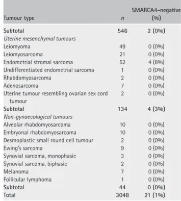
Table 1. SMARCA4 immunohistochemical analysis in ovarian, uterine, and selected non-gynaecological tumours
Tumour type n SMARCA4-negative (%) SCCOHT Primary tumours 46 42 (91%) Patient-derived xenografts 2 2 (100%) Cell lines 2 2 (100%) Subtotal 50 46 (92%)
Ovarian epithelial tumours
High-grade serous carcinoma 1198 0 (0%)
Clear cell carcinoma 360 15 (4%)
Endometrioid adenocarcinoma 258 0 (0%)
Endometrioid adenocarcinoma, mixed 9 0 (0%)
Endometrioid borderline tumour 1 0 (0%)
Low-grade serous carcinoma 53 0 (0%)
Serous borderline tumour 23 0 (0%)
Mucinous carcinoma 98 0 (0%)
Mucinous borderline tumour 6 0 (0%)
Mixed mucinous tumour 5 0 (0%)
Anaplastic carcinoma in mucinous carcinoma 1 0 (0%)
Mixed carcinoma 1 0 (0%)
Undifferentiated carcinoma 18 0 (0%)
Carcinosarcoma 27 0 (0%)
Benign Brenner tumour 3 0 (0%)
Borderline Brenner tumour 1 0 (0%)
Malignant Brenner tumour 2 0 (0%)
Small cell carcinoma, pulmonary type 3 0 (0%) Ovarian carcinoma, not otherwise specified 5 0 (0%)
Subtotal 2072 15 (1%)
Ovarian/adnexal sex cord-stromal tumours
Adult granulosa cell tumour 113 0 (0%)
Juvenile granulosa cell tumour 29 0 (0%)
Fibroma/fibrosarcoma 8 0 (0%)
Thecoma 7 0 (0%)
Sertoli–Leydig cell tumour 41 0 (0%)
Leydig cell tumour 2 0 (0%)
Sclerosing stromal tumour 6 0 (0%)
Sex cord tumour with annular tubules 2 0 (0%)
Steroid cell tumour 1 0 (0%)
Sex cord tumour, not otherwise specified 4 0 (0%) Female adnexal tumour of Wolffian origin 4 0 (0%)
Gynandroblastoma 3 0 (0%)
Endometrioid stromal sarcoma 1 0 (0%)
Subtotal 221 0 (0%)
Ovarian germ cell tumours
Dysgerminoma 5 0 (0%)
Yolk sac tumour 6 0 (0%)
Dysgerminoma and yolk sac tumour 1 0 (0%)
Mature teratoma 3 0 (0%)
Immature teratoma 6 0 (0%)
Carcinoid tumour 4 0 (0%)
Strumal carcinoid in a mucinous cystadenoma 2 0 (0%) Sebaceous carcinoma in a dermoid cyst 1 0 (0%)
Squamous cell carcinoma 3 0 (0%)
Subtotal 31 0 (0%)
Uterine epithelial tumours
Endometrioid adenocarcinoma 336 1 (0%)
Clear cell carcinoma 28 0 (0%)
High-grade serous carcinoma 94 0 (0%)
Mucinous carcinoma 3 0 (0%)
Mixed carcinoma 11 0 (0%)
Large cell carcinoma 1 0 (0%)
Small cell carcinoma 3 0 (0%)
Undifferentiated carcinoma 3 0 (0%) Dedifferentiated carcinoma 2 1 (50%) Carcinosarcoma 65 0 (0%) Table 1. Continued Tumour type n SMARCA4-negative (%) Subtotal 546 2 (0%)
Uterine mesenchymal tumours
Leiomyoma 49 0 (0%)
Leiomyosarcoma 21 0 (0%)
Endometrial stromal sarcoma 52 4 (8%)
Undifferentiated endometrial sarcoma 1 0 (0%)
Rhabdomyosarcoma 2 0 (0%)
Adenosarcoma 7 0 (0%)
Uterine tumour resembling ovarian sex cord tumour 2 0 (0%) Subtotal 134 4 (3%) Non-gynaecological tumours Alveolar rhabdomyosarcoma 10 0 (0%) Embryonal rhabdomyosarcoma 10 0 (0%)
Desmoplastic small round cell tumour 2 0 (0%)
Ewing’s sarcoma 9 0 (0%)
Synovial sarcoma, monophasic 3 0 (0%)
Synovial sarcoma, biphasic 2 0 (0%)
Melanoma 7 0 (0%)
Follicular lymphoma 1 0 (0%)
Subtotal 44 0 (0%)
Total 3048 21 (1%)
that retained SMARCA4 expression, three lacked SMARCB1 (also called BAF47 or INI1), a differ-ent core member of the SWI/SNF complex. One of the three SMARCB1-negative tumours showed biallelic inactivation of SMARCB1. In the remaining SMARCA4-positive case, exome sequencing revealed no mutations in SWI/SNF genes.
We examined SMARC4 expression in 3048 primary ovarian and uterine tumours and non-gynaecological mimics of SCCOHT. Among 2324 primary ovarian epithelial, sex cord-stromal, and germ cell tumours (including the previously reported 485 tumours [2]), SMARCA4 loss was only observed in clear cell car-cinoma (15/360, 4%) (Table 1), a tumour that is not in the histological differential diagnosis of SCCOHT. All ovarian tumours in the histological differential diag-nosis of SCCOHT (including small cell carcinoma of pulmonary type, adult and juvenile granulosa cell tumours, Sertoli–Leydig cell tumours, endometrial stro-mal sarcoma, and dysgerminoma) retained SMARCA4 expression.
Among 680 primary uterine tumours, SMARCA4 loss was observed in endometrioid carcinoma (2/338, 1%) and endometrial stromal sarcoma (ESS, 4/52, 4%; Table 1). Both endometrioid carcinomas were FIGO grade 3; one was a dedifferentiated carcinoma [16], a tumour composed of low-grade endometrioid carcinoma (FIGO grade 1 or 2) and undifferentiated carcinoma. Among the ESS cases, two were low-grade and two were high-grade. One high-grade ESS showed rhab-doid features. For the SMARCA4-negative dedifferen-tiated carcinoma and the high-grade ESS cases, only the high-grade/undifferentiated portions were represented on the TMAs.
We also examined SMARCA4 expression in 44 extra-ovarian, non-gynaecological tumours that resem-ble SCCOHT histologically and can present with intra-abdominal or ovarian disease mimicking ovarian carcinoma clinically, including alveolar and embryonal rhabdomyosarcoma, desmoplastic small round cell tumour, Ewing’s sarcoma, synovial sarcoma, follicu-lar lymphoma, and melanoma. All of these tumours retained SMARCA4 expression (Table 1).
In summary, 46/50 (92%) of all SCCOHT-derived samples in our series lacked SMARCA4, and 49/50 (98%) lacked a core member of the SWI/SNF complex (either SMARCA4 or SMARCB1). When combined with other published SCCOHT cases [3–5], SMARCA4 loss has now been observed in 83/91 cases (91% sensitivity). Among primary ovarian tumours and non-gynaecological tumours that can mimic SCCOHT, we observed SMARCA4 loss in only 15/2368 tumours (15/2324 ovarian and 0/44 non-gynaecological) for a specificity of 99% among the tumours examined, and the only ovarian tumour that lacked SMARCA4 in our study (clear cell carcinoma) is not in the differential diagnosis of SCCOHT. These data demonstrate that the lack of SMARCA4 in ovarian tumours is a highly sensitive and almost completely specific IHC marker of SCCOHT.
Absence of SMARCA2 protein in SCCOHT tumours
In order to determine SMARCA2 status in SCCOHT and the potential for its development as a therapeutic target, we examined its expression in 45 cases. All 45 tumours (43 lacking SMARCA4 and 2 lacking SMARCB1) were negative for SMARCA2.
How-ever, rare tumour cells (usually< 1% but up to 10%
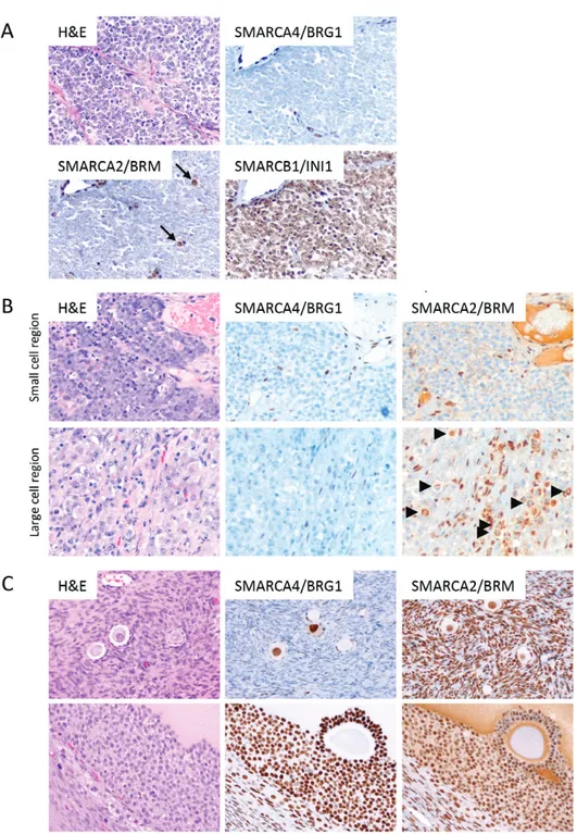
focally) still expressed SMARCA2. In areas containing only small cells, SMARCA2 staining was exclusively nuclear (Figure 1A, arrows). In tumours with a large cell component, the protein was exclusively localized to the cytoplasm of large cells (Figure 1B, bottom panels, arrowheads), with no cytoplasmic expression in the small cell component (Figure 1B, upper panels).
We did not identify mutations in the SMARCA2 gene in SCCOHT, implicating silencing during tumourigen-esis or in the cell of origin as the underlying mecha-nism explaining the absence of SMARCA2 protein. We therefore performed IHC for SMARCA2 in ovaries of premenopausal women and found uniform and strong expression in all ovarian cell types, including surface epithelial cells, stromal cells, granulosa cells, theca cells, and oocytes (Figure 1C). This result suggests that SCCOHT loses SMARCA2 expression during tumouri-genesis or, alternatively, SCCOHT arises from a rare SMARCA2-negative cell type in the ovary.
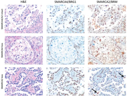
To determine whether dual loss of both SMARCA4 and SMARCA2 was specific for SCCOHT, we exam-ined SMARCA2 expression by IHC in ovarian clear cell carcinoma, the only other tumour lacking SMARCA4 expression (Table 1). Tumours were deficient either for SMARCA4 or for SMARCA2, but not both (Figure 2),
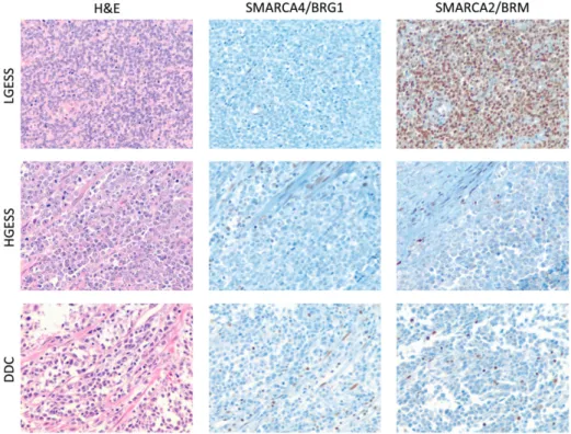
indicating that dual deficiency of the two SWI/SNF ATPases is specific for SCCOHT. In uterine tumours, combined SMARCA4 and SMARCA2 deficiency was only observed in dedifferentiated carcinoma (n = 1) and high-grade ESS (n = 2) (Figure 3).
Epigenetic silencing of SMARCA2 in SCCOHT cells
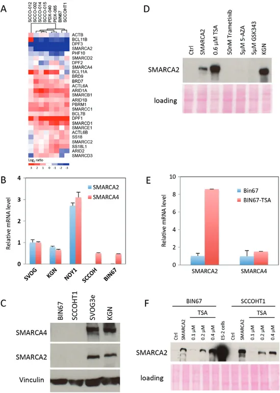
The lack of mutations or deletions involving SMARCA2 in SCCOHT and the expression of SMARCA2 protein in rare tumour cells suggest that the absence of SMARCA2 protein in most tumour cells arises from epigenetic silencing or mRNA degradation, as reported in other SMARCA2-deficient cell lines [17–21]. Compared with normal premenopausal ovary, SMARCA2 mRNA levels were decreased in seven SCCOHT tumour samples (four primary tumours and two mouse xenografts gen-erated directly from patient tumours, and one SCCOHT cell line), an effect that was specific for SMARCA2 and not observed with genes encoding most other SWI/SNF family members (Figure 4A). Similar to the tumour samples, no SMARCA2 mRNA (Figure 4B) or protein (Figure 4C, S1) was detected in two SCCOHT cell lines, BIN67 and SCCOHT1, and no underlying mutation was identified. SMARCA2 mRNA levels were significantly lower in BIN67 and SCCOHT1 cells than in NOY1 (a yolk sac tumour cell line), SVOG3e (an immortalized primary granulosa cell line) or KGN cells (an adult granulosa cell tumour cell line) (Figure 4B), suggesting epigenetically silencing of the SMARCA2 gene or degra-dation of SMARCA2 mRNA as the mechanism underly-ing the lack of SMARCA2 protein in SCCOHT cells.
To determine whether we could reactivate SMARCA2 expression in SCCOHT cell lines, we treated BIN67 cells with inhibitors that target epigenetic regulators including the HDAC inhibitor trichostatin A (TSA), the DNA methyltransferase inhibitor 5-azacytidine (5-AZA), and the EZH2 histone methyltransferase inhibitor GSK343. Of these reagents, only the HDAC inhibitor TSA induced the expression of SMARCA2 protein (Figure 4D), correlating with increased mRNA levels in BIN67 cells (Figure 4E). Furthermore, TSA stimulated the expression of SMARCA2 protein in a dose-dependent manner in BIN67 and SCCOHT1 cells (Figure 4 F). These data implicate HDAC-mediated epi-genetic silencing of the SMARCA2 gene in SCCOHT or, alternatively, an indirect inhibitory effect on SMARCA2 mRNA degradation.
Re-expression of SMARCA4 or SMARCA2 inhibits
cell growth
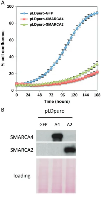
To assess whether the absence of SMARCA4 and SMARCA2 is crucial for tumour cell growth, we infected BIN67 cells with lentiviruses driving expres-sion of SMARCA4 or SMARCA2. Overexpresexpres-sion of either protein robustly suppressed the growth of BIN67 cells (Figure 5A). However, SMARCA4 expression did not significantly inhibit the growth of cells lacking only SMARCA4 (JHOC5 cells, ovarian
Figure 1. Immunohistochemical analysis of core SWI/SNF proteins in SCCOHT. (A) Dual loss of SMARCA4/BRG1 and SMARCA2/BRM in
SCCOHT. Endothelium and lymphocytes are internal positive controls for both proteins. Arrows denote rare tumour cells expressing SMARCA2. SMARCB1/INI1 protein expression serves as a positive control for tumour cell immunoreactivity. (B) SMARCA2/BRM expression and subcellular localization in regions of small cell versus large cell morphology. When present, SMARCA2 only showed nuclear localization in the small cells (arrows, A). In contrast, scattered tumour cells with large cell morphology also expressed SMARCA2 in the cytoplasm (arrowheads, B). (C) SMARCA4/BRG1 and SMARCA2/BRM expression in normal ovary from premenopausal women. SMARCA4 was expressed strongly in oocytes and granulosa cells but was either weak or absent in stromal cells. In contrast, SMARCA2 showed diffuse and strong expression in oocytes, granulosa cells, and stromal cells. In addition, both proteins showed uniform expression in ovarian surface epithelium (data not shown). H&E and SMARCA4 immunohistochemistry images are reproduced from our previous publication, Ramos et al [2] (Supplementary Figure 4, panels A–D).
clear cell carcinoma cell line [22]) or with normal SMARCA4 and SMARCA2 expression (ES-2 ovar-ian endometrioid adenocarcinoma cell line [22]) (data not shown). Previous studies have shown that the ability of SMARCA4 to induce cell cycle arrest
in SMARCA4/SMARCA2-deficient cell lines is
dependent on a functional retinoblastoma growth control pathway [17,19,23–26]. Therefore, the rapid growth arrest that SMARCA4 or SMARCA2 expression induced in SCCOHT cells is consistent with the absence of detectable RB1 gene mutations in all tumour samples and the presence of intact, full length RB1 protein (both
Figure 2. Immunohistochemical analysis of SMARCA4/BRG1 and SMARCA2/BRM in ovarian clear cell carcinoma. Tumours were negative for
either SMARCA4 (middle panels) or SMARCA2 (bottom panels) or showed intact expression of both proteins (upper panels). No dual deficient tumours were identified. Similar to SCCOHT, rare scattered SMARCA2-positive cells (arrows) were identified in some SMARCA2-negative tumours.
hypo- and hyper-phosphorylated forms) in the BIN67 cells (data not shown).
Discussion
SMARCA4 in ovarian and endometrial tumours
Almost all SCCOHTs contain inactivating somatic and germline mutations in SMARCA4 that result in loss of the SMARCA4 (BRG1) protein. We investigated SMARCA4 expression by IHC in a large collection of gynaecological and non-gynaecological tumours to determine the specificity of SMARCA4 loss for SCCOHT. All tumours in the differential diagnosis that we examined retained SMARCA4 expression. The only primary ovarian tumour other than SCCOHT that showed loss of SMARCA4 – clear cell carci-noma – does not enter the histological differential diagnosis of SCCOHT. Therefore, the absence of SMARCA4 is highly sensitive and essentially com-pletely specific for SCCOHT.
Though we only studied small numbers of germ cell tumours in the differential diagnosis of SCCOHT, our results are consistent with those of Witkowski et al, who found no SMARCA4 mutations in 106 germ cell tumours, including 25 dysgerminomas and seminomas [27]. A few ovarian tumours in the differential diagno-sis of SCCOHT were not studied, including metastatic small cell lung carcinoma and oxyphilic tumours that can resemble the large cell variant of SCCOHT. In
addition, we did not examine small cell carcinoma of the endometrium or cervix; notably, the latter has been reported to be deficient in SMARCA4 [28]. Though SCCOHT and cervical small cell carcinoma have dis-tinct clinical presentations, other IHC markers (eg WT1 and TTF1) [29] or molecular studies (HPV18 in the cer-vical tumours) [28,30] can also be used to distinguish between these entities.
The near universal expression of SMARCA4 as determined by IHC contrasts with the presence of SMARCA4 point mutations or deletions reported in several tumours in our study such as high-grade serous carcinomas [9,10,31–33]. Different mutation types in these tumours compared with those in SCCOHT likely underlie this apparent discrepancy. Of almost 100 SMARCA4 mutations reported in SCCOHT, all but three are destructive to the protein (truncating, frameshift, spice site or deletions) [15] with com-mon bi-allelic inactivation due to mutation or loss of heterozygosity of the second allele [3,5]. In contrast, approximately 80% of SMARCA4 mutations found in more than 500 other tumour samples are missense and do not typically involve inactivation of the second allele [9,10,31,32]. Therefore, several tumours in our study may harbour missense mutations in SMARCA4 that, by their nature, were not suggested by IHC. Interestingly, although ovarian high-grade serous carcinomas report-edly show mutually exclusive homozygous deletions in SMARCA4 and SMARCA2 [31,32], we did not observe SMARCA4 protein loss in any of our samples. This apparent discrepancy might be explained by the extreme
Figure 3. Immunohistochemical analysis of SMARCA4/BRG1 and SMARCA2/BRM in endometrial stromal sarcoma (ESS) and dedifferentiated
carcinoma (DDC) of the uterus. Three uterine tumour types showed SMARCA4/BRG1 deficiency: low-grade ESS (LGESS, n = 2, upper panels), high-grade ESS (HGESS, n = 2, middle panels), and DDC (n = 1, lower panels). Both cases of SMARCA4-negative LGESS maintained expression of SMARCA2. In contrast, both HGESS and the DDC that lacked SMARCA4 were also deficient in SMARCA2.
intratumoural heterogeneity [34] that characterizes this tumour. Accordingly, it seems unlikely that random selection of two different areas of a tumour for inclu-sion in a TMA would result in all tumour cells in both regions having homozygous deletions in SMARCA4.
Silencing of SMARCA2 in SCCOHT
Several groups recently demonstrated that SMARCA4-deficient lung cancer cells depend on SMARCA2, the other mutually exclusive ATPase in SWI/SNF complexes, for proliferation and survival [11,12,35], suggesting that SMARCA2 could be a therapeu-tic target in SMARCA4-mutant tumours. However, this requirement is not absolute, as some cell lines [11–13,17,19,23–26] and lung cancers [36,37] lack both ATPases. Similar to the lung cancers with poor prognosis that lack both SMARCA4 and SMARCA2 [37], all SMARCA4-deficient SCCOHTs were also deficient for SMARCA2. In addition, as previously reported for other SMARCA4-mutant tumours with loss of SMARCA2 expression [18–21], we could restore SMARCA2 expression by treatment of SCCOHT cell lines with the HDAC inhibitor TSA. We have found that HDAC inhibitors are potent inducers of SMARCA2 and growth arrest in SCCOHT cell lines (data not shown). Based on the pleotropic effects of HDAC inhibitors on global gene expression, we think that it is unlikely that SMARCA2 up-regulation is the sole mechanism by which these agents cause growth arrest. We are currently performing shRNA studies against SMARCA2 and with specific HDACs (guided by the known
HDAC specificity of the particular HDAC inhibitors) to determine the role that SMARCA2 plays in HDAC inhibitor-induced growth arrest and which HDACs mediate the growth inhibitory effect. Therefore, our results strongly support silencing, either transcriptional or post-transcriptional, as the mechanism underlying the absence of SMARCA2 gene expression in these tumours and implicate HDAC inhibitors as potential treatment options.
Among ovarian tumours, dual loss of SMARCA4 and SMARCA2 was only observed in SCCOHT. Ovarian clear cell carcinomas lacked SMARCA4 or SMARCA2 but not both. Similarly, Huang et al recently reported no loss of SMARCA4 in 68 ovarian clear cell carcino-mas, with only one tumour showing SMARCA2 loss [38], and although almost all SCCOHT tumour cells lacked SMARCA2, rare SMARCA2-positive tumour cells were identified and the subcellular localization correlated with cell morphology – exclusively nuclear in small cells and either nuclear or cytoplasmic in large cells. Cytoplasmic SMARCA2 in large cells of SCCOHT is reminiscent of the cytoplasmic SMARCB1 staining observed in some cases of epithelioid sarcoma [39]. These findings suggest that in addition to mutation and gene silencing, SWI/SNF defects may also be due to protein mislocalization.
Potential cells of origin
SCCOHT may arise from resident ovarian cells or from a cell type within an immature teratoma [4]. The conspicuous lack of SMARCA2 mutations in three
Figure 4. SMARCA2 silencing in SCCOHT tumours and cell lines and reactivation by the histone deacetylase inhibitor trichostatin A.
(A) Heat map depicting the expression of genes encoding SWI/SNF family members in SCCOHT tumours (SCCO-002, -012, -014, and -015), patient-derived mouse xenografts (PDX-040 and −065), and SCCOHT cell lines (BIN67 and SCCOHT1). SMARCA2 mRNA is strongly down-regulated in contrast to almost all other SWI/SNF genes. Premenopausal ovaries were used as a reference. (B) Real-time PCR of steady-state SMARCA4 and SMARCA2 mRNA levels in SCCOHT cell lines (BIN67 and SCCOHT1) compared with other ovarian cell lines. SVOG3e: immortalized granulosa cells; KGN: adult granulosa cell line; NOY1: yolk sac tumour cell line. Levels of mRNA were normalized to GAPDH. (C) Western blotting for SMARCA4 and SMARCA2 in SCCOHT cell lines. Vinculin served as a loading control. Both SCCOHT cell lines expressed SMARCB1/INI1 (data not shown), similar to primary tumours (Figure 1A). (D) Specific up-regulation of SMARCA2/BRM protein by the histone deacetylase inhibitor trichostatin A (TSA). BIN67 cells were treated for 72 h with TSA, GSK343, trametinib or 5-azacytidine, and SMARCA2 expression was analysed by western blotting. Ctrl denotes vehicle treatment. SMARCA2 (lane 2) denotes cells transduced with lentivirus expressing SMARCA2. KGN: adult granulosa cell tumour cells. Ponceau S staining served as a loading control. (E) Real-time PCR of SMARCA4 and SMARCA2 mRNA levels in BIN67 cells after 72 h of treatment with 0.6 μMTSA. Although absolute baseline SMARCA2 mRNA levels are significantly lower than SMARCA4 (Figure 4B), both are arbitrarily set to 1 in this experiment. SMARCA2 mRNA levels are strongly up-regulated by TSA compared with SMARCA4. (F) SMARCA2 protein up-regulation is TSA-dose-dependent in SCCOHT cell lines. BIN67 and SCCOHT1 cells were treated with increasing doses of TSA for 72 h, and SMARCA2 expression was analysed by western blotting. Ctrl denotes vehicle treatment. SMARCA2 (lane 2 for each cell line) denotes cells transduced with lentivirus expressing SMARCA2. ES-2: ovarian endometrioid adenocarcinoma cells. Ponceau S staining served as a loading control.
Figure 5. Re-expression of SMARCA4 or SMARCA2 suppresses
SCCOHT cell growth. (A) BIN67 cells were transduced with lentiviruses expressing GFP, SMARCA4 or SMARCA2 and seeded at a density of 4000 cells per well in 96-well plates for monitoring cell growth using an IncuCyte ZOOM® live cell monitor. The per-centage of confluence in each well was calculated and plotted to determine the effect of SMARCA4 or SMARCA2 re-expression on cell growth. (B) Re-expression of SMARCA4 or SMARCA2 was con-firmed by western blotting. Ponceau S staining served as a loading control.
papers that identified the SMARCA4 mutation in SCCOHT by next-generation sequencing [2,3,5], together with the up-regulation of SMARCA2 mRNA and translated protein by the histone deacetylase inhibitor trichostatin A, indicates that the lack of SMARCA2 protein expression is due to either epi-genetic or post-transcriptional silencing. The lack of SMARCA2 expression in tumours may reflect either its original absence in the cell of origin or silencing as a pathogenic event during tumourige-nesis. Lentivirus-mediated restoration of wild-type SMARCA4 does not result in re-expression of
endogenous SMARCA2 protein, whereas HDAC inhibitors do result in SMARCA2 protein expression (data not shown). This differential SMARCA2 protein up-regulation is not likely due to differences in the effects of SMARCA4 expression or HDAC inhibitor treatment on the cell cycle since both manipulations result in growth arrest. We speculate that the inabil-ity of exogenous SMARCA4 to restore endogenous SMARCA2 expression reflects the origin of SCCOHT from a rare SMARCA2-negative cell.
Molecular pathogenesis
The function of the SWI/SNF complex in transcriptional regulation, the absence of other recurrent mutations, and the diploid cytogenetic profile of SCCOHT indicate that the principal mechanism of oncogenesis is epigenetic dysregulation. The pathological features – poorly dif-ferentiated and highly proliferative – suggest that the principal defect is the failure to activate genes that pro-mote or maintain terminal differentiation.
The role of the SWI/SNF complex in regulating differ-entiation as a potential mechanism of SCCOHT patho-genesis is also suggested by the uterine tumours with combined loss of both SMARCA4 and SMARCA2 – dedifferentiated carcinoma and high-grade ESS. These tumours are conceptually similar in that they both have low- and high-grade areas separated by an abrupt transition indicating a dedifferentiation event and, like SCCOHT, they both can show small cell or rhabdoid fea-tures in the high-grade areas [16,40].
We propose that dual deficiency in core members of the SWI/SNF complex (eg SMARCA4 and SMARCA2 or SMARCB1 and SMARCA2) induces dedifferenti-ation from a normal cell or a low-grade tumour into an aggressive high-grade tumour with small cell and/or rhabdoid features. Similar to our findings in SCCOHT, malignant rhabdoid tumours also show dual deficiency for core SWI/SNF members: SMARCB1 by mutation and SMARCA2 by non-mutational silencing [41–43]. The shared clinicopathological features of SCCOHT and rhabdoid tumours of the brain and kidney – lethal behaviour, diploid cytogenetics, small cell/rhabdoid his-tology, and dual loss of core SWI/SNF components (one by mutation, one by silencing) – support the pro-posal that SCCOHT is a malignant rhabdoid tumour of the ovary, as proposed by Foulkes et al [44]. More importantly, the shared genetics of rhabdoid tumours will hopefully predict their response to drugs that tar-get epigenetic modifications. Our preliminary studies on HDAC inhibitors support this notion.
Acknowledgments
We thank the SCCOHT families and patients for their important contributions to TGen’s institutional review board-approved study. We thank Drs Ralf Hass and Barbara Vanderhyden for kindly providing SCCOHT-1 and BIN67 cells, respectively. Viable frozen SCCOHT
tumour cells used to establish patient-derived xenografts were obtained from Molecular Response (San Diego, CA, USA). This study was supported by grants from the National Institutes of Health (R01 CA195670-01; BEW, JMT, and DGH), the Terry Fox Research Initiative New Frontiers Program in Cancer (grant 1021; DGH), the Canadian Cancer Society Research Initiative (DGH and YW), the Terry Fox Foundation Strategic Health Research Training Program in Cancer Research at Cana-dian Institutes of Health Research (ANK), the Anne Rita Monahan Foundation (PR), the Marsha Rivkin Center for Ovarian Cancer Research, the Ovarian cer Alliance of Arizona, the Small Cell Ovarian Can-cer Foundation, and philanthropic support to the TGen Foundation.
Author contribution statement
The authors contributed in the following way: study design, data/case collection, performed experiments, data analysis and interpretation, literature search, gen-eration of figures, writing and editing of manuscript: ANK, YW, and PR; study design, data/case collection, data analysis and interpretation, literature search, gener-ation of figures, editing of manuscript: WPDH; data/case collection, data analysis and interpretation, writing and editing of manuscript: EO; data/case collection, data interpretation, editing of manuscript: ED, JP, MRN, and TON; performed experiments, data analysis, editing of manuscript: CC; data analysis, editing of manuscript: SL; data/case collection: FK, SK, AS, BMR, JTR, and DDB; study design, data/case collection, data analysis and interpretation, editing of manuscript: BEW, JMT, CBG, and DGH.
References
1. Young RH, Oliva E, Scully RE. Small cell carcinoma of the ovary, hypercalcemic type: a clinicopathological analysis of 150 cases. Am
J Surg Pathol 1994; 18: 1102–1116.
2. Ramos P, Karnezis AN, Craig DW, et al. Small cell carcinoma of the ovary, hypercalcemic type, displays frequent inactivating germline and somatic mutations in SMARCA4. Nature Genet 2014; 46:427–429.
3. Witkowski L, Carrot-Zhang J, Albrecht S, et al. Germline and somatic SMARCA4 mutations characterize small cell carcinoma of the ovary, hypercalcemic type. Nature Genet 2014; 46: 438–443. 4. Kupryjanczyk J, Dansonka-Mieszkowska A, Moes-Sosnowska J,
et al. Ovarian small cell carcinoma of hypercalcemic type – evidence
of germline origin and smarca4 gene inactivation. A pilot study. Pol
J Pathol 2013; 64: 238–246.
5. Jelinic P, Mueller JJ, Olvera N, et al. Recurrent SMARCA4 muta-tions in small cell carcinoma of the ovary. Nature Genet 2014; 46: 424–426.
6. Hargreaves DC, Crabtree GR. ATP-dependent chromatin remod-eling: genetics, genomics and mechanisms. Cell Res 2011; 21: 396–420.
7. Neigeborn L, Carlson M. Genes affecting the regulation of SUC2 gene expression by glucose repression in Saccharomyces cerevisiae.
Genetics 1984; 108: 845–858.
8. Stern M, Jensen R, Herskowitz I. Five SWI genes are required for expression of the HO gene in yeast. J Mol Biol 1984; 178: 853–868. 9. Kadoch C, Hargreaves DC, Hodges C, et al. Proteomic and bioin-formatic analysis of mammalian SWI/SNF complexes identifies extensive roles in human malignancy. Nature Genet 2013; 45: 592–601.
10. Shain AH, Pollack JR. The spectrum of SWI/SNF mutations, ubiq-uitous in human cancers. PloS One 2013; 8: e55119.
11. Oike T, Ogiwara H, Tominaga Y, et al. A synthetic lethality-based strategy to treat cancers harboring a genetic deficiency in the chro-matin remodeling factor BRG1. Cancer Res 2013; 73: 5508–5518. 12. Hoffman GR, Rahal R, Buxton F, et al. Functional epigenetics
approach identifies BRM/SMARCA2 as a critical synthetic lethal tar-get in BRG1-deficient cancers. Proc Natl Acad Sci U S A 2014; 111: 3128–3133.
13. Helming KC, Wang X, Roberts CW. Vulnerabilities of mutant SWI/SNF complexes in cancer. Cancer Cell 2014; 26: 309–317. 14. Alkushi A, Clarke BA, Akbari M, et al. Identification of
prognos-tically relevant and reproducible subsets of endometrial adenocar-cinoma based on clustering analysis of immunostaining data. Mod
Pathol 2007; 20: 1156–1165.
15. Ramos P, Karnezis AN, Hendricks WPD, et al. Loss of the tumor suppressor SMARCA4 in small cell carcinoma of the ovary, hyper-calcemic type (SCCOHT). Rare Diseases 2014; 2: e967148. 16. Silva EG, Deavers MT, Bodurka DC, et al. Association of low-grade
endometrioid carcinoma of the uterus and ovary with undifferentiated carcinoma: a new type of dedifferentiated carcinoma? Int J Gynecol
Pathol 2006; 25: 52–58.
17. Reisman DN, Strobeck MW, Betz BL, et al. Concomitant down-regulation of BRM and BRG1 in human tumor cell lines: dif-ferential effects on RB-mediated growth arrest vs CD44 expression.
Oncogene 2002; 21: 1196–1207.
18. Strobeck MW, DeCristofaro MF, Banine F, et al. The BRG-1 subunit of the SWI/SNF complex regulates CD44 expression. J Biol Chem 2001; 276: 9273–9278.
19. Strobeck MW, Reisman DN, Gunawardena RW, et al. Compensa-tion of BRG-1 funcCompensa-tion by Brm: insight into the role of the core SWI–SNF subunits in retinoblastoma tumor suppressor signaling.
J Biol Chem 2002; 277: 4782–4789.
20. Yamamichi N, Inada K, Ichinose M, et al. Frequent loss of Brm expression in gastric cancer correlates with histologic features and differentiation state. Cancer Res 2007; 67: 10727–10735. 21. Banine F, Bartlett C, Gunawardena R, et al. SWI/SNF
chromatin-remodeling factors induce changes in DNA methy-lation to promote transcriptional activation. Cancer Res 2005; 65: 3542–3547.
22. Anglesio MS, Wiegand KC, Melnyk N, et al. Correction: type-specific cell line models for type-type-specific ovarian cancer research.
PLoS One 2013; 8.
23. Dunaief JL, Strober BE, Guha S, et al. The retinoblastoma protein and BRG1 form a complex and cooperate to induce cell cycle arrest.
Cell 1994; 79: 119–130.
24. Muchardt C, Bourachot B, Reyes JC, et al. ras transformation is associated with decreased expression of the brm/SNF2α ATPase from the mammalian SWI–SNF complex. EMBO J 1998; 17: 223–231. 25. Strober BE, Dunaief JL, Guha, et al. Functional interactions between
the hBRM/hBRG1 transcriptional activators and the pRB family of proteins. Mol Cell Biol 1996; 16: 1576–1583.
26. Strobeck MW, Knudsen KE, Fribourg AF, et al. BRG-1 is required for RB-mediated cell cycle arrest. Proc Natl Acad Sci U S A 2000; 97:7748–7753.
27. Witkowski L, Lalonde E, Zhang J, et al. Familial rhabdoid tumour ‘avant la lettre’ – from pathology review to exome sequencing and back again. J Pathol 2013; 231: 35–43.
28. Kuo KT, Liang CW, Hsiao CH, et al. Downregulation of BRG-1 repressed expression of CD44s in cervical neuroendocrine carcinoma and adenocarcinoma. Mod Pathol 2006; 19: 1570–1577.
29. Carlson JW, Nucci MR, Brodsky J, et al. Biomarker-assisted diag-nosis of ovarian, cervical and pulmonary small cell carcinomas: the role of TTF-1, WT-1 and HPV analysis. Histopathology 2007; 51: 305–312.
30. Stoler MH, Mills SE, Gersell DJ, et al. Small-cell neuroendocrine carcinoma of the cervix. A human papillomavirus type 18-associated cancer. Am J Surg Pathol 1991; 15: 28–32.
31. Gao J, Aksoy BA, Dogrusoz U, et al. Integrative analysis of complex cancer genomics and clinical profiles using the cBioPortal. Sci Signal 2013; 6: pl1.
32. Cerami E, Gao J, Dogrusoz U, et al. The cBio cancer genomics portal: an open platform for exploring multidimensional cancer genomics data. Cancer Discov 2012; 2: 401–404.
33. Cancer Genome Atlas Research Network. Integrated genomic analy-ses of ovarian carcinoma. Nature 2011; 474: 609–615.
34. Bashashati A, Ha G, Tone A, et al. Distinct evolutionary trajectories of primary high-grade serous ovarian cancers revealed through spatial mutational profiling. J Pathol 2013; 231: 21–34.
35. Wilson BG, Helming KC, Wang X, et al. Residual complexes containing SMARCA2 (BRM) underlie the oncogenic drive of
SMARCA4 (BRG1) mutation. Mol Cell Biol 2014; 34: 1136–1144.
36. Reisman DN, Sciarrotta J, Wang W, et al. Loss of BRG1/BRM in human lung cancer cell lines and primary lung cancers: correlation with poor prognosis. Cancer Res 2003; 63: 560–566.
37. Matsubara D, Kishaba Y, Ishikawa S, et al. Lung cancer with loss
of BRG1/BRM, shows epithelial mesenchymal transition phenotype and distinct histologic and genetic features. Cancer Sci 2013; 104: 266–273.
38. Huang HN, Lin MC, Huang WC, et al. Loss of ARID1A expression and its relationship with PI3K–Akt pathway alterations and ZNF217 amplification in ovarian clear cell carcinoma. Mod Pathol 2014; 27: 983–990.
39. Agaimy A. The expanding family of SMARCB1(INI1)-deficient neoplasia: implications of phenotypic, biological, and molecular heterogeneity. Adv Anat Pathol 2014; 21: 394–410.
40. Lee CH, Marino-Enriquez A, Ou W, et al. The clinicopathologic fea-tures of YWHAE-FAM22 endometrial stromal sarcomas: a histolog-ically high-grade and clinhistolog-ically aggressive tumor. Am J Surg Pathol 2012; 36: 641–653.
41. Kahali B, Yu J, Marquez SB, et al. The silencing of the SWI/SNF subunit and anticancer gene BRM in Rhabdoid tumors. Oncotarget 2014; 5: 3316–3332.
42. Li L, Fan XS, Xia QY, et al. Concurrent loss of INI1, PBRM1, and BRM expression in epithelioid sarcoma: implications for the cocon-tributions of multiple SWI/SNF complex members to pathogenesis.
Hum Pathol 2014; 45: 2247–2254.
43. Yamamichi N, Yamamichi-Nishina M, Mizutani T, et al. The Brm gene suppressed at the post-transcriptional level in various human cell lines is inducible by transient HDAC inhibitor treatment, which exhibits antioncogenic potential. Oncogene 2005; 24: 5471–5481. 44. Foulkes WD, Clarke BA, Hasselblatt M, et al. No small
sur-prise – small cell carcinoma of the ovary, hypercalcaemic type, is a malignant rhabdoid tumour. J Pathol 2014; 233: 209–214.
SUPPORTING INFORMATION ON THE INTERNET
The following supporting information may be found in the online version of this article: Figure S1.Specificity of SMARCA4 and SMARCA2 antibodies by western blot.