1
UNIVERSITÀ DEGLI STUDI DI CATANIA
FACOLTÁ DI MEDICINA E CHIRURGIA
CORSO DI DOTTORATO IN MEDICINA MOLECOLARE
XXVI Ciclo
DIPARTIMENTO DI BIOMEDICINA MOLECOLARE E CLINICA Sezione di Malattie Respiratorie
Oxidative stress and anti-oxidant response in allergen, virus, and
corticosteroids withdrawal-induced asthma exacerbation.
THESIS presented for the
DEGREE of
DOCTOR OF PHILOSOPHY
By
Caterina Folisi
Supervisor: Prof. G. U. Di Maria
2
Preface
The experiments included in the current thesis have been performed during my appointment as PhD student in Molecular Medicine from November 2010 to November 2014 at:
Dipartimento di Bio-medicina Clinica e Molecolare Sezione Malattie Respiratorie Università di Catania Ospedale Garibaldi-Nesima, Catania, Italia,
Dept. of Respiratory Medicine, Academic Medical Centre (AMC), University of Amsterdam, Amsterdam, the Netherlands;
Centre for Proteomic Research, Life Sciences Building, University of Southampton, United Kingdom.
Under supervision of:
Professor Giuseppe Di Maria (Dipt. di Bio-medicina Clinica e Molecolare, University of Catania) and Professor Riccardo Polosa (Dipt. di Medicina Interna e Immunologia Clinica, University of Catania),
Professor Peter Sterk and Dr. Rene Lutter (Dept. of Respiratory Medicine, Univeristy of Amsterdam);
Professor Ratko Djukanovic and Dr. Paul Skipp (Dept. of Respiratory Medicine and Proteomics Centre, University of Southampton).
This thesis is based on the following manuscripts, which are under revision:
“Enhanced oxidative stress and reduced anti-oxidative capacity of airway
macrophages during rhinovirus 16-induced asthma exacerbation.” Caterina
Folisi, Suzanne M. Bal, Marianne van de Pol, Annemiek Dijkhuis, Koen F. van der Sluijs, Giuseppe Di Maria, Peter J. Sterk and René Lutter.
“Local and systemic increased oxidative stress and reduced anti-oxidant
capacity In House Dust Mite-induced asthma exacerbations.” Caterina Folisi,
Marianne van de Pol, Barbara S. Dierdorp, Jaring van der Zee, Guiseppe U. Di Maria, Peter J. Sterk and René Lutter.
3 “Susceptibility to allergies is associated with inadequate cellular anti-oxidant
responses”. Lara U.M. Gouveia, Caterina Folisi, J.H. Akkerdaas, Adrian
Logiantar, Marianne A. van de Po, Jaring van der Zee, Esmeralda J.M. Krop, René Lutter, Ronald van Ree, and Leonie van Rijt.
“Systemic increased oxidative stress and reduced anti-oxidant capacity In
corticosteroids withdrawal-induced asthma exacerbations.” Caterina Folisi,
Marianne van de Pol, Barbara S. Dierdorp, Guiseppe U. Di Maria, Peter J. Sterk and René Lutter.
I declare that the work presented in this thesis is my own, and that no part has been submitted for a degree or comparable award of this or any other university or institution. To the best of my knowledge and belief, the thesis contains no material previously published or written by another person except where due reference is made.
4
Acknowledgements
I have been able to complete this research program with the support and active co-operation of several persons who, now here, I wish to sincerely thank.
First of all, I would like to express special thanks to my supervisors for their support, constructive criticism and enthusiasm through all phases of the studies. It would have been quite impossible to carry on the research and make it into the final shape of a thesis without their guidance and sympathetic encouragement. I am, indeed, grateful for their inspiration, support, and valuable comments to improve the quality of my research. I am grateful to Dr. Susane Bal, Annemiek Dijkhuis, and Dr. Marianne Van Der Pol for the excellent collaboration in carrying out clinical examinations.
I do thank Dr. Lara U.M Gouveia, Dr. Lara Ravanetti and all the colleagues at the Department of Experimental Immunology in Amsterdam for professional and practical support.
I also would like to acknowledge Dr. Paul Skipp, Dr. Dominique Burg, Leanne Wickens, Dr. Erika Parkinson and all the colleagues at the Proteomic Centre of Southampton for helping out with experiments. I am intensely indebted for their extraordinary assistance in developing a proteomic methodological approach. I learned a lot from all of them. I am very grateful for my stay in Amsterdam and Southampton! I do appreciate all the encouraging support that I have been given by all colleagues at these departments. I have benefited from fruitful professional discussions and personal support by close colleagues who always had an open door and contributed to keeping up my spirit. I have been privileged to have the great opportunity of working with them.
Likewise, I appreciate the suggestions for improvement of the thesis made by the assessment committee.
Finally and above all, I would like to express gratitude to my family and friends for their emotional support, care and love throughout these years. I am truly blessed for their invaluable presence in my life.
5
Abstract
It is now estimated that over than 300 million people of all ages and races, suffer from asthma. The burden of this disease for governments, families, and patients is increasing globally. Asthma is a heterogeneous and complex condition caused by a combination of genetic and environmental factors that result in recurrent, reversible bronchial obstruction. Asthma is characterized by recurrent cough, wheeze, chest tightness, and responsive to bronchodilators. 1 Airway hyper-responsiveness, chronic airway inflammation, remodelling, and mucus hyper-secretion are important features of asthma. Oxidative stress is thought to play a central role in asthma. It occurs when the production of oxidative species overcomes the ability of the biological systems to readily detoxify them or repair the resulting cellular damage. Oxidative stress and a disturbed anti-oxidant status are well established in asthmatics. However, no systematic examination of protein oxidation and anti-oxidant defenses in asthmatics has been performed.
This thesis has been focused on the evaluation of oxidative stress and anti-oxidant response in asthma and during its exacerbation (worsening of symptoms). Specifically, the current thesis was aimed to assess the oxidative consequences of an asthma exacerbation on cellular proteins and to identify anti-oxidant pathways mainly involved in the protective response. The thesis also has had as object the relation between oxidative stress, anti-oxidant status, and asthma symptoms in adult patients. A comprehensive bio-chemical evaluation of oxidative status and anti-oxidant defenses is needed to identify the nature and extent of any possible anti-oxidant deficience or oxidative abnormality during asthma and its exacerbation. A full understanding of the redox control of asthma exacerbation could support the development of safe and effective therapeutic interventions. The current thesis also aims to highlight gaps in knowledge and potential avenues for further investigation.
The population that participated in the studies included in this thesis consists of 4 groups of asthmatics. The first group of nine mild asthmatics was challenged with Rhinovirus-16 in order to cause a virus-induced asthma exacerbation. The second group included twenty allergic asthmatics exposed to House Dust Mite (HDM) in order to provoke an allergen-induced asthma. The third group was composed of thirty-seven
6 laboratory animal workers exposed to occupational allergens from rodents over a period of two years; some of them did become allergic. The fourth group included twenty-three asthmatics under corticosteroids treatment whose withdrawal caused the asthma exacrbation on-set. In vivo, ex-vivo, and in-vitro experiments have been performed in different settings and with different purposes in order to elucidate the relation of oxidative status and asthma exacerbation. Protein oxidation has been evaluated as stable bio-marker of oxidative stress and the expression level was measured for several anti-oxidant and cyto-protective proteins in plasma and induced sputum from asthmatics. Pro-inflammatory mediator production has been also determined.
Patients during asthma exacerbation, as expected, showed higher level of oxidative stress. Interestingly, patients during an exacerbation were also more susceptible to oxidative protein damage; this was associated with a reduced anti-oxidant capacity, reduced nuclear translocation of the main anti-oxidant transcription factor, and enhanced pro-inflammatory mediator production. Furthermore, baseline levels of oxidative stress were able to predict which patients were more prone to develop exacerbation symptoms. Taken together these results suggest that enhancing local anti-oxidant mechanisms in asthmatics may attenuate airway inflammation and the exacerbation.
Keywords: Oxidative stress; Anti-oxidant response; Virus-induced asthma exacerbation; Allergen induced asthma exacerbation; Corticosteroids-induced asthma exacerbation; Induced sputum; Allergy; Sensitization; Rhinovirus-16; House Dust Mites (HDM);.Sirtuins; Carbonylation; Lipid peroxidation; Heat Shock Proteins; Acetylation.
7
Contents
Preface ... 2 Acknowledgements ... 4 Abstract ... 5 Chapter 1 ... 10 1. Asthma ... 10 1.1. History ... 10 1.2. Pathophysiology ... 11 1.2.1. Inflammation ... 111.2.1.1. Inflammatory cells involved in asthma ... 13
1.2.1.2. Inflammatory mediators ... 16 1.2.1.3. Effects of inflammation ... 19 1.2.1.4. Anti-inflammatory mechanisms ... 23 1.3. Epidemiology of asthma ... 25 1.4. Diagnosis ... 31 1.4.1. Classification ... 32 1.5. Prevention ... 35
1.6. Management and therapy... 36
1.7. Prognosis ... 40
Chapter 2 ... 41
2. Asthma and oxidative stress ... 41
2.2. Oxidative stress and redox systems in the lungs ... 41
2.2.1. Endogenous reactive oxygen species. ... 42
2.2.2. Biological oxidative processes in the lungs and anti-oxidant ... 45
2.2.2.1. Non-Enzymatic lung antioxidants ... 46
2.2.2.2. Enzymatic lung antioxidants. ... 46
2.3. Consequences of oxidative stress in asthma ... 53
2.4. Exacerbations of asthma and oxidative stress ... 54
Chapter 3 ... 55
3. Definition and classification of oxidative and anti-oxidative biomarkers used for the study of asthma ... 55
3.1. Markers of oxidative stress and protein oxidative damage ... 55
3.1.1. Lipid peroxidation and 4-Hydroxy-2-nonenal ... 55
3.1.2. Proteins carbonylation ... 59
3.2. Markers of anti-oxidant response ... 61
8
3.2.2. Heat shock proteins ... 63
3.2.3. Thioredoxin-2 and Thioredoxin Reductase ... 65
3.2.4. Heme oxygenase ... 66
3.2.5. Nuclear factor E2-related factor 2... 68
Experimental part ... 70
Chapter 4 ... 71
4. Oxidative stress and Rhinovirus-induced asthma exacerbation ... 71
4.1. Introduction ... 74
4.2. Material and Methods ... 76
4.3. Results ... 82
4.4. Discussion ... 101
Supplementary material ... 104
Chapter 5 ... 105
5. Oxidative stress and Allergen-induced asthma exacerbation ... 105
5.1. Introduction ... 108
5.2. Material and Methods ... 110
5.3. Results ... 112
5.4. Discussion ... 134
5.5. Supplementary material ... 137
Chapter 6 ... 142
6. Oxidative stress and allergic sensitization ... 142
6.1. Introduction ... 143 6.2. Methods ... 145 6.2.1. Murine studies ... 145 6.2.2. Human studies ... 147 6.3. Results ... 149 6.4. Discussion ... 161 Supplementary Material ... 164 Chapter 7 ... 169
7. Oxidative stress and corticosteroids withdrawal-induced exacerbation ... 169
7.1. Introduction ... 171
7.2. Material and Methods ... 172
7.3. Discussion of results ... 176
Chapter 8 ... 181 8. Oxidative stress and asthma: clinical implication, conclusion and future directions
9
8.1. Clinical implication ... 181
8.2. Conclusion and future directions ... 182
Chapter 9 ... 184
9. Proteomic investigation of N-Lysin Acetilation and Carbonlylation... 184
9.1. Introduction ... 184
9.2. Methods and Results ... 186
9.3. Results ... 190
10. List of Tables and Figures ... 201
11. Abbreviations ... 204
10
Chapter 1
1. Asthma
Asthma is one of the most prevalent chronic diseases worldwide. This condition is characterized by a complex inter-relation of airflow obstruction, bronchial hyperresponsiveness and airway inflammation.1 Several aetiological risk factors have been identified for this disease, including genetic and environmental causes. Due to the rapid increase in the prevalence of asthma observed over the past three decades, it can be hypothesized that biological, life style and environmental factors play a role in the susceptibility of individuals. In this first chapter the main pathophysiological and aetiological factors thought to play a role in asthma will be described, as well as its epidemiological, diagnosis and prognosis.
1.1. History
The term Asthma comes from the Greek verb aazein, meaning to pant, exhale with open mouth, and sharp breath. Asthma has been already known from ancient Egyptian times. Indeed, the Georg Ebers Papyrus encompasses prescriptions for over 700 remedies for asthma as to heat a mixture of herbs and inhale their fumes.
Hundred years ago it was common in China to treat a person with asthma using herbs containing ephedrine.
It was in the Iliad, a Greek epic poem attributed to Homer, that the expression asthma appeared for the first time. However, the Corpus Hippocraticum is the first manuscript where the term is used as a medical term. Hippocrates assumed that spasms associated to asthma were more expected to occur amongst anglers, tailors and metal-workers. Aretaeus of Cappadocia (100 AD) composed a clinical description of asthma.
Galen (130-200 AD) defined asthma as bronchial obstructions and treated it with owl's blood in wine. Moses Maimonides (1135-1204 AD), the philosopher from Andalucia (Spain), wrote Treatise of Asthma for Prince Al-Afdal. Maimonides showed that his
11 patient's symptoms often started as a common cold. Eventually the patient gasped for air and coughed until mucus was expelled. Maimonides recommended avoidance of strong medication, plenty of rest, fluids, moderation of sexual activity, and warm soups. Jean Baptiste Van Helmont (1579-1644 AD), a physician from Belgium, assumed that asthma initiates in the pipes of the lungs. Bernardino Ramazzini (1633-1714 AD), the predecessor of sports medicine, identified a link between asthma and organic dust. He, moreover, recognized and defined the exercise-induced asthma.
At the beginning of the 20th century asthma was considered as a psychosomatic illness with management frequently involving psychoanalysis and 'talking cures'. This psychiatric model was disproved and asthma became recognized as a physical condition. Asthma, as an inflammatory disease, was not really accepted until the 1960s.
1.2. Pathophysiology
Asthma can be considered the result of chronic inflammation of the airways which causes an increase in the contractibility of the surrounding smooth muscles and narrowing of the airway. The constriction is normally reversible. Changes in the airways include an increase in eosinophils and thickening of the lamina reticularis. The airways' smooth muscle can increase in size together with an increase of mucous glands. Cell types involved include: T lymphocytes, macrophages, and neutrophils. There is also the contribution of cytokines, chemokines, histamine, and leukotrienes .2The next paragraphs will describe in detail the mechanisms of inflammation, cellular and soluble mediators involved in the pathophysiology of asthma.
1.2.1. Inflammation
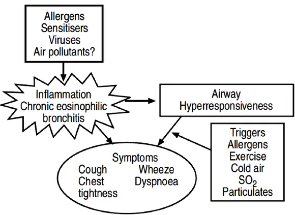
The pathophysiology of asthma is characterized by airway inflammation. Indeed, patients with acute asthma have extensively inflamed airways often reddened and swollen. The lumen is obstructed by mucus composed of proteins exuded from airway vessels and secreted from epithelial cells. The airway wall is infiltrated with inflammatory cells, mainly eosinophils and lymphocytes. Broncho Alveolar Lavage
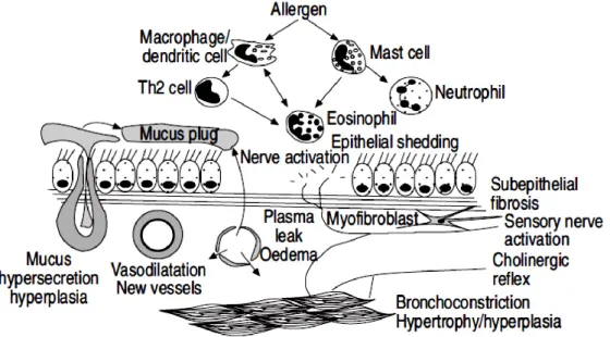
12 (BAL) from asthmatics has shown an increase in lymphocytes, mast cells, eosinophils and activated macrophages. Biopsies have shown augmented stimulated mast cells, macrophages, eosinophils and T-lymphocytes.3 These changes are found even in mild asthma. The inflammation in allergic asthma is determined by exposure to allergens through immunoglobulin E (IgE)-dependent mechanisms and is mainly characterized by eosinophils infiltration. Acute inflammatory response is converted into a chronic inflammation which structural consequences. The degree of inflammation is related to airway hyper-responsiveness (AHR), as measured by histamine or methacholine challenge. The severity of AHR in turn is related to asthma symptoms and to the necessity for treatment. Inflammation may increase AHR by stimulation of airway sensory nerve endings (Fig. 1.1).
Fig. 1.1 Inflammation in the airways of asthmatic patients leads to airway hyperresponsiveness and
13
1.2.1.1. Inflammatory cells involved in asthma
In the inflammatory pathophysiology of asthma are involved different cell types among which the most important are: mast cells, airway T cells, CD (+) (T helper) cells, basophils, macrophages, and eosinophils. In the next paragraphs the role of these cells in asthma pathophysiology will be briefly described.
Mast cells are derived from the myeloid stem cells and contain granules rich in histamine and heparin.4 Mast cells are important in initiating the acute broncho-constrictor responses to allergens, exercise, hyperventilation, etc. These cells release neurotrophins, pro-inflammatory cytokines, chemokines and growth factors.5Asthmatics are characterized by a marked increase in mast cells in airway smooth muscle (ASM).6 Treatment with prednisone results in a decrease in mast cells.7 Furthermore, mast cells stimulate human lung fibroblast proliferation.8 Mast cells secrete interleukin (IL)-4 and tumor necrosis factor (TNF)-α.9These cells are activated by an IgE-dependent mechanism. Humanized anti-IgE antibodies inhibit IgE-mediated effects. 10,11 Although this treatment shows marginal improvements in severe steroid-dependent asthma.12,13 Macrophages are activated by allergen via low affinity IgE receptors (FceRII).14 ,15 Alveolar macrophages have a suppressive effect on lymphocyte function which appears to be reduced after allergen exposure.16 In asthma the secretion of the anti-inflammatory protein IL-10 is reduced in alveolar macrophages.17 Macrophages also inhibit the secretion of IL-5 but this is defective in allergic asthmatics.18 These cells act as antigen-presenting cells to T-lymphocytes.19 No changes in the macrophage sub-populations in induced sputum of allergic asthmatic have been identified.20
Dendritic cells induce a T-lymphocyte mediated immune response21 acting as antigen-presenting effectors.22,23 Myeloid dendritic cells promote the differentiation of T-helper (Th) 2 cells24 and eosinophilia.25Immature dendritic cells require cytokines such as IL-12 and TNF-α to promote the normally preponderant Th1 response.26
Eosinophils play a cardinal role in asthma. Indeed, allergen inhalation results in a marked increase in eosinophils and there is a correlation between blood eosinophil or
14 bronchial lavage and AHR. Eosinophils release basic proteins and oxygen-derived free radicals.27,28 Activated eosinophils induce airway epithelial damage.29
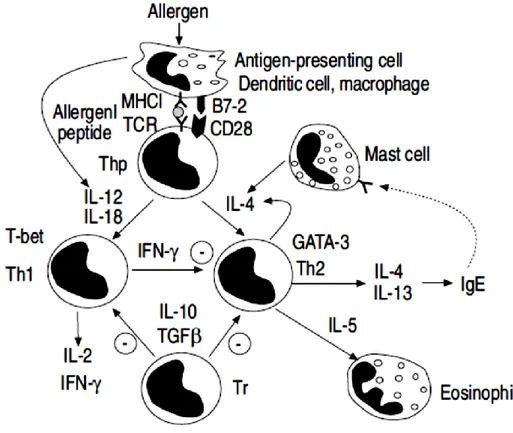
Neutrophils are prominent in severe asthma.30 , 31 , 32 , 33 High doses of corticosteroids inhibit neutrophils’ apoptosis.34,35 When neutrophils are recruited an increase of IL-8 in induced sputum occurs possibly due to the increased oxidative stress. Neutrophilia is also associated with a reduced responsiveness to corticosteroids and acute asthma. T-lymphocytes release cytokines promoting the recruitment of eosinophils and mast cells.36,37 The balance between Th1 cells and Th2 cells is determined by locally released cytokines. IL-12 promotes Th1 cells whereas IL-4 or IL-13 favour Th2 cells (Fig. 1.2). Steroids effect the balance between IL-12 and IL-13.38 , 39 Regulatory T (Tr) cells suppress the immune response through the secretion of IL-10 and transforming growth factor (TGF)-β. 40,41
Fig. 1.2 Asthmatic inflammation is characterised by a preponderance of T-helper (Th) 2 lymphocytes.
The transcription factors T-beta and GATA-3 may regulate the balance between Th1 and Th2 cells. Regulatory T-cells (Tr) have an inhibitory effect. Source: Pathophysiology of asthma P.J. Barnes.
15 B-lymphocytes secrete IgE.42 IL-4 is crucial in switching B-cells to IgE production, and CD40 on T-cells is an important signal through interaction with CD40-ligand on B-cells. Basophils have uncertain role in asthma.43An increase in basophils has been documented in the airways of asthmatics after allergen challenge. 44,45
Platelets fall in circulating after allergen challenge with increased release of the chemokine RANTES. 46 , 47 Chemokines associated with Th2-mediated inflammation activate and aggregate platelets.48
Epithelial cells, endothelial cells, fibroblasts and airway smooth muscle cells are also an important source of inflammatory mediators. 49 , 50 ,51 , 52 Epithelial cells are important target of inhaled glucocorticoids (Fig. 1.3).
Fig. 1.3 Airway epithelial cells and inflammatory mediators’release. O2: oxygen; NO2: nitrogen dioxide; TNF: tumour necrosis factor; IL: interleukin; GM-CSF: granulocyte-macrophage colony-stimulating factor; RANTES: regulated on activation T-cell expressed and secreted; MCP: monocyte chemotactic protein; TARC: thymus and activation regulated chemokine; PDGF: platelet-derived growth factor; EGF: endothelial growth factor; FGF: fibroblast growth factor; IGF: insulin-like growth factor. Source: Pathophysiology of asthma P.J. Barnes.
16
1.2.1.2. Inflammatory mediators
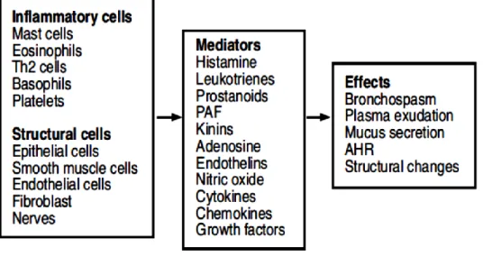
Different mediators are implicated in asthma showing a variety of effects (Fig. 1.4).53
Fig. 1.4 Cells and mediators involved in asthma. Source: Pathophysiology of asthma P.J. Barnes.
Histamine, prostaglandine, leukotrienes and kinins contract airway smooth muscle, increase microvascular leakage and airway mucus secretion, and attract other inflammatory cells.
The cysteinyl-leukotrienes, LTC4, LTD4 and LTE4, are potent constrictors of human airways.54 Potent LTD4 antagonists protect against exercise- and allergen-induced broncho-constriction. Chronic treatment with anti-leukotrienes improves lung function and asthma symptoms.55 Cys-LTs increase in eosinophils in induced sputum.56,57 Platelet-activating factor (PAF) is a potent inflammatory mediator.58 A genetic mutation of the PAF metabolising enzyme is associated with severe asthma.59 However PAF antagonists, such as modipafant, do not control asthma symptoms.60
Prostaglandins (PG) have potent effects on airway function.61 Nevertheless, the inhibition of their synthesis with COX inhibitors does not have any effect in most patients. Aspirin-sensitive asthmais associated with increased formation of cys-LTs.62,63 PGD2 stimulates the chemo-attractant receptor of Th2 cells (CRTH2), which is
17 expressed on Th2 cells, eosinophils and basophils. Deletion of the PGD2 receptors in mice significantly inhibits inflammatory responses to allergen and AHR.64
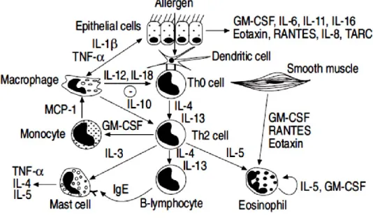
Cytokines play a critical role in orchestrating the inflammatory response (Fig. 1.5).65 IL-3 is important for the survival of mast cells. IL-4 is critical in switching B lymphocytes to produce IgE and for expression of VCAM-1 on endothelial cells.66 IL-5 is important in the differentiation, survival and priming of eosinophils.67The administration of an anti-IL-5 antibody (mepolizumab) is associated with a significant decrease in eosinophil.
IL-9 may play a critical role in sensitising responses to the cytokines IL-4 and IL-5.68 IL-1β, IL-6, TNF-α and GM-CSF are released from a variety of cells. TNF-α is increased in asthmatic airways. 69 Inhalation of TNF-α increased airway responsiveness.70 TNF-α and IL-1β activate the pro-inflammatory transcription factors,
nuclear factor-kB (NF-kB) and activator protein-1 (AP-1). Interferon (IFN)-ɤ, IL-10, IL-12 and IL-18, play a regulatory role and inhibit the allergic inflammatory process.
Fig. 1.5 The cytokine network in asthma. TNF: tumour necrosis factor; IL: interleukin; GM-CSF:
granulocyte-macrophage colony-stimulating factor; RANTES: regulated on activation T-cell expressed and secreted; MCP: monocyte chemotactic protein; TARC: thymus and activation regulated chemokine; PDGF: platelet-derived growth factor; EGF: endothelial growth factor; FGF: fibroblast growth factor; IGF: insulin-like growth factor; Th: T-helper. Source: Pathophysiology of asthma P.J. Barnes.
18 Chemokines are a large superfamily of mostly small, secreted chemotactic cytokines that function in leukocyte trafficking, recruitment and activation. The actions of chemokines are important for a wide range of processes such as allergic responses, infectious and autoimmune diseases. Over 50 different chemokines are now recognised.71 There is increased expression of eotaxin, eotaxin-2, MCP- 3, and MCP-4 in the airways of asthmatics. 72,73,74 These molecules activate a common receptor on eosinophils termed CCR3. 75 , 76 A neutralising antibody against eotaxin reduces eosinophil recruitment in to the lung after allergen.77 RANTES also activates CCR3.78 MCP-1 activates CCR2 on monocytes and T-lymphocytes. MCP-1 levels are increased in BAL fluid of asthmatics. Blocking MCP-1 results in a marked reduction of AHR.79 CCR4 are selectively expressed on Th2 cells and are activated by the chemokines monocyte-derived chemokine (MDC) and thymus activation regulated chemokine (TARC).80 Epithelial cells of patients with asthma express TARC.81 Increased concentrations of TARC are found in BAL fluid of asthmatic.82
Endothelins are potent peptide mediators that are vaso-constrictors and broncho-constrictors.83 Endothelin-1 levels are increased in the sputum of asthmatics depending on allergen exposure and steroid treatment.84 , 85 Endothelins induce ASM cell proliferation promoting a pro-fibrotic phenotype.
NO is produced by several cells in the airway by NO synthases.86,87,88 The level of NO in the exhaled air of asthmatics is increased especially during an acute exacerbations.89
90,91 Measurement of exhaled NO in asthma is increasingly used as a noninvasive way
of monitoring the inflammatory process.92,93 Under oxidative stress the formation of the potent radical peroxynitrite may result in nitrosylation of proteins in the airways.94
19
1.2.1.3. Effects of inflammation
The acute and chronic allergic inflammatory responses have several effects (Fig. 1.6 A
and B). The structural changes that occur in the airways are named "remodelling".95
Fig. 1.6 A Acute and chronic inflammatory effects on the airway in asthma. Barnes. Source:
Pathophysiology of asthma P.J. Barnes.
Fig. 1.6 B Acute and chronic inflammatory effects on the airway in asthma. A: location of lungs in the
body and airways in the lungs. B: a normal, non-asthmatic airway. C: an airway during asthmatic symptoms. The airway is narrowed, limiting air flow. Tightened muscles constrict air flow, as do inflamed and thickened airways. Excess mucus clogs the airway. Image: http://www.nhlbi.nih.gov/health/dci/Diseases/Asthma/Asthma_WhatIs.html
20 Airway epithelial shedding is a characteristic feature of asthma. Ozone-exposure, viruses, chemicals and allergens can lead to its development as a consequence of inflammatory mediator’s production. Epithelial damage results in loss of its barrier function to allow penetration of allergens, loss of enzymes which normally degrade inflammatory mediators, loss of a relaxant factor, and exposure of sensory nerves. Several inhaled allergens activate protease-activated receptor (PAR)-2, which shows increased expression in airway epithelial cells of asthmatics.96 Epithelial cells may also release growth factors that stimulate structural changes in the airways.97
A thickened bronchial epithelial basement membrane has long been regarded as a histo-pathologic characteristic of asthma. Sub-epithelial fibrosis has been observed even in mild asthmatics.98 The basement membrane appears thickened due to the deposition of Type III and V collagen.99 , 100 TGF-β, platelet-derived growth factor (PDGF), and
endothelin-1 can be produced by epithelial cells or macrophages in the inflamed airway.101 There is also evidence for fibrosis in ASM.102
ASM contraction has a key role in the symptomatology of asthma. Many inflammatory mediators have broncho-constrictor effects.103. Reduced responsiveness to β-adrenergic agonists has been reported in post mortem bronchi from asthmatics.104 Chronic exposure to inflammatory cytokines, such as IL-1β, down-regulates the response of ASM to β2-adrenergic agonists.105 , 106 , 107 In asthmatics has been documented a characteristic hypertrophy and hyperplasia of ASM.108,109
Allergic inflammation has several effects on blood vessels in the respiratory tract. Recent studies have revealed an increased airway mucosal blood flow in asthma.110 An increase in the vascular volume contributes to airway narrowing and exercise-induced asthma.111 The increase in blood vessels in asthmatics may also be due to the release of VEGF and TNF-α.112 , 113 Microvascular leakage is an essential component of the inflammatory response in asthma.114,115
In asthmatics has been reported hyperplasia of sub-mucosal glands.116,117 Th2 cytokines IL-4, IL-13 and IL-9 induce mucus hypersecretion.118 The epithelial growth factor (EGF) stimulates the expression of the mucin gene MUC5AC. 119,120 This is associated with the
21 expression of a specific calcium-activated chloride channel in goblet cells designated gob-5.121
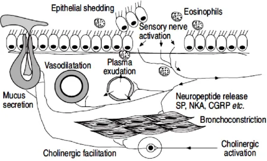
Inflammatory products sensitize sensory nerve to become hyperalgesic. Neurotrophins, such as nerve growth factor (NGF), may be released from inflammatory and structural cells in asthmatic airways. 122,123,124 Neurotrophins cause proliferation and sensitisation of airway sensory nerves.125 Bronchodilator nerves have been shown to be defective in asthma.126 Lack of vasoactive intestinal peptide (VIP)-immuno-reactive nerves has been reported in severe asthma.127 Airway nerves release also neurotransmitters which have inflammatory effects (Fig. 1.7).128An increase in SP-immuno-reactive nerves has been
described in severe asthma.129 A reduction in the activity of enzymes which degrade neuropeptides 130 and an increased gene expression of the receptors which mediate the inflammatory effects and bronchoconstrictor effects of SP have been descibed.131
Fig. 1.7 Possible neurogenic inflammation in asthmatic airways. Substance P (SP) causes vasodilatation,
plasma exudation and mucus secretion, whereas neurokinin A (NKA) causes bronchoconstriction and enhanced cholinergic reflexes and calcitonin generelated peptide (CGRP) vasodilatation. Source: Pathophysiology of asthma P.J. Barnes.
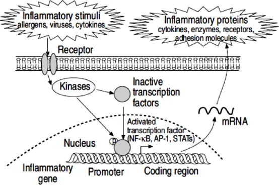
22 A number of transcription factors are involved in the regulation of the expression of inflammatory proteins in asthma (Fig. 1.8).132
NF-Kb is triggered by multiple stimuli including protein kinase C activators, oxidants and proinflammatory cytokines.133 Activation of NF-kB has been shown increased in asthmatic airways.134 NF-kB regulates the expression of several pro-inflammatory cytokines (IL-1β, TNF-α, GM-CSF), chemokines (RANTES, MIP-1a, eotaxin), adhesion molecules (ICAM-1, VCAM-1) and inflammatory enzymes (cyclooxygenase-2 and iNOS).
The c-Fos component of AP-1 is also activated in asthmatic airways.135
GATA-3 determines the differentiation of Th2 cells and is increased expression in asthmatics. 136,137 The differentiation of Th1 cells is regulated by the transcription factor T-bet.138 In a murine model the deletion of the T-bet gene is associated with asthma-like
phenotypes.139
Fig. 1.8 Transcription factors activated by inflammatory stimuli and responsible for increase the
expression of multiple inflammatory genes. Nuclear factor kappa-B (NF-kB), activator protein-1 (AP-1),
signal transduction-activated transcription factors (STATs), messenger ribonucleic acid (mRNA). Source: Pathophysioloof gy of asthma P.J. Barnes.
23
1.2.1.4. Anti-inflammatory mechanisms
A numbr of anti-inflammatory mechanisms have been shown defective in asthma.140 Cortisol regulates the allergic inflammatory response. Inhibition of endogenous cortisol secretion by metyrapone results in an increase in the late response to allergen in the skin.141 Cortisol is converted to the inactive cortisone by the enzyme 11-β-hydroxysteroid dehydrogenase.142 This enzyme seems to function abnormally in asthma.143
IL-1 receptor antagonist (IL-1ra) inhibits the binding of IL-1 to its receptors and therefore has a potential anti-inflammatory potential. It is reported to be effective in an animal model of asthma.144
IL-12 and IFN-ɤ enhance Th1 cells and inhibit Th2 cells. IL-12 infusions in patients with asthma inhibit peripheral blood eosinophilia.145 The IL-12 expression seems impaired in asthma.
IL-10 inhibits the expression of multiple inflammatory mediators. IL-10 secretion and gene transcription are defective in macrophages and monocytes from asthmatics (Fig.
1.9).146 , 147 , 148 PGE2 has inhibitory effects on macrophages, epithelial cells and eosinophils. 15-hydroxyeicosatetraenoic (15-HETE) and lipoxins inhibit cysteinyl-leukotriene effects on the airways.149 Lipoxins have also strong anti-inflammatory effects.150
The peptide adrenomedullin, which is expressed in high concentrations in the lung, has bronchodilator activity151 and inhibit the secretion of cytokines from macrophages.152 Plasma concentrations are no different in patients with asthma.153
24
Fig. 1.9 Transcription factors play a key role in amplifying and perpetuating the inflammatory response
in asthma. IL-10 secretion is deficient in macrophages from patients with asthma, resulting in increased
release of inflammatory mediators. NF-kB: nuclear factor kappa-B; LPS: lipopolysaccharide; inducible nitric oxide synthase; COX: cyclooxygenase; TNF: tumour necrosis factor; GM-CSF: granulocyte-macrophage colony-stimulating factor; RANTES: regulated on activation T-cell expressed and secreted; MIP: macrophage inflammatory protein. Source: Pathophysiology of asthma P.J. Barnes.
25
1.3. Epidemiology of asthma
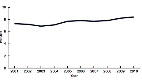
In 2011 ~235 million people worldwide were affected by asthma, and approximately 250,000 people die per year from the disease. 154 To date, most of the epidemiological evidence on the burden of asthma comes from developed populations. In the next paragraphs epidemiological data from a study on asthma prevalence in the United States from 2001 to 2010 will be shown. This study has shown an increase from 7.3% in 2001 to 8.4% in 2010 (Fig. 1.10).155 In the United States in 2010, an estimated 25.7 million people had asthma: 18.7 million adults aged 18 and over, and 7.0 million children aged 0–17 years.155
Fig. 1.10 Asthma prevalence in the United States, 2001-2010. Source: CDC/NCHS, National Health
Interview Survey.
The study showed that children aged 0–17 years had higher asthma prevalence (9.5%) than adults aged 18 and over (7.7%) for the period 2008–2010. Females had higher asthma prevalence than males (9.2% compared with 7.0%).155 Persons of multiple race had the highest asthma prevalence (14.1%), while Asian persons had the lowest rates (5.2%). Persons of black (11.2%) and American Indian or Alaska Native (9.4%) races had higher asthma prevalence compared with white persons (7.7%). Among Hispanic groups, asthma prevalence was higher among persons of Puerto Rican (16.1%) than Mexican (5.4%) descent (Fig. 1.11).155
26
Fig. 1.11 Asthma prevalence, by selected demographic characteristics: Sources: CDC/NCHS, Health
Data Interactive and National Health Interview Survey.155
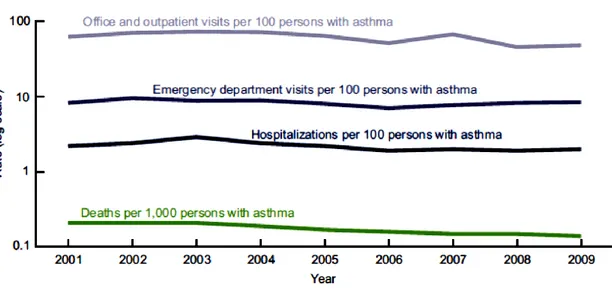
In the United States asthma prevalence from 2001 to 2010 was higher for groups with lower income-to-poverty level ratios. While 11.2% of those with incomes less than 100% of the poverty level had asthma, asthma prevalence was 8.7% for persons with incomes 100% to less than 200% of the poverty level, and 7.3% for persons with incomes at least 200% of the poverty level.155 Asthma death rates per 1,000 persons with asthma declined from 2001 to 2009 (Fig. 1.12).
27
Fig. 1.12 Asthma health care encounters per 100 persons with asthma, and asthma deaths per 1,000
persons with asthma:United States, 2001–2009. Access at: http://www.cdc.gov/nchs/data/databriefs/db94
The rates of health care encounters per 100 persons with asthma across all health care settings (Fig. 1.13) were similar for males and females, and for black and white persons, but the rate for children was higher than that for adults.155
Fig. 1.13. Asthma health care encounters per 100 persons with asthma: United States, 2001–2009. NOTE:
Access data table for at: http://www.cdc.gov/nchs/data/databriefs/db94_tables.pdf#4
Children aged 0–17 years with asthma had a higher asthma visit rate for primary care and a higher ED visit rate than adults aged 18 and over.155
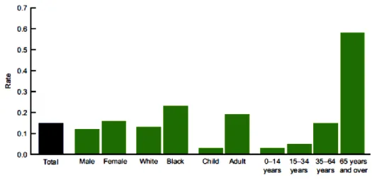
28 In the United States the asthma death rate per 1,000 persons with asthma was 0.15 for the period 2007–2009. The highest rate was for adults aged 65 and over (0.58 per 1,000 persons with asthma) (Fig. 1.14).155
Fig. 1.14 Asthma deaths per 1,000 persons with asthma, by selected demographic characteristics: United
States, average annual 2007–2009.155
Asthma prevalence also differs between populations of the same ethnicity. U.S.-born Mexican populations, for example, have higher asthma rates than non-U.S. born Mexican populations that are living in the U.S.156
Asthma affects approximately 5% of the United Kingdom’s population.157 In England, an estimated 261,400 people were newly diagnosed with asthma in 2005; 5.7 million people had an asthma diagnosis and were prescribed 32.6 million asthma-related prescriptions.158
In Italy from 1990 to 2010 the national median prevalenceof asthma and allergic rhinitis increased from 4.6% to 6.6% and from 19.4% to 25.8%, respectively.159 Antonicelli et al in 2014 illustrated the overall costs of asthma in Italy with highest values for severe persistent asthma, Fig. 1.15.
29
Fig. 1.15 Overall costs of asthma Italy. Adapted from Antonicelli L. et al. Eur Respir J 2004. GINA 2002
classification.
In spite of the epidemiological suggestion for an increase in the prevalence of asthma in several countries, the bases of the increase are still debated. While the exact cause of asthma is not known, it is thought that a variety of factors interacting with one another, early in life, result in the development of asthma. It has been hypothesized that asthmatic subjects may have a genetic predisposition to develop the disease. Elements of the pathogenesis of asthma, including the immune response and the regulation of pro-inflammatory cytokines, are also under genetic control and are activated under environmental factors in genetically predisposed subjects.
The fast increase detected in asthma prevalence cannot be explained on the basis of genetic predisposition only. Therefore, attention has been centred on a number of environmental factors. Indoor and outdoor allergens, such as domestic mites, animal allergens, pollens, fungi and molds, have been suggested to have a role in the manifestation and persistence of asthma. Environmental pollutants, mainly industrial smog and those derived from ozone and nitrogen oxides, may intensify clinical manifestations of asthmatic subjects.
As seen previously developed countries with a higher socio-economic level have the highest prevalence of asthma. It has been proposed that better hygienic conditions
30 derived from this affluent status may be in part related to the increase in allergic diseases. One of the underlying mechanisms hypothesized for the rise of atopy and asthma in industrialised countries, is the reduction in the incidence of early childhood infections and the consequent expansion of T helper type 2 lymphocytes, which would lead to an imbalance in the regulatory mechanisms of the inflammatory response later on in life.
Children with siblings are more likely to acquire infections during their childhood and consequently they would be protected against allergic diseases later on in life. This has contributed to the hypothesis that family size and, specifically number of older siblings may be related to asthma. Nevertheless, changes in family size over the past 30 years do not explain the growth in asthma observed in the same period in the United Kingdom or New Zealand, two of the countries with highest prevalence.
31
1.4. Diagnosis
The diagnosis of asthma typically is based on family history, the pattern of symptoms and response to therapy (Fig. 1.17). 160 A diagnosis of asthma should be supposed if there is a history of recurrent wheezing, coughing or difficulty breathing and these symptoms worsen due to exercise, viral infections, allergens or air pollution.161 Spirometry is used to confirm the diagnosis. 162 Spirometry measures the lung function and specifically the amount (volume) and/or speed (flow) of air that can be inhaled and exhaled. In children under the age of six the diagnosis is more difficult as they are too young for spirometry. If the Forced Expiratory Volume in 1 second (FEV1) measured by
this technique improves more than 12% following administration of a bronchodilator such as salbutamol, this is supportive of the diagnosis (Fig 1.16).163 As caffeine is a bronchodilator its use before a lung function test may interfere with the results.164 Diffusing capacity of the lung (DL) measures the transfer of gas from air in the lung, to
the red blood cells in lung blood vessels. Single-breath diffusing capacity helps to differentiate asthma from Chronic Obstructive Pulmonary Diseases (COPD).
32 Bronchial hyper-responsiveness (BHR) can be defined as the tendency for the airways of asthmatic subjects to broncho-constrict when exposed to various chemical and physical stimuli. Exposure to stimuli, such as allergens, which are specific for an individual, produce a different effect, in that the non-specific stimuli generally cause a short-lived period of broncho-constriction without inducing significant airway inflammation whilst antigenic stimuli cause more prolonged bronchoconstriction with an immediate response lasting for 1-2 hours that may follow a late response at 4-8 hours, which is characterized by inflammatory cell recruitment to the airways. Many broncho-constrictor stimuli can be used to measure the degree of BHR, including inhaled histamine or methacholine, inhaled hypertonic saline or distilled water, exercise or cold air. During obstructive processes, the reduction of FEV1 is bigger than the reduction of
FVC and the FEV1/FVC ratio is reduced; contrariwise, in restrictive lung disease the
reduction in FVC is greater than in FEV1 and the ratio is augmented or normal. The
assessmenot f BHR has been done mainly through challenge with histamine and more recently methacholine. The methacholine challenge consist of the inhalation of increasing concentrations of a methacholine that causes airway narrowing in those predisposed. If negative a person does not have asthma; if positive, however, it is not specific for the disease.
Other supportive indications for asthma includes: a ≥20% difference in peak expiratory flow (PEF) rate on at least three days in a week for at least two weeks, a ≥20% improvement of PEF following treatment with either salbutamol, inhaled corticosteroids or prednisone, or a ≥20% decrease in PEF following exposure to a trigger. Testing PEF may be useful for daily self-monitoring of asthma and in guiding treatment in those with acute exacerbations.
1.4.1. Classification
Although asthma is a chronic obstructive condition, it is not considered as a part of chronic obstructive pulmonary diseases.165 Unlike to these diseases, the airway obstruction in asthma is usually reversible.166Two main factors, severity and control, determine asthma classification, which in turn affect the type of therapy initiated (depending on the severity) and how therapy should be adjusted over time (based on the
33 control level). Severity and control should be assessed separately. Asthma is clinically classified according to the frequency and severity of symptoms, FEV1, and PEF
rate.167,168 Asthma may also be classified as atopic (extrinsic) or non-atopic (intrinsic), when symptoms are precipitated by allergens.169 Based on severity level, asthma can be classified as intermittent or persistent. Patients with intermittent asthma usually have minimal asthma symptoms and no interference with normal activity, whereas patients with persistent asthma have more severe symptoms and limitations in normal activity due to reduced lung function
In acute asthma exacerbation is commonly referred to asthma attacks with worsening of the classic symptoms are shortness of breath, wheezing, and chest tightness.170 In severe cases, air motion may be significantly impaired.171 During an attack can occur the use of accessory muscles of respiration, a paradoxical pulse, and over-inflation of the chest.
172 A blue color of the skin and nails may occur from lack of oxygen.173 In a mild
exacerbation the peak expiratory flow rate (PEFR) is ≥200 L/min or ≥50% of the predicted best.174 Moderate is defined as between 80 and 200 L/min or 25% and 50% of the predicted best while severe is defined as ≤ 80 L/min or ≤25% of the predicted best. Acute severe asthma is an acute exacerbation of asthma that does not respond to standard treatments. Risk factors for exacerbations include:
• Ever intubated for asthma, • Uncontrolled asthma symptoms;
• Having ≥1 exacerbation in last 12 months;
• Low FEV1 (measure lung function at start of treatment, at 3-6 months to assess
personal best, and periodically thereafter);
• Incorrect inhaler technique and/or poor adherence; • Smoking;
34 Brittle asthma is distinguishable by recurrent, severe attacks. Type 1 brittle asthma is a disease with wide peak flow variability, despite intense medication. Type 2 brittle asthma is background well-controlled asthma with sudden severe exacerbations.
Exercise can trigger bronchoconstriction.175 It occurs in most people with asthma and up to 20% of people without asthma.It is more common when it is dry and cold. Inhaled β2-agonists do not improve athletic performance among those without asthma.176 However oral doses may improve endurance and strength.177
Asthma when is a result of workplace exposures is commonly reported as occupational disease. It is estimated that 5–25% of asthma cases in adults are work–related. Isocyanates, grain and wood dust, colophony, soldering flux, latex, animals, and aldehydes have been implicated as most common agents.178
Many other conditions can cause symptoms analogous to those of asthma. In children allergic rhinitis and sinusitis should be considered as well as foreign body aspiration, tracheal stenosis, vascular rings, enlarged lymph nodes, etc. In adults, COPD, congestive heart failure, airway masses, as well as drug-induced coughing due to ACE inhibitors should be considered. COPD can coexist with asthma and can occur as a complication of chronic asthma. When older than 65 years most people with obstructive airway disease develop also asthma. A deep level of investigation is not performed due to COPD and asthma sharing similar principles. 179,180,181
35
1.5. Prevention
The evidence for the effectiveness prevention of asthma is not strong.182 Limiting smoke exposure both in utero and after delivery, breastfeeding, and increased exposure to daycare are not well supported. Early pet exposure may be useful. Dietary restrictions during pregnancy or breast feeding have not been found to be effective. Removing compounds known to sensitive people from the work place may be effective. Annual influenza vaccinations may affect the risk of exacerbations.183 Immunization, however, is recommended by the World Health Organization.184 Smoking prohibition is effective in decreasing exacerbations of asthma.185
The Global Initiative for asthma recommends:1
- Provide skills and support for guided asthma self-management:
This comprises self-monitoring of symptoms and/or PEF, a written asthma action plan and regular medical review
- Prescribe medications or regimen that minimize exacerbations: ICS-containing controller medications reduce risk of exacerbations
For patients with ≥1 exacerbations in previous year, consider low-dose ICS/formoterol maintenance and reliever regimen
- Encourage avoidance of tobacco smoke:
Provide smoking cessation advice and resources at every visit - For patients with severe asthma
Refer to a specialist center, if available, for consideration of add-on medications and/or sputum-guided treatment
- For patients with confirmed food allergy: Appropriate food avoidance
36
1.6. Management and therapy
Despite the fact there is no cure for asthma, symptoms can usually be improved (Fig.
1.17 and 1.18). 186
Fig. 1.17 GINA 2014, Box 1-1
Ineffective management of asthma significantly influences morbidity, mortality and health care utilization, resulting in increased health care costs. A precise, detailed, and customized plan for monitoring and managing of the symptoms is firmly necessary. This should comprise the reduction of exposure to allergens, testing the severity of symptoms, and the usage of medications. The treatment should be adjusted according to
37 changes in symptoms. The effective management for asthma should include identifying and eliminating triggers, such as cigarette smoke, pets, or aspirin. 187 Exercise is beneficial in people with stable asthma.188 Medications are selected based on the severity of illness and the frequency of symptoms (Fig. 1.17).189
Fig.1.18 Stepwise approach to control asthma symptoms and reduce risk. GINA 2014, Box 3-5
Short-acting beta-2-adrenoceptor agonists (SABA), such as salbutamol (albuterol USAN) represent the the most effective agents for quick symptom relief as they rapidly reverse airflow obstruction for all patients with asthma. Bronchodilation occurs due to blocking β2-adrenergic receptors, which antagonize bronchoconstriction. The most commonly used SABAs are albuterol, levalbuterol, and pirbuterol. It is recommended using SABAs only as needed for symptom relief, but not for regular use. Tachycardia,
38 tremor, and anxiety are the most common dose-dependent side effects. They are recommended before exercise in those with exercise induced symptoms.190 No-selective adrenergic agonists191 are not recommended due to their excessive cardiac stimulation. Anticholinergic medications, such as ipratropium bromide, provide additional benefit when used in combination with SABA in those with moderate or severe symptoms.192 Ipratropium bromide is used to overcome acute bronchospasms by blocking muscarinic cholinergic receptors. Common side effects associated with ipratropium bromide use are dry mouth, increased wheezing, and blurred vision.
Corticosteroids are generally considered the most effective treatment available for long-term control. OCSs are used for exacerbation management. These medications reverse inflammation and decrease relapse occurrences. Systematic corticosteroids have a potent anti-inflammatory effect, but should be used with caution due to complex adverse effects such as abnormalities in glucose metabolism, fluid retention, weight gain and hypertension. Methylprednisolone, prednisolone and prednisone are oral corticosteroids used for asthma exacerbations management and severe persistent asthma Inhaled corticosterois (ICS) such as beclomethasone are generally used except in severe persistent disease, in which oral corticosteroids are required.
Long-acting beta-adrenoceptor agonists (LABA) such as salmeterol and formoterol can improve asthma control, once given in combination with inhaled corticosteroids.193 LABAs have a bronchodilator effect, but do not affect airway inflammation. LABAs activate adenylate cyclase and produce functional antagonism of bronchoconstriction providing a bronchodilator effect When used without steroids they increase the risk of severe side-effects. 194 Available combinations of ICS/LABA inhalers are fluticasone/salmeterol, budesonide/formoterol and mometasone/formoterol. Potential life-threatening exacerbations associated with LABA use include tachycardia, skeletal muscle tremor and hypokalemia.
Leukotriene antagonists (such as montelukast and zafirlukast) are used in addition to inhaled corticosteroids, usually also in conjunction with LABA.195 In children they appear to be of little advantage when added to ICS.196 Leukotriene modifiers include
39 two groups of agents: leukotriene receptor antagonists (LTRAs) (i.e., montelukast, zafirlukast) and leukotriene synthesis inhibitors (LTSIs) (i.e., zileuton).
Mast cell stabilizers (such as cromolyn sodium) are another non-preferred alternative to corticosteroids. The mechanism of anti-inflammatory action is determined by blocking early and late reactions to allergens and by stabilizing mast cells membranes. The anti-inflammatory effect and excellent safety profile of these agents provide symptom control, along with a decrease in the number of exacerbations compared to placebo. Potential side effects are cough and throat irritation.
Emergency management of asthma includes oxygen to alleviate hypoxia.197 Oral corticosteroids are recommended with five days of prednisone.198 Magnesium sulfate intravenous provide a bronchodilating effect in severe acute asthma attacks.199 Heliox, a mixture of helium and oxygen, may also be considered in severe unresponsive cases. The use of Methylxanthines (such as theophylline) in acute exacerbations is controversial. It has bronchodilator and mild anti-inflammatory effects. Theophylline provides muscle relaxation by inhibition of phosphodiesterase. It is not preferred therapy since it can lead to frequent adverse events (e.g., severe headache, tachycardia, nausea, vomiting) and it is not as effective in asthma as low dose ICSs. Theophylline is used when asthma is not well-controlled with ICS, LABAs or LTRAs
Ketamine is theoretically useful when intubation and mechanical ventilation is needed.200 In severe not controlled and persistent asthma bronchial thermo-plasty represent an option.201 Sublingual immuno-therapy in allergic rhinitis and asthma improve outcomes.
Many asthmatics use alternative treatments and approaches. 202,203,204 Complementary and alternative medicine (CAM) asthma treatment ranges from breathing exercises to herbal remedies. Unfortunately, a lack of well-designed clinical trials makes it difficult to assess the safety and efficacy of these treatments. There is insufficient evidence to support the use of acupuncture, osteopathic, chiropractic, physiotherapeutic and respiratory therapeutic maneuvers in asthma. 205,206,207 Air-ionisers show no evidence that they improve asthma symptoms or benefit lung function.208
40
1.7. Prognosis
The prognosis for asthma is usually good, especially for children with mild disease.209 Mortality has decreased over the last few decades due to better recognition and improvement in therapeutic intervention.210 Globally it causes moderate or severe disability in 19.4 million people.211 Of asthma diagnosed during childhood, half of cases will no longer carry the diagnosis after a decade.212 Airway remodeling is observed, but it is unknown whether these represent harmful or beneficial changes.213 Early treatment with corticosteroids seems to prevent the decline in lung function.214
41
Chapter 2
2. Asthma and oxidative stress
2.1. Introduction
Oxidative stress is the condition characterize by an overproduction of Reactive Oxygen Species (ROS) and/or antioxidant decreases. At physiological levels, ROS function as “redox messengers” in intracellular signaling. Excess ROS induce oxidative modification of cellular macromolecules, inhibit protein function and promote cell death. The alteration of intracellular redox homeostasis, and irreversible oxidative modifications of lipid, protein or DNA accompanies a wide spectrum of clinical disorders including asthma. As described in Chapter 1 asthma is a chronic inflammatory disorder of the airways involving interaction of cells and mediators. The increase of inflammatory processes in asthma ultimately result in high levels of reactive oxygen and nitrogen species (ROS, RNS).215,216,217,218 In asthma the increased oxidative species and the deficiency of anti-oxidant capacity lead to modifications of proteins and alterations in their function that are biologically relevant to the initiation and maintenance of inflammation. This chapter will first explain the process of oxidative stress, then focus on the redox abnormalities in asthma and finally elucidate the consequences on molecular processes.
2.2. Oxidative stress and redox systems in the lungs
The lungs show a vast mucosal epithelial surface directly exposed to inhaled oxygen and airborne reactive pollutants and microorganisms. This makes the lungs particularly susceptible to oxidant-mediated damage. Also endogenously are generated high levels of RNS and ROS to maintain a sterile internal environmental. Altogether, endogenous RNS and ROS produce an oxidizing lung environment (Fig. 2.1). However, because of the abundance of antioxidant systems available to the lung the redox state in the healthy lung is reducing.219
42
Fig. 2.1 Sources of exogenous inhalational and endogenous reactive oxygen species (ROS) and reactive
nitrogen species (RNS) in the lung. Environmental sources are ozone, air pollutants, particulates
containing metals, and cigarette smoke. Endogenous ROS are produced as byproducts of mitochondrial respiration. Inflammatory cells can produce high levels of ROS and RNS in response to allergens and microbial infections. Source: Redox Control of Asthma: Molecular Mechanisms and Therapeutic Opportunities Suzy A.A. Comhair and Serpil C. Erzurum. Antioxidants & redox signaling volume 12, number 1, 2010
2.2.1. Endogenous reactive oxygen species.
The tetravalent reduction of oxygen during mitochondrial electron transport can result in formation of the radical superoxide (O2·).220 Another source for intracellular
generation of O2 is the NADPH oxidase found in neutrophils, monocytes, and
macrophages.221 , 222 , 223 , 224 O2· can be also produced by molybdenum hydroxylase
43 and react with proteins that contain transitionmetal prosthetic groups, such as heme or iron/sulfur groups. 226,227,228 The main reaction of superoxide is to react with itself to produce hydrogen peroxide and oxygen (Reaction 1).229 Superoxide dismutation can be spontaneous or can be catalyzed by the enzymes (SOD).
Once formed, the oxidizing potential of H2O2 may be amplified by eosinophil and
neutrophil derived peroxidases eosinophil peroxidase (EPO) and myeloperoxidase (MPO), respectively (Reaction 2). 230,231,232,233 MPO is the most abundant protein stored in neutrophil granules, and secreted during cell activation.234
Kinnula et al. has shown that alveolar macrophages and Type II cells produce high levels of H2O2.235
Onother extremely reactive oxidizing is the hydroxyl radical (.OH).236 The .OH can be formed by Haber–Weiss Reaction followed by the Fenton Reaction.237
An alternative pathway for OH formation in vivo may involve MPO and EPO. Under physiological concentrations of halides, MPO produces hypochlorous acid (HOCl) and EPO produces hypobromous acid (HOBr). Hypohalous acids can generate .OH after reacting with O2. (Reaction 3). .OH can react with different molecules such as protein,
44 In the lung is widely produced nitric oxide (.NO) by nitric oxide synthases (NOS).242 All NOS convert L-arginine to NO and L-citrulline. There are three forms of NOS, the inducible NOS (iNOS or NOS2), neuronal NOS (nNOS or NOS1), and endothelial NOS
(eNOS or NOS3).243 nNOS and eNOS are constitutively expressed in neuronal and
endothelial cells.244 In the airway NOS3 is primarily localized in pulmonary endothelial
cells, and NOS1 in non-adrenergic, non-cholinergic inhibitory neurons.245246 NOS2 is
continuously expressed in normal human airway epithelium.247 , 248 . 249 . 250NO is also produced by the upper respiratory tract epithelium within the nasopharynx and paranasal sinuses.251 Epithelial NOS2 activity is a major determinant of NO present in
exhaled breath.252 The iNOS is regulated at the level of transcription and mRNA stability, is calcium independent, and produces nanomolar levels of NO. Regulation of iNOS expression is increased by cytokines and proinflammatory factors, interferon gamma, TNF-α, and IL1-β.253 iNOS is also regulated by availability of arginine and cofactor tetrahydrobiopterin. Conditions that decrease arginine will lead to greater superoxide formation.254 Auto-oxidation of .NO with O2 results in the formation of
nitrite (NO2-). NO2- is also a substrate for hemeperoxidases such as MPO and EPO.
Peroxidase-catalyzed oxidation of NO2- results in the formation of nitrogen dioxide
radical (NO2·).255 NO reacts with superoxide to form peroxynitrite (ONOO.). ONOO.
can nitrate tyrosine residues and alter levels or function of enzymes, structural and signaling proteins.256
2.2.1.1. Environmental exposures.
Because the lung interfaces with the external environment, it is frequently exposed to airborne oxidants. Ozone, particulate matter and cigarette smoke represent the most common air pollution problems.
Ozone is formed from volatile hydrocarbons, halogenated organics, and oxides of nitrogen in the presence of sunlight.257 Ambient ozone levels usually vary between 20 and 40 parts per billion (ppb).258 High concentrations of ozone can be harmful to the lung.259,260,261,262,263,264 Ozone reacts with unsaturated fatty acids and cell membranes to produce lipid ozonation products.265,266
45 Particulate matter pollution is one of the most serious air pollution problems in urban environments. One of the most dangerous forms of particulate matter pollution is diesel exhaust particle. Diesel exhaust particles are a polyaromatic hydrocarbon, a hydrophobic molecule that can diffuse easily through cell membranes. Diesel exhaust particles may therefore modify cell growth and differentiation.
Environmental tobacco smoke is a complex mixture of gases and particles. Cigarette smoke contains >4,000 chemicals including 50 that are known to cause cancer. Some of them are carbon monoxide, cyanide, arsenic, mercury, and NO. Furthermore, cigarette smoke generates or contains*1014 oxidative molecules per puff such as hydrogen peroxide and superoxide. Tobacco smoke leads to activation of phagocytes augmenting release of free radicals.267
2.2.2. Biological oxidative processes in the lungs and anti-oxidant
The formation of ROS and RNS is an essential for neutrophils, monocytes, macrophages, and eosinophils in order to kill bacteria. These phagocytic cells use NADPH oxidase enzymatic systems to generate O2·-.268 They can also form HOCl through myeloperoxidase-catalyzed oxidation of the Cl- ion by H2O2. .NO is also
involved in mononuclear cell-mediated killing of Mycobacterium tuberculosis and other pathogens in rodents and is toxic to tumor cell lines in vitro.269 Cytochrome P450 also exploits the reactivity of the iron–oxygen complex to catalyze oxidation of a number of endogenous compounds and xenobiotics.270
The balance between physiologic functions and damage is determined by the relative rates of formation and the removal of free radicals. The lungs have developed several endogenous antioxidant systems. These systems may be divided into enzymatic and nonenzymatic groups.
46
2.2.2.1. Non-Enzymatic lung antioxidants
The most well-researched nonenzymatic antioxidants include lipid-soluble vitamin E (tocopherol), vitamin A, and carotenoids (including beta-carotene), and water-soluble vitamin C and glutathione (GSH).
Vitamin E is an important hydrophilic antioxidant. It protects the cell membrane from oxidation by reacting with lipid radicals, such as lipid peroxyl radicals (LOO·) that are produced during lipid peroxidation reactions.271
Vitamin C is a hydrophilic vitamin that can directly scavenge O2 ·- and ·OH by forming the semidehydroascorbate free radical that subsequently is reduced by GSH.272 Vitamin C, however, is usually not considered a major antioxidant because it also has pro-oxidant properties.273
Glutathione (GSH) is the predominant protein for maintenance of the cellular redox.274 GSH is a cysteine-containing peptide found in most forms of aerobic life, and is present in high concentration in blood and lung.275 Lung epithelial lining fluid contains up to 300 micromolar concentration of GSH,276 and >90% of the GSH is maintained in the reduced form. ROS increase GSH through induction of g-glutamyl cysteine synthetase, the ratelimiting enzyme of GSH biosynthesis.277
Other non-enzymatic antioxidants include β-carotene, uric acid, bilirubin, taurine, albumin, cysteine and cysteamine.
2.2.2.2. Enzymatic lung antioxidants.
The enzymatic antioxidants include superoxide dismutases (SOD), catalase, glutathione peroxidases, heme oxygenase, glutaredoxin, thioredoxin, and peroxiredoxin. These antioxidant enzymes usually require trace metal cofactors. SOD, for example, consists of proteins co-factored with copper, zinc, or manganese.278 Iron is required as a co-factor for catalase.279
Superoxide dismutases (SOD) are ubiquitous enzymes with an essential function in protecting aerobic cells against oxidative stress. They catalyze the reaction of