1
University of Catania
International PhD Program in
Basic and Applied Biomedical Sciences
XXIX Cycle
PhD thesis
Marta Anna Szychlinska
“Study of Morpho-Molecular Mechanisms and Tissue
Engineering involved in the Musculoskeletal Disorders”
Coordinator: Prof. Ferdinando Nicoletti
Tutor: Prof. Giuseppe Musumeci
2
Copyright © M.A. Szychlinska, 2016
All rights reserved. No part of this book may be reproduced, stored in a retrieval system or transmitted in any form or by any means, without prior permission of the author. The copyright of
3
Table of contents CHAPTER I
1. Summary ... 12
2. Generic Introduction ... 13
2.1. Structure, Function and Composition of Articular Cartilage ... 13
2.2. Muskuloskeletal Disorders: Osteoarthritis ... 15
2.3. Mesenchymal Stem Cells and Tissue Engineering in Cartilage Regeneration ... 16
2.4. Physical Activity ... 17
2.5. The purpose and design of the present research... 18
2.5.1. Aims of the single researches ... 18
Age-related degeneration of articular cartilage in the pathogenesis of osteoarthritis: molecular markers of senescent chondrocytes ... 18
Physical activity ameliorates cartilage degeneration in a rat model of aging: a study on lubricin expression ... 18
Altered joint tribology in osteoarthritis: Reduced lubricin synthesis due to the inflammatory process. New horizons for therapeutic approaches ... 19
Expression of CHI3L1 and CHIT1 in osteoarthritic rat cartilage model. A morphological study... 19
Co-Expression and Co-Localization of Cartilage Glycoproteins CHI3L1 and Lubricin in Osteoarthritic Cartilage: Morphological, Immunohistochemical and Gene Expression Profiles ... 19
Biosynthesis of collagen I, II, RUNX2 and lubricin at different time points of chondrogenic differentiation in a 3D in vitro model of human mesenchymal stem cells derived from adipose tissue ... 20
Assessment of osteoblast and chondrocyte biomarkers in adipose tissue derived-mesenchymal stem cells and relative study of Lubricin and Caspase-3 expression .. 20
4
CHAPTER II
Age-related degeneration of articular cartilage in the pathogenesis of osteoarthritis: molecular markers of senescent chondrocytes
1. Summary ... 23
2. Introduction ... 23
3. Osteoarthritis ... 25
4. Cellular aging ... 28
5. Age-related articular cartilage degeneration ... 30
6. Chondrocyte senescence... 32
7. Chondrocyte senescence markers... 33
8. Conclusions ... 35
9. References ... 36
CHAPTER III Physical activity ameliorates cartilage degeneration in a rat model of aging: a study on lubricin expression 1. Abstract ... 43
2. Introduction ... 43
3. Materials and methods ... 45
3.1. Animals ... 45
3.2. Experimental design ... 45
3.3. Histochemistry ... 47
3.4. Immunohistochemistry ... 47
3.5. Evaluation of immunohistochemistry ... 48
5
3.7. Biochemical studies ... 49
3.8. Western blot analysis ... 49
3.9. Statistical analysis ... 50
4. Results ... 50
4.1. Histochemistry ... 50
4.2. Observations ... 52
4.3. Biochemical analysis ... 55
4.4. Western blot analysis ... 55
5. Discussion ... 56
6. Perspectives ... 58
7. References ... 58
CHAPTER IV Altered joint tribology in osteoarthritis: Reduced lubricin synthesis due to the inflammatory process. New horizons for therapeutic approaches 1. Abstract ... 62 2. Introduction ... 62 3. Inflammatory theory of OA ... 63 3.1. Inflammatory cytokines ... 64 3.2. Synovitis ... 64 3.3. Inflammaging... 65 3.4. Chitinases ... 66
4. Inflammation and lubricin synthesis ... 68
4.1. Lubricin ... 68
4.2. Lubricin and OA ... 68
6
5. Therapeutic approaches for OA ... 71
5.1. Anticytokine therapy ... 71 5.2. Recombinant lubricin ... 72 5.3. Physical activity ... 73 6. Conclusions ... 74 7. References ... 75 CHAPTER V Expression of CHI3L1 and CHIT1 in osteoarthritic rat cartilage model. A morphological study 1. Abstract ... 81
2. Introduction ... 81
3. Materials and methods ... 83
3.1. Breeding and housing of animals ... 83
3.2. Histomorphometric analysis ... 84
3.3. Histology ... 84
3.4. Immunohistochemistry ... 84
3.5. Evaluation of immunohistochemistry ... 85
3.6. Computerized morphometric measurements and image analysis ... 86
3.7. Statistical analysis ... 86 4. Results ... 86 4.1. Histomorphometric analysis ... 86 4.2. Histology ... 88 4.3. Immunohistochemical observations ... 89 5. Discussion ... 92 6. References ... 95
7
CHAPTER VI
Co-Expression and Co-Localization of Cartilage Glycoproteins CHI3L1 and Lubricin in Osteoarthritic Cartilage: Morphological, Immunohistochemical and Gene Expression Profiles
1. Abstract ... 100
2. Introduction ... 100
3. Results ... 101
3.1. Radiographic analysis ... 101
3.2. Histomorphometric analysis ... 102
3.3. Immunohistochemistry (IHC) observations ... 103
3.4. Double immunostaining observations ... 106
3.5. Chitinase 3-Like-1 (CHI3L1) and lubricin mRNA expression in osteoarthritic rat cartilage model ... 107
4. Discussion ... 108
5. Materials and methods ... 110
5.1. Breeding and housing of animals ... 110
5.2. Radiographic analysis ... 112
5.3. Histomorphometric analysis ... 112
5.4. Histology and histochemistry analysis... 113
5.5. Immunohistochemistry (IHC) analysis ... 114
5.6. Double immunostaining analysis ... 115
5.7. Evaluation of immunohistochemistry ... 116
5.8. Computerized morphometric measurements and image analysis ... 116
5.9. RNA isolation and preparation ... 117
5.10. Gene expression analysis by Real-Time PCR (qRT-PCR) ... 117
5.11. Statistical analysis ... 118
8
7. References ... 119
CHAPTER VII Biosynthesis of collagen I, II, RUNX2 and lubricin at different time points of chondrogenic differentiation in a 3D in vitro model of human mesenchymal stem cells derived from adipose tissue 1. Abstract ... 124
2. Introduction ... 125
3. Materials and methods ... 126
3.1. Patients ... 126
3.2. Culture of human mesenchymal stem cells (MSCs) from adipose tissue... 126
3.3. Determination of markers for MSCs ... 127
3.4. Differentiation of human MSCs in chondrocytes... 127
3.5. Histology and histochemistry ... 128
3.6. Immunohistochemistry ... 128
3.7. Evaluation of immunohistochemistry ... 129
3.8. Computerized morphometric measurements and image analysis ... 129
3.9. Western blot analysis ... 130
3.10. Statistical analysis ... 130
4. Results ... 131
4.1. Identification of MSCs markers ... 131
4.2. Histology and histochemistry ... 131
4.3. Immunohistochemistry ... 134
4.4. Western blot analysis ... 138
5. Discussion ... 138
9
CHAPTER VIII
Assessment of osteoblast and chondrocyte biomarkers in adipose tissue derived-mesenchymal stem cells and relative study of Lubricin and Caspase-3 expression
1. Abstract ... 145
2. Introduction ... 145
3. Results ... 147
3.1. Identification of adipose tissue-derived MSC markers ... 147
3.2. Differentiation of AMSCs into chondrogenic lineage ... 149
3.3. Biochemical analysis (ELISA) ... 149
3.4. Gene expression analysis by Real-Time PCR (qRT-PCR)... 150
3.5. Western blot analysis ... 151
3.6. Histochemistry ... 152
3.7. Immunohistochemistry (IHC)... 153
4. Discussion ... 157
5. Materials and methods ... 159
5.1. Animals ... 159
5.2. Culture of AMSCs ... 160
5.3. Determination of markers for AMSCs ... 161
5.4. Differentiation of AMSCs in chondrogenic lineage ... 161
5.5. CCC pre-treatment for cell culture experiments ... 162
5.6. CCC scaffold seeding... 162
5.7. Biochemical analysis (ELISA) ... 163
5.8. RNA isolation and preparation ... 163
5.9. Gene expression analysis by Real-Time PCR (qRT-PCR)... 164
5.10. Western blot analysis ... 164
10
5.12. Immunohistochemistry (IHC) ... 165
5.13. Evaluation of immunohistochemistry ... 166
5.14. Computerized morphometric measurements and image analysis ... 167
5.15. Statistical analysis ... 167 6. Conclusions ... 168 7. References ... 170 CHAPTER IX 1. General discussion ... 174 2. General conclusion ... 175
3. General list of References ... 176
4. List of publications and scientific contributions ... 178
4.1. Other Publications ... 178
4.2. Posters ... 179
4.3. Awards ... 181
11
12
1. Summary
Osteoarthritis (OA), one of the most commonly occurring musculoskeletal conditions, is a nearly universal, slowly progressive degenerative condition of articular cartilage, affecting of men and women as they age. Articular cartilage is characterized by its extreme fragility, due to the fact that it is neither innervated nor vascularized, so once injured it is not able to repair and it undergoes the degenerative process. Although a lot is known about this complex and multifactorial disease, the only existing therapy consists in the attenuation of its symptoms but no resolutory therapy is available. Considering numerous aspects and multifactorial nature of the OA disease, the aim of this research was to improve the knowledge of the morpho-molecular mechanisms occurring in the osteoarthritic and normal cartilage and to find the possible therapeutic solutions for this complicated disease. The present study focused on the most important aspects of OA. Primarily, we took into account its principal risk factor and triggering element represented by aging process, to pass subsequently to investigate about its main cause given by the inflammatory process and altered lubricating ability of the cartilage tissue, to end with the investigation of the solution to the problem given by the tissue engineering approach based on adipose tissue-derived MSCs. In conclusion, the present research highlights some important aspects concerning molecular alterations of articular cartilage tissue occurring in the pathological conditions and provides new insights for the treatments aimed to prevent, attenuate or solve the osteoarthritic process, as well as to restore the lost cartilage tissue.
Keywords: Osteoarthritis, Articular cartilage, Aging, Inflammation, Lubricin, Chitinases, Tissue
13
2. General Introduction
2.1. Structure, function and composition of articular cartilage
Articular cartilage is a specialized connective tissue that covers the opposing ends of the bones of diarthrodial joints (1). It is also found in the growth plate of the metaphysis (2). Its principal function is to provide a continuous and almost frictionless surface for articulation and to allow the transmission of low friction loads. Articular cartilage is avascular and aneural so it has a limited capacity for intrinsic healing and repair. Therefore, since this tissue is continuously subjected to a severe biomechanical environment, its preservation and health are fundamental for the joint health. Articular cartilage is a hyaline cartilage and is composed of highly specialized cells called chondrocytes. Chondrocytes are metabolically active and play a fundamental role in the development, maintenance, and repair of the extracellular matrix (ECM). They originate from mesenchymal stem cells through the process of chondrogenesis and constitute about 2% of the total volume of articular cartilage. Depending on the anatomical zone, these cells vary in shape, number, and size. Articular cartilage thickness is about 2 to 4 mm. It is composed of a dense ECM with a sparse distribution of chondrocytes within it. The ECM is principally composed of water, collagen type II, and proteoglycans such as aggrecan, with other non-collagenous proteins and glycoproteins in smaller quantities (3,4). Type II collagen is responsible for the tensile strength of the cartilage, while aggrecan provides the osmotic resistance for cartilage, which is critical to maintain its unique viscoelastic and mechanical properties (1). In the hyaline cartilage, the chondrocytes are located in lacunae within the ECM and represent only 5% to 10% of the total cartilage volume, but they are crucial to the maintenance of a stable cartilage. Nutrients and cellular repair components are
transported to the chondrocytes by diffusion from the synovial fluid, generated by joint motion. Thanks to histochemistry and microscopy, it is possible to highlight the characteristic organization
of articular cartilage that shows a heterogeneous distribution of cell and matrix components through its width (5). Four different zones are identified in the articular cartilage 1) superficial zone; 2) intermediate (or middle zone); 3) radial zone (or deep zone); 4) calcified cartilage (or calcified zone) (Figure 1).
14
Figure 1. Safranin-O staining (cartilaginous proteoglycans detection) of the healthy hyaline cartilage layers. Scale bars:
100 µm. The figure taken from the paper by Musumeci et al., 2014 (5).
1) The thin superficial zone, where chondrocytes produce and lay mainly collagen type II and IX in a parallel direction to the surface of the tissue, protects deeper layers from shear, tensile, and compressive stresses imposed by articulation. It contains a relatively high number of flattened chondrocytes. It is in direct contact with synovial fluid and is responsible for most of the tensile properties of cartilage.
2) A middle zone is characterized by randomly oriented collagen fibres. It provides a functional and anatomic link between the superficial and deep zones. It represents the major part of the total cartilage volume and contains more proteoglycans, which maintain the fluid within the tissue and are responsible of the hyaline cartilage‘s distinctive compression resistance properties. In this layer, the collagen is organized obliquely and the chondrocytes are spherical and at low density.
3) A deep zone, located at the cartilage-bone interface where the collagen fibres are aligned perpendicular to the surface. Thanks to this collagen fibres distribution this zone is responsible of providing the greatest resistance to compressive forces. Here we can find the highest proteoglycan content, and the lowest water concentration. The chondrocytes are typically arranged in columnar orientation and parallel to the collagen fibres. The deep zone is separated from the calcified one by the tide mark as we can observe in the Figure 1.
15
4) The calcified zoneplays a basic role in connecting the cartilage to the bone, by implanting the collagen fibrils of the deep zone to the subchondral bone. In this zone, we can notice a scarce number of chondrocytes, which become hypertrophic.
It is important to mention that only some minor defects in articular cartilage‘s ECM can be regenerated by chondrocytes. With the avascular and aneural features of articular cartilage, it should resists to the not indifferent mechanical load over years without degenerative changes. Due to its unique properties, cartilage shows little or no intrinsic repairing capacity for an effective healing response. When more extensive defects exceed the reparative capacity, the damage can become permanent and triggers the development of pathological conditions such as osteoarthritis.
2.2. Musculoskeletal disorders: Osteoarthritis
Over the past century, with the world's population growth, increased average age and decreased death rates, people are becoming increasingly susceptible to the age-related diseases, including musculoskeletal disorders. The recent Global Burden of Disease (GBD) Study estimated that the burden disability from musculoskeletal conditions worldwide is exceptionally high. Osteoarthritis, in particular, as well as rheumatoid arthritis and gout are significant contributors to the global disability burden (6). The impact of this disability on the life quality, economic independence and the costs to society due to health and social care and work loss, are not indifferent.
Osteoarthritis (OA), one of the most commonly occurring musculoskeletal conditions, is a nearly universal, slowly progressive degenerative condition affecting of men and women as they age. OA of the hip and knee represent two of the most significant causes of pain and physical disability in adults. In the United States, OA is the second most common form of disability. OA is a clinical condition of joints involved in the movement of limbs. OA is a degenerative and slowly progressing joint disease, regarding the entire joint and especially the articular cartilage tissue (7). The latter is characterized by its extreme fragility, due to the fact that it is neither innervated nor vascularized, so once injured it is not able to repair and it undergoes the degenerative process. The condition is characterized by degeneration of the cartilage with reactive formation of new bone at the articular margins. This degeneration and new bone formation result in pain and stiffness of the affected joints (8). All joints can be affected, but OA is seen most commonly in the hand, hip, and knee joints (9). Primary OA is the most common form and has no known cause, although it is often related to aging and heredity. Aging is the most prominent risk factor triggering the initiation of OA. A fundamental mechanism that mediates age-related dysfunctions is represented by cellular senescence, characterized by the phenomenon of telomere shortening, resulting in cell proliferation
16
arrest. The telomeres are replicated by a special reverse transcriptase called telomerase, which level in normal human somatic tissues is insufficient to prevent telomere shortening. Secondary OA may occur in any joint as a result of articular injury, such as fracture, repetitive joint use, obesity, or metabolic disease, and may occur at any age (10).
It has been widely demonstrated that inflammatory mediators participate in the onset and progression of OA after joint injury (11). Moreover, the evidences from literature indicate that the flow of pro-inflammatory cytokines following the acute injury seems to be directly associated with the altered lubricating ability occurring in the joint tissue. The latter is associated with the reduction of lubricin, which is one of the major joint lubricants (12).
Actually, although a lot is known about this complex and multifactorial disease, the only existing therapy consists in the attenuation of its symptoms but no resolutory cure is available.
2.3. Mesenchymal stem cells and tissue engineering in cartilage regeneration
OA disease is represented by the poor regenerative properties of the articular cartilage once injured. For this reason the field of articular cartilage tissue engineering, which aims to repair, regenerate, and/or improve injured or diseased articular cartilage functionality, evokes intense interest and holds great potential for improving articular cartilage therapy. As a rapidly expanding field, tissue engineering may provide alternative solutions for articular cartilage repair and regeneration through developing biomimetic tissue substitutes. In the orthopedic field the bioinspired scaffolds are designed to mimic structural and biological cues of the native cartilage unit, supporting both cartilaginous repair and the integration of the newly formed matrix with the surrounding tissues. Successful articular cartilage tissue engineering strategy relies largely on several essential components including cellular component, supporting 3D carrier scaffolding matrix, bioactive agent and proper physical stimulants. Utilization of mesenchymal stem cells (MSCs) for articular cartilage tissue engineering is continuously increasing compared to use of autologous chondrocytesthanks to their high proliferative capacity and chondrogenic differentiating potency (13). Chondrogenesis of MSCs involves several processes such as cell recruitment, migration, expansion, differentiation and maturation, which are regulated by an amount of growth and transcriptional factors. Various sources of MSCs have been investigated including adipose tissue, amniotic fluid, blood, bone marrow, dermis, embryonic stem cells, infrapatellar fat pad, muscle, periosteum, placenta, synovium, trabecular bone, and umbilical cord (14). A wide range of matrices have been investigated to develop tissue engineering-based strategies including hydrophilic polymers, carbohydrate-based scaffolds, protein-based scaffolds, etc. TGF-β1, TGF-β3, BMP-2, and hypoxic environment are the recommended bioactive agents to induce optimum
17
chondrogenesis of MSCs and the combination of shear forces/dynamic compression is designed as the best maturation-promoting physical stimulant (15-17).
The perfect cartilage for articular repair should be characterized by the phenotypic stable chondrocytes that remain in their quiescent state without differentiating in hypertrophic chondrocytes with inferior biological and mechanical features. Unfortunately, the progression of MCS-derived chondrocytes to hypertrophic phenotype under in vitro chondrogenic culture conditions seems to be unavoidable. Maintaining MSC-derived chondrocytes in their quiescent healthy state represents then the biggest challenge for clinical application of MSCs in articular cartilage repair. The further studies in this field seem necessary for providing new insights in understanding the regulation of stem cell function and potentially open novel therapeutic avenues for OA treatments.
2.4. Physical activity
Nowadays the concept of health is closely related with proper nutrition and adequate physical activity. Several lines of evidence have confirmed the beneficial role of both, but especially their synergy in gets well-being. The information about the best way to feed and about the proper physical activity is not always based on scientific evidence and often the general population, more and more interested in their own well-being, falls victim of misunderstandings or wrong informations. Physical activity increases energy metabolism, contributes to a healthy energy balance and can be used to increase lean mass, bone, strengthen joints, can be useful in controlling pain and improves cardio-respiratory efficiency by promoting psychological well-being. Physical activity is also useful to mitigate or improve the condition of chronic diseases, for example people with diabetes and with osteoarthritis (18). OA Research Society International (OARSI) recommends physical activity programs after surgical interventions in cases of severe and advanced cases of OA, to maintain joint health and keep the patient mobile. Indeed, physical therapy is crucial for the success of any surgical procedure and can promote recovery of muscle strength, range of motion, coordinated walking, proprioception and mitigate joint pain (19). The beneficial effects of the conjugation of physical activity and Mediterrean Diet, based on the consumption of extra virgin olive oil, has been also investigated and confirmed. It was shown that this conjugation was able to improve the articular cartilage condition by increasing the expression of lubricin, the major lubricating molecule present in synovial fluid and cartilage, and by decreasing the expression of pro-inflammatory mediators such as interleukin 1, responsible of the degenerative responses that follow the acute injury (20).
18
2.5. The purpose and design of the present research
Having regard of what said above and considering a numerous aspects and multifactorial nature of the OA disease, the aim of this research was to improve the knowledge of the morpho-molecular mechanisms occurring in the osteoarthritic and normal cartilage and to find the possible therapeutic solutions for this complicated and multifactorial disease.
2.5.1. Aims of the single researches:
Age-related degeneration of articular cartilage in the pathogenesis of osteoarthritis:
molecular markers of senescent chondrocytes.
Aging is the most prominent risk factor triggering the initiation and progression of OA. Many hypothesis link aging and OA and the most accredited one is that chondrocytes undergo premature aging due to several stressors, such as excessive mechanical loading or oxidative stress, inducing the so called "stress-induced senescent state". The aim of the review was to focus on molecular markers and mechanisms implicated in chondrocyte aging and the pathogenesis of OA. We discussed the most important age-related morphological and biological changes that affect articular cartilage and chondrocytes with aging and identified their main senescence markers. Published in Musumeci G, Szychlinska MA, Mobasheri A. Age-related degeneration of articular cartilage in the pathogenesis of osteoarthritis: molecular markers of senescent chondrocytes. Histol Histopathol. 2015;30:1-12.
Physical activity ameliorates cartilage degeneration in a rat model of aging: a study on
lubricin expression.
Since aging is primarily implicated in the OA onset, we wondered whether OA might be prevented by undertaking regular physical activity, since its beneficial effects have been widely reported in literature. The aim of the research was to investigate if the moderate physical activity would improve the articular cartilage condition of aged rats by increasing the lubricin expression. Published in Musumeci G, Castrogiovanni P, Trovato FM, Imbesi R, Giunta S, Szychlinska MA, Loreto C, Castorina S, Mobasheri A. Physical activity ameliorates cartilage degeneration in a rat model of aging: a study on lubricin expression. Scand J Med Sci Sports. 2015;25:e222-30.
19
Altered joint tribology in osteoarthritis: Reduced lubricin synthesis due to the
inflammatory process. New horizons for therapeutic approaches.
It is evident that inflammatory process is strongly implicated in the progression of articular cartilage degeneration and OA onset. Moreover, the flow of pro-inflammatory cytokines following an acute injury seems to be associated also with the altered lubricating capacity of the tissue. Thus, the aim of this review was to consolidate the evidence that implicates the inflammation in the attenuation of the joint tribology and synovial lubrication leading to the decreased joint function and pain. Published in Szychlinska MA, Leonardi R, Al-Qahtani M, Mobasheri A, Musumeci G. Altered joint tribology in osteoarthritis: Reduced lubricin synthesis due to the inflammatory process. New horizons for therapeutic approaches. Ann Phys Rehabil Med. 2016;59:149-56.
Expression of CHI3L1 and CHIT1 in osteoarthritic rat cartilage model. A morphological
study.
Recently, the biological role of chitinases such as human cartilage glycoprotein 39 (CHI3L1) and chitotriosidase (CHIT1) has been studied in relation to several inflammatory and degenerative disorders. Hence, the aim of this study was to evaluate for the first time the expression of these two chitinases in the osteoarthritic and normal rat articular cartilage, to assess their potential involvement in the OA disease. Published in Di Rosa M, Szychlinska MA, Tibullo D, Malaguarnera L, Musumeci G. Expression of CHI3L1 and CHIT1 in osteoarthritic rat cartilage model. A morphological study. Eur J Histochem. 2014;58:2423.
Co-Expression and Co-Localization of Cartilage Glycoproteins CHI3L1 and Lubricin in
Osteoarthritic Cartilage: Morphological, Immunohistochemical and Gene Expression Profiles.
Since CHIT3L1, studied in the previous research, was shown to be implicated in the OA disease, and is also known as a potential marker for the activation of chondrocytes, while lubricin was shown to be chondroprotective, the aim of this study was to investigate the co-expression and co-localization of CHIT3L1 and lubricin in normal and osteoarthritic rat articular cartilage and to correlate their altered expression to the specific grade of OA. Published in Szychlinska MA, Trovato FM, Di Rosa M, Malaguarnera L, Puzzo L, Leonardi R, Castrogiovanni P, Musumeci G. Co-Expression and Co-Localization of Cartilage Glycoproteins
20
CHI3L1 and Lubricin in Osteoarthritic Cartilage: Morphological, Immunohistochemical and Gene Expression Profiles. Int J Mol Sci. 2016;17:359.
Biosynthesis of collagen I, II, RUNX2 and lubricin at different time points of
chondrogenic differentiation in a 3D in vitro model of human mesenchymal stem cells derived from adipose tissue.
Since there is no resolutory therapy for the OA disease and the MSC-based approaches and tissue engineering for the cartilage regeneration seem to be very promising, we wanted to further investigate the potential use of adipose tissue-derived MSCs for the cartilage regeneration. The first aim of this study was to assess the best time for differentiation of adipose tissue derived MSCs to chondrocytes, through the self-assembly process. The second aim was to demonstrate that the expression of lubricin, such as the expression of collagen type II, could be a possible biomarker for the detection of chondrocytes well-being and viability in the natural self-assembling constructs, called 'cell pellets'. Published in Musumeci G, Mobasheri A, Trovato FM, Szychlinska MA, Graziano AC, Lo Furno D, Avola R, Mangano S, Giuffrida R, Cardile V. Biosynthesis of collagen I, II, RUNX2 and lubricin at different time points of chondrogenic differentiation in a 3D in vitro model of human mesenchymal stem cells derived from adipose tissue. Acta Histochem. 2014;116:1407-17.
Assessment of osteoblast and chondrocyte biomarkers in adipose tissue
derived-mesenchymal stem cells and relative study of Lubricin and Caspase-3 expression.
As the continuation of the previous study, the aim of the present research was to demonstrate the morphological and biochemical structure of a healthy hyaline cartilage constituted by chondrocytes differentiated from adipose tissue derived-MSCs growing on a Collagen Cell Carrier (CCC) scaffold, through the evaluation of the expression of some osteoblasts (RUNX2 and osteocalcin), chondrocytes (collagen I, II and lubricin) and apoptosis (caspase-3) biomarkers in undifferentiated MSCs, chondrocytes cultured in 2D and chondrocytes grown on the CCC scaffolds. In submission (Szychlinska MA, Castrogiovanni P, Nsir H, Di Rosa M, Guglielmino C, Parenti R, Calabrese G, Pricoco E, Salvatorelli L, Magro G, Mobasheri A, Musumeci G. Assessment of osteoblast and chondrocyte biomarkers in adipose tissue derived-mesenchymal stem cells and relative study of Lubricin and Caspase-3 expression. A bioengineering model for cartilage regeneration. International Journal of Molecular Sciences).
21
22
Histol Histopathol 2015:30:1-12. doi: 10.14670/HH-30.1.
Histology and Histopathology
Cellular and Molecular Biology
REVIEW
Age-related degeneration of articular cartilage in the pathogenesis of
osteoarthritis:
molecular markers of senescent chondrocytes
Giuseppe Musumeci1*, Marta Anna Szychlinska1, Ali Mobasheri2,3,4
1Department of Bio-medical Sciences, Human Anatomy and Histology Section, School of Medicine, University of
Catania, Via S. Sofia 87, 95125 Catania, Italy
2School of Veterinary Medicine, Faculty of Health and Medical Sciences, University of Surrey, Duke of Kent Building,
Guildford, Surrey GU2 7XH, United Kingdom
3Medical Research Council and Arthritis Research UK Centre for Musculoskeletal Ageing Research, Arthritis Research
UK Centre for Sport, Exercise and Osteoarthritis, Arthritis Research UK Pain Centre, University of Nottingham, Queen's Medical Centre, Nottingham, NG7 2UH, United Kingdom
4Center of Excellence in Genomic Medicine Research (CEGMR), King Fahd Medical Research Center (KFMRC), King
AbdulAziz University, Jeddah, 21589, Kingdom of Saudi Arabia
*Corresponding author: G. Musumeci Ph.D., Department of Bio-medical Sciences, Human Anatomy and Histology Section, School of Medicine, University of Catania, Via S. Sofia 87, 95125 Catania, Italy. Tel: +39 095 3782043; Fax:
23
1. Summary
Aging is a natural process by which every single living organism approaches its twilight of existence in a natural way. However, aging is also linked to the pathogenesis of a number of complex diseases. This is the case for osteoarthritis (OA), where age is considered to be a major risk factor of this important and increasingly common joint disorder. Half of the world's population, aged 65 and older, suffers from OA. Although the relationship between the development of OA and aging has not yet been completely understood, it is thought that age-related changes correlate with other risk factors. The most prominent hypothesis linking aging and OA is that chondrocytes undergo premature aging due to several factors, such as excessive mechanical load or oxidative stress, which induce the so called ―stress-induced senescent state‖, which is ultimately responsible for the onset of OA. This review focuses on molecular markers and mechanisms implicated in chondrocyte aging and the pathogenesis of OA. We discuss the most important age-related morphological and biological changes that affect articular cartilage and chondrocytes. We also identify the main senescence markers that may be used to recognize molecular alterations in the extracellular matrix of cartilage as related to senescence. Since the aging process is strongly associated with the onset of osteoarthritis, we believe that strategies aimed at preventing chondrocyte senescence, as well as the identification of new increasingly sensitive senescent markers, could have a positive impact on the development of new therapies for this severe disease.
Keywords: Osteoarthritis, Aging, Cartilage, Chondrocyte, Lubricin, Senescence, Senescence
markers
2. Introduction
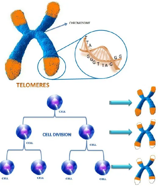
Our body is made up of an incredibly large number of cells, around 100 billion, some of them have a rather short life, others such as chondrocytes remain for a lifetime, but at a certain point, the mechanism slows down, cell duplication starts to fail and the cells are no longer replaced by other ones. The cells arrest their cell cycle progression. Each cell has a limited number of possible divisions, which is fixed between 50 and 70. The reason lies in the structure of chromosomes that are duplicated at each division in order to obtain one copy for itself and another for the daughter cell that will play the same role in the body. The division process, however, is not perfect, the cellular mechanism fails to copy the ends of chromosomes, the so-called telomeres (Fig. 1). After each cell division, the telomeres become shorter and shorter and are eventually completely worn out and parts of chromosomes that contain essential genetic information begin to erode. At
24
this point the cell is at the end of its life and it approaches death by activating specific mechanisms. In order to avoid the aging process, a ―magic‖ molecule able to stretch the ends of chromosomes would be necessary.
Figure 1. Telomere shortening. A telomere is a region of repetitive DNA (TTAGGG repeats) at the end of a
chromosome, which protects the end of the chromosome from deterioration. Each time cells divide, telomeres shorten, and there is a limit to the number of times a given cell can go on dividing, the so-called ―Hayflick limit‖. When the
length of the telomeres is too short cell division stops.
This normally happens in cells and it is due to telomerase. However, telomerase is active only in embryonic cells and in a few adult cells such as cells of the immune system, in the other ones its activity runs out over time. It is for this reason that we talk about ―cell senescence‖. Cellular senescence is strongly associated with the development of several serious diseases such as
25
cancer, diabetes, cardiovascular and neurodegenerative diseases, and osteoarthritis (OA) (Burnet and Berger, 2014). OA is a degenerative joint disease, which affects especially the articular cartilage, leading to pain and stiffness of the affected joint. OA is one of the most disabling musculoskeletal disorders in the world and it affects mostly the elderly population (Musumeci et al., 2014a). In this review we discuss how cellular senescence can influence the onset of a complex joint disease such as OA. We will discuss the most important features of cellular senescence and how the age-related changes, which arise at cellular and tissue level, influence the development and progression of OA. Lastly, we will list some of the most important senescence markers used to evidence the senescence of chondrocytes.
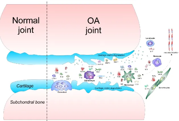
3. Osteoarthritis
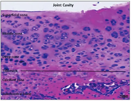
OA is the most common form of joint disease and affects mainly hips, knees, hands, and feet, leading to severe disability and loss of quality of life, particularly in the elderly population (Musumeci et al., 2013a). Indeed, half of the world's population, aged 65 and older, suffers from OA. It has been estimated that 9.6% of men and 18% of women in that age group, have symptomatic OA. Moreover, OA is considered the most important cause of impaired mobility and contributes to 50% of all the musculoskeletal diseases worldwide (Wolf and Pfleger, 2003). The disease is characterized by joint dysfunction due to gradual changes in several structures of the joint such as synovium, subchondral bone and especially articular cartilage (Fig. 2) (Buckwalter and Mankin, 1998a). Progressive wear and tear on articular cartilage can lead to a progressive cartilage tissue loss, further exposing the bony ends, leaving them without protection (Buckwalter and Mankin, 1998b). This finally deteriorates into the most common form of arthritis, or rather moderate OA at early stage (Fig. 3) and severe OA at advanced stage (Fig. 4) (Musumeci et al., 2014a).
The degradation, consequent loss of articular cartilage and formation of osteophytes lead to chronic pain and functional restrictions in the affected joint. Unfortunately, articular cartilage has a limited regenerative capacity (Musumeci et al., 2013b; Iwamoto et al., 2013;). Consequently, once injured, cartilage is much more difficult to self-heal and the only way to improve the patient‘s condition is therapeutic intervention. Different factors can be involved in the development of OA, such as joint injury, genetic predisposition, defective position of joints, obesity, malnutrition and excessive mechanical load, which all lead to similar alterations of the articular cartilage (Musumeci et al., 2014b,c; Goldring and Goldring, 2007; Lee et al., 2013).
26
Figure 2. Healthy articular knee cartilage from rat. Hematoxylin and Eosin staining. Normal articular knee hyaline
cartilage layers: superficial zone, intermediate (or middle zone), radial zone (or deep zone) and calcified cartilage (or calcified zone). Tidemark, the border between non-calcified and calcified cartilage. In the superficial zone, cells are flat
and small; in the middle and deep zone, cells are organized in columns; the tidemark is evident. Magnification x20. Scale bars: 100 µm.
However, the prevalence of OA rises directly with age, which represents the most prominent risk factor for the initiation and progression of primary OA, but it is important to underline that OA is not a simple ―wearing out in time‖ of the joints and the degenerative changes related to age can be distinguished from those due to the disease (Musumeci et al., 2013c). T he relationship between the development of OA and aging is not completely understood and it is thought that the age-related changes are correlated to other risk factors, which may occur concurrently or in conjunction with it. In reality, not all older adults develop OA and OA-like changes can also develop without a significant contribution of aging. Thus, aging and OA are inter-related but not inter-dependent (Loeser, 2004). However, there is also a possibility that the chondrocytes undergo premature aging due to several factors, such as excessive mechanical load or oxidative stress. In the latter case, aging and the development of OA are both inter-related and inter-dependent (Martin et al., 2004a; Akagi et al., 2010).
27
Figure 3. Moderate OA articular knee cartilage from rat. Hematoxylin and Eosin staining. Articular knee hyaline
cartilage layers at early OA stage: superficial zone, intermediate (or middle zone), radial zone (or deep zone) and calcified cartilage (or calcified zone). Tidemark, the border between non-calcified and calcified cartilage. Clear deep fissures in the articular surface, the cells from the superficial, intermediate and deep zone, where chondrocytes are not
arranged in columns. The tidemark is not intact in all its extension and the subchondral bone shows little fibrillation. Magnification x20. Scale bars: 100 µm.
Figure 4. Severe OA articular knee cartilage from rat. Hematoxylin and Eosin staining. Articular knee hyaline cartilage
layers at advanced OA stage, due to aging: superficial zone, intermediate (or middle zone), radial zone (or deep zone) and calcified cartilage (or calcified zone). Tidemark, the border between non-calcified and calcified cartilage. Cells are
arranged in clusters especially around fissures or disappear completely as the disease progresses. The organization of cartilage is completely disordered and replaced by fibrocartilaginous, scar-like tissue with fibroblast like cells.
28
4. Cellular aging
Cellular aging, or cell senescence, refers to the limited capacity of mitotic cells to further multiply in time (over 30–40 divisions). This limit is known as the ―Hayflick limit‖ (Hayflick, 1984). This form of senescence is called ―replicative senescence‖, also known as intrinsic senescence, which results from an arrest in cell-cycle progression. Some of the changes exhibited by cells, which have undergone replicative senescence can be found in cells in older adults, such as shortened telomeres, formation of senescence-associated (SA) heterochromatin (Muller, 2009) and changes of phenotype with an alteration in gene expression (Bodnar et al., 1998). It has been hypothesized that the telomere length could be considered as a marker for replicative senescence. Telomeres cannot be completely replicated in primary cells and become shorter with each round of cell division. Telomeres are nucleoprotein structures (TTAGGG repeats) that cap the ends of the linear eukaryotic chromosomes and thereby protect their stability and integrity during replication by protecting chromosome ends against exonucleases (Fig. 1). Telomeres are replicated by a special reverse transcriptase called telomerase, in a complex mechanism that is coordinated with the genome's replication. Telomerase is an RNA-dependent DNA polymerase that synthesizes telomeric DNA sequences and comprises two essential components. One is the functional RNA component (in humans called hTERC), which serves as a template for telomeric DNA synthesis. The other is a catalytic protein (hTERT) with reverse transcriptase activity and the primary determinant for the enzyme activity (Bryan and Cech, 1999; Kupiec, 2014). The level of telomerase in normal human somatic tissues is insufficient to prevent telomere shortening. Telomeres can be lengthened through increasing telomerase activity by exogenous expression of hTERT or hTR (the RNA template)(Greider, 1998). As proof of this concept the chondrocytes transduced with hTERT proved to be able to increase telomere length and therefore to prolong cell lifespan, increasing in this way the efficacy of cartilage repair (Martin and Buckwalter, 2003). Another type of senescence is ―stress-induced senescence‖, also known as extrinsic senescence, which is independent of telomere length. In quiescent cells such as chondrocytes, this type of senescence may be more important than the replicative version, because progressive telomere shortening cannot completely explain senescence in these post-mitotic cells (Ben-Porath and Weinberg, 2005; Chen and Goligorsky, 2006). The various types of stress responsible for this kind of senescence include DNA damage, oxidative stress, oncogene activity, ultraviolet radiation and chronic inflammation (Itahana et al., 2004; Campisi, 2005). Oxidative stress is thought to play a major role as a stressor. It results when the amount of reactive oxygen species (ROS) exceeds the anti-oxidant capacity of the cell. ROS are generated by intracellular enzymes such as nicotine amide adenine dinucleotide phosphate (NADPH) oxidase and 5-lipoxygenase in response to activation of specific cell signaling pathways
29
(Kamata and Hirata, 1999; Finkel and Holbrook, 2000). A direct role for increased ROS levels in promoting cell senescence is a positive feedback activation of the ROS-protein kinase C delta (PKCδ) signaling pathway, which cooperates with the p16INK4A-retinoblastoma protein (Rb) pathway, which plays an important role in the control of cell-cycle progression (Takahashi et al., 2006). Telomere shortening is also observed in stress-induced senescence and it is due to oxidative damage to DNA caused by ROS. The ends of chromosomes are particularly sensitive to oxidative damage, which causes telomere erosion similar to that seen with replicative senescence (Yudoh et al., 2005). Also, the ROS generated from excessive mechanical loading and stimulation of cytokines contribute to DNA damage, which subsequently results in telomere shortening (Tomiyama et al., 2007; Davies et al., 2008). Cellular senescence, as well as apoptosis, can be viewed as a powerful tumor-suppressor mechanism that withdraws cells with irreparable DNA damage from the cell cycle (de Lange and Jacks, 1999; Artandi and DePinho, 2000; Puzzo et al., 2014) through the intrinsic or mitochondrial (Caltabiano et al., 2013; Loreto et al., 2011a) and extrinsic apoptosis pathway (Cardile et al., 2013; Loreto et al., 2011b). Several recent studies report that cartilage degeneration also coincides with increased apoptotic chondrocytes (Musumeci et al., 2011a; Musumeci et al., 2011b; Galanti et al., 2013). Therefore, the senescence signals, that is, a telomere-based one or a stress-based one, trigger a DNA damage response and this response shares a common signaling pathway that converges on either or both of the well-established two tumor- suppressor proteins, p53 (the p53-p21-pRb pathway) (Martin and Buckwalter, 2003; Herbig and Sedivy, 2006) and RB and pRb proteins (the p16-pRb pathway) (Musumeci et al., 2010; Musumeci et al., 2011c). In the p53-p21-pRb pathway, senescence stimuli activate the p53, which then can induce senescence by activating pRb through p21, which is a transcriptional target of p53. This senescence can be reversed upon subsequent inactivation of p53. In the p16-pRb pathway, senescence stimuli induce p16, which activates pRb. Once the pRb pathway is engaged by p16, the senescence cannot be reversed by subsequent inactivation of p53, silencing of p16 or inactivation of pRb (Beauséjour et al., 2003). The difference between these two pathways is that the p53 -p21-pRb pathway mediates the senescence due to telomere shortening and the p16-pRB pathway is thought to mediate premature senescence (Beauséjour et al., 2003). Once cells have entered senescence, they are arrested in the G1 phase of the cell cycle and display a characteristic morphology (vacuolated, flattened cells) and altered gene expression (Cristofalo et al., 2004). The senescent cells exhibit the so-called ―senescent secretory phenotype‖ (SSP), which could be also correlated with the development of OA. It is interesting to note that the senescent cells, which are mitotically inactive, are biologically active (Campisi, 2005). These cells are able to increase the expression of genes that inhibit proliferation and to increase the secretion of several proteins, including
30
inflammatory cytokines such as interleukin-6 (IL-6) and interleukin-1 (IL-1), degradative enzymes such as metalloproteinases (MMPs) and growth factors such as epidermal growth factor (EGF) that regulate cell proliferation and all of which may stimulate tissue aging and tumorigenesis (Zhang et al., 2003). Recent studies reported that the increased expression of the IL-8 receptor CXCR2 and insulin-like growth factor binding protein 7 (IGFBP7) could also contribute to cell aging (Acosta et al., 2008; Wajapeyee et al., 2008). The accumulation of cells expressing the SSP can also contribute to tissue senescence by impairing the extracellular matrix due to the increased secretion of degradative enzymes (Campisi and d‘Adda di Fagagna, 2007). Another important feature of senescent cells is represented by the epigenetic changes related to the formation of foci of heterochromatin, referred to as senescence-associated heterochromatin foci (SAHFs), which include histone variants such as the macro-H2A (Zhang and Adams, 2007).
5. Age-related articular cartilage degeneration.
Articular cartilage matrix undergoes several changes with age, including structural, molecular and mechanical ones, surface fibrillation, alterations in structure and composition of proteoglycans, increased collagen cross-linking and decreased tensile strength and stiffness (Hollander et al., 1995; Squires et al., 2003) (Fig. 5). Deterioration in chondrocyte function accompanies these changes also in the extracellular matrix (Aurich et al., 2002). Several reports revealed that chondrocyte senescence contributes to the risk for cartilage degeneration by the decreased ability of chondrocytes to maintain and repair the articular cartilage tissue (Martin and Buckwalter, 2001a; Aigner et al., 2002). There is clinical evidence from Magnetic Resonance Imaging (MRI) studies that the articular cartilage in the knee thins with aging, especially at the patella and at the femoral side of the joint (Hudelmaier et al., 2001; Ding et al., 2005). The progressive articular cartilage thinning with age is related to gradual loss of cartilage matrix and decrease in cartilage hydration and cellularity. This kind of damage stimulates a chondrocyte specific synthetic and proliferative response that may maintain or even restore the articular cartilage. This response may continue for years. However, in instances of progressive joint degeneration the anabolic response eventually declines and the imbalance between chondrocyte synthetic activity and degradative activity leads to progressive thinning of articular cartilage. These alterations may further accelerate the loss of articular cartilage (Buckwalter et al., 2000). Different changes observed in articular cartilage with aging are probably due to chondrocyte senescence, which results in the progressive decrease in cell function.In fact, the mitotic and synthetic activity of human chondrocytes decline with age. They become less responsive to anabolic mechanical stimuli, to anabolic cytokine and to insulin-like growth factor I (IGF-I). The cells synthesize smaller
31
aggrecans and less functional link proteins leading to the formation of smaller and more irregular proteoglycan aggregates. The latter is the most striking change in articular cartilage matrix related with age. Aggrecans are the molecules that give articular cartilage its stiffness to compression, resilience and durability, thus their alteration makes the tissue more vulnerable to injury and development of progressive degeneration (Buckwalter et al., 1986; Buckwalter and Rosenberg, 1988; Bolton et al., 1999; Martin and Buckwalter, 2000).
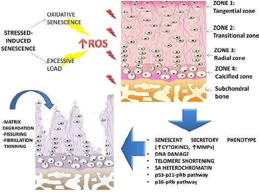
Figure 5. Stress-induced senescence and Osteoarthritis. The telomere shortening process in senescent chondrocytes is
more probably due to the stress-induced type of senescence. Oxidative stress and excessive mechanical loading are thought to be the major stressors that induce the increased production of ROS, which are responsible for DNA damage and for the subsequent senescence of the cells. Once cells have entered senescence, they are arrested in the G1 phase of
the cell cycle and they display a characteristic gene expression called ―senescent secretory phenotype‖, which is strongly correlated with the development of OA.
Moreover, the senescent cartilage matrix appears more susceptible to the accumulation of advanced glycation end-products (AGEs) in cartilage collagen, which results in increased cross-linking and in subsequent increased stiffness, making the cartilage more susceptible to fatigue failure (Verzijl et al., 2002). In addition, the increased levels of AGEs in articular cartilage may also affect chondrocyte function by decreasing its anabolic activity. The mechanism proposed to be responsible for this alteration is the expression of the receptor for the advanced glycation end-products (RAGE) by chondrocytes, which proves to be increased both with aging and in development of OA (DeGroot et al., 1999; Loeser et al., 2005). Stimulation of chondrocyte RAGE results in increased production of MMPs and in modulation of the chondrocyte phenotype to
32
hypertrophy, which represent two hallmarks of OA (Cecil et al., 2005; Yammani et al., 2006). Furthermore, RAGE signaling is also associated with increased levels of ROS, providing another link between oxidative stress, aging and OA (Loeser, 2004). Another important feature of the aged articular cartilage is its increased calcification, as demonstrated radiographically (Felson et al., 1989). This could be associated with the increased activity of transglutaminase, involved in the biomineralization process (Rosenthal et al., 1997) and with increased production of the inorganic pyrophosphate in response to transforming growth factor β (TGF-β) stimulation (Felson et al., 1989). Chondrocalcinosis is strongly associated with OA, but there is evidence of older people with asymptomatic chondrocalcinosis, thus it proves not to be inter-dependent with the development of OA and its role is not completely clear (Doherty and Dieppe, 1988; Rosen et al., 1997).
6. Chondrocyte senescence
Chondrocytes from older adults exhibit many changes, typical of cell senescence, when compared with cells isolated from young adults. The most evident change is represented by telomere shortening, characteristic of replicative senescence. This evidence is controversial as adult articular chondrocytes rarely, if ever, divide in normal tissue in vivo. The lack of cell division in normal adult articular cartilage suggests that the chondrocytes present in the cartilage of an older adult are likely to be the same cells that were present decades earlier. This fact makes these cells more susceptible to the accumulation of changes from both aging and extrinsic stress. In fact, it is most likely that chondrocyte senescence is the extrinsic type, induced by different stressors. The telomere shortening in adult chondrocytes could be due to DNA damage from ROS as discussed further above (Mankin, 1963; Martin and Buckwalter, 2001b; Martin et al., 2004b). The increased ROS levels could be both age-related (Del Carlo and Loeser, 2003) and generated from excessive mechanical loading and/or stimulation by cytokines (Kurz et al., 2005; Davies et al., 2008). There is also evidence for reduced levels of antioxidant enzymes in cartilage with aging and in OA that would contribute to chondrocyte senescence and oxidative damage. In human articular chondrocytes, decreased levels of mitochondrial superoxide dismutase were found both with aging and in OA cells (Finkel and Holbrook, 2000). Moreover, it has been hypothesized that joint injury accelerates chondrocyte senescence and that this acceleration plays a role in the joint degeneration responsible for post-traumatic OA. Indeed, excessive loading of articular surfaces due to acute joint trauma or post-traumatic joint instability, incongruity or mal-alignment increases release of ROS, and the increased oxidative stress on chondrocytes accelerates chondrocyte senescence (James et al., 2004).Other important features of chondrocyte senescence are the exhibition of SSP, which has important implications in development and progression of OA and the decline in the proliferative
33
and anabolic response to growth factors, as well as their reduction in cartilage. It has been noted that senescent chondrocytes lose the ability in response to: IGF-I, which is known to be an important autocrine survival factor that stimulates cartilage matrix synthesis (Martin et al., 1997); TGF-β, an important cartilage anabolic factor (Scharstuhl et al., 2002) and to bone morphogenetic protein 6 (BMP-6), known to stimulate proteoglycan synthesis (Bobacz et al., 2003). Chondrocyte senescence also contributes to the decline in the cell number within the cartilaginous tissue, due to increased cell death. Several studies demonstrated the loss of cellular density in cartilage with aging or/and in OA (Vignon et al., 1976; Adams and Horton, 1998; Horton et al., 1998; Aigner et al., 2004a; Kuhn et al., 2004). These findings provide evidence to support the concept that chondrocyte senescence may be involved in the progression of cartilage degeneration, because of their decreased ability to maintain or restore the articular cartilage (Fig. 5).
7. Chondrocyte senescence markers
An altered gene expression pattern on the cellular level appears to be one potentially important facet of chondrocyte behavior in OA cartilage (Aigner et al., 2004b; Aigner et al., 2007). The diversification of gene expression in senescent chondrocytes is due to stochastic DNA damage, which represents a core mechanism in cellular aging in general and in OA cartilage degeneration in particular. The evidence of senescence in chondrocytes can be investigated using several senescence markers, such as senescent-associated-β-galactosidase (SA-βgal), highly condensed domains of facultative heterochromatin SAHF, increased p53, p21, pRb and p16INK4a (Fig. 5). Staining for SA-βgal has been shown to be present in articular chondrocytes from older adults and in OA chondrocytes (Martin and Buckwalter, 2001b; Price et al., 2002). SA-βgal is related to the detection of increased levels of the lysosomal enzyme β-galactosidase at pH 6.0 rather than at the normal pH 4.5. Detection of its activity at pH 6.0 is thought to be due to an increase in lysosomal mass (Itahana et al., 2007). Chondrocyte SA-βgal staining, as well as telomere shortening, has also been noted after treatment in vitro with IL-1β or H2O2 consistent with stress-induced senescence
(Dai et al., 2006). Although SA-βgal is a useful senescence marker, its activity is critically dependent on the detection conditions, and SA-βgal is also expressed in the non-senescent cells that have a high lysosomal content (Kurz et al., 2000; Matthews et al., 2006). Multiple markers of senescence are therefore recommended to demonstrate senescence in vivo. SAHF are thought to repress expression of proliferation-promoting genes, thereby contributing to senescence-associated proliferation arrest.Inclusion of proliferation-promoting genes, such as cyclin A, into these compact chromatin foci is thought to silence expression of those genes, which are associated with cell cycle arrest (Adams, 2007). Ink4a encodes an archetypical cyclin-dependent inhibitor (CKI) associated
34
with senescence. Indeed, the over-expression of p16INK4a in chondrocytes is associated with SSP, which includes increased production of pro-inflammatory cytokines (such as IL-6, IL-8, IL-1β) and matrix remodeling regulatory metalloproteinases (such as MMP1, MMP13, etc) (Leonardi et al., 2008; Loreto et al., 2013). As mentioned above, all these factors are deleterious for cartilage integrity. According to this finding, the repressed levels of miR-24, a negative regulator of p16INK4a, was also found in OA cartilage (Philipot et al., 2014). Recently, the expression of Caveolin1, a protein that participates in premature cellular senescence, was also investigated in human OA cartilage. It was observed that the treatments with catabolic factors of oxidative stress (H2O2) and
IL-1β, which simulate the OA environment, was able to up-regulate the expression of caveolin1. The over-expression of caveolin1 is associated with cartilage degeneration and the mediation of the premature senescence in OA chondrocytes by activating p38 MAPK, which impair the ability of chondrocytes to produce type II collagen and aggrecan (Dai et al., 2006). Other important senescence markers are represented by telomere length and telomerase activity. As discussed in detail above, telomere shortening is the most representative feature of cellular aging, and it is due to the decreased expression of telomerase with time which leads to telomere instability. Telomere length can be measured by using the Single Telomere Length Assay (STELA), Southern blot analysis, Q-PCR and the more recent Quantitative-Peptide Nucleic Acid-Fluorescent in situ Hybridization assay (Q-PNA-FISH) (Cukusic et al., 2014). Telomerase activity can be measured for example by using a Telomere Repeat Amplification Protocol (TRAP) (Zhou and Xing, 2012) (Table 1). Recently, several studies have been focused on the expression of lubricin, also known as proteoglycan 4 (PRG4) or superficial zone protein (SZP), in different experimental conditions, in particular in conjunction with physical activity (Musumeci et al., 2013d). Lubricin is a chondroprotective glycoprotein that serves as a critical boundary lubricant between opposing cartilage surfaces (Musumeci et al., 2013e; Leonardi et al., 2011). It has a majorprotective role in preventing cartilage wear and synovial cell adhesion, proliferation, and in reducing the coefficient of friction of the articular cartilage surface (Musumeci et al., 2013f; Leonardi et al., 2012a,b). Since the lubricin has a fundamental role in maintaining the homeostasis of the articular cartilage and in preventing its degeneration, we hypothesized that its expression would decrease in senescent chondrocytes and that it could be evaluated as a new specific chondrocyte senescence marker as confirmed in our recent and interesting study (Musumeci et al., 2014d).
35
Table 1. An overview of the key markers for the senescent chondrocytes and their functions.
8. Conclusions
Although the direct relationship between the aging process and the development of OA is not completely understood, we may surmise that chondrocyte senescence contributes to cartilage degeneration by impairing the ability of these cells to maintain and repair the cartilage tissue. Moreover, we have also seen that these two processes (aging and OA) could be inter-dependent. There are several lines of evidence that suggest chondrocytes exposed to the ―osteoarthritic environment‖ are characterized by oxidative stress and production of cytokines, and this induces the so-called stress-induced senescent state, which may contribute to cartilage degeneration as we have discussed above. All these observations suggest that a better understanding of the changes arising with age in articular cartilage and how they influence the response of the tissue to different stressors, as well as the identification of new increasingly sensitive senescence markers, would be very useful in the preventive detection of the disease and in its consequent treatment. Further research is required to unravel the detailed mechanisms of senescence related to the pathogenesis of OA. Strategies aimed at preventing chondrocyte senescence could have a positive impact on the development of new therapies for OA and on halting the progression of this severe disease.