Novel Acylguanidine Derivatives Targeting
Smoothened Induce Antiproliferative and
Pro-Apoptotic Effects in Chronic Myeloid
Leukemia Cells
Alessandra Chiarenza1, Fabrizio Manetti2, Elena Petricci2, Martial Ruat3, Antonella Naldini1, Maurizio Taddei2, Fabio Carraro1,4*
1 Department of Molecular and Developmental Medicine, University of Siena, Siena, Italy, 2 Department of Biotechnology Chemistry and Pharmacy, University of Siena, Siena, Italy, 3 CNRS, UMR-9197,
Neuroscience Paris- Saclay Institute, Molecules Circuits Department, Signal Transduction and
Developmental Neuropharmacology Team, Gif-sur-Yvette, France, 4 Istituto Toscano Tumori, Siena, Italy *[email protected]
Abstract
The most relevant therapeutic approaches to treat CML rely on the administration of tyro-sine kinase inhibitors (TKIs) like Imatinib, which are able to counteract the activity of Bcr-Abl protein increasing patient’s life expectancy and survival. Unfortunately, there are some issues TKIs are not able to address; first of all TKIs are not so effective in increasing survival of patients in blast crisis, second they are not able to eradicate leukemic stem cells (LSC) which represent the major cause of disease relapse, and third patients often develop resis-tance to TKIs due to mutations in the drug binding site. For all these reasons it’s of primary interest to find alternative strategies to treat CML. Literature shows that Hedgehog signaling pathway is involved in LSC maintenance, and pharmacological inhibition of Smoothened (SMO), one of the key molecules of the pathway, has been demonstrated to reduce Bcr-Abl positive bone marrow cells and LSC. Consequently, targeting SMO could be a promising way to develop a new treatment strategy for CML overcoming the limitations of current ther-apies. In our work we have tested some compounds able to inhibit SMO, and among them MRT92 appears to be a very potent SMO antagonist. We found that almost all our com-pounds were able to reduce Gli1 protein levels in K-562 and in KU-812 CML cell lines. Fur-thermore, they were also able to increase Gli1 and SMO RNA levels, and to reduce cell proliferation and induce apoptosis/autophagy in both the tested cell lines. Finally, we dem-onstrated that our compounds were able to modulate the expression of some miRNAs related to Hedgehog pathway such as miR-324-5p and miR-326. Being Hedgehog pathway deeply implicated in the mechanisms of CML we may conclude that it could be a good thera-peutic target for CML and our compounds seem to be promising antagonists of such pathway.
OPEN ACCESS
Citation: Chiarenza A, Manetti F, Petricci E, Ruat M, Naldini A, Taddei M, et al. (2016) Novel
Acylguanidine Derivatives Targeting Smoothened Induce Antiproliferative and Pro-Apoptotic Effects in Chronic Myeloid Leukemia Cells. PLoS ONE 11(3): e0149919. doi:10.1371/journal.pone.0149919 Editor: Persio Dello Sbarba, Università degli Studi di Firenze, ITALY
Received: November 10, 2015 Accepted: February 5, 2016 Published: March 2, 2016
Copyright: © 2016 Chiarenza et al. This is an open access article distributed under the terms of the Creative Commons Attribution License, which permits unrestricted use, distribution, and reproduction in any medium, provided the original author and source are credited.
Data Availability Statement: All relevant data are within the paper and its Supporting Information files. Funding: This work has received financial support from the Istituto Toscano Tumori.
Competing Interests: The authors have declared that no competing interests exist.
Introduction
Chronic myelogenous leukemia (CML) is a clonal myeloproliferative malignancy that arises in hematopoietic stem cells harboring the reciprocal translocation between chromosomes 9 and 22, thus resulting in the formation of the Philadelphia chromosome [1]. This translocation fuses the breakpoint cluster region (Bcr) and the Abelson kinase (Abl) genes, forming the Abl oncogene that encodes the constitutively active cytoplasmatic tyrosine kinase (TK) Bcr-Abl [2,3], present in>90% of CML cases. The aberrant kinase activity of Bcr-Abl is responsible for CML initiation [4], and the consequent disease progresses through three phases (chronic proliferative phase, accelerated phase, and blast crisis phase), becoming more resistant to treat-ment in each successive phase. The last phase is also characterized by the presence of genomic instability and is ultimately fatal.
The finding that Bcr-Abl is the cause of the leukemic phenotype and that the TK activity of Abl is fundamental for Bcr-Abl-mediated transformation, make this kinase an important target for the development of specific therapies [5]. The advent of TK inhibitors (TKI) targeting Bcr-Abl has revolutionized the treatment of CML. Imatinib [6,7], which was the first Bcr-Abl inhib-itor approved for CML therapy [8,9], has improved patients’life expectance and survival espe-cially in the chronic phase. The occurrence of relapse, resistance [10–13], and the necessity of a continued chemotherapy led to the discovery of nilotinib [14,15], dasatinib [16], and bafetinib [17] that are much more active toward Bcr-Abl and are able to block imatinib-resistant CML, with the sole exception of the T315I Bcr-Abl mutation that is recognized by ponatinib [18], a third generation TKI. Dasatinib was approved by FDA in 2006 for adult patients (chronic phase CML) with resistance or intolerance to prior therapies, nilotinib was approved in 2010 for chronic phase CML patients, and ponatinib was approved in 2012 for T315I CML patients. At the end of 2012, also bosutinib, a dual Bcr-Abl/Src inhibitor, was approved by FDA for the treatment of adult patients with resistant CML in chronic, accelerated or blast phase [9]. Although such compounds demonstrated clinical efficacy in some cases of imatinib resistance, the problem of LSC insensitivity remained unsolved.
On the basis of these considerations, treatment of CML with currently available TKIs suffers from three major limitations. In fact, although Bcr-Abl expression is deeply reduced or abro-gated in the majority of patients, the anti-CML drugs have not significantly improved survival in patients in blast crisis (BC) [19]. Moreover, imatinib is unable to kill leukemic stem cells (LSC) in CML [20,21] because LSC do not depend on Bcr-Abl activity for survival [22]. Finally, kinase domain mutations confer resistance to imatinib in several patients. Therefore, treatment of the blast crisis, eradication of LSC, and the insensitivity of resistant cells to imatinib still remain the major unsolved problems in the treatment of CML [19]. In this perspective, finding alternative strategies to overcome limitations of current therapies has acquiring growing importance.
Currently, several investigational approaches are under study in the attempt to prevent BC and to deplete LSC population. A potential approach for BC prevention is to interfere with the self-renewal properties of LSC [23]. In this context, a pivotal role for survival maintenance of LSC has been found for BCL6 [24], HIF1α [25], and Smoothened (SMO) [21,26,27]. Recent lit-erature shows that Hedgehog (Hh) signaling pathway is clearly involved in expansion of Bcr/ Abl-positive stem cells [27,28], and in functional regulation of CML in terms of self-renewal, proliferation, and apoptosis. Moreover, loss or pharmacological inhibition of SMO, an essential component of the Hh pathway, impairs LSC renewal, decreases the propagation of Bcr-Abl-driven CML, and reduces the growth of resistant CML [27].
Hh signaling pathway is highly conserved in vertebrates and it is involved in embryonic development, organogenesis and cell proliferation. In the absence of Hh ligands, Patched (Ptch) receptor inhibits the activation of the downstream protein SMO by keeping it blocked
in an intracellular vescicle [29]. Upon binding of Hh ligands to Ptch receptor, such inhibition is released and the activation of SMO, followed by its migration on primary cilium cell mem-brane, leads to pathway activation which culminates in a signal transduction cascade. These events cause the nuclear translocation of the Gli family of transcription factors (Gli1-3), and the subsequent activation or inhibition of various cell cycle, proliferation and survival regulat-ing genes such as the D-type cyclins, c-Myc and Bcl-2 [30–34]. As part of a feedback mecha-nism, Gli target genes also comprise members of the Hh pathway, such as Gli1 and Ptch1 [35,36]. Among the inhibitors of the Hh pathway that interact directly with SMO, vismodegib and sonidegib are the two compounds approved by FDA in 2012 and 2015, respectively, for the treatment of basal cell carcinoma (BCC). Other hedgehog inhibitor compounds have been studied for BCC, like CUR61414 [37], but though they showed a good activity both in vitro and on mice, they failed the clinical phase I studies [38].
On the other hand, preclinical studies are checking the possibility to induce apoptosis in BC cells by treatment with various drugs, alone or in combination. As an example, inhibition of Mek and farnesyl transferase [39], or treatment with a dual Bcr-Abl/Jak2 inhibitor [40], as well as p53 stabilization [18] induce apoptosis and death in human BC CML K-562 cells. Impor-tantly, this cell line expresses all the Hh signaling molecules, including sonic Hh (Shh), Ptch, SMO and Gli1 [41].
Taken together, these results suggest that small molecules able to inactivate the Hh pathway by blocking SMO could be in principle useful either to inhibit BC cell proliferation or, at the same time, to deplete population of LSC whose survival, self-renewal, and expansion is strongly dependent on the Hh pathway [27]. Moreover, a combination therapy comprised of the cur-rently available Bcr-Abl inhibitors and small molecules able to block SMO could represent a very promising and effective tool to deplete CML cells, overcome chemotherapy resistance, eradicate LSC, and thus potentially cure CML.
MicroRNAs (miRNAs) are a class of small non-coding cellular RNAs that are responsible for messenger RNA translational inhibition or degradation. In several human cancers, a down-regulated miRNA signature with high Hh signaling does exist. Downregulation of these miRNAs allows high levels of expression of Hh-dependent genes leading to tumour cell prolif-eration. As an example, miR-324-5p was shown to target the activator components of the Hh pathway, SMO and Gli1, thereby suppressing progenitor and tumour cell growth [42]. More-over, it has been demonstrated that upregulation of SMO is associated with reduced expression of miRNA-326 [43].
Within a drug design project aimed at identifying new small molecules acting as Hh inhibi-tors, we have recently found a class of compounds with a very impressive ability to inhibit SMO by direct interaction [44,45]. In particular, in previous studies, compound MRT-83, belonging to the chemical class of acylguanidines substituted with a phenyl group, appeared to be one of the most potent SMO antagonists known so far [46]. On this basis, we planned to design new MRT-83 analogues and to check their ability to block growth and proliferation of two human BC CML cell lines (namely, K-562 and KU-812) and among the tested compounds we found that the one which possessed the phenylethyl terminal group (MRT92) was particu-larly active toward Hh pathway.
Results and Discussion
Specificity for Hh pathway
Western blotting analysis was applied to evaluate the ability of the compounds to affect Gli1 and suppressor of fused (SuFu) protein expression. Compounds effects were compared with the activity of CUR61414 used as reference compound and AT43 (a known SMO agonist) [37].
As shown inFig 1aand quantified inFig 1ctreatment of K-562 and KU-812 (Fig 1b and 1d) cells with the compounds MRTX, MRT94, MRT92, MRT83, at 20μM for 24 h, significantly reduced Gli1 protein concentration in comparison with non-treated control. A similar decrease of Gli1 protein was found by treatment with CUR61414. We were not able to determine any significant effect of MRTY on both cell lines. While on the contrary, as expected, AT43 was able to significantly increase Gli1 expression. These results showed a negative modulation of Hh pathway by our SMO inhibitors through the reduction of Gli1 protein expression. We also investigated the effect of our compounds on SuFu which is known to be a regulator of Gli pro-teins counteracting Spop activity, thus preventing Gli factors degradation [47]. On K-562 cells our compounds were not able to induce any significant modification of SuFu and even AT43, despite inducing a significant increase of Gli1, did not produce an increase in SuFu. On the contrary, on KU-812 cells the compounds MRT92 and CUR61414 were able to significantly reduce the amount of expressed protein, while AT43 induced a significant increase that corre-lated with Gli1 increase. The reduction of SuFu, even if not always significant, is a further con-firmation of the ability of tested compounds to interact with Hh pathway. Given the specificity of these compounds to target Hh pathway, their effects on RNA expression were evaluated for the most important pathway components (i.e., Gli1 and SMO). As expected, no significant changes of the Gli1 and SMO RNA expression levels were found in both cell lines after a 3 h incubation with the studied compounds (data not shown). On the contrary, Gli1 RNA expres-sion was increased in K-562 cells by a 24 h treatment with 10μM inhibitor (Fig 2a), with the
Fig 1. Effects of compounds and controls on Gli1 and SuFu protein expression. Effects of compounds MRT83, MRTX, MRT92, MRTY, MRT94 and control compounds after a 24 h treatment at 20μM on Gli1 and SuFu protein expression in K-562 cells (a) and KU-812 cells (b) and quantification relative to medium expression (c-d).β-actin was used as loading control. Data are representative images, quantifications are expressed as the means ± SEM of three independent experiments.*p<0.05 vs medium.
doi:10.1371/journal.pone.0149919.g001
sole exception of MRT83. This enhanced expression could be considered as a compensatory effect that balances Gli1 protein level reduction consequent to pathway blockade. Only MRTX and MRTY showed a residual effect after 72 h (Fig 2b). SMO RNA levels were unchanged by treatment of K-562 cells for 24 (Fig 2c) and 72 h (Fig 2d), probably because the compounds act only by binding the protein and preventing its ciliar translocation, without effectively reducing the cellular amount of SMO. In this way, no compensatory mechanisms are required to be acti-vated by the cell.
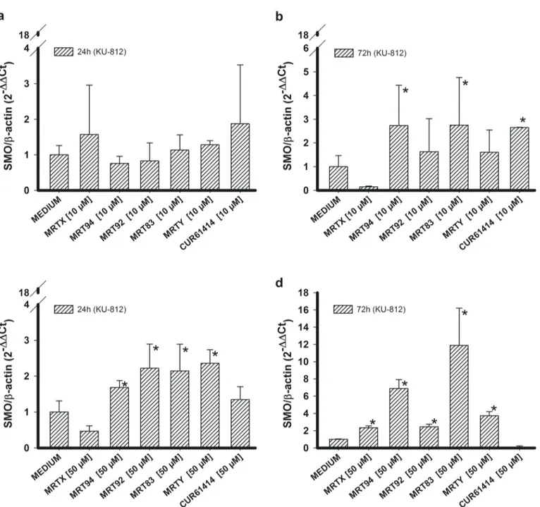
On the other hand, KU-812 cells responded to the studied compounds in a different way. In fact, only 10μM MRTY induced an increase of Gli1 RNA expression after 24 h treatment (Fig 3a) and its effect was maintained at 72 h (Fig 3b). Increasing compound concentrations to 20 and 50μM led MRTX and MRT92 (in addition to MRTY) to produce an effect after 24 h (Fig 3c), not maintained at 72 h (Fig 3d). Compounds MRT94 and MRT83 were tested only at 50μM since they did not show any significant activity even at this concentration. These results suggest that KU-812 cells are less sensitive to the studied compounds in comparison to K-562 cells. Con-sequently, higher compound concentrations were required to determine a biological effect.
A compound concentration of 10μM did not affect SMO RNA expression after a 24 h treat-ment (Fig 4a), while increased SMO RNA levels were found after a 72 h treatment with MRT94 and MRT83 (Fig 4b). Increased concentrations (50μM) of MRT94-Y led to higher SMO RNA expression levels after 24 h (Fig 4c), while all tested compounds were able to increase SMO RNA expression after a 72 h treatment (Fig 4d). In summary, differently from what found in K-562 cells, an increase of SMO RNA expression was found in KU-812 cells after a 10μM treatment for 72 h, and after a 50μM treatment after 24 and 72 h. These results suggest that a 10μM concentration is too low to induce a rapid effect (within 24 h), while higher concentra-tions (50μM) exhibit their effects already after 24 h and maintain them for 72 h at least.
Effects on miRNAs expression
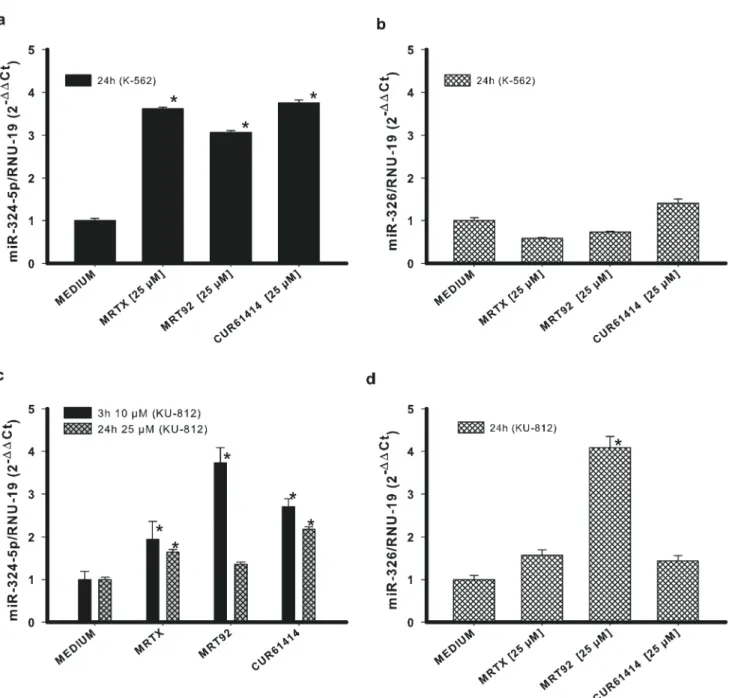
To confirm the specificity of our compounds towards Hh pathway, we investigated the effects they exerted on two miRNAs closely related to the Hedgehog pathway: miRNA-324-5p and miRNA-326. They both suppress the pathway activator SMO, and miR-324-5p also regulates the downstream transcription factor Gli1[42]. For this analysis, the two compounds with the best activity for both cell lines have been selected. Both K-562 and KU-812 cells were treated with 25μM MRTX and MRT92 for 24 h. Results show a significant increase of miR-324-5p in both cell lines and the greatest effect toward KU-812 cells was found after 3 h of treatment (Fig 5a and 5c). In addition, MRT92 at 25μM after 24h of treatment was able to increase miR-324-5p but did not reach significativity. These results are in agreement with the hypothesis that compounds negatively regulate SMO and inactivate the Hh pathway by the reduction of Gli1, thus inducing an increase of miRNA324-5p level. According to previous results showing that SMO expression was not affected in K-562 cells, miR-326 expression did not change in the same cell line (Fig 5b). On the contrary, a significant increase of its expression was found in MRT92-treated KU-812 cells (Fig 5d).
It is noteworthy that changes of the miRNAs levels are in agreement with Gli1 and SMO RNA variations in both tested cell lines. In fact, the levels of both miRNA-324-5p and its target (Gli1) increase after a 24 h treatment in the tested cell lines; the amounts of miRNA-326 and its target (SMO) remain both unaltered in K-562 cells and increase in KU-812 cells with the only exception of miRNA-326 in the sample treated with compound MRTX. This parallel level of expression of the miRNAs and the respective target genes is due to the activation of a control mechanism which promotes miRNAs increase to prevent the uncontrolled production of the target gene RNA.
Antiproliferative activity
Ability of the new compounds and CUR61414 to affect viability of K-562 and KU-812 cell lines (Fig 6) was checked by resazurin proliferation assay. The IC50values obtained for each
com-pound are listed inTable 1. A concentration-dependent antiproliferative activity of compounds
Fig 2. Gli1 and Smo RNA expression in K-562 cells. Effects of compounds MRTX, MRT94, MRT92, MRT83, MRTY and control compound in K-562 cells on Gli1 RNA expression after a 24h (a) or 72h (b) treatment and on SMO RNA expression after a 24h (c) or 72h (d) treatment at 10μM. Data are expressed as the means± SEM of four independent experiments performed in triplicate. *p<0.05 vs medium.
doi:10.1371/journal.pone.0149919.g002
comparable to or better than that of CUR61414 was found toward both cell lines. In particular, MRT92 and MRTY showed IC50values lower than 10μM in K-562 cells (7.7 and 7.8 μM,
respectively, versus 14μM found for CUR61414). IC50values for MRTX and MRT92 toward
KU-812 cells were respectively 5.5 and 7.2μM (27 μM for CUR61414). The remaining com-pounds showed a two-digit micro molar IC50.
Pro-apoptotic activity and autophagy
Prompted by their ability to reduce CML cell line proliferation, the new compounds were also checked for pro-apoptotic activity versus poly-ADP-ribose-polymerase (PARP). We used a
Fig 3. Gli1 RNA expression in KU-812 cells. Effects of compounds MRTX, MRT94, MRT92, MRT83, MRTY and control compound in KU-812 cells on Gli1 RNA expression after 24h (a) or 72h (b) treatment at 10μM and treatment at 20 and 50 μM after 24h (c) or 72h (d). Data are expressed as the means ± SEM of three independent experiments performed in triplicate.*p<0.05 vs medium.
single representative compound concentration of 10μM. Immunoblot analysis of uncleaved and cleaved PARP indicated that a significant PARP cleavage did not occur in K-562 cells after 72 h of treatment except for MRTX treated cells (Fig 7a), while tested compounds, with the exception of MRT94, led to an enhancement of the cleaved PARP in KU-812-cells that were potently induced to apoptosis after 72 h of treatment (Fig 7b).
Moreover, the expression of Bax and Bcl-2 RNA levels was also investigated. In fact, the ratio between Bax and Bcl-2 RNA expression is a critical determinant to induce cells toward
Fig 4. SMO RNA expression in KU-812 cells. Effects of compounds MRTX, MRT94, MRT92, MRT83, MRTY and control compound in KU-812 cells on SMO RNA expression after 24h (a) or 72h (b) treatment at 10μM and treatment after 24h (c) or 72h (d) at 50 μM. Data are expressed as the means ± SEM of three independent experiments performed in triplicate.*p<0.05 vs medium.
doi:10.1371/journal.pone.0149919.g004
apoptosis and represents a direct index of the induction of the apoptotic process [48], thus helping in exploring the apoptosis induction. In agreement with results of the PARP assay, no significant pro-apoptotic effect was measured in K-562 cells after 72h of treatment except for samples treated with MRTY (Fig 8a), while MRTX, MRT94, and MRT92 were able to induce apoptosis in KU-812 cells after 72h (Fig 8b).
We further evaluated the expression of BNIP3, a protein that has been shown to be corre-lated to autophagy [49]. Seventy-two hours exposure to compounds lead to an increase in BNIP3 expression, particularly this was significant in samples treated with MRT94, MRT92,
Fig 5. Changes in miRNAs expression. Effects of compounds MRTX, MRT92 and control compound in K-562 cells (a-b) or KU-812 cells (c-d) on miRNA324-5p and miRNA-326 expression. Data are expressed as the means± SEM of three independent experiments performed in triplicate. *p<0.05 vs medium.
Fig 6. Effects of compounds in K-562 cells and KU-812 cells viability. Effects of compounds MRTX (a), MRT94 (b), MRT92 (c) MRT83 (d), MRTY (e) and CUR61414 (f) (0.5–150 μM) in K-562 cells (black dots) and KU-812 cells (white dots) viability Values are the mean ± SEM for three independent experiments performed in triplicate.
doi:10.1371/journal.pone.0149919.g006
and MRTY in K-562 cells (Fig 9a). This result is in agreement with previous findings [49] that show an induction of autophagy in Bcr-Abl-positive CML cells by inhibition of Hh pathway. Differently from K-562 cells, BNIP3 expression in KU-812 cells was not significantly increased (Fig 9b), on the contrary the level of BNIP3 was reduced by the same compounds that elicited apoptosis.
Even if it is reported in literature [50] that the Hh pathway blockade produce an apoptotic response in K-562 cell line, this is not accordant with our results. But since we know that an increase of BNIP3 indicate an early cell damage which most likely will lead to an apoptotic response in longer time frames the differences are probably only due to experimental detection timing.
The reported results demonstrated that some of the tested compounds were able to induce autophagy in K-562 cell line mediated, as shown, by an increase of BNIP3 RNA levels. MRTX and MRT92 induced apoptosis on KU-812 cells as shown by the increase of cleaved PARP and the ratio of Bax/Bcl2. MRT94 was able to significantly increase the ratio of Bax/Bcl2 but did not show any increase in PARP cleavage; probably this compound may require a longer time to fully activate the caspase cascade.
Proliferation comparison on K-562 cells
We evaluated the inhibition of proliferation on K-562 cells after either Gli1 gene silencing or treatment with compounds that have proven to be able to reduce Gli1. For this experiment, we
Table 1. Antiproliferative effects of the new compounds toward K-562 and KU-812 cell lines.
Compounds IC50(μM)
K-562 CELL LINE KU-812 CELL LINE
MRTX 26.49± 2.4 5.48± 1.65 MRT94 22.99± 4.8 20.02± 14.96 MRT92 7.67± 2.62 7.22± 1.52 MRT83 58.64± 15.19 55.07± 9.25 MRTY 7.82± 4.79 >100 CUR61414 14.41± 5.91 27.11± 12.94 doi:10.1371/journal.pone.0149919.t001
Fig 7. Pro-apoptotic activity of the compounds on PARP cleavage. Effects of compounds MRTX, MRT94, MRT92, MRT83, MRTY and control compound after a 72h treatment at 10μM on PARP cleavage in K-562 cells (a) and KU-812 cells (b).β-actin was used as loading control. Data are representative images of three independent experiments.
chose MRTX and MRT92. Gli1 siRNA was inserted by electroporation in K-562 cells and this elicited a reduction on Gli1 expression (Fig 10a). Blocking Hh pathway by Gli1 gene silencing led to a significant reduction on cells viability that was comparable with the reduction of viabil-ity in non-silenced K-562 cells induced by MRTX and MRT92 (Fig 10b). In both cases inhibi-tion of cell proliferainhibi-tion was about 90%. It is of particular interest that pathway blockade with
Fig 8. Pro-apoptotic activity of the compounds expressed as ratio between Bax and Bcl-2 RNA levels. Effects of compounds MRTX, MRT94, MRT92, MRT83, MRTY and control compound after a 72h treatment at 10μM on Bax/Bcl2 RNA ratio in K-562 cells (a) and KU-812 cells (b). Data are expressed as the means± SEM of three independent experiments performed in triplicate. *p<0.05 vs medium.
doi:10.1371/journal.pone.0149919.g008
Fig 9. Pro-autophagic activity of the compounds expressed as BNIP3 RNA levels. Effects of compounds MRTX, MRT94, MRT92, MRT83, MRTY and control compound after a 72h treatment at 10μM on BNIP3 RNA ratio in K-562 cells (a) and KU-812 cells (b). Data are expressed as the means ± SEM of three independent experiments performed in triplicate.*p<0.05 vs medium.
doi:10.1371/journal.pone.0149919.g009
our compounds showed the same inhibition of proliferation of biological pathway blockade through Gli1 gene silencing.
The current treatment for CML is based on TKIs [6,7,11]. Despite their efficacy, TKIs pres-ent several limitations as their inability to improve survival in patipres-ents in BC [19], the develop-ment of resistance [19] and their inability to kill LSC which represent the reservoir of the disease and the major cause of relapse [20,21].
Given the relationship between Hh-SMO pathway activation and CML progression from LSC to BC, combined with the ability of our compounds either to block SMO or to inhibit CML cell growth and proliferation, our compounds seem to be particularly suitable for a prom-ising therapeutic approach toward CML. A combination therapy comprised of the currently available Bcr-Abl inhibitors (such as ponatinib that is also able to target imatinib-resistant cells) and new small molecules that are able to block SMO could represent a very promising and effective tool to deplete CML cells also in blast crisis, overcome chemotherapy resistance, and eradicate LSC, as already reported in some literature for vismodegib and ponatinib [51].
Among our compounds the one with phenylethyl terminal group (MRT92) appears to be very specific towards Hh pathway as it strongly decreases Gli1 protein expression and modu-lates Gli1 and SMO RNA levels and miRNAs in both tested cell lines. Furthermore it demon-strated an impressive ability to inhibit proliferation in both tested cell lines with IC50values far
below 10μM, it induced apoptosis in KU-812 and seems to provoke autophagy in K-562 cell line. In conclusion our study has proven that MRT92 is certainly a promising therapeutic com-pound, and the best candidate for further experimental investigations.
Fig 10. Effects Gli1 siRNA and compounds in K-562 cells viability. Effect of Gli1 siRNA and Ctr siRNA on Gli1 protein and RNA expression (a). Percent viability of non silenced K-562 cells treated with MRTX and MRT92 and Gli1 silenced K-562 cells (b). Data are expressed as the means± SEM of three independent experiments performed in triplicate.
Experimental Section
Synthesis of SMO antagonists is described inS1 Appendixand illustrated inS1 Figof Support-ing Information.
Cell lines and treatments
Human CML K-562 cells (American Type Culture Collection) in blast crisis and human CML KU-812 cell line (American Type Culture Collection) in myeloid blast crisis, both expressing Hh signaling pathway and carrying Philadelphia chromosome, were employed for biological assays. Cell lines were cultured in RPMI 1640 medium (Euroclone, Devon, UK), supplemented with 10% or 20% FCS, respectively, 1% L-glutamine 2 mM, streptomycin 100μg/ml, and peni-cillin 100 U/mL (Euroclone, Devon, UK), and were maintained in a humidified atmosphere at 37°C and 5% CO2.
When indicated, K-562 and KU-812 cells were treated with compounds (MRTX, MRT94, 2, MRT83, MRTY), with the reference compound CUR61414, or with an agonist of the Hh path-way (AT43) for 3, 24 or 72 h at indicated compound concentration.
Proliferation assay
Cell proliferation was evaluated by resazurin fluorescent method. Cells were starved overnight with RPMI 1640 culture medium supplemented with 0.5% FCS, 1% L-glutamine, and antibiot-ics (100μg/mL streptomycin and 100 U/ml penicillin), and maintained in a humidified atmo-sphere at 37°C and 5% CO2.
Later, the medium was removed and the culture was refreshed with new medium at the usual concentration of FCS. Cells were plated at a concentration of 105cells/well in a 96 multiwell plate. Then, scalar concentrations of each compound ranging from 0.5μM to 150 μM or a fixed concen-tration of compound (10μM), or no compounds were added to the cells and the plate was incu-bated in a humidified atmosphere at 37°C and 5% CO2for 72 h. Six hours before the end of
incubation, resazurin was added at a final concentration of 320μM and fluorescence was evalu-ated by fluorimetric analysis employing FLUOstar OPTIMA plate reader (BMG LABTECH, Offenburg, Germany) at an excitation wavelength of 530 nm and emission wavelength of 590 nm.
RNA isolation and quantitative real time PCR
To determine Gli1 and SMO expression, K-562 and KU-812 cells were starved overnight with RPMI 1640 culture medium supplemented with 0.5% FCS, 1% L-glutamine, and antibiotics (100μg/mL streptomycin and 100U/mL penicillin), and maintained in a humidified atmo-sphere at 37°C and 5% CO2. Later, the medium was removed and the culture was refreshed
with new medium at the usual concentration of FCS. Cells were plated at a concentration of 3.5 x 10⁵cells/ml and added with each compound (10 or 50μM) for 24 or 72 h.
Total RNA isolation was performed by cell lysis with TRI-Reagent (Ambion, Foster City, USA) by taking the upper aqueous phase obtained after centrifugation at 1000g for 10 min. RNA was then washed in isopropanol and cool 75% ethanol, resuspended in nuclease-free water and kept at -20°C for further analysis.
MicroRNA isolation was performed by miRCURY™ RNA Isolation Kit—cell & plant (EXI-QON, Vedbaek, Denmark) according to manufacturer’s instruction.
cDNA from total RNA extracted in TRI-Reagent was then synthesized using the iScript™ cDNA Sinthesis Kit (Bio-Rad Laboratories, Hercules, USA) and qRT-PCR analysis of Bax, Bcl-2, BNIP3, SMO and Gli1 RNA expression was performed on cDNAs by using iQ™ SYBR Green Supermix (Bio-Rad Laboratories, Hercules, USA).
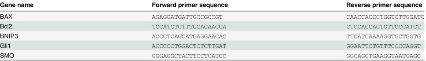
Primer were designed using Primer3 [52,53] and purchased from Invitrogen (Carlsbad, USA), sequences are reported inTable 2. Data were analyzed with iQ™ 5 Optical System Soft-ware, Security Edition (Bio-Rad Laboratories, Hercules, USA). All values were normalized to β-actin endogenous control and RNA relative expression was measured using the 2-ΔΔCt
method.
cDNA from RNA isolated by miRCURY™ RNA Isolation Kit—cell & plant was synthesized using miRCURY™ LNA Universal RT microRNA PCR (EXIQON, Vedbaek, Denmark) accord-ing to manufacturer’s instruction and qRT-PCR analysis of Gli1, SMO, miR-324-5p, miR-326 RNA expression was performed on cDNAs by using ExiLENT SYBR1Green master mix (EXI-QON, Vedbaek, Denmark) and MicroRNA LNA™ PCR primers. Data were analyzed with iQ™ 5 Optical System Software, Security Edition. All values were normalized to non-coding RNA U6, and RNA spike-ins were used as controls for isolation, cDNA synthesis and PCR. RNA rel-ative expression was measured using the 2-ΔΔCtmethod.
Protein expression
To assess Gli1, SuFu and ß-actin protein production and PARP cleavage, K-562 and KU-812 cells were starved overnight with RPMI 1640 culture medium supplemented with 0.5% FCS, 1% L-glutamine and antibiotics (100μg/ml streptomycin and 100 U/ml penicillin), and main-tained in a humidified atmosphere at 37°C and 5% CO2. Later, the medium was removed and
the culture was refreshed with new medium with 10% FCS. Cells were plated at a concentration of 3.5 x 10⁵cells/mL and added with each compound (10 or 20μM) for 24 or 72 h. Later, cells were harvested and lysed in an appropriate buffer containing 1% Triton X-100 and protease inhibitors. Proteins were quantitated by the BCA method (Pierce, Rockford, USA). Equal amounts of total cellular protein were resolved by SDS-polyacrylamide gel electrophoresis, with 10% acrylamide for PARP and 8% for Gli1 and SuFu. Blotted proteins were transferred by electroblotting to a PVDF membrane (Hoefer Pharmacia Biotech, San Francisco, USA) for 1 h at 100 v and 4°C. After a saturation step of 1 h with a solution of 5% nonfat dry milk and 0.1% TBST 10X in agitation at room temperature, anti-PARP, anti-β-actin, anti-Gli1 or anti-SuFu (Cell Signaling Technology, Boston, USA) antibodies were added to the PVDF membrane according to manufacturer’s instruction. On the day after, incubation with HRP-linked second-ary antibodies was carried out for 1 h in agitation at room temperature and then HRP substrate was added (Bio-Rad Laboratories, Hercules, USA). Nonsaturated, immunoreactive bands were detected with a CCD camera gel documentation system (ChemiDocXRS, Bio-Rad Laboratories, Hercules, USA) and then quantitated with Image Lab ver.5.1 analysis software (Bio-Rad Labo-ratories, Hercules, USA).β-actin was used as loading control.
Gene silencing
Gli1 gene silencing on K-562 cells was performed by inserting into cells a siRNA (AUAUCUU GCCCGAAGCAGGUAGUGC) towards Gli1 or a control scrambled siRNA owning the same CG
Table 2. Primer sequences.
Gene name Forward primer sequence Reverse primer sequence
BAX AGAGGATGATTGCCGCCGT CAACCACCCTGGTCTTGGATC
Bcl2 TCCATGTCTTTGGACAACCA CTCCACCAGTGTTCCCATCT
BNIP3 ACCCTCAGCATGAGGAACAC TTCATCAAAAGGTGCTGGTG
Gli1 ACCCCCTGGACTCTCTTGAT GGAATTCTGTTTCCCCAGGT
SMO GGGAGGCTACTTCCTCATCC GGCAGCTGAAGGTAATGAGC
ratio, at a final concentration of 30 nM by means of electroporation. Briefly, cells were centri-fuged at 200g for 10 min. at room temperature, washed with sterile PBS and centricentri-fuged again. Then, cells were resuspended in resuspension buffer R (Invitrogen, Carlsbad, USA) at a final density of 1 x 10⁷cells/ml and siRNA towards Gli1 (or scrambled siRNA) was added at a final concentration of 30 nM. Then cells were electroporated using Neon™ Trasfection System (Invi-trogen, Carlsbad, USA) and according to the following parameters: Pulse Voltage: 1350 v; Pulse Width: 10 ms; Pulse Number: 4; Cell Density: 3 x 10⁷. After electroporation, cells were plated on a 24-well plate at a concentration of 3 x 10⁵cells/well in a final volume of 500μL RPMI supplemented with 1% glutamine without antibiotics and with 10% FCS and incubated for 24 h in a humidified atmosphere at 37°C, 5% CO2for further analysis.
Statistical analysis
Reported data are Mean ± SEM of at least three independent experiment performed in tripli-cate. The statistical analysis was performed by Student’s t test using the Bonferoni correction for multiple test when appropriate. In all cases, only probability (p) values below 0.05 were con-sidered significant.
Supporting Information
S1 Appendix. Synthesis of Smo antagonists. (PDF)
S1 Fig. Synthesis of Smo antagonists.i. HCl, MeOH, r.t; ii. toluene, reflux; iii. NH4SCN,
ace-tone, reflux. (TIF)
Author Contributions
Conceived and designed the experiments: AC FC. Performed the experiments: AC. Analyzed the data: AC FM AN FC. Contributed reagents/materials/analysis tools: EP MR MT FC. Wrote the paper: AC FM FC.
References
1. Rowley JD. Letter: A new consistent chromosomal abnormality in chronic myelogenous leukaemia identified by quinacrine fluorescence and Giemsa staining. Nature 1973 Jun 1; 243(5405):290–3. PMID:4126434
2. Shtivelman E, Lifshitz B, Gale RP, Canaani E. Fused transcript of abl and bcr genes in chronic myelog-enous leukaemia. Nature 1985 Jun 13; 315(6020):550–4. PMID:2989692
3. Ben-Neriah Y, Daley GQ, Mes-Masson AM, Witte ON, Baltimore D. The chronic myelogenous leuke-mia-specific P210 protein is the product of the bcr/abl hybrid gene. Science 1986 Jul 11; 233 (4760):212–4. PMID:3460176
4. Daley GQ, Baltimore D. Transformation of an interleukin 3-dependent hematopoietic cell line by the chronic myelogenous leukemia-specific P210bcr/abl protein. Proc Natl Acad Sci U S A 1988 Dec; 85 (23):9312–6. PMID:3143116
5. Lambert GK, Duhme-Klair AK, Morgan T, Ramjee MK. The background, discovery and clinical develop-ment of BCR-ABL inhibitors. Drug Discov Today 2013 Oct; 18(19–20):992–1000. doi:10.1016/j.drudis. 2013.06.001PMID:23769978
6. Buchdunger E, Zimmermann J, Mett H, Meyer T, Muller M, Druker BJ, et al. Inhibition of the Abl protein-tyrosine kinase in vitro and in vivo by a 2-phenylaminopyrimidine derivative. Cancer Res 1996 Jan 1; 56(1):100–4. PMID:8548747
7. Druker BJ, Tamura S, Buchdunger E, Ohno S, Segal GM, Fanning S, et al. Effects of a selective inhibi-tor of the Abl tyrosine kinase on the growth of Bcr-Abl positive cells. Nat Med 1996 May; 2(5):561–6. PMID:8616716
8. FDA approves Gleevec for leukemia treatment. FDA Consum. 35 (6). 2001.
9. Drugs Approved for Chronic Myelogenous Leukemia (CML). Available: http://www.cancer.gov/about-cancer/treatment/drugs/leukemia#7.
10. Mahon FX, Deininger MW, Schultheis B, Chabrol J, Reiffers J, Goldman JM, et al. Selection and char-acterization of BCR-ABL positive cell lines with differential sensitivity to the tyrosine kinase inhibitor STI571: diverse mechanisms of resistance. Blood 2000 Aug 1; 96(3):1070–9. PMID:10910924 11. de LH, Apperley JF, Khorashad JS, Milojkovic D, Reid AG, Bua M, et al. Imatinib for newly diagnosed
patients with chronic myeloid leukemia: incidence of sustained responses in an intention-to-treat analy-sis. J Clin Oncol 2008 Jul 10; 26(20):3358–63. doi:10.1200/JCO.2007.15.8154PMID:18519952 12. Jabbour E, Kantarjian HM, Abruzzo LV, O'Brien S, Garcia-Manero G, Verstovsek S, et al.
Chromo-somal abnormalities in Philadelphia chromosome negative metaphases appearing during imatinib mesylate therapy in patients with newly diagnosed chronic myeloid leukemia in chronic phase. Blood 2007 Oct 15; 110(8):2991–5. PMID:17625066
13. O'Hare T, Eide CA, Deininger MW. Abl kinase domain mutations and the unsettled problem of Bcr-AblT315I: looking into the future of controlling drug resistance in chronic myeloid leukemia. Clin Lym-phoma Myeloma 2007 Mar; 7 Suppl 3:S120–S130. PMID:17382021
14. Weisberg E, Manley PW, Breitenstein W, Bruggen J, Cowan-Jacob SW, Ray A, et al. Characterization of AMN107, a selective inhibitor of native and mutant Bcr-Abl. Cancer Cell 2005 Feb; 7(2):129–41. PMID:15710326
15. Jorgensen HG, Allan EK, Jordanides NE, Mountford JC, Holyoake TL. Nilotinib exerts equipotent anti-proliferative effects to imatinib and does not induce apoptosis in CD34+ CML cells. Blood 2007 May 1; 109(9):4016–9. PMID:17213283
16. Lombardo LJ, Lee FY, Chen P, Norris D, Barrish JC, Behnia K, et al. Discovery of N-(2-chloro-6-methyl-phenyl)-2-(6-(4-(2-hydroxyethyl)- piperazin-1-yl)-2-methylpyrimidin-4- ylamino)thiazole-5-carboxamide (BMS-354825), a dual Src/Abl kinase inhibitor with potent antitumor activity in preclinical assays. J Med Chem 2004 Dec 30; 47(27):6658–61. PMID:15615512
17. Asaki T, Sugiyama Y, Hamamoto T, Higashioka M, Umehara M, Naito H, et al. Design and synthesis of 3-substituted benzamide derivatives as Bcr-Abl kinase inhibitors. Bioorg Med Chem Lett 2006 Mar 1; 16(5):1421–5. PMID:16332440
18. O'Hare T, Shakespeare WC, Zhu X, Eide CA, Rivera VM, Wang F, et al. AP24534, a pan-BCR-ABL inhibitor for chronic myeloid leukemia, potently inhibits the T315I mutant and overcomes mutation-based resistance. Cancer Cell 2009 Nov 6; 16(5):401–12. doi:10.1016/j.ccr.2009.09.028PMID: 19878872
19. Hehlmann R. How I treat CML blast crisis. Blood 2012 Jul 26; 120(4):737–47. doi: 10.1182/blood-2012-03-380147PMID:22653972
20. Barnes DJ, Melo JV. Primitive, quiescent and difficult to kill: the role of non-proliferating stem cells in chronic myeloid leukemia. Cell Cycle 2006 Dec; 5(24):2862–6. PMID:17172863
21. Long B, Zhu H, Zhu C, Liu T, Meng W. Activation of the Hedgehog pathway in chronic myelogeneous leukemia patients. J Exp Clin Cancer Res 2011; 30:8. doi:10.1186/1756-9966-30-8PMID:21235817 22. Hamilton A, Helgason GV, Schemionek M, Zhang B, Myssina S, Allan EK, et al. Chronic myeloid
leuke-mia stem cells are not dependent on Bcr-Abl kinase activity for their survival. Blood 2012 Feb 9; 119 (6):1501–10. doi:10.1182/blood-2010-12-326843PMID:22184410
23. Crews LA, Jamieson CH. Selective elimination of leukemia stem cells: hitting a moving target. Cancer Lett 2013 Sep 10; 338(1):15–22. doi:10.1016/j.canlet.2012.08.006PMID:22906415
24. Hurtz C, Hatzi K, Cerchietti L, Braig M, Park E, Kim YM, et al. BCL6-mediated repression of p53 is criti-cal for leukemia stem cell survival in chronic myeloid leukemia. J Exp Med 2011 Oct 24; 208(11):2163– 74. doi:10.1084/jem.20110304PMID:21911423
25. Zhang H, Li H, Xi HS, Li S. HIF1alpha is required for survival maintenance of chronic myeloid leukemia stem cells. Blood 2012 Mar 15; 119(11):2595–607. doi:10.1182/blood-2011-10-387381PMID: 22275380
26. Jagani Z, Dorsch M, Warmuth M. Hedgehog pathway activation in chronic myeloid leukemia. Cell Cycle 2010 Sep 1; 9(17):3449–56. PMID:20928937
27. Zhao C, Chen A, Jamieson CH, Fereshteh M, Abrahamsson A, Blum J, et al. Hedgehog signalling is essential for maintenance of cancer stem cells in myeloid leukaemia. Nature 2009 Apr 9; 458 (7239):776–9. doi:10.1038/nature07737PMID:19169242
28. Dierks C, Beigi R, Guo GR, Zirlik K, Stegert MR, Manley P, et al. Expansion of Bcr-Abl-positive leuke-mic stem cells is dependent on Hedgehog pathway activation. Cancer Cell 2008 Sep 9; 14(3):238–49. doi:10.1016/j.ccr.2008.08.003PMID:18772113
29. Merchant AA, Matsui W. Targeting Hedgehog—a cancer stem cell pathway. Clin Cancer Res 2010 Jun 15; 16(12):3130–40. doi:10.1158/1078-0432.CCR-09-2846PMID:20530699
30. Regl G, Kasper M, Schnidar H, Eichberger T, Neill GW, Philpott MP, et al. Activation of the BCL2 pro-moter in response to Hedgehog/GLI signal transduction is predominantly mediated by GLI2. Cancer Res 2004 Nov 1; 64(21):7724–31. PMID:15520176
31. Kenney AM, Rowitch DH. Sonic hedgehog promotes G(1) cyclin expression and sustained cell cycle progression in mammalian neuronal precursors. Mol Cell Biol 2000 Dec; 20(23):9055–67. PMID: 11074003
32. Kenney AM, Cole MD, Rowitch DH. Nmyc upregulation by sonic hedgehog signaling promotes prolifer-ation in developing cerebellar granule neuron precursors. Development 2003 Jan; 130(1):15–28. PMID:12441288
33. Bigelow RL, Chari NS, Unden AB, Spurgers KB, Lee S, Roop DR, et al. Transcriptional regulation of bcl-2 mediated by the sonic hedgehog signaling pathway through gli-1. J Biol Chem 2004 Jan 9; 279 (2):1197–205. PMID:14555646
34. Duman-Scheel M, Weng L, Xin S, Du W. Hedgehog regulates cell growth and proliferation by inducing Cyclin D and Cyclin E. Nature 2002 May 16; 417(6886):299–304. PMID:12015606
35. Lee J, Platt KA, Censullo P, Altaba A. Gli1 is a target of Sonic hedgehog that induces ventral neural tube development. Development 1997 Jul; 124(13):2537–52. PMID:9216996
36. Goodrich LV, Johnson RL, Milenkovic L, McMahon JA, Scott MP. Conservation of the hedgehog/ patched signaling pathway from flies to mice: induction of a mouse patched gene by Hedgehog. Genes Dev 1996 Feb 1; 10(3):301–12. PMID:8595881
37. Frank-Kamenetsky M, Zhang XM, Bottega S, Guicherit O, Wichterle H, Dudek H, et al. Small-molecule modulators of Hedgehog signaling: identification and characterization of Smoothened agonists and antagonists. J Biol 2002 Nov 6; 1(2):10. PMID:12437772
38. Tang T, Tang JY, Li D, Reich M, Callahan CA, Fu L, et al. Targeting superficial or nodular Basal cell car-cinoma with topically formulated small molecule inhibitor of smoothened. Clin Cancer Res 2011 May 15; 17(10):3378–87. doi:10.1158/1078-0432.CCR-10-3370PMID:21558397
39. Pellicano F, Simara P, Sinclair A, Helgason GV, Copland M, Grant S, et al. The MEK inhibitor
PD184352 enhances BMS-214662-induced apoptosis in CD34+ CML stem/progenitor cells. Leukemia 2011 Jul; 25(7):1159–67. doi:10.1038/leu.2011.67PMID:21483442
40. Samanta AK, Chakraborty SN, Wang Y, Schlette E, Reddy EP, Arlinghaus RB. Destabilization of Bcr-Abl/Jak2 Network by a Jak2/Abl Kinase Inhibitor ON044580 Overcomes Drug Resistance in Blast Cri-sis Chronic Myelogenous Leukemia (CML). Genes Cancer 2010 Apr; 1(4):346–59. doi:10.1177/ 1947601910372232PMID:20798787
41. Liao HF, Su YC, Zheng ZY, Jhih CC, Hou MH, Chao KS, et al. Sonic hedgehog signaling regulates Bcr-Abl expression in human chronic myeloid leukemia cells. Biomed Pharmacother 2012 Jul; 66(5):378– 83. doi:10.1016/j.biopha.2011.12.008PMID:22397755
42. Ferretti E, De SE, Miele E, Laneve P, Po A, Pelloni M, et al. Concerted microRNA control of Hedgehog signalling in cerebellar neuronal progenitor and tumour cells. EMBO J 2008 Oct 8; 27(19):2616–27. doi: 10.1038/emboj.2008.172PMID:18756266
43. Babashah S, Sadeghizadeh M, Hajifathali A, Tavirani MR, Zomorod MS, Ghadiani M, et al. Targeting of the signal transducer Smo links microRNA-326 to the oncogenic Hedgehog pathway in CD34+ CML stem/progenitor cells. Int J Cancer 2013 Aug 1; 133(3):579–89. doi:10.1002/ijc.28043PMID: 23341351
44. Manetti F, Faure H, Roudaut H, Gorojankina T, Traiffort E, Schoenfelder A, et al. Virtual screening-based discovery and mechanistic characterization of the acylthiourea MRT-10 family as smoothened antagonists. Mol Pharmacol 2010 Oct; 78(4):658–65. doi:10.1124/mol.110.065102PMID:20664000 45. Solinas A, Faure H, Roudaut H, Traiffort E, Schoenfelder A, Mann A, et al. Acylthiourea, acylurea, and
acylguanidine derivatives with potent hedgehog inhibiting activity. J Med Chem 2012 Feb 23; 55 (4):1559–71. doi:10.1021/jm2013369PMID:22268551
46. Roudaut H, Traiffort E, Gorojankina T, Vincent L, Faure H, Schoenfelder A, et al. Identification and mechanism of action of the acylguanidine MRT-83, a novel potent Smoothened antagonist. Mol Phar-macol 2011 Mar; 79(3):453–60. doi:10.1124/mol.110.069708PMID:21177415
47. Chen MH, Wilson CW, Li YJ, Law KK, Lu CS, Gacayan R, et al. Cilium-independent regulation of Gli protein function by Sufu in Hedgehog signaling is evolutionarily conserved. Genes Dev 2009 Aug 15; 23(16):1910–28. doi:10.1101/gad.1794109PMID:19684112
48. Naldini A, Morena E, Pucci A, Miglietta D, Riboldi E, Sozzani S, et al. Hypoxia affects dendritic cell sur-vival: role of the hypoxia-inducible factor-1alpha and lipopolysaccharide. J Cell Physiol 2012 Feb; 227 (2):587–95. doi:10.1002/jcp.22761PMID:21448921
49. Lee J, Giordano S, Zhang J. Autophagy, mitochondria and oxidative stress: cross-talk and redox signal-ling. Biochem J 2012 Jan 15; 441(2):523–40. doi:10.1042/BJ20111451PMID:22187934
50. Warzecha J, Bonke L, Koehl U, Munkelt D, Gottig S, Percic D, et al. The hedgehog inhibitor cyclopa-mine induces apoptosis in leukemic cells in vitro. Leuk Lymphoma 2008 Dec; 49(12):2383–6. doi:10. 1080/10428190802510315PMID:19052992
51. Katagiri S, Tauchi T, Okabe S, Minami Y, Kimura S, Maekawa T, et al. Combination of ponatinib with Hedgehog antagonist vismodegib for therapy-resistant BCR-ABL1-positive leukemia. Clin Cancer Res 2013 Mar 15; 19(6):1422–32. doi:10.1158/1078-0432.CCR-12-1777PMID:23319824
52. Untergasser A, Cutcutache I, Koressaar T, Ye J, Faircloth BC, Remm M, et al. Primer3—new capabili-ties and interfaces. Nucleic Acids Res 2012 Aug; 40(15):e115. PMID:22730293
53. Koressaar T, Remm M. Enhancements and modifications of primer design program Primer3. Bioinfor-matics 2007 May 15; 23(10):1289–91. PMID:17379693