Short Report
Mol Syndromol 2010;1:239–245 DOI: 10.1159/000328135
Unmasking of a Recessive SCARF2 Mutation
by a 22q11.12 de novo Deletion in a Patient
with Van den Ende-Gupta Syndrome
M.F. Bedeschi
a
L. Colombo
b
F. Mari
c
K. Hofmann
d
A. Rauch
d, e
B. Gentilin
a
A. Renieri
c
D. Clerici
b
a
Medical Genetic Unit and b NICU, Fondazione IRCCS Cà Granda Ospedale Maggiore Policlinico, Università degli
Studi di Milano, Milano , and c
Medical Genetics, Molecular Biology Department, University of Siena, Siena , Italy;
d
Institute of Human Genetics, Friedrich-Alexander University of Erlangen-Nuremberg, Erlangen , Germany;
e
Institute of Medical Genetics, University of Zurich, Zurich , Switzerland
Van den Ende-Gupta syndrome (VDEGS; MIM 600920) is a very rare autosomal recessive disease char-a cterized by distinct crchar-aniofchar-acichar-al char-and skeletchar-al char-anomchar-alies such as blepharophimosis, down-slanted eyes, a flat and wide nasal bridge, malar and/or maxillary hypoplasia, prominent ears, a narrow and beaked nose, an everted lower lip, palatal abnormalities, camptodactyly, arachno-dactyly, long thumbs, hallux valgus, flexion contractures, slender ribs, hooked clavicles, and bowed long bones [van den Ende et al., 1992; Bistritzer et al., 1993; Gupta et al., 1995; Phadke et al., 1998; Schweitzer et al., 2003; Guerra et al., 2005; Carr et al., 2007; Leal and Silva, 2009]. In 1992, van den Ende and colleagues first reported a 10-yeaold girl, born to consanguineous Brazilian pa r-ents, with characteristic features. She had normal intel-ligence, blepharophimosis, malar hypoplasia, a beaked nose, an everted lower lip, and arachnocamptodactyly of fingers and toes. The authors suggested a ‘new’ autosomal recessive Marden-Walker-like syndrome. Gupta et al. [1995] reported a 3-year-old girl with similar features whose parents were first cousins. X-ray showed maxillary hypoplasia with a small anterior cranial fossa, slender ribs with lateral ends hooked to the clavicles, an absent glenoid fossa, bowed humeri, ulnae and femora, and Key Words
Arachnocamptodactyly ⴢ Blepharophimosis ⴢ Congenital
contractures ⴢ SCARF2 ⴢ Van den Ende-Gupta syndrome
Abstract
Van den Ende-Gupta syndrome (VDEGS) is a congenital con-dition characterized by craniofacial and skeletal manifesta-tions, specifically blepharophimosis, malar and maxillary hy-poplasia, distinctive nose, arachnocamptodactyly, and long slender bones of the hands and feet. To date, only 24 patients have been described. It is generally thought that the syn-drome is transmitted by an autosomal recessive mode of in-heritance, although evidence for genetic heterogeneity has recently been presented. We report on a girl followed from birth up to 3 years of life with a set of peculiar minor anoma-lies, arachnocamptodactyly of hands and feet, characteris-tic of VDEGS in association with a 22q11.12 deletion. Recent-ly, the VDEGS gene was mapped to the DiGeorge syndrome region on 22q11.2, and homozygous mutations in the SCARF2 gene were identified. We now report the first patient with VDEGS due to compound heterozygosity for the common 22q11.2 microdeletion and a hemizygous SCARF2 splice site
mutation. Copyright © 2011 S. Karger AG, Basel
Accepted: March 28, 2011 by A. Rauch
Published online: May 18, 2011
Maria Francesca Bedeschi © 2011 S. Karger AG, Basel
tively long fibulae. The bones of the hands and feet were relatively long, with the exception of the terminal phalan-ges which were shortened.
Following these initial observations, Phadke et al. [1998] reported 2 unrelated Indian girls with some fea-tures of the condition. Bistritzer et al. [1993] reported 2 double second cousins from an inbred pedigree suspect-ed to have VDEGS. Cardiac examination and general de-velopment were normal. One female infant had a promi-nent clitoris and fused labia. Schweitzer et al. [2003] re-ported 2 Hispanic brothers born to unrelated parents, who both had distinctive cerebellar enlargement, a new finding for this disorder. Ali et al. [2010] observed cuta-neous syndactyly of toes 2 and 3 as a consistent feature in their patients. Further patients with this constellation of anomalies have been reported, reinforcing the hypothesis of an autosomal recessive mode of inheritance [Bistritzer et al., 1993; Schweitzer et al., 2003; Carr et al., 2007; Ali et al., 2010]. Only one report by Leal and Silva [2009] sug-gested genetic heterogeneity and an autosomal dominant trait based on the observation of 3 affected individuals, 2 brothers and their half-sister, assuming gonadal mosa-icism.
The patients affected by VDEGS had a normal karyo-type, and FISH for specific 22q11.12 abnormalities performed in 3 cases was normal [Gupta et al., 1995; Schweitzer et al., 2003; Carr et al., 2007]. Recently, Anas-tasio et al. [2010] mapped VDEGS to the DiGeorge syn-drome region on 22q11.2 and identified homozygous mu-tations in the SCARF2 gene as the underlying cause in 4 patients from 3 highly inbred Bedouin families. We now report the first patient with VDEGS due to compound heterozygosity for the common 22q11.2 microdeletion and a hemizygous SCARF2 splice site mutation.
Clinical Report
The female proband is the first child born to a 30-year-old mother and a 32-year-old father. Both parents were healthy and non-consanguineous, and no drugs had been taken during the gestational period. Pregnancy was uneventful except for bilateral clubfeet, mild bilateral pyelectasis, and lack of visualization of opening-closing hand movements observed at 21+6 weeks of ges-tation by prenatal ultrasound scan. Growth parameters were: bi-parietal diameter 52 mm (25th–50th centile), head circumference 193 mm (25th–50th centile), abdominal circumference 160 mm (25th–50th centile), femur length 38 mm (50th centile). Cytoge-netic analysis performed on a chorionic villous sample was nor-mal fenor-male (46,XX).
The girl was delivered by caesarean section at 38 weeks of ges-tation with an APGAR score of 7–9 at the first and fifth minute.
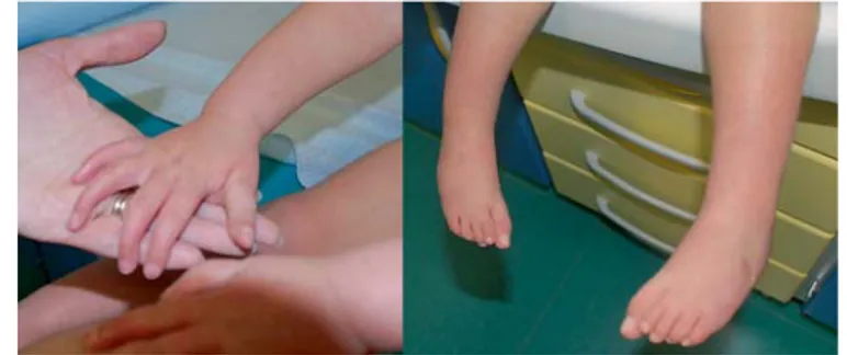
Birth weight was 2,450 g (3rd–10th centile), length 50 cm (50th centile), and head circumference 34 cm (10th centile). At birth, minor facial anomalies were evident including hypertelorism, blepharophimosis, unilateral microphtalmia, corneal opacity, broad nasal bridge, narrow nose, malar hypoplasia, long philtrum, low set and posterior angulated ears, thin vermillion of the lips, microretrognathia, and microstomia. Camptodactyly and arach-nodactyly of fingers and toes, proximally set thumb, bilateral club-feet (most marked on right foot), and cutaneous syndactyly of 2nd/3rd toes bilaterally were also present. In addition, hypo-ex-tensibility of the proximal interphalangeal joints between the sec-ond and fifth fingers and self-limiting joint contractures without stiffness at knee and elbow level were observed ( fig. 1 a, b).
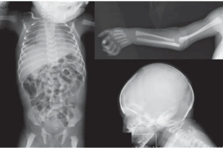
At day 2 of life, she had a transient episode of hypocalcemia (7.8 mg/dl). Cerebral and abdominal ultrasound scans were per-formed and were both normal. No anomalies were evident in the brainstem auditory evoked response. An ophthalmological ex-amination showed blepharophimosis, right sclerocornea, bilat-eral microcornea and cataracts. Fundus oculi was normal. A to-tal-body skeletal X-ray survey was performed and revealed mild dolichocephaly, long and thin phalanges and metatarsals, 11 pairs of thin ribs, and a bilateral clavicle hook ( fig. 2 ).
On follow-up at 1 year and at 2 years 9 months, her growth and development were normal. At the latter examination she mea-sured: height 87 cm (10th centile), weight 11.9 kg (10th centile), and head circumference 48.3 cm (10th centile).
The same minor facial anomalies were evident, as was arach-nodactyly of hands and feet, but camptodactyly and bilateral clubfeet had improved ( fig. 3, 4 ). A cardiac ultrasound scan was performed and showed an ostium secundum atrial septal defect. The child had a mild motor developmental delay that improved progressively with physiotherapy. The major motor milestones were sitting at 8 months and walking at 22 months. Speech devel-opment was normal.
Molecular Analysis
Array-comparative genomic hybridization (array-CGH) anal-ysis was performed using commercially available oligonucleotide microarrays containing about 99,000 60-mer probes with an esti-mated median spatial resolution of nearly 10 kb and a functional resolution close to 35 kb (Human Genome CGH Microarray 105A Kit, Agilent Technologies). Labeling and hybridization were per-formed following the protocols provided by the manufacturer (Agilent Technologies according to the Agilent protocol Oligo-nucleotide Array-Based CGH for Genomic DNA Analysis v 2.0). Slides were dried and then scanned using an Agilent G2565BA DNA microarray scanner.
Image analysis was performed by CGH Analytics software v. 3.4.40 with default settings. The software automatically deter-mines the fluorescence intensities of the spots for both fluoro-chromes, performs background subtraction and data normaliza-tion, and compiles the data into a spreadsheet that links the fluo-rescent signal of every oligo on the array to the oligo name, its position on the array, and its position in the genome. The linear order of the oligos is reconstituted in the ratio plots consistent with an ideogram. The ratio plot is arbitrarily assigned such that gains and losses in DNA copy number at a particular locus are
observed as a deviation of the ratio plot from a modal value of 1.0. DNA sequence information is taken from the public UCSC data-base (Human Genome Browser, http://genome.ucsc.edu, March 2006 assembly).
Mutational analysis of the SCARF2 gene in the patient was performed by Sanger sequencing using an ABI 3730 capillary se-quencer after PCR amplification with intronic primers for the 11 coding exons. In addition, both parents were sequenced for exon 4 and flanking intronic regions.
Results
A 22q11.1–q11.21 microdeletion was identified with the proximal breakpoint in 22q11.1 located between 17.08 and 17.27 Mb (last oligonucleotide present and first de-leted, respectively) and the distal breakpoint between 19.83 and 19.89 Mb in 22q11.21 (last oligonucleotide
de-a b
Fig. 1. Clinical features of the proband include a blepharophimosis, flat wide nasal bridge, malar hypoplasia, and prominent ears, and b arachnocamptodactyly of fingers and toes, proximally set thumb, and bilateral club-feet.
Fig. 2. Radiographs of the patient showing hooked clavicles, slen-der ribs, and long bones.
leted and first present, respectively, referring to hg18). Thus the deletion corresponds to the common DiGeorge/ VCFS ‘3-Mb’ deletion between low copy repeats LCR-A (LCR22–2ⴕ) and LCR-D (LCR22–4ⴕ) [Rauch et al., 2005; Guo et al., 2011]. To confirm the array data, a second ar-ray-CGH experiment was performed in the patient and parents. The deletion was confirmed in the patient, while the parents showed a normal result. Analysis of the de-leted region suggested the absence of at least 39 known genes ( fig. 5 ).
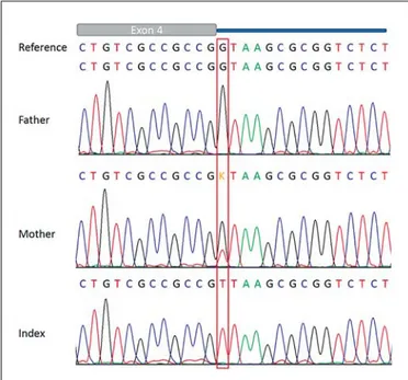
Sanger sequencing of the SCARF2 gene revealed the c.854 + 1G 1 T (intron 4) splice acceptor mutation hemi-zygous in the patient and heterohemi-zygous in the healthy
mother, while the father showed the wild-type sequence ( fig. 6 ). Online bioinformatics tools such as ‘Mutation taster’ and ‘Human splicing finder’ both indicated loss of the splicing site.
Discussion
The facial phenotype of our case and the pattern of congenital anomalies are similar to that of patients previ-ously reported by van den Ende et al. [1992] and Gupta et al. [1995]. Since the VDEGS was considered an autosomal recessive condition, detection of a heterozygous 22q11.2
Fig. 3. Proband at 1 year of age and at 2 years 9 months. Fig. 4. Hands and feet of the proband at 2 years 9 months.
Fig. 5. Results of array-CGH. On the left the chromosome 22 ideo-gram is shown indicating the 22q11.21 microdeletion in the ratio profile. On the right, the deletion indicated by the array-CGH ex-periment is mapped against the corresponding genomic region in
the UCSC genome browser build 36.1 (2006). In the lower part, the extent of the deletion in the patient described in this report, which represents the common ‘3-Mb’ deletion size, is indicated (green line).
deletion in our patient was surprising. The recent identi-fication of homozygous mutations in the SCARF2 gene in inbred families with VDEGS [Anastasio et al., 2010] sug-gested unmasking of a recessive mutation of SCARF2, which is located within the 22q11.2 common deletion re-gion. Subsequent sequencing of SCARF2 in our patient revealed indeed a maternally inherited splice site muta-tion in addimuta-tion to the 22q11.2 microdelemuta-tion and hence absence of a functional SCARF2 gene. SCARF2 contains putative epidermal growth factor-like domains in its ex-tracellular domain, along with a number of positively charged residues in its intracellular domain, indicating that it may be involved in intracellular signaling. Scarf2 is expressed in mouse branchial or pharyngeal arches and mandibular maxillary and urogenital ridge tissues [Anastasio et al., 2010].
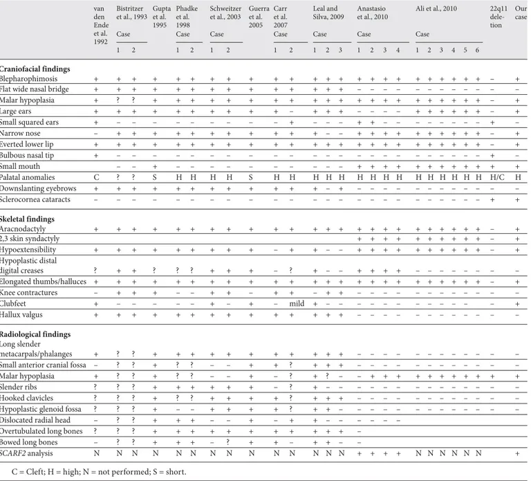
Comparison of the clinical findings of our case with clinical characteristics of 24 patients described with this condition ( table 1 ) [van den Ende et al., 1992; Bistrit-zer et al., 1993; Gupta et al., 1995; Phadke et al., 1998; Schweitzer et al., 2003; Guerra et al., 2005; Carr et al., 2007; Leal and Silva 2009; Ali et al., 2010; Anastasio et al.,
2010] confirms considerable overlap of our case with the published patients with VDEGS, particularly the facial appearance and the arachnocamptodactyly. The most common anomalies are indeed arachnodactyly, campto-dactyly, an unusual facial appearance with blepharophi-mosis, beaked nose, malar hypoplasia, everted lips, and prominent ears. Growth and intelligence are normal in all cases. The finger contractures usually gradually im-prove and do not cause functional limitations. In all cas-es, radiographic features showed slender long boncas-es, metacarpals, metatarsal, and phalanges. Further investi-gation is warranted concerning the relevance of cerebel-lar encerebel-largement and learning difficulties reported by Schweitzer et al. [2003].
In addition, the patient reported here had sclerocornea and cataracts never described previously in VDEGS. In-stead, ocular findings such as sclerocornea, microphtal-mia, and cataract have been described in the 22q11 dele-tion syndrome [Binenbaum et al., 2008; Casteels et al., 2008]. The patient here reported presented with a 2.56– 2.8 Mb deletion that represents the typical 3-Mb deletion at 22q11.2 that is usually observed. In addition to the oc-ular anomalies, she showed some typical clinical features of the 22q deletion phenotype such as ostium secundum atrial septal defect and transient neonatal hypocalcemia [McDonald-McGinn and Sullivan, 2011]. The facial fea-tures such as small mouth, prominent nose, hyper-telorism, and narrow palpebral fissures, although over-lapping with the 22q11 deletion syndrome, were clearly dominated by the characteristics of VDEGS. She had no other 22q11.2 deletion features, in particular no velopha-ryngeal insufficiency, no thymic hypoplasia, and no evi-dence of immunodeficiency.
Limb anomalies are uncommon in 22q11.2 deletion patients. A few studies reported patients with a 22q11.2 deletion and polydactyly, ectrodactyly, thumb anoma-lies, minor upper/lower limb skeletal anomaanoma-lies, synos-tosis, and contractures, but neither arachnodactyly nor hypo-extensibility of fingers are described so far [Kokit-su-Nakata et al., 2008]. On the contrary, arachnocamp-todactyly is the most characteristic clinical feature of VDEGS.
To our knowledge, no published patient with a 22q11.2 deletion showed clinical features similar to a VDEGS phenotype. On the other hand, cases with VDEGS had a normal karyotype, and FISH for specific 22q11.2 abnor-malities performed in 3 cases was normal [Gupta et al., 1995; Schweitzer et al., 2003; Carr et al., 2007].
VDEGS has been generally considered to be an auto-somal recessive entity, given that 3 affected individuals
Fig. 6. Electropherograms showing the SCARF2 c.854 + 1G 1 T (intron 4) splice acceptor mutation hemizygous in the patient (in-dex) and heterozygous in the healthy mother. The relatively small mutation versus wild-type peak in the mother may be caused by preferential amplification of the wild-type allele or by mosaicism for the mutation.
from different families were born to normal and consan-guineous parents [van den Ende et al., 1992; Bistritzer et al., 1993; Gupta et al., 1995]. The identification of homo-zygous mutations in the SCARF2 gene as the underlying cause reported by Anastasio et al. [2010] in VDEGS pa-tients from 3 consanguineous families further supports the conclusion that VDEGS is an autosomal recessive en-tity. Our case, though, demonstrates that sequencing alone might falsely indicate a homozygous mutation and
that the recurrence risk is not necessarily 25% in all cases. After exclusion of a compensating deletion/duplication event by FISH in the parents [Alkalay et al., 2011], we would assume an approximately 1% recurrence risk for the 22q11.2 deletion with reference to the possibility of a germ line mosaicism. Accordingly, recurrence risk for VDEGS would be approximately 0.5%.
Recently, because 3 affected individuals, 2 brothers and their half-sister, were reported, Leal and Silva [2009]
Table 1. C linical features in VDEGS van den Ende et al. 1992 Bistritzer et al., 1993 Gupta et al. 1995 Phadke et al. 1998 Schweitzer et al., 2003 Guerra et al. 2005 Carr et al. 2007 Leal and Silva, 2009 Anastasio et al., 2010 Ali et al., 2010 22q11 dele-tion Our case
Case Case Case Case Case Case C ase
1 2 1 2 1 2 1 2 1 2 3 1 2 3 4 1 2 3 4 5 6
Craniofacial findings
Blepharophimosis + + + + + + + + + + + + + + + + + + + + + + + + – +
Flat wide nasal bridge + + + + + + + + + + + + + + – – – – – – – – – – – –
Malar hypoplasia + ? ? + + + + + + + + + + + + + + + + + + + + + – +
Large ears + + + + + + + + + + – + + + – – – – + + + + + + – +
Small squared ears + – – – – – – – – – + – – – + + – – – – – – – – + –
Narrow nose – + + + + + + + + + + + – – + + + + + + + + + + – +
Everted lower lip + + + + + + + + + + + + + + + + + + + + + + + + – +
Bulbous nasal tip + – – – – – – – – – – – – – – – – – – – – – – – + –
Small mouth – – + – – – – – – – – – – + + + + + + + + + + + + Palatal anomalies C ? ? S H H H H S H H H H H H H H H H H H H H H H/C H Downslanting eyebrows + + + + + + + + + + + + – + – – – – – – – – – – – – Sclerocornea cataracts – – – – – – – – – – – – – – – – – – – – – – – – + + Skeletal findings Aracnodactyly + + + + + + + + + + + + + + + + + + + + + + + + – + 2,3 skin syndactyly + + + + + + + + + + – + Hypoextensibility + + + + + + + + + – + + – – + + + + + + + + + + – + Hypoplastic distal digital creases ? + + ? ? ? + + + – ? + – – + + + + – – – – – – – – Elongated thumbs/halluces + + + + + + + + + + + + + + + + + + + + + + + + – + Knee contractures – + + + – – + + – + + – + + – – – – – – – – – – – – Clubfeet + – – – – – + – + – mild + – – – – – – – – – – – – + Hallux valgus + + + + + + + + + + + + + + – – – – – – – – – – – – Radiological findings Long slender metacarpals/phalanges + ? ? + + + + + + + + + + + – – – – – – – – – – – –
Small anterior cranial fossa – ? ? + ? ? – – + + ? + + + – – – – – – – – – – – –
Malar hypoplasia + ? ? + ? ? – – + – ? + ? – – + + + + + + + + + + +
Slender ribs ? ? ? + + + + + + – ? + – – – – – – – – – – – – – –
Hooked clavicles ? ? ? + ? ? + + + + ? + + + – – – – – – – – – – – –
Hypoplastic glenoid fossa ? ? ? + – – + + + + ? + + – – – – – – – – – – – – –
Dislocated radial head – ? ? + + + – – + – + + – – – – – –
Overtubulated long bones ? ? ? + + + + + + + + + + + –
Bowed long bones – ? ? + + + – ? + + – + + – –
SCARF2 analysis N N N N N N N N N N N N N N + + + + N N N N N N +
hypothesized an autosomal dominant transmission and gonadal mosaicism, suggesting genetic heterogeneity. However, we consider it more likely, that all 3 half-sib-lings are by change carriers of recessive mutations, a hy-pothesis that could now be proven.
Acknowledgments
The authors would like to gratefully thank the patient and her family for their consent to publish this data. We thank Dr. Filo-mena Papa, UOD Medical Genetic Unit, University of Siena, for her molecular contribution.
References
Ali R, Almureikhi M, Al-Musaifri F, Bhat V, Tee-bi A, Ben-Omran T: Further delineation of the Van den Ende-Gupta syndrome. Am J Med Genet A 152: 3095–3100 (2010). Alkalay AA, Guo T, Montagna C, Digilio MC,
Dallapiccola B, et al: Genetic dosage com-pensation in a family with velo-cardio-fa-cial/DiGeorge/22q11.2 deletion syndrome. Am J Med Genet A 155: 548–554 (2011). Anastasio N, Ben-Omran T, Teebi A, Ha KC,
Lalonde E, et al: Mutations in SCARF2 are responsible for Van den Ende-Gupta syn-drome. Am J Hum Genet 87: 553–9 (2010). Binenbaum G, McDonald-McGinn DM, Zackai
EH, Walker BM, Coleman K, et al: Sclerocor-nea associated with the chromosome 22q11.2 deletion syndrome. Am J Med Genet A 146: 904–909 (2008).
Bistritzer T, Fried K, Lahat E, Dvir M, Goldberg M: Congenital contractural arachnodactyly in two double second cousins: possible ho-mozygosity. Clin Genet 44: 15–19 (1993). Carr CW, Carron JD, Lachman RS,
Abdul-Rah-man OA: Van den Ende-Gupta syndrome: laryngeal abnormalities in two siblings. Am J Med Genet Part A 143A:2706–2711 (2007).
Casteels I, Casaer P, Gewillig M, Swillen A, Devriendt K: Ocular findings in children with a microdeletion in chromosome 22q11.2. Eur J Pediatr 167: 751–755 (2008). Guerra D, Sanchez O, Richieri-Costa A: Van den
Ende-Gupta syndrome of blepharophimosis, arachnodactyly, and congenital contrac-tures. Am J Med Genet Part A 136A:377–380 (2005).
Guo X, Freyer L, Morrow B, Zheng D: Charac-terization of the past and current duplication activities in the human 22q11.2 region. BMC Genomics 12: 71 (2011).
Gupta A, Hall CM, Ransley YF, Murday VA: A new autosomal recessive syndrome of char-acteristic facies, joint contractures, skeletal abnormalities, and normal development: second report with further clinical delinea-tion. J Med Genet 32: 809–812 (1995). Kokitsu-Nakata NM, Guion-Almeida ML,
Rich-ieri-Costa A: 22q11 deletion syndrome and limb anomalies: report on two Brazilian pa-tients. Cleft Palate Craniofac J 45: 561–566 (2008).
Leal GF, Silva EO: Van den Ende-Gupta syn-drome: evidence for genetic heterogeneity. Am J Med Genet part A 149A:1293–1295 (2009).
McDonald-McGinn DM, Sullivan KE: Chromo-some 22q11.2 deletion syndrome (DiGeorge syndrome/velocardiofacial syndrome). Med-icine (Baltimore) 90: 1–18 (2011).
Phadke SR, Gulati R, Agarwal SS: Further delin-eation of a new (van den Ende-Gupta) syn-drome of blepharophimosis, contractural arachnodactyly, and characteristic face. Am J Med Genet 77: 16–18 (1998).
Rauch A, Zink S, Zweier C, Thiel CT, Koch A, et al: Systematic assessement of atypical dele-tions reveals genotype-phenotype correla-tion in 22q11.2. J Med Genet 42: 871–876 (2005).
Schweitzer DN, Lachman RS, Pressman BD, Graham JM Jr: Van den Ende-Gupta syn-drome of blepharophimosis, arachnodacty-ly, and congenital contractures: clinical de-lineation and recurrence in brothers. Am J Med Genet Part A 118A:267–273 (2003). van den Ende J, van Bever Y, Rodini ESO,
Rich-ieri-Costa A: Marden-Walker-like syndrome without psychomotor retardation: report of a Brazilian girl born to consanguineous par-ents. Am J Med Genet 42: 467–469 (1992).