DOCTORAL SCHOOL IN BIOLOGY
Section: Biology Applied to Human Health
CYCLE XXVII
Evolution of virulence in pathogenic
Escherichia coli strains:
Impact on public health
Laura Grande,
MSc. (Hons), Biology
Tutor
Prof. Paolo Visca
Co-Tutor
Dr. Stefano Morabito
PhD Coordinator
Prof. Paolo Visca
Abstract
Diarrheagenic Escherichia coli (DEC), including Verocytotoxin-producing E. coli (VTEC), are a significant
public health issue worldwide. The management of the infections caused by these bacterial pathogens is
complicated by their extreme heterogeneity, including strains causing a plethora of symptoms spanning from
uncomplicated diarrhoea to life-threatening systemic sequelae such as the haemolytic-uremic syndrome
(HUS).
Considerable efforts have been devoted by the scientific community to understand the evolutionary forces
leading to the emergence of new E. coli pathogenic clones. However, E. coli genome is very dynamic and
evolves continuously through horizontal gene transfer. The main objective of the present piece of research
was to investigate the molecular bases of the evolution of pathogenic E. coli, mainly DEC.
We focused on VTEC as a model of dynamic pathogenic group (pathotype) encompassing many established
Escherichia coli
pathotypes. VTEC pathogenicity mainly relies on two aspects, the ability to efficiently
colonize the host gut and the production of the Verocytotoxins (VTs). Genes encoding the VTs (vtx) are
carried by lambdoid bacteriophages whose genome is normally integrated into the E. coli chromosome.
We have investigated the distribution of several Mobile Genetic Elements (MGEs) encoding virulence
features in different VTEC subpopulations and studied their evolution through the differentiation of the
virulence genes into different allelic variants. Additionally, we studied the different bacteriophages
transporting the vtx genes into different VTEC types and their role in manipulating the host biology. Finally,
we assessed the possibility that the spreading of VT-phages may or may not be subjected to host-related
barriers by probing the possible acquisition of such phages by E. coli strains with a genetic background
different from VTEC.
The work presented here has been largely based on the genomic comparison of VTEC strains isolated from
human and animal sources and held in the collections of the EU RL VTEC and of the collaborating
institutions. This approach led to the identification of a pathogenicity island encoding an allelic variant of the
Subtilase cytotoxin (subAB2, Chapter 3) and to the description of the allelic variants of the
colonization-associated virulence factor toxB (toxB1 and toxB2, Chapter 4). Moreover, the distribution analysis of the two
virulence factors on a large panel of E. coli strains allowed making inference on their role in the VTEC
pathogenetic process.
The genomic approach has also been used to identify and characterize different VT-phages present in E. coli
strains associated to the most severe form of infection, the HUS. In detail, a VT-phage present in VTEC
O157 strains and able to influence the regulation of genes involved in the colonization mechanism has been
described (Φ-8, Chapter 5). Additionally, the complete sequence of the VT-phage isolated from a recently
described E. coli pathotype, the Enteroaggregative Haemorrhagic E. coli (EAHEC) has been obtained and
compared to other VT-phages sequences with the aim to help unravelling their complicate biology (Phi-191,
Chapter 5). With the same aim, the evaluation of the stable acquisition of VT-phages by non and pathogenic
E. coli
strains belonging to all the known pathotypes led to the conclusion that such phages show a host
range broader than expected (Chapter 6). This observation, together with other evidences from the literature
raises the hypothesis that probably any Enterobacteria equipped with an efficient colonization machinery
could be potentially acquire a VT-phage generating clones with augmented virulence potential for humans,
as it happened with the EAHEC O104:H4 that caused a large outbreak of HUS in Germany in 2011.
Acknowledgements
Looking back, I can confirm this is the most difficult part of my PhD thesis to be written.
This piece of Research is the result of a three-years period spent at the Unit of Foodborne Zoonoses at the Istituto Superiore di Sanità (ISS), where periods of pressure were often alternated by pleasant moments. I’m really grateful and at the same time conscious I could never hope for myself a better place to spend this period, from both a human and a scientific side.
Many thanks to prof. Paolo Visca, who acted as a supervisor before and during my PhD program.
My warm thanks goes to dr. Alfredo Caprioli, first of all for guesting me at the Unit he heads at ISS, for always being encouraging during this period and always listening to me whenever I needed, no matter how busy he could be. I owe much gratitude to my supervisor dr. Stefano Morabito, who patiently tried to teach me how to move in the fascinating world of Research and for always encouraging me to give the best. I also thank him for his passionate attitude in work and life, for always being a enjoyable and optimistic person, and for often representing a friend, other than a supervisor. I just hope to make him proud of his investment on me...
I would really want to warmly thank the “droplets”, Valeria, Rosy, and Antonella. Vale, for taking care of me in my really first steps in a lab, for being a strong and precious support even in my worst moments, for our gossip time, both hilarious and serious. Rosy, thanks for being first of all a guide and a great example to follow, an advisor, a precious friend, depending on my needs. Anto, for her freshness and spontaneity, for often being a “life coach” for all of us, for her special way to help me in my toughest moments, and for the tears she will shed while reading these words. I’m also very grateful to all the people I met during this period, students, trainees and lab guests, they unconsciously and always spur on me to be better. Among them, a special thanks goes to Paola, for her extraordinary sensitivity, to Fernanda, for always making me laugh, to Federica, for her hilarious tales.
A really important step for my education was the possibility I had to go abroad and spend some periods in different labs, where I met a lot of great scientists and good people with whom I shared my time. Thanks to Maite, Eelco, Lucas for taking care of me and to the nice people from their labs which gave me the possibility to feel home where I wasn’t. A really special thanks to the people, flatmates and friends, that took part in my “roman phase”. Among them my gratitude especially goes to Carletta, for our infinitely long lunch breaks, Rossella and Stefano, for the plethora of crazy sketches I could use to write a book, to Iris, for her special enthusiastic way of living, to Fefi and Alessio, who shared with me labs, classes, and sometimes shopping times, to Roslen, for our doctoral gossip time, to Deborha, Ila, Luca, Daniela and Andrea, and to all those friends I forgot to mention.
My biggest debt of gratitude is for my family, mum and dad, for giving me and my sisters all they could, for not being stressful to me, for always giving me the freedom to choose whatever I wanted, to my sister Paola, you were, you are and you will always be a solid point of reference in my life, to my sister Elisa, for always taking care of me, no matter how distant we could be. A special thanks to my brother-in-law Fra, for supporting my sister all the time. A really special thanks goes to my nephew Samu, who still doesn’t know how great is the joy he gave me while calling me “zia Lalla”.
A tremendous thanks to my really big family, the multitude of uncles, cousins, relatives of every degree and type (don’t get upset if I’m not mentioning all of you), which always were present in my life and always will be, I’m pretty sure. A special mention goes to my “cummarella” Nata, for her contagious laughter and her will to live, and my grandmothers, for being great women. Thanks to all of you for loving me the way I am.
I owe much also to Sandra e Bartolo, for welcoming me as a daughter and always make me feeling home.
Last but not least, Fabio, I don’t have good words to sum up here my gratitude to you. Thanks for taking part of my life and my projects no matter how distant we could be, always making me feel the prettiest, the smartest, the best.
Ringraziamenti
Guardando indietro, posso quasi affermare con certezza che questa, per me, rappresenta la parte più complessa della stesura della tesi. Questo lavoro rappresenta la conclusione di un percorso di tre anni presso il reparto di Zoonosi Trasmesse di Alimenti dell’Istituto Superiore di Sanità, dove accanto ai momenti di sacrificio e impegni non sono mai mancati quelli di piacevoli distrazioni. Nella fase conclusiva di questo progetto, mi guardo indietro con gratitudine consapevole del fatto che non avrei potuto desiderare di meglio per la mia formazione, tanto scientifica, quanto umana. Un grazie al prof. Paolo Visca, professore all’Università di Roma Tre e supervisore prima e durante il mio dottorato di Ricerca.
Un sentito ringraziamento va al dr. Alfredo Caprioli, per avermi innanzitutto ospitata nel suo Reparto presso l’Istituto Superiore di Sanità, per avermi incoraggiata nel mio percorso, e sempre ascoltata in caso di necessità,
indipendentemente dalla sua mole di impegni.
Un grazie speciale va al mio supervisore dr. Stefano Morabito, per aver tentato di insegnarmi pazientemente come muovermi nell’affascinante mondo della Ricerca, per avermi sempre spronata a mettermi alla prova e a fare di meglio. Non ultimo lo ringrazio per il suo appassionato atteggiamento nel lavoro e nella vita, sempre divertente e positivo, e per aver spesso anche rappresentato un amico, oltre che un supervisore. Mi auguro solo di poterlo rendere orgoglioso del suo investimento..
Vorrei inoltre ringraziare di cuore le “droplets” Valeria, Rosy e Antonella. Valeria, per essersi presa cura di me nei miei primi passi in laboratorio, per aver rappresentato un solido e prezioso aiuto nei momenti di difficoltà, non solo
lavorativi, per aver riempito di chiacchere, tanto scherzose quanto serie, il tempo trascorso insieme. Rosy, per essere stata una guida e un riferimento anzitutto, una consigliera, una preziosa amica, a seconda delle necessità. Antonella, per la sua freschezza e spontaneità, per aver spesso agito da “motivatore” per tutte noi, per il suo modo speciale di starmi vicina nei momenti difficili, e per le lacrime che verserà leggendo queste righe.
Un grande ringraziamento va a tutte le persone che hanno incrociato il mio percorso lavorativo spronandomi a dare e fare sempre meglio, dai numerosi ospiti stranieri e non che hanno frequentato il laboratorio, agli studenti, tirocinanti e quant’altro. Tra loro un grazie speciale va a Paola, per la sua sensibilità fuori dal comune, a Fernanda, per avermi fatta ridere come non mai, a Federica, per i suoi racconti esilaranti.
Devo molto della mia formazione umana e lavorativa alle mie esperienze all’estero e alle numerose persone che ne hanno fatto parte, dai supervisori Maite, Eelco, Lucas e alle persone che quotidianamente hanno incrociato e condiviso il mio cammino: grazie per avermi ospitata, seguita e spronata, e spesso fatta sentire a casa quando non lo ero.
Un grazie speciale alle persone che fanno parte o hanno fatto parte della mia fase “romana”, i miei coinquilini e amici. Tra loro un grazie speciale a Carletta, per i nostri pranzi infiniti, a Rossella e Stefano, per avermi fornito tale quantità di spunti di ordinaria follia da poter scrivere un libro, a Iris, per il suo influsso sempre positivo, a Fefi e Alessio, che tra una lezione, un laboratorio, una spedizione di shopping, non sono mai mancati, a Roslen, per i nostri sfoghi sulle attività dottorali e non solo, a Deborha, Ila, Luca, Daniela e Andrea, e tutti gli altri che non sono in questa lista..
Un sentito grazie va poi alla mia famiglia, a mia madre e mio padre anzitutto, per aver dato tutto quello che avevano per le loro figlie, per non essere mai stati pressanti, per avermi lasciato sempre la libertà di scegliere quello che volevo, a
mia sorella Paola, per aver rappresentato sempre e comunque un punto di riferimento nella mia vita, a mia sorella Elisa, per essersi sempre presa cura di me, indipendentemente dalla distanza. Un grazie inoltre va a mio cognato Francesco, che sopporta mia sorella tutti i giorni…Il mio grazie più sentito va però a mio nipote Samuele, che nel chiamarmi “zia Lalla” non sa ancora di avermi procurato una delle gioie più grandi della terra. Un enorme grazie va poi alla mia enorme famiglia allargata, la carrellata di zii, cugini, parenti di ogni grado e categoria (non me ne vogliano se non li nomino tutti), che sono sempre stati presenti nella mia vita e che sono sicura, sempre ne faranno parte. Una menzione speciale merita la mia “cummarella” Nata, per la sua risata e la sua voglia di vivere contagiose, e le mie nonne, per il loro esempio di donne fuori dal comune. Grazie a tutti per avermi sempre voluta bene per quella che sono.
Devo molto a Sandra e Bartolo, per avermi accolta come una figlia e fatto sempre sentire a casa.
Non ultimo, Fabio, non so come riassumere qui in due righe la gratitudine nei tuoi confronti. Grazie per aver preso parte alla mia vita e ai miei progetti indipendentemente da quanto fossimo distanti, facendomi sempre sentire la più bella, la più intelligente, la più capace.
E infine, un monito a me stessa, spero che questa tesi possa sempre ricordarmi che posso contare su una forza d’animo che non credevo nemmeno di avere.
Table of Contents
Chapter 1: Introduction ... 1
1.1 Escherichia coli ... 3
1.2 Diarrheagenic Escherichia coli pathotypes ... 3
1.2.1 Enterotoxigenic Escherichia coli ... 3
1.2.2 Enteroinvasive Escherichia coli ... 4
1.2.3 Diffusely Adherent Escherichia coli ... 4
1.2.4 Enteroaggregative Escherichia coli ... 4
1.2.5 Enteropathogenic Escherichia coli ... 5
1.2.6 Verocytotoxin-producing Escherichia coli ... 6
1.2.6 Enteroaggregative Haemorrhagic Escherichia coli ... 8
1.3 Epidemiology of diarrheagenic Escherichia coli infections ... 9
1.4 Mobile Genetic Elements and their role in bacterial evolution ... 10
1.4.1 Genomic and Pathogenicity Islands ... 11
1.4.2 Bacteriophages with emphasis on VT-phages ... 11
1.4.3 Other MGEs ... 13
Chapter 2: Aims of the work ... 15
Chapter 3: Characterisation of the genetic determinants encoding a novel allelic variant of the
Subtilase cytotoxin (SubAB ) ... 19
3.1 Publication:
A new pathogenicity island carrying an allelic variant of the Subtilase cytotoxin is
common among Shiga toxin producing Escherichia coli of human and ovine origin ... 23
Chapter 4: Identification of an allelic variant of the virulence associated gene toxB in
Verocytotoxin-producing Escherichia coli serogroups associated with severe human disease ... 31
4.1 Publication:
Identification of two allelic variants of toxB gene and investigation of their distribution
among Verocytotoxin-producing Escherichia coli... 35
Chapter 5: Identification and characterisation of VT2-phages present in Verocytotoxin-producing
Escherichia coli strains causing the Haemolytic-Uremic Syndrome………. 41
5.1 Publication: Identification and characterization of a peculiar vtx2-converting phage frequently present
in Verocytotoxin-Producing Escherichia coli O157 Isolated from Human Infections ... 47
5.2 Publication: Whole genome sequence comparison of vtx2-converting phages from Enteroaggregative
Haemorrhagic Escherichia coli strains ... 59
Chapter 6: Study of the VT-phages host range and the emergence of new pathogenic Escherichia coli
clones with augmented virulence……… ………...71
6.1 Publication: Shiga toxin-converting phages and the emergence of new pathogenic Escherichia coli: a
world in motion ... 75
Chapter 7: Discussion and Conclusions... 83
References ... 97
Appendices ... 103
APPENDIX 1:LIST OF ABBREVIATIONS
... 104
CHAPTER 1
Chapter 1
1.1 Escherichia coli
Escherichia coli is a Gram-negative facultative anaerobe bacterium that colonizes the intestine of mammals during the first months of life becoming an important member of the gut microflora (Tenaillon et al., 2010). The relationship between E. coli and its host is based on mutual benefits, in which the former obtains a steady supply of nutrients, protection from stresses and the possibility to be disseminated in the environment and to other hosts with faeces (Tenaillon et al., 2010), while favouring the turnover of intestinal epithelium in the latter, promoting an healthy immune response against pathogens.
E. coli has a biphasic lifestyle, being able to persist for prolonged periods outside the host in environmental niches, such as water and soil (Vogeleer et al., 2014).
When living within the human body, E. coli is generally confined to the intestinal lumen; however in the immune-suppressed host and/or when intestinal barriers are injured, E. coli strains can reach other body compartments causing disease (Nataro et al., 1998a). Additionally, some strains evolved the ability to cause disease in healthy individuals in their own right, ranging from mild, self-limiting illness to life-threatening forms of infection (Nataro et al., 2001).
Pathogenic E. coli cause three general syndromes including urinary tract infections, sepsis/meningitis and diarrheal diseases and are divided into two main categories: Extraintestinal Pathogenic E. coli (ExPEC) and Diarrheagenic E. coli (DEC). More in detail, strains presenting similar mechanisms of pathogenesis or causing similar clinical symptoms have been collected into sub-groups called pathotypes (Donnenberg et
al., 2001). Intuitively, ExPEC are responsible of infections occurring outside the gastrointestinal tract. These include the genito-urinary tract, the central nervous system, the circulatory and the respiratory systems (Russo et al., 2003). ExPEC pathotypes include uropathogenic E. coli (UPEC), strains associated with bacteraemia and sepsis (SePEC) and neonatal meningitis-associated E. coli (NMEC). UPEC are responsible for 70–95% of community-acquired urinary tract infections in the general population in the USA and approximately 50% of nosocomial infections, hence accounting for substantial morbidity, and burden of disease (Foxman, 2010). NMEC cause 20-40% of neonatal meningitis occurring in the USA with a mortality rate of approx. 8% (Smith et al., 2007).
The DEC group includes strains causing gastroenteritis in humans and animals and are responsible for a range of diseases causing mild to moderate symptoms, including watery and protracted diarrhoea, to more severe forms such the haemorrhagic colitis and the life-threatening haemolytic uremic syndrome (Tozzoli et
al., 2014b). Being the main focus of this thesis, the DEC group and the related pathotypes will be treated more in detail in the next sections.
1.2 Diarrheagenic Escherichia coli pathotypes
The currently recognized DEC pathotypes are: enterotoxigenic E. coli (ETEC), enteroinvasive E. coli (EIEC), diffusely adherent E. coli (DAEC), enteroaggregative E. coli (EAggEC), enteropathogenic E. coli (EPEC), and Shiga toxin-producing E. coli (STEC), also referred to as Verocytotoxin-producing E. coli (VTEC).
1.2.1 Enterotoxigenic Escherichia coli
ETEC strains are a prevalent cause of diarrhoea among children in low-income countries and an important cause of the traveller’s diarrhoea, a disease affecting people from industrialized countries travelling to developing regions (Northey et al., 2007). In addition, infections caused by these microorganisms
contribute substantially to delayed childhood growth and malnutrition in developing countries (Qadri et al., 2007). ETEC elaborate at least one member of two defined groups of enterotoxins: STs (heat-stable toxins) and LTs (heat-labile toxins), both causing an increase of intracellular level of the second messengers’ cyclic nucleotides and an unbalanced movement of ions in small intestine epithelial cells, which determines the secretory watery diarrhoea (Kaper et al., 2004). Besides toxins production, ETEC pathogenicity relies upon the presence of virulence factors such as adhesins, generically named as colonization factors (CFs or
Chapter 1
CFAs). More than twenty CFs variants have been described until now, displaying different types of structures (fimbriae, fibres and non-fimbrial) (Del Canto et al., 2014). To date there is no effective
prophylaxis for ETEC infections, even though CFs seem to be suitable antigens and are under investigation for vaccines formulations (Gaastra et al., 1996).
ETEC strains require a high infectious dose to cause infections, thus making direct person-to-person transmission rare. On the other hand, poor drinking water quality, lack of a sewage system and feeding of supplementary foods are risk factors for ETEC infection. Additionally, contaminated food, mainly raw vegetables and soft cheeses, have been reported as common sources of sporadic cases and outbreaks in industrialized countries (Tozzoli et al., 2014b).
1.2.2 Enteroinvasive Escherichia coli
EIEC are biochemically, genetically and pathogenically closely related to Shigella spp. from which are distinguished by a few minor biochemical tests (Kaper et al., 2004). EIEC may cause an inflammatory colitis and dysentery. However, in most cases the strains belonging to this pathotype elicit a watery diarrhoea often indistinguishable from that due to infection by other pathogenic E. coli (Nataro et al., 1998a). EIEC strains present a peculiar mode of pathogenesis characterised by the vacuole-mediated penetration of epithelial cells of the distal large bowel followed by the lysis of the endocytic vacuole, intracellular multiplication and movement into adjacent cells (Sansonetti, 2002). The ability of EIEC to invade and destroy the intestinal tissue is largely associated with the presence of a plasmid (pINV), also present in Shigella spp., which harbours virulence factors such as the invasion antigens named as IpaA to IpaH (Lan et al., 2004) and a type III secretion system (T3SS), responsible for the translocation of multiple proteins into the eukaryotic cells which trigger signalling events, cytoskeleton rearrangements and lysis of the endocytic vacuole (Tran Van Nhieu et al., 2000).
EIEC are human pathogens and infection occurs via the oral-faecal route following a person-to-person transmission (Tozzoli et al., 2014b).
1.2.3 Diffusely Adherent Escherichia coli
DAEC are defined by the presence of a diffuse pattern of adhesion to HEp-2 cells. These strains induce a cytopathic effect characterised by the development of long cellular extensions, which wrap around the adherent bacteria. This requires the production of proteins belonging to the Dr family of adhesins, termed Dr Adhesion factor (DAF) (Kaper et al., 2004). DAEC strains have been implicated as a cause of diarrhoea particularly in children elder than 12 months (Scaletsky et al., 2002). However, their precise role in diarrheal diseases, their classification and epidemiology of the infections are still unclear (Tozzoli et al., 2014b).
1.2.4 Enteroaggregative Escherichia coli
EAggEC strains can cause watery, mucoid, secretory and persistent diarrhoea with low-grade fever and little or no vomiting in children and adults of both developing and industrialized countries (Bhan et al., 1989). Such strains are able to grow and survive embedded in a mucus-containing biofilm, thus strongly adhering to the human intestines (Nataro et al., 1998b). The striking feature of these strains is the “stacked-brick” adhesion pattern to epithelial cells, in which the bacteria both bind to the epithelial cells’ surface and aggregate one another due to the production of a wide range of fimbriae (Harrington et al., 2006). These include the aggregative adhesion fimbriae (AAF/I to AAF/V), whose genes are encoded on 55-65 MDa plasmids (pAAs). AAF genes expression, as well as that of other virulence factors, is regulated by the master and plasmid-conveyed transcriptional activator AggR (Dudley et al., 2006). The same plasmid carries other virulence factors such as the gene coding for the antiaggregation protein, or dispersin (Aap), able to neutralize the LPS negative charges and thus favouring the adhesion, and the aat operon encoding the ABC transporter responsible for dispersin secretion. Beside the colonization and the biofilm production,
Chapter 1
EAggEC pathogenicity relies on the secretion of toxins, which play an important role in causing the watery diarrhoea (Harrington et al., 2006). These include the serine protease autotransporters of the
Enterobateriaceae (SPATEs), the EAST-1 cytotoxin, ShET1 (Hebbelstrup Jensen et al., 2014). EAggEC reservoir has not been determined, but it is generally accepted to be human (Huppertz et al., 1997). Infections are transmitted via the oral-faecal route through inter-human contacts or the ingestion of contaminated water or food (Jiang et al., 2002).
1.2.5 Enteropathogenic Escherichia coli
EPEC, the first DEC pathotype ever described, is the main causative agent of infantile diarrhoea in developing countries (Nataro et al., 1998a). An EPEC strain is defined by its ability to cause the “attaching and effacing” (A/E) lesion to the epithelial cells (Kaper, 1996). Two sub-groups can be individuated: typical EPEC (tEPEC) and atypical EPEC (aEPEC), genetically distinguished by the presence, in the former, of a large plasmid called EAF (EPEC Adherence Factor) and in the majority of the latter, of a large virulence plasmid resembling that present in some strains belonging to another DEC pathotype, the Verocytotoxin-producing E. coli (VTEC; see below). The two sub-groups also present differences in the epidemiology of the infections, with aEPEC being characterised by transmission routes more similar to those of the infections caused by VTEC, which are typically foodborne zoonoses, and the tEPEC infections being mainly spread through inter-human contacts and the usual oral-faecal routes. Both EPEC groups may cause persistent chronic diarrhoea, (Donnenberg, 1995) with more severe acute forms sometimes reported (Bower et al., 1989). Vomiting and low-grade fever are also common symptoms of EPEC infection (Boisen
et al., 2014).
The histopathology of EPEC infection includes the formation of a peculiar lesion to the enterocyte, termed the attaching and effacing (A/E), which results from a multi-steps mechanism. The bacteria first interact with the enterocyte layer by means of a bundle-forming pilus (BFP), whose gene is encoded on the EAF plasmid. In the second step an adhesin, the intimin, is produced and exposed on the bacterial surface, while several effectors are released into the host cell via a T3SS, determining the disruption of microvilli, accumulation of actin and the formation of cup-like pedestals upon which the bacteria sit (Fig. 1.1). In the third step, the intimate attachment is mediated by the strong interaction between the intimin and Tir, the intimin receptor produced by the bacterium, translocated through the T3SS and eventually exposed on the host cell surface. The ability to cause this lesion is conferred by the presence of 41 genes conveyed by a 35-40 Kb pathogenicity island, called the Locus for Enterocyte Effacement (LEE) (Donnenberg et al., 2001, Kaper et al., 2004).
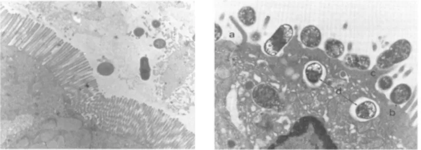
Fig. 1.1. Attaching and effacing lesions produced by an enteropathogenic E. coli (EPEC) in the ligated loop intestinal assay in rabbit. Healthy enterocytes with microvillus layer (left); A/E lesion (right) from: a) few residual microvilli; b) intimately adherent E. coli; c) pedestal formation beneath the adherent bacteria; d)
sometimes internalization of the bacteria into the enterocytes (Piérard et al., 2012).
Chapter 1
Additional virulence factors of both tEPEC and aEPEC have been described outside the LEE locus such as the large protein lymphostatin (LifA), also known as Efa1, that inhibits lymphocyte activation (Klapproth et
al., 2000) and enhances the bacterial adhesion (Tatsuno et al., 2000). In the tEPEC, the large plasmid EAF encodes, besides the BFP, the per locus (plasmid-encoded regulator), whose genes are involved in the regulation of several virulence factors.
1.2.6 Verocytotoxin-producing Escherichia coli
VTEC were first recognized as cause of human infections in the early ‘80s during two outbreaks of bloody diarrhoea occurred in the USA and associated to the consumption of undercooked hamburgers at a fast-food chain (Riley et al., 1983). Stool cultures from patients yielded E. coli isolates belonging to a rare serotype, O157:H7. The disease was designated haemorrhagic colitis (HC) and the causative agents
Enterohaemorrhagic E. coli (EHEC).
VTEC are zoonotic pathogens with cattle being recognized as the major reservoir (Caprioli et al., 2005). Transmission of VTEC infections occurs through the consumption of contaminated food and water, but person-to-person transmission or by direct contact with animals or animal manure have also been reported, although rarely (Heuvelink et al., 2002).
Nowadays, a wide variety of vehicles for VTEC infections has been described, including hamburger and other meat products, water and unpasteurised milk, cantaloupe melon, apple juice, leafy vegetables and several types of sprouts among others (Kaper et al., 2004). VTEC cause large community outbreaks, sometimes with severe outcomes, facilitated by a very low infectious dose estimated to be around ten colony-forming units (CFU) for VTEC O157. In 1996 in Japan about 10,000 people became infected with VTEC O157:H7 following the consumption of radish sprouts (Watanabe et al., 1999). In 2000, in Canada 2,300 people acquired VTEC O157:H7 through the consumption of contaminated water and seven died (Holme, 2003). In 2011, in Germany and France, more than 4,000 people were infected by a VTEC O104:H4 strain, 900 developed HUS, 54 died (Karch et al., 2012).
The main virulence feature of VTEC strains is the ability to elaborate potent cytotoxins known as Verocytotoxins (VTs). These are AB5 toxins, consisting of five identical B subunits responsible for the
binding to the cellular receptor and a single A subunit, which cleaves the rRNA of target cells, causing cell death by blocking protein synthesis (Melton-Celsa et al., 1998). VTs are also called Shiga-Toxins (Stxs) because of their similarity to the Shiga-toxin produced by Shigella dysenteriae type I, and VTEC are therefore also termed STEC (Shiga-toxin producing E. coli). There are two main antigenic types of VTs, VT1 and VT2, including three subtypes of VT1 (VT1a, VT1c and VT1d) and seven subtypes of VT2 (VT2a, VT2b, VT2c, VT2d, VT2e, VT2f and VT2g) (Scheutz et al., 2012). The VT-coding genes (vtx) are conveyed by lambdoid bacteriophages integrated in the bacterial chromosome (Ogura et al., 2007), which are usually maintained in a lysogenic state but retain the capability to enter the lytic cycle, multiply and move to other hosts spreading the vtx genes (Plunkett et al., 1999). Each VT-phage carries one vtx operon consisting of the two subunit genes, vtxA and vtxB, which encode the complete AB5 holotoxin. VTEC can
produce either VT1 or VT2 alone or both and in different subtypes combination (Muniesa et al., 2014). VTs are produced in the colon and may reach the kidney’s glomerular microvasculature via the bloodstream, where through direct apoptotic activity and pro-inflammatory actions damage the endothelial cells causing occlusion. This can lead to the Haemolytic Uremic Syndrome (HUS), whose main symptoms are
haemolytic anaemia, thrombocytopenia and potentially fatal acute renal failure (Andreoli et al., 2002). The estimated rate of fatality due to HUS is 3-5% (Karch et al., 2005). Other sequelae of VTEC infection may include neurological consequences due to the action of VTs on the endothelium of the brain vessels. The treatment of VTEC infections is mainly supportive and consists in rehydration and in the dialysis for the treatment of HUS. In fact, administration of antibiotics is not recommended, since it has been proposed
Chapter 1
to cause the progression of infection towards the most severe forms, probably due to the antibiotic-dependent enhanced VTs production and its massive release into the bloodstream (Kimmitt et al., 2000). The presence of VT-bacteriophages seems not to be sufficient for VTEC to cause disease; VTEC strains isolated from the most severe forms of infection, HC and HUS, also carry additional virulence factors involved in the colonization, and more in general, in the pathogenetic mechanism (Tozzoli et al., 2014b). Most of VTEC strains associated with HUS and with epidemic diseases possess the LEE locus, a genetic element shared with EPEC and responsible for the induction of the histopathological lesion called “attaching and effacing” (A/E) (McDaniel et al., 1995, Karmali et al., 2003).
Although serotype O157:H7 has been implicated in the largest VTEC outbreaks (Karmali, 1989, Griffin et
al., 1991), there is growing concern about the risk posed to human health by non-O157 VTEC serotypes, more than 400 of which have been so far associated with human illness (Tozzoli et al., 2014b).
LEE-negative VTEC strains, in some cases, have also been associated with serious human disease (Nataro
et al., 1998a, Johnson et al., 2006, Käppeli et al., 2011). Such strains usually possess alternative virulence-associated genes, such as the adhesin Saa, encoded on the plasmid pO113 described in LEE-negative VTEC of serogroup O113 (Paton et al., 2001). In addition to saa, the genes sab, epeA and subAB, encoding respectively a SPATE exhibiting protease and mucinase activity, a protein contributing to adherence and biofilm formation, and the Subtilase cytotoxin, are carried by the same plasmid (Paton et al., 1999, Steyert
et al., 2012).
The continuously growing use of genome sequencing technologies paved the way to a more comprehensive knowledge of the VTEC genomes. The determination of the complete genome sequence of E. coli O157:H7 strains EDL 933 and Sakai at the beginning of 21st century, showed that in these VTEC strains, the
chromosome contains more than 170 genomic islands which are not present in the E. coli K-12 MG1655 laboratory strain genome and that 33% of them harbour genes with unknown functions (Hayashi et al., 2001, Perna et al., 2001). Among these genomic islands, a 22-kb PAI designated OI-122 in VTEC O157:H7 EDL933 strain and SplE3 in the Sakai strain, carries the 5’ of the efa1/lifA gene (Morabito et al., 2003), which is involved in the repression of host lymphocyte activation (Klapproth et al., 2000). PAI OI-122 is strongly associated with the LEE locus in both VTEC and EPEC strains, and in many of them is physically linked to the LEE locus itself in a mosaic PAI (Morabito et al., 2003). It has been proposed that the LEE locus and PAI OI-122 may have been acquired as a unique larger PAI and that they separated later on in some strains, following genetic rearrangement events (Morabito et al., 2003).
Most virulent VTEC strains also possess a large virulence plasmid, called pO157 in VTEC O157 strains, conveying the genes for the production of the enterohaemolysin, a toxin favouring the release of
haemoglobin from red blood cells during infection, thus providing a source of iron for the bacteria (Schmidt
et al., 1995) and other putative virulence genes such as espP and katP encoding a serine protease and a catalase-perossidase, respectively (Brunder et al., 2006). The pO157 also hosts the gene toxB (Tozzoli et
al., 2005, Michelacci et al., 2014), whose product proved to contribute to VTEC O157 adherence to Caco-2 cultured cells by promoting the production and/or the secretion of type III secreted proteins (Stevens et al., 2004).
In the attempt to come to a classification of the various VTEC types, the VTEC serotypes have been distributed into five categories termed “seropathotypes” (SPTs) and indicated with the letters from A to E, in a descending order of pathogenicity (Karmali et al., 2003). Such a distinction has been based on their reported frequency in the human illness, the association with severe disease and outbreaks, and the presence of mobile genetic elements conferring virulence genes such as the LEE locus and the PAI OI-122 (Karmali
et al., 2003).
Studies aiming at comparing the different SPTs for the identification of the whole genomic asset conferring to VTEC the full pathogenicity, have led to the description of another PAI, PAI OI-57, which seems to be
Chapter 1
associated with the most virulent SPTs, A and B (Imamovic et al., 2010). This PAI harbours two genes whose products are annotated with a putative function: adfO, coding for a factor described to increase the ability of VTEC O157 to adhere to cultured HeLa cells (Ho et al., 2008), and cfk, a phage-associated bacterial cell killing factor (Perna et al., 2001).
1.2.7 Enteroaggregative-Haemorrhagic Escherichia coli
Although not yet recognized as an official E. coli pathotype, the Enteroaggregative Haemorrhagic
Escherichia coli group (EAHEC) deserves to be mentioned. Such an E. coli subpopulation showcases virulence features transversal to two different DEC types: VTEC and EAggEC. The proposal for such a pathotype was ratified in 2011 after a large outbreak of haemorrhagic colitis and HUS that plagued Germany and, to a lesser extent France, with more than 4,000 cases of infection, 900 HUS, and 54 deaths (Frank et al., 2011). The outbreak was characterised by an unusual high rate of infections progressing to HUS (22% in spite of the usual 5-10%) and the infectious agent belonged to a before rarely reported E. coli serotype, O104:H4 (Frank et al., 2011). The outbreak strain carried the VT2a-phage and induced the “stacked-brick” pattern of adhesion to cultured Hep-2 cells, typical of EAggEC strains (Scheutz et al., 2011). In fact, it carried genes peculiar of the EAggEC pathotype, such as the aggregative adherence fimbriae type 1 and several SPATEs (Steiner, 2014).
After the German outbreak in 2011, the scientific community looked retrospectively at the reported HUS cases linked to infections with atypical VTEC types or browsed the scientific literature with the aim to assess if other EAHEC cases of infection could be retrieved. It turned out that the first ever reported EAHEC strain caused eight HUS cases in France in 1992 and belonged to O111:H10 serotype (Morabito et
al., 1998). Additionally, in the time-span 1992-2012, at least six sporadic cases of EAHEC-associated HUS were observed as caused by EAHEC strains belonging to three different serotypes such as O86: HNM, O104:H4 and O111:H21 (Morabito et al., 1998, Iyoda et al., 2000, Scavia et al., 2011, Dallman et al., 2012). Furthermore, an O15 VT2-producing strain positive for the presence of Enteroaggregative markers has been described in a patient with septicaemia (Wester et al., 2013). Finally, an EAHEC strain O127:H4 strain has been recently isolated from four HUS cases occurred in northern Italy in 2013 (Tozzoli et al., 2014a).
The appearance of the first EAHEC raised the question if it was indeed a new pathogenic group of E. coli or we were rather observing accidental sporadic events of VT-bacteriophages acquisition by classical EAggEC strains.
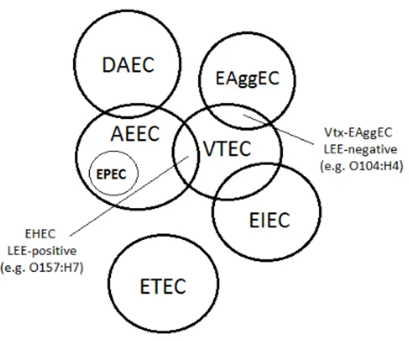
The experience of the German outbreak and the following reported existence of at least six different EAHEC serotypes, as well as the growing number of reports of E. coli other than VTEC producing VTs, such as the ExPEC strains isolated from patients with bacteraemia (Wester et al., 2013) or ETEC strains displaying the presence of the vtx-coding genes (Tozzoli et al., 2014a), seem to indicate that E. coli pathotypes should not be considered as rigidly separate entities but rather E. coli sub-populations dynamically exchanging part of their genomic traits causing the continuous emergence of hybrid groups, sometimes with augmented pathogenicity, as in the cases of EHEC and EAHEC (Fig. 1.2).
It´s now generally accepted that E. coli genome as a whole, also termed pangenome, is constituted of a ¨backbone¨ of around 2,000 genes shared by all E. coli and a supplement of accessory genes, generally carried by Mobile Genetic Elements (MGE), peculiar to each subpopulation (Franz et al., 2014). MGEs strongly contribute to the high flexibility and dynamicity of E. coli genome and constituted the engine that powered the evolution of the different DEC pathotypes, therefore will be treated more in detail in following sections.
Fig. 1.2. Relationships between human DEC pathotypes. DAEC: Verocytotoxin-producing E.
EPEC: Enteropathogenic E. coli; EHEC: Enterohaemorrhagic E. coli; ETEC: Enterotoxigenic E. coli; EAEC: Enteroaggregative E. coli; Vtx
overlapping between circles represent the fractions of the genome shared by the related pathotypes
1.3 Epidemiology of diarrheagenic
VTEC represent the only pathogenic group of recognized as the major reservoir
numerous animal species, ruminants are considered as the major an humans (Caprioli et al., 2005).
Transmission of VTEC infections to man occurs through the consumption of food and water, including in the latter the exposure linked to recreational activities. Food of animal origin can be primaril
during the transformation process, e.g. the carcasses and meat at the slaughterhouses or milk during milking procedure. Vegetables can become contaminated following an environmental pathway including the use of ruminants’ manure to fertilize fields where crops are grown. Although most of VTEC infections, especially those caused by O157 strains have been linked to exposure to a food vehicle or water, person
transmission or by direct contact with animals or animal manure have also bee 2002).
VTEC infections are not mandatorily notified in most of the countries worldwide, resulting in a massive underestimation. In the European Union, the cases of infections caused by VTEC are communicated, voluntarily by the EU Member States, to the European Centre for Disease Prevention and Control (ECDC), which collects the information and elaborate trends and
Epidemiological Report, referring to the data of 2011 year, 9,534 VTEC infections have been reported from 27 European countries (ECDC, 2014)
occurrence of the large German outbreak of VTEC O104:H4 infections (See section 1.2.7). A total of 1,006 of the reported cases were HUS, showing an incidence of 11% for this severe for
Twenty-eight per cent of HUS cases were reported in 0 prevalent serogroups, followed by 25
(ECDC, 2014).
As for the other diarrheagenic E. coli
circulation and the oral-faecal route of transmission of the infections (See section 1.2), their circulation is
Fig. 1.2. Relationships between human DEC pathotypes. DAEC: Diffusely-adherent E. coli; VTEC:
coli; AEEC: Attaching and effacing E. coli; EIEC: Enteroinvasive E. coli; EPEC: Enteropathogenic E. coli; EHEC: Enterohaemorrhagic E. coli; ETEC: Enterotoxigenic E. coli; EAEC: Enteroaggregative E. coli; Vtx-EAggEC: Shiga toxin-producing Enteroaggregative
overlapping between circles represent the fractions of the genome shared by the related pathotypes (adapted from Franz et al., 2014).
1.3 Epidemiology of diarrheagenic Escherichia coli infections
VTEC represent the only pathogenic group of E. coli that has a definite zoonotic origin with cattle being recognized as the major reservoir (Caprioli et al., 2005). Even though VTEC can be found in the gut of numerous animal species, ruminants are considered as the major animal source of strains highly virulent to Transmission of VTEC infections to man occurs through the consumption of food and water, including in the latter the exposure linked to recreational activities. Food of animal origin can be primaril
during the transformation process, e.g. the carcasses and meat at the slaughterhouses or milk during milking procedure. Vegetables can become contaminated following an environmental pathway including the use of
fields where crops are grown. Although most of VTEC infections, especially those caused by O157 strains have been linked to exposure to a food vehicle or water, person
transmission or by direct contact with animals or animal manure have also been reported
VTEC infections are not mandatorily notified in most of the countries worldwide, resulting in a massive underestimation. In the European Union, the cases of infections caused by VTEC are communicated, voluntarily by the EU Member States, to the European Centre for Disease Prevention and Control (ECDC), which collects the information and elaborate trends and incidence data. In the last published Annual Epidemiological Report, referring to the data of 2011 year, 9,534 VTEC infections have been reported from
(ECDC, 2014). This number is 2.5 higher than the previous year (Fig 1.3) due to the occurrence of the large German outbreak of VTEC O104:H4 infections (See section 1.2.7). A total of 1,006 of the reported cases were HUS, showing an incidence of 11% for this severe form of the infection.
eight per cent of HUS cases were reported in 0-4 years old children with O157 and O26 as prevalent serogroups, followed by 25-44 years old adults with O104 being the predominant serogroup
E. coli, ETEC, EAggEC, EPEC and EIEC, given their inter
faecal route of transmission of the infections (See section 1.2), their circulation is
Chapter 1
adherent E. coli; VTEC: coli; AEEC: Attaching and effacing E. coli; EIEC: Enteroinvasive E. coli; EPEC: Enteropathogenic E. coli; EHEC: Enterohaemorrhagic E. coli; ETEC: Enterotoxigenic E. coli;
producing Enteroaggregative E. coli. Areas of overlapping between circles represent the fractions of the genome shared by the related pathotypes
that has a definite zoonotic origin with cattle being . Even though VTEC can be found in the gut of
imal source of strains highly virulent to Transmission of VTEC infections to man occurs through the consumption of food and water, including in the latter the exposure linked to recreational activities. Food of animal origin can be primarily contaminated during the transformation process, e.g. the carcasses and meat at the slaughterhouses or milk during milking procedure. Vegetables can become contaminated following an environmental pathway including the use of
fields where crops are grown. Although most of VTEC infections, especially those caused by O157 strains have been linked to exposure to a food vehicle or water, person-to-person
n reported (Heuvelink et al., VTEC infections are not mandatorily notified in most of the countries worldwide, resulting in a massive underestimation. In the European Union, the cases of infections caused by VTEC are communicated, voluntarily by the EU Member States, to the European Centre for Disease Prevention and Control (ECDC),
incidence data. In the last published Annual Epidemiological Report, referring to the data of 2011 year, 9,534 VTEC infections have been reported from
. This number is 2.5 higher than the previous year (Fig 1.3) due to the occurrence of the large German outbreak of VTEC O104:H4 infections (See section 1.2.7). A total of 1,006
m of the infection. 4 years old children with O157 and O26 as 44 years old adults with O104 being the predominant serogroup
EPEC and EIEC, given their inter-human faecal route of transmission of the infections (See section 1.2), their circulation is
Chapter 1
more common in developing countries where the high incidence of the disease caused by these pathotypes of E. coli, in conjunction with the poor hygienic conditions and the lack of effective human sewage treatments is responsible for their environmental diffusion and persistence. ETEC, EPEC, EAggEC are the most important enteric pathogens in children below five years of age and are responsible for roughly a half of all the diarrheal episodes in developing countries (Clarke, 2001).
Fig.1.3 Trend and number of VTEC cases reported in the EU, 2007-2011 (ECDC, 2013).
Dutta and colleagues (Dutta et al., 2013) reported the results of an investigation on 3,826 stool specimens collected from acute diarrheal patients hospitalized at the Infectious Diseases Hospital in Kolkata, India, and found that the major prevalence of infections was due to EAggEC (5.7%), followed by ETEC (4.2%) and EPEC (1.8%). Other authors reported variable rates of EPEC isolation ranging from 12.6 to 44.9% in children under five years with diarrhoea in Iran (Jafari et al., 2012). In the same country, the same study reported ETEC detection rates from less than 10% up to 33% (Jafari et al., 2012).
A recent case-control study described a prevalence of EAggEC of 41% in children under five years in north-eastern Brazil compared to previous data ranging from 2 to 12% (Lima et al., 2013). In Guinea-Bissau the most common bacteria isolated from children under two years with persistent diarrhoea was EAggEC (Valentiner-Branth et al., 2003).
Among the human-borne E. coli pathotypes, the EAggEC show the more variable epidemiology. This pathotype has been reported as a leading cause of travellers’ diarrhoea, a common cause of persistent diarrhoea among HIV-infected people as well as of endemic paediatric diarrhoea in industrialized and developing countries (Boisen et al., 2014). In a large study in England, including more than 3,600 cases of diarrhoea, EAggEC were the second most common bacterial cause of gastroenteritis (Tompkins et al., 1999).
1.4 Mobile Genetic Elements and their role in bacterial evolution
E. coli genome vary in size from 4.7 Mb to 6.5 Mb (Hazen et al., 2012). The studies on the E. coli pangenome indicate that this species is continuously evolving through horizontal gene acquisition and diversification (Dobrindt et al., 2010). Changes occur also through point mutations, deletions and single nucleotide variations (Ochman et al., 2000). These events contribute to the genome optimization (Fig. 1.4) and eventually lead to the adaptation of the microbe, which gains new niches, escape from host immune defences and acquire new resistances to antimicrobials.
The genome plasticity and the resulting variability have made a significant contribution to the successful emergence of new pathogenic E. coli (Leopold et al., 2014).
Chapter 1
A striking feature of pathogenic E. coli is the presence of genes that encode virulence factors on mobile genetic elements (MGEs) (Kaper et al., 2004). MGEs include plasmids, genomic islands (GIs),
pathogenicity islands (PAIs), bacteriophages, integrons, transposons and insertion sequence elements (ISs) (Dobrindt et al., 2010).
Fig.1.4 Mechanisms of genome optimization (Leopold et al., 2014).
1.4.1 Genomic and Pathogenicity Islands
PAIs and GIs form a distinct super-family of MGEs (Hacker et al., 1997) that integrate into and excide from the host chromosome making the occurrence of horizontal gene transfer possible.
GIs typically integrate in the host chromosome in the proximity of tRNA genes, possess a different G+C content compared to the average of the genome backbone, and often carry cryptic or functional genes encoding factors related to the self-mobilization, such as integrases and transposases (Kaper et al., 2004). Different types of GIs have been described so far, some convey genes involved in the metabolism and enhancing the microbial fitness, other confer drug resistance or are involved in the pathogenetic
mechanism. The latter are termed Pathogenicity Islands (PAIs). PAIs encode a wide variety of virulence factors, such as toxins, adhesins, iron uptake systems, secretion systems and strategies to escape from the host immune defence mechanisms (Dobrindt et al., 2010).
Some PAIs contain regions showing homology to other MGEs, such as bacteriophages and plasmids, suggesting that they may have derived from events of integration of such MGE in the host chromosome leading to a more stable association and vertical replication (Dobrindt et al., 2004).
The presence in the GIs of genes displaying a certain homology in the DNA sequence with other ORFs facilitates the genetic re-arrangement events between different islands and within the same island, thus boosting the plasticity of E. coli genome.
1.4.2 Bacteriophages with emphasis on VT-phages
MGEs are the engine of bacterial diversity and bacteriophages in particular seem to have the deepest impact on the evolution of pathogenic bacteria (Campbell, 1996). As a matter of fact, the spread of virulence-associated genes by lysogenic phages is a well-known phenomenon in both negative and Gram-positive bacteria (Boyd et al., 2002). The availability of complete E. coli genome sequences shows a high diversity in the amount of prophage-related sequences within this species (Dobrindt et al., 2010). The modular structure of bacteriophages genome has been proposed since the early studies on this topic (Susskind et al., 1978). According to this theory, bacteriophages are constituted by several functional
Chapter 1
modules that can be interchanged between each other either by homologous recombination at specific sequences or by non-homologous recombination at random points (Muniesa et al., 2014).
Among the several phages and phage-related sequences in E. coli genome, the Verocytotoxin encoding-bacteriophages (VT-phages) played, and may still be playing, a pivotal role in the evolution of the different
E. coli pathotypes. In fact, besides VTEC, the ability of producing VT seems to be shared with almost all the pathogenic groups of strains belonging to this species (Scavia et al., 2011, Wester et al., 2013, Tozzoli
et al., 2014a). VT-phages constitute a heterogeneous group of temperate lambdoid phages that harbour vtx genes. VT-phages described so far belong to the Myoviridae and Siphoviridae virus families (Schmidt, 2001), vary in size genomes and composition, morphology, host range, integrate in different host
chromosomal loci and differentially influence the host pathogenicity (Muniesa et al., 2014, Tozzoli et al., 2014a).
VT-phages alternate a lysogenic state, in which the phage genome is silently integrated into the host chromosome, with a lytic cycle, characterised by replication and phage particle formation, followed by the lysis of the host cell. VT-phages are then spread to other host cells.
VT-phages genomes have been characterised by restriction analysis and mapping experiments since the early ‘80s and lately by whole genome sequencing. Reported genome sizes range from 42 Kb to more than 60 Kb in phages isolated from different VTEC strains (Schmidt, 2001, Muniesa et al., 2014).
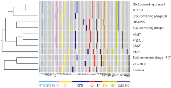
The number of available VT-phages genome sequences is growing and several comparative genomics studies have been published with the aim of highlighting similarities and differences between them and understanding the basis and the factors influencing their heterogeneous biology (Fig. 1.5) (Ahmed et al., 2012, Laing et al., 2012, Smith et al., 2012, Steyert et al., 2012, Grande et al., 2014). The genetic structure of VT-phages mainly corresponds to that of phage lambda, with an early region comprising genes involved in the regulation of lysogenic-lytic cycle and a late region containing all the genes necessary for packaging and phage particle formation. Vtx-coding genes are located in the late region downstream the
anti-terminator site of Q gene and upstream the lysis cassette. The position of vtx genes is conserved and their transcription is boosted when lytic cycle is induced.
Fig. 1.5 Multi-genome comparison of VT-phages. Variants of the same gene are indicated with the same colour (Smith et al., 2012).
Chapter 1
Lytic cycle can be induced by exposure to stress inducing agents, such as many classes of antibiotics, UV light, high hydrostatic pressure, H2O2 or occur spontaneously following SOS-independent mechanisms
(Imamovic et al., 2012).
VT-phages have been described in a number of Enterobacteriaceae including, beside different pathotypes of
E. coli (Karch et al., 1999, Tozzoli et al., 2014a), Shigella dysenteriae type I, Shigella sonnei, Shigella
flexnerii (Strauch et al., 2001, Gray et al., 2014), Citrobacter freundii (Schmidt et al., 1993, Tschape et al., 1995) and Enterobacter cloacae (Paton et al., 1996).
Free VT-phages can be found in the environment, including water and sewage systems, in which they can survive for prolonged periods (Muniesa et al., 2014). Their presence in the environment increases the risk of the emergence of bacterial pathogens, especially new pathogenic E. coli strains.
1.4.3 Other MGEs
Other chromosomally-borne MGEs include the insertion elements, transposons and integrons. These are segments of DNA that frequently change location in the chromosome. Different categories of such molecules are recognized: (i) insertion sequences with terminal inverted repeats sequences which encode the information for the self-mobilization; (ii) composite transposons flanked on both sides by insertion sequences; (iii) non composite transposons which contain no insertion sequences (Leopold, 2014). Integrons are well known for their ability to carry and spread antibiotic-resistance cassettes. Recently, super-integrons containing up to eight different cassettes for antibiotic resistance and flanked by insertion sequences targeted by integrases have been described (Cambray et al., 2010, Hall, 2012a, Hall, 2012b). Plasmids are the only extra-chromosomal MGE. Nonetheless, some of them can also integrate into the host chromosome. Plasmids can exchange genetic information among different strains, thus contributing to the genome evolution by horizontal gene transfer. E. coli plasmids can vary in size from few Kb up to more than 150 Kb. Larger plasmids encode virulence factors, such as fimbriae, serine proteases, enterotoxins and haemolysins, among others. Also in plasmids, repeated regions and insertion sequences serve as
homologous points for genetic recombination explaining the observed variability in sizes and gene content (Leopold et al., 2014). Several groups of genes have been shown to be originated from plasmid integration into the chromosome and subsequent events of deletion, insertion and stabilization (Brzuszkiewicz et al., 2009).
Horizontal gene transfer operated through MGEs strongly contributes to genome optimization, with potential alterations ranging from the addition of virulence genes to the modification, loss or gain of entire genomic regions. These rapid changes are referred as microevolution and become apparent within a few generations, in the environmental, animal or human reservoir (Leopold et al., 2014).
CHAPTER 2
Chapter 2
The aim of the present work was to investigate the molecular bases of the evolution of pathogenic E. coli. The increasing availability of information on the E. coli genomes has led to the description of a wealth of mobile genetic elements (MGEs) conveying genes encoding factors involved in the E. coli pathogenetic mechanism, but at the same time highlighted that the asset of virulence genes conferring to E. coli the full pathogenicity might be by far more complex and is not completely unravelled yet.
The current classification of the E. coli pathotypes has been long based on the presence of defined PAIs and virulence genes and the related mechanisms of host colonization or toxin production. However, the
boundaries between the different pathogenic types have become less sharp following the observation that part of the accessory DNA, mainly that associated with virulence, seems to overlap different pathotypes causing the concept of E. coli pathotypes themselves to be re-considered.
Examples of such an overlapping are the presence of the LEE locus in EPEC and EHEC, or the observed presence of VT-converting phages, hallmark of the VTEC pathotype, into strains belonging to all the other known E. coli pathotypes, bringing into question if VTEC are a pathotype in its own right or rather the VT-phage should be considered as being the real pathogen, exploiting the colonization machinery of the pathogenic E. coli to generate augmented pathogenicity variants.
Despite the considerable efforts devoted by the scientific community, the evolutionary forces leading to the emergence of new pathogenic clones or variants of E. coli are not completely understood. As a matter of fact, E. coli genome is very dynamic and evolves continuously through horizontal gene transfer. Indeed, it has been shown that the gain and loss of virulence factors, such as the LEE locus, the VT-converting phages, the Enterohaemolysin-coding gene, among others, have occurred several times and in parallel in separate lineages, thus suggesting that many forces intervened in the E. coli convergent evolution (Franz et
al., 2014). In this piece of research we focused on VTEC as a model of dynamic pathotype encompassing many established E. coli pathogenic groups. We have investigated the distribution of MGEs encoding virulence features in different VTEC subpopulations and studied their evolution through the differentiation of the virulence genes into different allelic variants. Additionally, we studied the different bacteriophages transporting the vtx genes into different VTEC types and their role in manipulating the host biology. Finally, we assessed the possibility that the spreading of VT-phages may or may not be subjected to host-related barriers by probing the possible acquisition of such phages by E. coli strains with a genetic background different from VTEC.
The work plan has been divided in the following work packages, listed here along with the corresponding detailed objectives:
1. Characterisation of the genetic determinants encoding a novel allelic variant of the Subtilase cytotoxin (SubAB):
• Analysis of the genetic determinant encoding a new SubAB variant (SubAB2);
• Study of structure of the MGE carrying the SubAB2-coding genes;
• Study of the distribution of SubAB-coding genes and their localization onto specific MGEs in VTEC strains from human and animal sources.
One publication in Chapter 3 (Clin Microbiol Infect. 2013 Mar;19(3):E149-56)
2. Identification of an allelic variant of the virulence-associated gene toxB in VTEC serogroups associated with severe human disease:
• Characterisation of the toxB gene in the large virulence plasmid of a VTEC O111 strain; • Description of the toxB gene allelic variants;
• Study of the distribution of the two variants in VTEC strains belonging to different serogroups and pathogroups.
One publication in Chapter 4 (Int J Med Microbiol. 2014 Jul;304(5-6):730-4).
Chapter 2
3. Identification and characterisation of VT2-phages present in pathogenic E. coli strains causing HUS: • Identification of a peculiar VT2-phage, Φ-8, present in VTEC O157 strains isolated from severe
human disease;
• Functional analysis of the presence of Φ-8 and its influence on the regulation/assembly of the Type III secretion system;
• Comparison between the WGSs of VT2-phage from Enteroaggregative Haemorrhagic E. coli (EAHEC) strains and the VT-phages sequences available in the public repositories;
• Identification of peculiar EAHEC VT-phages sequences.
Two publications in Chapter 5 (Infect Immun. 2014 Jul; 82(7): 3023-32; BMC Genomics. 2014 Jul 8;15:574).
4. Study of the VT-phages host range and the emergence of new pathogenic E. coli clones with augmented virulence:
• Determination of the WGSs of VT2-phages from Enteroaggregative Haemorrhagic E. coli (EAHEC) and their comparison;
• Analysis of the experimental ability of pathogenic and non-pathogenic E. coli strains to be infected with VT2-phages;
• Evaluation of the stable acquisition of VT-phages by E. coli strains other than VTEC following experimental infections.
One publication in Chapter 6 (Front Cell Infect Microbiol. 2014 Jun 20;4:80).
CHAPTER 3
Characterisation of the genetic determinants
encoding a novel allelic variant of the Subtilase
cytotoxin (SubAB)
Chapter 3
Introduction
Subtilase (SubAB) is an AB5 cytotoxin produced by some VTEC strains usually lacking the LEE locus.
The production of SubAB has been first described in a VTEC O113 strain isolated from a human case of HUS in Australia in 2003 and later identified in other VTEC serogroups. SubAB is delivered to the host cell endoplasmic reticulum where it causes the inhibition of the protein synthesis, resulting in the unfolded protein response and finally apoptosis. SubAB has been supposed to contribute to the pathogenetic process through a synergistic action with the VTs. In the prototype Subtilase-producing strain, subAB operon is located on the large virulence plasmid, named pO113, also carrying the saa gene, encoding an
autoagglutinating adhesin. Two major allelic variants of the subAB genes, termed subAB1 and subAB2, and a
minor subtype, termed subAB2-2, have been described so far.
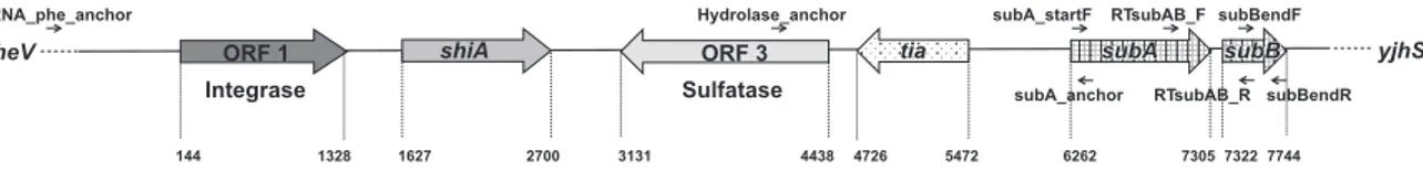
In the present work we determined the complete sequence of the PAI conveying the subAB2 genes from a
VT-negative E. coli strain isolated from a case of diarrhoea in Italy and investigated the distribution of both the major variants in human E. coli strains belonging to different pathotypes and in a large collection of LEE-negative VTEC of human and ovine origin.
Results
The entire nucleotidic sequence of the chromosomal locus containing the subAB2operon was determined,
consisting in an 8 kb-long pathogenicity island that has been termed Subtilase-Encoding PAI (SE-PAI). The PAI was integrated in the pheV-tRNA locus and, besides subAB and a gene encoding an integrase, it harboured the tia gene, encoding an invasion determinant first described in an ETEC strain isolated from a human case of disease. Additionally, it presented an ORF showing great sequence similarity with the gene
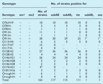
shiA of Shigella. The study of the distribution of the two subAB allelic variants confirmed that subAB genes are commonly found in LEE-negative VTEC, showing their presence in 72% and 86% of the strains from human cases of diarrhoea and healthy sheep examined, respectively. Most of the subAB-positive strains identified (98%) possessed the subAB2allelic variant and were also positive for tia, suggesting the presence
of the entire SE-PAI.
Conclusions
The SE-PAI carrying the subAB2allelic variant has been characterised and fully sequenced. It is inserted in
the E. coli chromosome in the pheV-tRNA locus, which represents a hotspot of integration for genomic islands. The SE-PAI also transports genes, beside subAB, which could play a role in the pathogenesis, such as shiA, a Shigella gene whose product has been described to attenuate the inflammatory response induced in the host upon infection, and tia, which encodes an invasion factor of ETEC. The latter genes could play a role in the colonization process by SubAB2-producing E. coli. The use of pheV as integration site by
SE-PAI could provide an explanation for its wide presence in the LEE-negative VTEC strains. In fact it could be the result of a mutual exclusion between this PAI and the LEE locus, which is often integrated in this chromosomal site.
The distribution of the two subAB variants has been analysed in a large panel of LEE-negative VTEC strains isolated from human and ovine sources. The results showed that subAB was present in the vast majority of LEE-negative VTEC strains isolated from both the ovine source and the human cases of diarrhoea and that subAB2was the prevalent allele.
Altogether these findings indicate that small ruminants may represent an important reservoir for human infections with VTEC subAB2-producing strains. Moreover, our findings suggest that LEE-negative VTEC
strains may express their virulence through the action of additional virulence features including either toxins (SubAB) or factors involved in the colonization mechanism, such as the products of tia and shiA genes.
Chapter 3
Candidate’s contributions to the present work:
Laura Grande contributed to the determination of the complete nucleotide sequence of the SE-PAI and helped in the screening of E. coli strains held at the Istituto Superiore di Sanità for the presence of subAB,
saa and tia genes. Moreover, she revised critically the manuscript for the publication.