UNIVERSITA’ POLITECNICA DELLE MARCHE
Scuola di Dottorato di Ricerca della Facoltà di Medicina e Chirurgia
Curriculum “Salute dell’Uomo”
Mesoderm stem cells and inflammation: role in the
pathogenesis and potential therapy of selected gynecological
diseases and primary myopathies
Ph.D. Candidate Tutor
Miriam Caffarini Dr. Monia Orciani
1
SUMMARY
ABSTRACT……….3
1.
Introduction……….……….4
1.1. Mesenchymal stem cells (MSCs)………4
1.2. Inflammation.……….6
1.3. Role of MSCs in the inflammation………..………9
1.4. STUDY 1: Uterine leiomyoma……….………..11
1.4.1. Role of MSCs in uterine leiomyomas………13
1.5. STUDY 2: Cervical intraepithelial neoplasia (CIN) ………..14
1.5.1. The role of inflammation and MSCs in age-related regression of CIN………..17
1.6. STUDY 3: Duchenne muscular Dystrophy (DMD) ………..18
1.6.1. Gene therapy……….21
1.6.2. Cell therapy……….23
2. Aim of the study………26
3. Materials and methods……….27
3.1. Human tissues collection……….27
3.2. Cell culture………....28
3.3. Characterization of MSCs from different tissues……….28
3.4. Doubling time………..30
3.5. STUDY 1………...31
3.5.1. Immunocytochemistry………31
3.5.2. Immunofluorescence………..31
3.5.3. Western blot………..32
3.5.4. Expression of inflammation-related cytokines by RT-PCR and ELISA……….32
3.6. STUDY 2………35
3.6.1. Proliferation assay……….35
3.6.2. Senescence associated β-galactosidase assay………35
3.6.3. PCR array for the senescence………35
3.6.4. Indirect co-culture with HeLa cells………..……….35
3.6.5. Expression of selected genes in co-cultured HeLa cells by RT-PCR………...36
3.6.6. Analysis of the effects of age and co-culture in secretion of inflammatory Cytokines……….37
3.6.7. Statistical Analysis……….37
2
3.7.1. Sample collection and cell culture……….38
3.7.2. Myogenic differentiation………...38
3.7.3. Immunofluorescence assay……….38
3.7.4. Protein extraction and quantification……….39
3.7.5. Western blot………..39
4. Results………...41
4.1. STUDY 1………41
4.1.1. Cell isolation and characterization……….41
4.1.2. MDR1, α-SMA, Collagen Type 1, and Fibronectin Expression……….43
4.1.3. Expression Profile of Inflammatory Cytokines………..47
4.2. STUDY 2………50
4.2.1. Cell isolation and characterization……….50
4.2.2. PCR Array for senescence in oC-MSCs and yC-MSCs……….56
4.2.3. Proliferation of HeLa cells after co-cultures with MSCs………..56
4.2.4. Gene expression……….56
4.2.5. Expression profile of inflammatory cytokines………58
4.3. STUDY 3………60
4.3.1. Cell culture and myogenic differentiation………60
4.3.2. Determination of expression of MyHC and DyS on differentiated cells………..60
4.3.3. Western blot analysis ……….60
5. Discussion……….63
5.1. STUDY 1………63
5.2. STUDY 2………66
5.3. STUDY 3………69
3
Abstract
Mesenchymal stem or stromal cells (MSCs) are a specific type of adult stem cells with an extensive proliferation and differentiation potential towards specialized cells developing from the mesoderm. MSCs are also characterized by paracrine function through the release of multiple growth factors, chemokines and cytokines. MSCs play as sentinel that feel the microenvironment and act consequently, switching from a pro-inflammatory phenotype to an immunosuppressive phenotype according to the signals they receive. In the present work the existence and the role of MSCs in the pathogenesis and potential therapy of selected gynecological diseases with an inflammatory component as uterine leiomyoma, cervical intraepithelial neoplasia (CIN), and in primary myopathies, as Duchenne Muscular Dystrophy (DMD) were evaluated. In the first study, progenitor cells were identified both in leiomyomas and normal myometrium, and the correlation between these cells and inflammation in leiomyoma onset has been investigated. The data suggest that the upregulation of cytokines related to chronic inflammation in leiomyoma progenit ors could favour a microenvironment suitable for the onset of this pathology. In the second study, MSCs from cervix of young (yC-MSCs) and old patients (oC-MSCs) were isolated and results show as their immunobiology is affected by the age of donors, influencing in turn the regression rate of CIN. In addition, in the crosstalk with HeLa cells, yC-MSCs play an anti-tumoral role sustaining an acute inflammatory environment. The goal of the third study was to find a correct strategy to enhance the production of dystrophin protein in DMD through gene therapy. Therefore, myoblasts isolated from DMD donor were transduced with green fluorescent protein (GFP) and a lentiviral vector expressing the snRNA to induce exon skipping; data indicate that transduced myoblasts were able to perform myogenic differentiation expressing a functional dystrophin protein.
4
1.
Introduction
1.1. Mesenchymal stem cells (MSCs)
Mesenchymal stem or stromal cells (MSCs) are a specific type of adult stem cells (SC) able to generate the different cell types of the mesenchyme (Caffarini M et al. 2018, Ullha I et al. 2015).
Due to their ability to differentiate into specialized cells developing from mesoderm, they were named as mesenchymal stem cells (MSCs). MSCs, also known as multipotent cells, exist in adult tissues of different sources, ranging from murine to humans.
After their discovery in bone marrow more than 40 years ago (in 1976) (Friedenstein et al. 1976), MSCs have been isolated from other many tissues, such as adipose tissue, periosteum/ synovial fluid, Wharton’s jelly, umbilical cord blood, placenta, amniotic fluid, skin, dental pulp, and skeletal muscle (Toyserkani NM et al. 2015, Zuk PA et al. 2002, Nemeth K et al 2015, Troyer DL et al. 2008, Nakahara H et al. 1991, Kim KH et al. 2017, Kern S et al. 2006, Brooke G et al. 2009, Shi C et al. 2006, Sellheyer K et al. 2010). At the beginning, they were called osteogenic stem cells or bone marrow stromal cells. Because of their plastic‐adherent capacity and their fibroblast-like cells morphology, they were identified as distinct from the hematopoietic stem cells (HSCs). In 1991, Caplan coined the name “mesenchymal stem cells” to define the multilineage potential of these cells and the ability to renew themselves for long periods of time without significative variations in their properties (Caplan AI. 1991). To better classify these multipotent cells as mesenchymal stem/stromal cells, Dominici et al. introduced three minimal criteria approved by the International Society for Cellular Therapy (ISCT): 1. First, cells must be plastic adherent when grown in culture under standard conditions. 2. Second, they must be positive (expression) for CD73, CD90, CD105 and negative for CD45, CD34, CD14 or CD11b, CD19 or CD79α, and HLA-DR surface molecules (tested by flow cytometry). 3. Third, MSCs must be able to differentiate into osteoblasts, adipocytes and chondrocytes in vitro (Dominici M et al. 2006, Soundararajan M et al. 2018).
Despite the mesodermal origin, MSCs have displayed the capacity of trans-differentiation into ectodermal lineages (Ullha I et al. 2015). In particular, MSCs isolated from different sources have shown trans-differentiation into neural cells as a result of exposure to neural induction media supplemented with cocktails of growth factors (Ullha I et al. 2015).
More in details, adding several growth factors to the neuronal medium cocktail, such as hepatocyte growth factor (HGF), FGF and EGF, it was possible to obtain specific neurons
5
phenotypes: oligodendrocytes, cholinergic and dopaminergic neurons (Naghdi M et al. 2009, Datta I et al. 2011, Barzilay R et al. 2009, Safford KM et al. 2002, Kang SK et al. 2003). It was also proved that MSCs were able to generate hepatocytes and pancreocytes upon induction with their corresponding conditioned media (Ullha I et al. 2015), revealing that hMSCs can also trans-differentiate into endodermal lineages. For what concern human MSCs, it is supposed that they require two passages to trans-differentiate into hepatocytes: differentiation and maturation steps, both included the addition of specific growth factors in the culture medium (Lee KD et al. 2004, Stock P et al. 2014). Therefore, differentiated cells into hepatocytes were able to express liver-specific transcription markers. So far, MSCs derived from adipose, dental, umbilical cord, amnion, Wharton jelly and placental tissues seem to have successfully differentiated into insulin producing β-cells (Kim et al. 2012, Criscimanna et al. 2012, Kanafi et al. 2013).
Besides their extensive proliferative and differentiation potential, it was demonstrated that MSCs lack the teratoma formation in vivo (Jiang Y et al 2007). This gives MSCs the advantage of being a good candidate for replacing embryonic stem cells (ESCs) and induced pluripotent stem cells (iPSC) in regenerative and cell therapies. MSCs are also characterized by paracrine function through the release of multiple growth factors, chemokines and cytokines (Jiang Y et al 2007).
Figure 1. Representation of Mesenchymal stem cells (MSCs) and their differentiation
6
Cytokines and chemokines are redundant secreted proteins with growth, differentiation, and activation functions that regulate and control immune cell trafficking and the cellular arrangement of immune organs (Borish LC et al. 2003).
1.2. Inflammation
Inflammation is a biological response of the organism to harmful stimuli. It happens when the immune system fights against something that could be injurious. The most common causes of inflammation are pathogens, as bacteria, viruses or fungi, external injuries and effects of chemicals and radiations. There are five signs that may indicate an inflamed process, such as redness, swelling, heat, pain and loss of function (rubor, tumor, calor, dolor et functio laesa) (Ferrero-Miliani L et al. 2007). During an inflammatory process, the innate immune system plays a pivotal role, as it mediates the first response and many cells are involved and release different factors, the inflammatory mediators. Inflammation can be classified as acute or chronic. Acute inflammation is a short-term process occurring in response to tissue injury, usually appearing within minutes or hours and it may be regards as the first line of defence against wound (Ramzi S Cotran et al. 1999). Acute inflammatory response is the result of a series of biochemical events that involves the local vascular system, the immune system and infiltration of neutrophils from the blood to the injured tissue. In some disorders the inflammatory process, which in normal conditions is self-limiting, becomes prolonged developing chronic inflammation (Ferrero-Miliani L et al. 2007). Chronic inflammation involves an infiltration of macrophages, T lymphocytes and plasma cells indicating a progressive shift in the type of cells present at the site of inflammation (Ferrero-Miliani L et al. 2007). Loss of tissue function due to fibrosis represents the final consequence of chronic inflammation. The immune system is capable to recognize and respond to pathogenic insult (Akira S et al. 2006) through specific pathogen recognition receptors (Chen GY et al. 2010). Then, a signalling cascade promotes the release of complement components, acute phase proteins, pro inflammatory cytokines and chemokines recruiting innate immune cells (neutrophils and macrophages) (Medzhitov R. 2010). This process is named adaptive response to foreign antigens and it is the other main immune strategy (besides the innate system).
Neutrophils and monocytes/macrophages co-express similar antigens and these innate phagocytes can readily produce effector molecules such as granular proteins, oxidants, chemokines and cytokines (Daley JM et al. 2008, Nauseef WM 2007, Sunderkötter C et
7
al. 2004). Regardless of their similarities, emerging evidence indicates that neutrophils and monocytes/macrophages have distinct roles as innate immune cells and therefore are indispensable as key players against infection. Typically, neutrophils are the first responders to be recruited and have a higher microbicidal activity; whereas monocytes/macrophages are recruited later on. Despite this, monocytes/macrophages are able to digest and present antigens to other immune cells, thereby allowing them to interact with the adaptive immune system (Silva MT and Correia-Neves M 2012). Neutrophils and monocytes/macrophages share a complex relationship and; together, they orchestrate a more enhanced immune response by regulating other immune cells as well as each other.
Whenever T cell are activated, antigen-presenting cells, with dendritic cells (DCs), macrophages, and B cells, reduce foreign proteins to small peptides that are presented in major histocompatibility complexes (MHC) (English K. 2013). Therefore, MHC-I stimulates cluster of differentiation (CD)8+ T cells, whereas MHC-II activates CD4+ T cells.
We now know that inflammation comes in many different forms and modalities, which are governed by different mechanisms of induction, regulation, and resolution (Auletta JJ et al. 2012, Medzhitov R. 2010). Between the variety of inflammatory phenomena, inflammation is an adaptive response to noxious conditions.
8
Figure 2. Upon infection, antigen presenting cells (APC) such as macrophages and dendritic cells are sequestered to the site of infection where they begin to
release pro-inflammatory cytokines such as IFNγ and IL-12. The chemokines in turn activate natural killer (NK) cells and induce the maturation of T cells into either CD8+ or CD4+ T cells. CD4+ T cells go on to form either T-helper 1 (Th1) or T-helper 2 type (Th2) T cells. Th1 cell interact with B cells via the T cell receptor
9
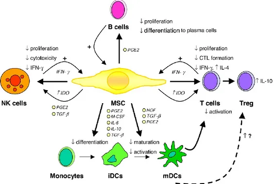
1.3. Role of MSCs in the inflammation
It is believed that MSCs possess many immunosuppressive mechanisms involved in the modulation of inflammation. They respond to specific signals produced by inflamed tissues both through paracrine effect and through their ability to migrate to the site of inflammation (Waterman RS et al. 2010). MSCs have sentinel functions that allow them to feel their microenvironment and act consequently (Aggarwal S et al. 2005) and they are able to switch from a pro-inflammatory phenotype to an immunosuppressive phenotype according to the signals they receive (Soundararajan M et al. 2018). Indeed, in conditions where there are large numbers of inflammatory cytokines, MSCs suppress immune responses by acting on a variety of immune cells (Jiang XX et al. 2005). Immunomodulation can be divided in some steps: 1) MSCs react to inflammation and move to the site of tissue injury, 2) receive the stimuli to be activated, 3) allow the pathogen clearance if it is required and 4) modulate inflammation (Waterman RS et al. 2010). So if on one hand, many studies confirmed that MSCs exert their immunosuppressive effects at a distance, on the other hand it has been demonstrate that MSCs require contact with immune cells to exert their effect (Waterman RS et al. 2010).They are able to directly modulate the T cell functions, inhibiting the proliferation and the activation of T-cells and also induce the generation of T-reg cells (Corcione A et al. 2006).
Figure 3. Immunomodulatory effects of MSCs. CTL indicates cytotoxic T cell; HGF, hepatocyte
growth factor; IDO, indoleamine 2,3-dioxygenase; PGE2, prostaglandin E2; and TGF-β, transforming growth factor β. Illustration by Paulette Dennis.
10
It has been proven that MSCs hinder dendritic cell (DC) maturation from monocytes, which is necessary for antigen presentation and T cell response (Spaggiari GM et al. 2008). This process is considered as an important checkpoint in assembling an immune response because immature DC not only fail to trigger T cells efficiently but also support tolerance induction (Akiyama K et al. 2012).
Corcione et al. observed that MSCs dissuade the proliferation, differentiation, and chemotactic properties of B cells in vitro, and suppress the cytokine-induced proliferation and cytotoxicity, and cytokine production of natural killer (NK) cells (Kong QF et al. 2009, Ghannam S et al. 2010). MSCs modulation of immune responses is mediated through several mechanisms, however, most of them involve the production of immunosuppressive factors such as prostaglandin E2, indoleamine 2,3-dioxygenase (IDO), and nitric oxide (even if the exact mechanism of action is still unclear.) The majority of these soluble factors are not constitutively produced by MSCs but are induced through the licensing or activation of MSCs as described before (Waterman RS et al. 2010).
MSCs are also able to alter the helper T-cell balance (Th1/Th2/Th17). CD4+ T helper cells become activated in response to pathogen- or danger- associated signals and can differentiate into various T-cell subsets with distinct cytokine and gene expression profiles: Th1, Th2, and Th17 subsets (English K. 2013). Depending on the circumstances, MSCs can modulate T- cell proliferation and function mediating their protective effect through shifting the balance from Th1 phenotype secreting IFNγ and TNFα to a more anti-inflammatory Th2 profile secreting increased levels of IL4, IL5, IL10, Il13 and other chemokines (Bai L et al. 2009, Fiorina P et al. 2009, Batten P et al. 2009). MSCs have the capacity to modulate Th17 differentiation in favour of IL4 producing Th2 cells or the generation of Treg (Bai L et al. 2009, Ghannam S et al. 2010). MSCs allow the generation of Treg corresponding with a decrease in Th1, Th2 and Th17 lymphocytes (Akiyama K et al. 2012, Kong QF et al. 2009, Ghannam S et al. 2010, Rafei M et al. 2009). In particular, TGFβ is the major soluble factor involved in MSC promotion of Treg in vivo (Akiyama K et al. 2012, Kong QF et al. 2009, Nemeth K et al. 2010, Zhao W et al. 2008). Many studies describe the activation of MSCs to produce soluble factors, but the sequence of events that lead to the generation of Treg and subsequence induction of tolerance is quite different and influenced by the particular microenvironment (allergic/alloreactive/allotollerant) (Waterman RS et al. 2010).
11
1.4. STUDY 1: Uterine leiomyoma
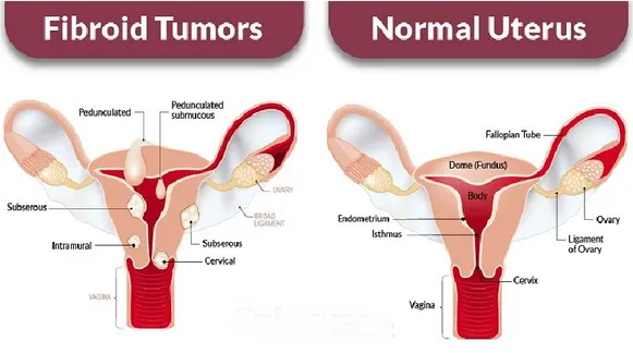
Uterine leiomyomas (also known as uterine fibroid) is benign tumor originated from the uterine smooth-muscle tissue and are characterized by the production of excessive quantities of extracellular matrix (ECM) (Aleksandrovych V et al. 2015). Uterine leiomyoma (UL) affects up to 80% of all women in their reproductive age (Aleksandrovych V et al. 2015), but it has slight difference between women with different skin color, (nearly 70% of white women and more than 80% of black women). These benign tumors can develop into significantly sized lesions (from 10 mm to 20 cm).
The main clinical symptoms are bleeding, pelvic pain, recurrent abortions and adverse obstetric outcomes often resulting in the infertility (Stewart EA. 2007, Parker WH. 2007, Ciavattini A et al. 2015). Traditionally classification of leiomyomas is based on the type of growth and location within the uterus (Brosens I. 2006). To date, there is no effective medical
Figure 4. Schematic representation of the main type of uterine fibroids (subserous, intramural,
cervical, pedunculated and submucous) and relative comparison with normal uterus. Subserosal fibroids grow mostly outside of the uterus and into the abdominal cavity. Intramural fibroids originate and grow within the wall of the uterus, and may negatively impact fertility and increase the risk of pregnancy loss. Submucosal fibroids grow and develop on the inside of the uterus and may also cause infertility and miscarriage. Subesrosal and submucosal types may also be pedunculated, meaning they have a stalk of tissue.
12
drug therapy against uterine leiomyomas but some medical treatments have been shown to be effective in reducing the volume.
Hysterectomy represents the main surgery approach even if it leads to a loss of female reproductive potential (Wallach and Vlahos 2004).
Histologically, leiomyomas appear with a different phenotype from the adjacent normal myometrium. Leiomyomas are known to be fibrotic disorders displaying excessive and continuous wound healing triggered by tissue injury and characterized by disorganized distribution of collagen fibers in extracellular matrix (ECM). However, excessive deposition and remodeling of extracellular matrix (ECM) are thought to play an important role in forming the main structure of leiomyoma (Islam MS et al. 2018). Leiomyomas typically have 50% more collagen than normal myometrium and present as encapsulated collagen-rich masses of smooth muscle cells. Moreover, ECM serve as reservoir for growth factors, cytokines, angiogenic and inflammatory response mediators (Islam MS et al 2014). The autocrine/ paracrine signaling has a significative influence in events involved in myometrium cellular transformation and turnover that are implicate din leiomyoma pathophysiology (Ciarmela P et al. 2011). Fibroids growth is affected by hormones during pregnancy and postpartum periods. Estrogen/progesterone- dependent in vivo growth of human leiomyoma tissue requires the presence of these multipotent tissue-specific stem cells (Bunting KD. 2002, Sozen I et al. 2002).
Despite their high prevalence, the cellular and molecular origins of uterine leiomyomas are not well understood. Recent studies suggest the involvement of epigenetic mechanisms such as DNA methylation and micro-RNA and histone modification in leiomyoma (Izadpanah R et al. 2006, Panepucci RA et al. 2004, Orciani M et al. 2017, McWilliams MM et al. 2017, Gargett CE et al. 2012, Hubbard SA et al. 2009). At the genetic level, several mutations, such as germline mutations causing fumarate hydratase deficiency, have been associated with leiomyoma formation (Ono M et al. 2014).
Therefore, the question about the origin and pathophysiology of leiomyoma is still open and unclear. Furthermore, several factors, such as genetic aberrations (Bulun SE. 2013) and undifferentiated cell population that could give rise to them, has been investigated (Zhang P et al. 2006, Canevari RA et al. 2005). The latter theory is sustained by the uterine tissue remodeling that happens during life in physiological (Carneiro MM. 2016) and pathological conditions (Blake RE. 2007).
13
1.4.1. Role of MSCs in uterine leiomyomas
MSCs could be involved in the onset and development of uterine leiomyomas. Indeed, some studies (Mas A et al. 2014, Mas A et al. 2015) proposed that undifferentiated cells are involved in myometrial pathologies, and also leiomyoma onset may be the results of impaired function, proliferation, and differentiation of progenitor cells inside the myometrium that are under the effect of ovarian hormones (Kurita T et al. 2001, Flake GP et al. 2003). The dysregulation of mesenchymal stem cells activity could be a possible explanation for the development of leiomyoma (Maruyama T et al. 2013). Furthermore, this hypothesis is strongly supported by the ability of leiomyomas to originate from a single altered cell derived from myometrial smooth muscle and to maintain the replicability (Mas A et al. 2014, Chang HL et al. 2010, Ono M et al. 2012). Their growth requires the presence of mature myometrial or leiomyoma cells with higher levels of steroid receptors and their ligands. This is based on the postulate that steroid hormone action on leiomyoma stem cells is mediated by mature myometrial cells (tumor initiation) or mature leiomyoma cells (growth maintenance) in a paracrine fashion (Ono M et al. 2014).
As demonstrated for other types of tumors, also in leiomyomas (Ono M et al. 2013, Ono M et al. 2014, Yin P et al. 2015, Wegienka G. 2012) microenvironment seems to play a key role in relationship with inflammation. If acute inflammation results in chronic inflammation, tumor onset and development are promoted (Orciani M et al. 2018). In this situation, cytokines secreted by the immune system and undifferentiated cells (Protic O et al. 2016, Islam MS et al. 2014, Weiss G et al. 2009, Elinav E et al. 2013, Ma S et al. 2014), which are involved in a complex crosstalk with neoplastic cells, are able to carry on a chronic inflammation. These cytokines influence proliferation, fibrosis, and angiogenesis, which in turn sustain fibroid formation and growth (Orciani M et al. 2013, Orciani M et al. 2016, Orciani M et al. 2017).
14
1.5. STUDY 2: Cervical intraepithelial neoplasia (CIN)
Cervical intraepithelial neoplasia (CIN) is a pre-cancerous lesion of uterine cervix epithelium and a surgical treatment is recommended for women diagnosed with high-grade CIN (CIN2-3). It is associated with several risk factors such as persistent human papillomavirus (HPV) infection (Vasiljević N et al. 2013), immunosuppression (Lima MI et al. 2009, Lodi CT et al. 2011), genetic mutations and epigenetic events (Virmani AK et al. 2001, Widschwendter A et al. 2004, Narayan G et al. 2003), sexual behavior and smoking (Luhn P et al. 2013). In particular, HPV infection seems to be the strongest factor influencing the natural history of CIN and increasing the risk for persistent disease. CIN can either resolve spontaneously or persist or progress if not treated properly.
Depending on the severity of the lesion type, CIN may occur in grade I (CIN1), II (CIN2) and III (CIN3). CIN 2, 3 are correlated with a risk of developing cervical cancer, and are typically treated with conisation. The societal importance is highlighted by the fact that the annual incidence of CIN1, 2 and 3 among young women (under 30 years) is increasing. The loop electrosurgical excision procedure (LEEP) is an usual method for treating high-grade CIN (Bae JH et al 2007). Despite this treatment was shown to not adversely affect fertility, it was associated with an increased risk of miscarriage in the second trimester (Kyrgiou M et al. 2015) and with enhanced chance for prematurity/higher risk for preterm birth and adverse pregnancy outcomes (Kyrgiou M et al. 2015, Liverani CA et al. 2016, Ciavattini A et al. 2015). For this reason, each patient needs an individual evaluation, especially for young women in a reproductive age, with a careful selection of patients to be treated surgically and of those that can be managed conservatively, also considering the possibility of regression of the lesions (Bekos C et. al 2018). In recent studies (Himes KP et al. 2007, Ciavattini A et al. 2015), it was demonstrated the correlation between a shortened cervical length treated with LEEP procedures and an increased risk of preterm birth (Song T et al. 2016). Therefore, it is important to consider how much cone depth increase in relationship with the amount of the volume of tissue excised. About that, Founta et al evaluated the change in cervical volume with the magnetic resonance imaging reporting that the mean cervical volume at 6 months post-procedure corresponded to 97.8% of the baseline cervical volume (Founta C et al. 2010). Furthermore, several studies have suggested that younger women generally seem to have higher rates of spontaneous regression and remission (Bekos C et al. 2018, Cox JT et al. 2003, Moscicki AB et al. 2010).
15
In particular, Bekos and colleagues investigated about age-dependent regression and progression rates in a large number of patients (783) using histologic data. They demonstrated that the regression rates were higher in young women with CIN and, in agreement with those reported in literature, regression was more likely in patients with low- grade CIN compared to high- grade dysplasia. All of these studies provides clinically relevant findings on the influence of age on the natural history of CIN and the rates of regression are notably high in young women with CIN and with a lower risk of progression.
16
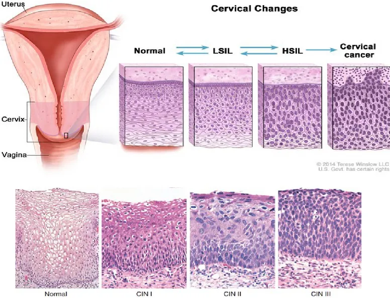
Figure 5. Representation of cervical changes in cervical intraepithelial neoplasia (CIN). Cervical dysplasia is the precursor to cervical cancer and it is characterized by
transformation and abnormal growth of cervical keratinocytes. Based on histopathology, three CIN (cervical intraepithelial neoplasia) grades are defined: CIN1 (mild dysplasia), CIN2 (moderate dysplasia) and CIN3 (severe dysplasia) lesions Based on cytology, two grades are defined: LSIL (low grade squamous intra-epithelial lesions) or HSIL (high-grade squamous intraepithelial lesions). Usually LSIL and CIN-1 overlap, and HSIL and CIN-2 and CIN-3 overlap. About 10% of LSIL/CIN-1 eventually progress to cervical cancer, while about 50% of HSIL/CIN-2/CIN-3 progress to cervical cancer.
17
1.5.1. The role of inflammation and MSCs in age-related regression of CIN
Several researches (Bekos C et al. 2018, Cox JT et al. 2003, McCredie MR et al. 2008) have evaluated the possible correlation between patient’s age and regression/progression rates of CIN, finding that younger women generally seem to have higher rates of spontaneous regression and remission. The underlying mechanisms are not yet fully known but, as for other solid tumors, inflammation may play a pivotal role in CIN fate (Hammes LS et al. 2007). The hypothesis of an involvement of inflammation in CIN progression is enforced also by studies aimed to evaluate the use of anti-inflammatory drugs in the treatment of CIN (Grabosch SM et al. 2018).
Inflammation is orchestrated by different cells of the immune system as well as by mesenchymal stem cells (Kyurkchiev D et al. 2014). The existence of MSCs in cervical tissue has been demonstrated by Eirò et al. in a study in which they isolated for the first time MSCs from the human uterine cervix (human uterine cervical stromal cells, HUCESCs) by exfoliation PAP smear (a minimally invasive procedure). HUCESCs and their secretome were able to inhibit the aggressive behavior of cancer cells (cell lines and primary tumors) in vitro and to reduce tumor growth in vivo in a mouse xenograft tumor model. They also showed an inhibitory effect on cancer associated fibroblast (CAFs) proliferation and invasion, and can inhibit and revert macrophage differentiation (Eirò N et al. 2014).
Further support has been obtained from the evidence that, within 6 months after LEEP treatment, cervix shows a regenerative process, reporting values of 71-98% of post-excision tissue deficiency (Song T et al. 2016, Papoutsis D et al. 2012, Ciavattini A et al. 2018). As inflammation is supported by undifferentiated mesenchymal stem cells (Orciani M et al. 2018), they plays a crucial role in the regression and progress of CIN. It is believed that the existence of a reservoir of MSCs inside cervical tissue represent the trigger for cervical regeneration after conization (Orciani M et al. 2018).
18
1.6. STUDY 3: Duchenne muscular Dystrophy (DMD)
Muscular dystrophy is a group of inherited diseases characterized by skeletal muscle deficiency and degeneration. Muscular dystrophies are progressive disorders caused by the loss of healthy muscle fibers over time that are replaced by fibrosis and fat, making muscle tissues less able to generate force for everyday activity (Gao QQ et al. 2015). Duchenne muscular dystrophy (DMD) is a severe X-linked recessive disorder and the most prevalent inherited myopathy affecting one in 3,500 live male births (Bushby K et al. 2010). DMD is usually diagnosed during early childhood, between 2 and 7 years of age, in which the first signs of muscle weakness appear evident. DMD patients present progressive muscular deficiency, in addition to orthopaedic, respiratory, and cardiac complications that lead to their death around the third or fourth decade of life (McNally EM. 2007, Bushby K et al. 2010).
At the molecular level, DMD is caused by mutations in the 2.2-Mb dystrophin gene which disrupts the protein’s reading frame causing premature stop codons (Koenig et al. 1987, Kunkel et al. 1987). These mutated transcripts produce truncated proteins that are unstable and subject to degradation, leaving the cells devoid of protein products. Therefore, the final result of all these mutations is an absence of functional dystrophin in the skeletal muscle. This gene is one of the biggest genes in the human genome with more than 2 million base pairs in Xp21.2-p21.1. The dystrophin coding sequence (11kbp) contains 79 exons encoding a 427 kDa dystrophin protein (Guiraud et al. 2015).
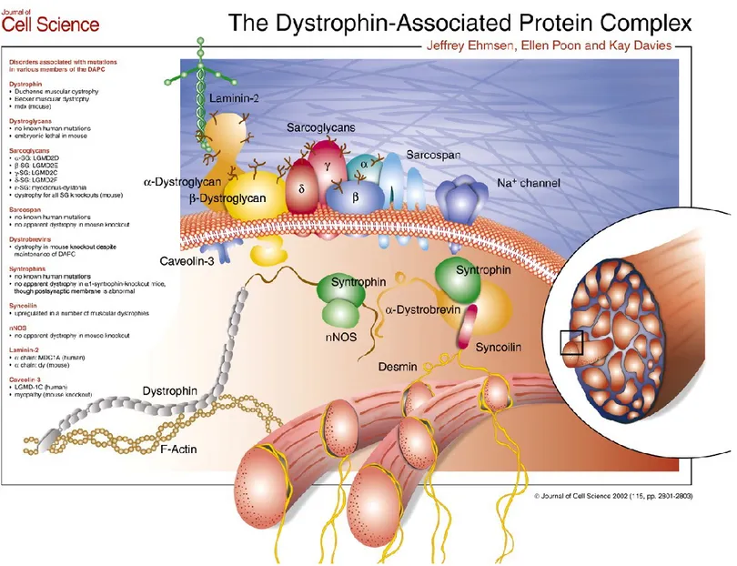
In skeletal and cardiac muscle, dystrophin binds to several proteins forming the Dystrophin-Associated Protein Complex (DAPC). The structure of dystrophin protein consists on an N-terminal actin-binding domain, a central rod-like domain with 24 spectrin-like triple helical coiled coils, and with a cysteine-rich C-terminus that permit aggregation of the DAPC (Ehmsen J et al. 2002). The DAPC has a pivotal role to link the actin cytoskeleton to the extracellular matrix, stabilizing the sarcolemma during contraction and relaxation, and sending force from the muscle sarcomeres to the extracellular matrix (Petrof BJ et al. 1993). Besides the fracture of the muscle fiber during the contraction, the DMD brings a modification in the intracellular signaling that contribute the progression of the disorder (Constantin B. 2014).
Most of the DMD patients come through the frame and non-sense mutations leding to reduction of the transcript level and truncation of translation (Monaco AP et al. 1988; Roberts RG et al. 1994).Dystrophin is a cytoskeletal protein crucial for the stability and function of myofibers in muscle.
19
It creates a mechanical link between the extracellular matrix and the cytoskeletal actin in muscle fibers through the dystrophin-associated protein complex (DAPC) (Ervasti JM et al. 2008).
Therefore, at the cellular level, the muscle of DMD patients exhibit necrosis, degeneration and regeneration, myofiber atrophy, fatty accumulation, fibrosis, and inflammation (Spencer and Tidball, 2001; Alvarez et al., 2002; Desguerre et al., 2009a,b; Serrano and Muñoz-Cá-noves, 2010; Zhou and Lu, 2010; Villalta et al., 2011).
In the last few years, different approaches (gene-based, cell-based, nano-particles, and pharmacological) have been developed to restore a functional dystrophin to DMD muscles (Negroni et al., 2016; Chamberlain and Chamberlain, 2017; Nance et al., 2017) and many of them are tested in clinical trials that are currently ongoing on DMD patients. Since 1995, among the main treatment choices for dystrophic patients in clinical trials, pharmacological approaches were found to be the primary with 57%, followed by 28% gene-based (22% antisense oligonucleotide-based exon skipping, 6% AAV gene addition), and 3% cell-base approaches (Cordova et al., 2018). The only standard therapy approved for dystrophic patients is the administration of steroids that have a number of side effects and only slow down the progression of DMD (Griggs et al. 2013). Furthermore, the clinical experimentation with novel approach reached encouraging results but none of them has achieved significant and long lasting clinical efficacy (Guirad et al. 2015). Currently steroids represent the only standard therapy for dystrophic patients but only delay the progression of the disease and come with serious side effects (Griggs et al. 2013). Many novel therapeutic approaches have entered clinical experimentation with encouraging results but none has yet reached significant and long lasting clinical efficacy (Guirad et al. 2015). These include new drugs, gene therapy, exon skipping, PTC124 (which triggers premature STOP-codon read-through), and cell therapy (Galli et al. 2018).
20
21
1.6.1. Gene therapy
The basic theory of in vivo gene therapy is the therapeutic delivery of nucleic acid into the patient cells, tissues, and organs as a drug to treat disease (Kaji E et al 2001). In details, a vector carries inside the diseased cell either a wild-type (wt) copy of the mutated gene (to replace it) or molecules that repair the DNA, or the mRNA, leading in all cases to the production of a normal or semi-normal protein at a sufficient level to carry on its specific function. In DMD specific case, to obtain a structural protein like dystrophin, it has been estimated that approximately 20-30% of the normal level is the minimum level necessary to restore function (Galli F et al. 2018).
The first step for gene therapy requires the right choice of the vector. Currently, adeno-associated vectors (AAV) are center-stage in gene therapy for muscular dystrophies (Chamberlain JR et al. 2017) as for other genetic diseases. AAV are small vectors and this feature presents pros and cons: in fact, the small dimension allow them to spread easily into the tissue but at the same time they are not able to accommodate sufficiently large enough cDNAs to encode a protein such as dystrophin. A second hurdle is the immune response of the host to the AAV capsid protein and to the gene products eventually expressed by the vector (Hareendran S et al. 2013). It is estimated that about half of the human population has been exposed to one or more serotypes of AAV, therefore the patients need to be screened preliminarily to warrant that pre-existing neutralizing antibodies may avoid any effect of the vector. Moreover, it is retained that the immune system may not see a part or whole gene product, which may evoke an immune response.
Another issue is related to the nature of the AAV vector it is the inability to integrate into the host cell genome, and cells during their divisions lose the vector. In case of striated muscle the problem is solved by the fact that differentiated cells don’t divide for long passages and persist during all the life of the host. Furthermore, the distribution of these vectors is considered a very important aspect, as it is related to the route of administration and on the structure of the target tissue. Skeletal muscle is the most abundant tissue of our body and the systemic way, especially the intra-venous one, is almost always the first choice of administration because represent the easiest way to distribute the vectors in this tissue. Moreover, this way permits to achieve wider vector distribution respect the intra-arterial characterization and the intra-muscular injection (Le Guiner C et al. 2014, Le Guiner C et al 2017).
22
Other aspects that should not be underestimated are complexity and costs that are necessary to produce large amount of AAV vectors to get significant dystrophin expression (Cohen-Haguenauer O et al. 2010).
At last, in this big scenario, gene replacement is the main method of gene correction. It was demonstrated that optimized versions of micro-dystrophin (Reza et al. 2016) and functional domains are now accessible and tests in few clinical trials with good results in patients affected by spinal muscular atrophy. However, a recent study in primates and piglets showed significant liver and neural toxicity. Another valid option is to use AAV delivered small nuclear RNA, such as U7, able to skip the mutated exon in order to restore the reading frame and produce a short but functional dystrophin (Le Guiner 2014, Vulin 2012). Currently, the strategy of using AAV as an enzyme carrier that correct the genome and repair the mutation is one of the most promising. CRISPR-Cas9 is the most innovative and current strategy that cut the damaged region restoring it with the correct sequence (Tabebordbar et al. 2016; Bengtsson et al. 2017). The fact that Cas9 is a bacterial protein is a potential cause of a strong immune reaction in vivo that could be solved using transient immune suppression or new version of the enzyme, maybe short lived to reduce exposition to the immune system.
23
1.6.2. Cell therapy
Nowadays, cell therapy is considered less popular and full of barriers that are more difficult to solve respect to gene therapy.
First of all, it is fundamental to choose the right type of cell that has a strong ability to differentiate into skeletal muscle cells and is also able to maintain this potential for more generations. Satellite cells are the first choice considering that they are the main stem cells of skeletal muscle responsible for both post-natal muscle growth and regeneration. In 1961, satellite cells were described for the first time and studied for their role in regeneration and senescence (Mauro et al). It has been shown that these cells, when transplanted in a mouse muscle, preserve their stemness (Rocheteau et al. 2012; Partridge et al. 1989), encouraging few clinical trials of cell transplantation for DMD. The theory at the base of this trial is that myogenic progenitors derived from satellite cell are expanded in vitro and then directly injected in some patient muscles without toxicity or efficacy. However, this route has led to two hurdles: massive death of transplanted cells and the fact that once injected, these cells remain in the same area without spreading to the whole muscle, making less favorable the possibility of intra-muscle transplant to larger muscles (Mouly et al. 2005; Peault et al. 2007). Many years were necessary to optimize the intramuscular transplantation protocol, and this approach has now become part of the clinical trial for the treatment of localized forms such as the Oculo-Pharyngeal Muscular Dystrophy (Pèrié et al. 2014). Until now, we can state that intra-muscular injection of satellite cells seems the therapy of choice for localized forms of muscular dystrophy or other muscle diseases, but not for forms affecting most of the body muscles (Galli F et al 2018). The second major issue for cell therapy is delivery. For a long time, systemic injection of myogenic cells was considered the best option to overcome this obstacle, but satellite cells are unable of extravasation and amass together in the capillaries creating micro-thrombi. However, a possible way to enhance diffusion involved the use of blood-born progenitor cells, which in bone marrow could participate to muscle regeneration in bone marrow transplantation (Ferrari et al. 1998). Nevertheless, this approach flunked to restore dystrophin expression in a considerable portion of muscle fibers in the receiving animals (Gussoni et al. 1999).
Another interesting approach consist on the use of mesangioblasts, that are mesenchymal-like cells, associated with the walls of large vessels, considered in vitro counterparts of muscle perivascular cells (Dellavalle et al. 2007).
24
In a clinical trial, Cossu and his group performed intra-arterially transplant mesangioblasts from human donor, usually an HLA-matched brother, in the femoral and subclavian arteries of five DMD patients in specific conditions in four consecutive injections at increasing cell dosage, resulted safety but lack of clear clinical efficacy. Indeed, expression of donor dystrophin was detected in the youngest patient, but only the 0.7% of donor DNA has been found in the biopsy of the same patient leading to the probably most difficult hurdle, the low engraftment (Cossu et al. 2015). Recently, the same authors confirmed that it is possible reach good results in disease of tissues like the blood or the epithelia, where damaged cells can be remove giving space for donor cells (Cossu et al. 2018).
Another way to act is the possibility to engineer donor cells, since muscle fibers have multinucleated cells, that may enter regenerating fibers and then also correct neighboring resident nuclei through overexpression of microdystrophin or sarcoglycan from a strong muscle promoter. It was recently shown that also MSC exosomes promote dystrophic muscle regeneration (Bier et al 2018).
An entirely innovative and different strategy consist on transducing donor myogenic cells with a lentivector expressing the U7 snRNA, engineered to skip exon 51, with the goal to exploit the diffusion of snRNA to neighboring nuclei and induce exon skipping there and thus amplify the production of dystrophin (Galli et al. 2018). Alternatively, transient expression of CRISPR Cas9 from a non integrating lentivector, may genetically correct also neighboring nuclei, thus amplifying the genetic correction. It gives the advantage of a permanent correction of the genetic defect respect to the exon skipping, but with the risk of the induction of an immune response that can be or not controlled by immune suppression.
In the last years, induced Pluripotent Stem cells (iPSc) represent another promising approach (Hotta and Yamanaka, 2015). Adult cells from the patient can be reprogrammed until a stage very close to embryonic stem cells, genetically corrected with CRISPR Cas9 for a definitive effect, and then differentiated to muscle progenitor cells. This technology provides the possibility to bypass an immune reaction, and clones with appropriate correction and genome integrity may be selected before induction of differentiation into the target cell type. Therefore, it can give good hopes for the cell therapy but the problems related to engraftment still remain.
25
Figure 7. (A) Muscular dystrophies fibers, including loss of mass, weakness, fat, and extracellular matrix accumulation. Gene and cell based therapies will have to
overcome the progressive degeneration of muscle fibers. When these histological changes become prominent, combined strategies are needed. (B) Muscle pre- or co-treatment may target inflammation, atrophy, membrane fragility, muscle weakness, and/or atrophy to pre-condition the tissue to increase efficiency of gene and cell therapy. (Cordova et al. 2018).
26
2.
Aim of the study
The main aim of these projects is to evaluate the existence of mesoderm stem cells (MSCs) and understand their role in the pathogenesis and potential therapy of selected gynecological diseases with an inflammatory component as uterine leiomyoma, cervical intraepithelial neoplasia (CIN), and in primary myopathies, as Duchenne Muscular Dystrophy (DMD).
To study gynaecological diseases, we isolate MSCs from uterine leiomyomas, healthy myometrium and cervixes. Then, the relationship between MSCs and specific inflammatory pathways, such as Th1, Th17 and Th2 will be investigated. In details, in study 1 we evaluate if: 1) the existence of undifferentiated cells may correlate with leiomyoma onset, 2) inflammation may sustain leiomyomas, and 3) cytokines secreted by undifferentiated cells may create an inflammatory microenvironment.
In study 2, the intention is to understand if: 1) age may affects the behaviour of MSCs from cervixes (C-MSCs) derived from young and old patients, 2) C-MSCs in turn may modulate inflammation and regression rate of CIN, and 3) in the crosstalk with HeLa cells, C-MSCs could play an anti-tumoral role through their paracrine effect.
About study 3, the goal is to find a correct strategy to enhance the production of dystrophin protein in Duchenne Muscular Dystrophy (DMD) through gene therapy. Therefore, to address to this question, myoblasts will be isolated from DMD donor, transduced with green fluorescent protein (GFP) and a lentiviral vector expressing the snRNA to induce exon skipping, and promoted the myogenic differentiation in order to check if they express a functional dystrophin protein.
27
3.
Materials and methods
3.1. Human tissues collection
For the first study, twelve samples of tissue respectively of leiomyoma and healthy myometrium were collected from women of childbearing age (between 30 to 35 years) by the Gynecology and Obstetrics Unit, Department of Clinical Sciences, Polytechnic University of Marche (Ancona, Italy). After a histologically confirmed diagnosis of leiomyoma, the patients underwent surgical operation such as hysterectomy or laparoscopy to remove the fibroid tissue. All tissue samples were collected in the operating room from a trained operator. One small fragment of 3-5 mm from leiomyoma and one from normal myometrium has been obtained by a cold-blade scalpel. The samples were placed in MSCGM medium (Mesenchymal Stem Cell Growth Medium, Lonza, Basel, Switzerland) and sent to our laboratory for processing. We reported the size (in cm), topographic site (anterior, posterior, left lateral, right lateral, and fundal), and location (subserosal, intramural or submucosal) of fibroids from where the samples were obtained. The removal of the sample did not alter the histopathological analysis in any case. All patients displayed good general condition; none of them had a history of myomectomy or uterine surgery, had received medical therapy or oral contraceptives in the previous three months, or had evidence of genital tract infection, endometriosis, or ovarian disease. All had a negative cervical vaginal swab collected prior to surgery, which was performed in the proliferative phase of the cycle. Adenomyosis or other uterine disorders demonstrated on histopathological examination were exclusion criteria.
For the second study, fourteen women undergoing cervical excision for high-grade cervical intraepithelial neoplasia (CIN 2-3) were recruited. The diagnosis was confirmed after colposcopy-directed cervical punch biopsy following an abnormal cervical cytology by the Gynecology and Obstetrics Unit, Department of Clinical Sciences, Polytechnic University of Marche (Ancona, Italy). The patients were divided into two groups according to age: 7 of them defined “young” (mean age 28±2) and the other 7 defined “old” (mean age 45±3).
The cervical excisional procedures were performed with the loop electrosurgical excision procedure (LEEP) technique, in an outpatient setting under local anesthesia and strict colposcopic guidance, with 1.5 – 2.0 cm rounded loops, chosen according to the type of the transformation zone and the area of cervical tissue to remove.
28
All patients provided their written informed consent to participate in the studies, which was approved by the institutional ethics committees and was conducted in accordance with the Declaration of Helsinki.
3.2. Cell culture
Tissue fragments (2-3 mm3) from leiomyomas and myometrium were firstly subjected to
mechanical digestion then to enzymatic digestion with 0.2% type II collagenase (Sigma-Aldrich, Milan, Italy) at 37°C for 4 hours; subsequently, partially digested solution was placed into 6-well plates containing MSCGM medium which enhances the growth of undifferentiated cells and maintained in culture using same media at 37°C in 95% air and 5% CO2. Tissue fragments,
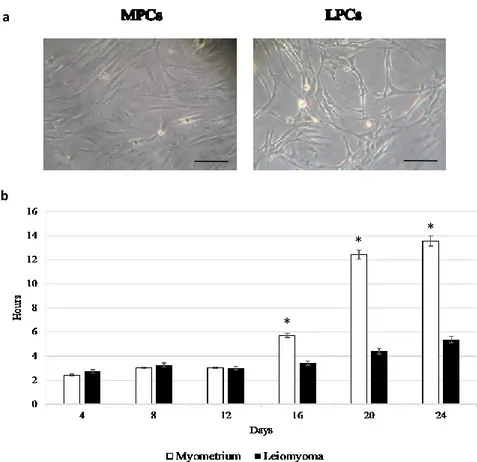
obtained from cone specimen (cervixes), were directly placed into 6-well plates containing MSCGM medium. The growth medium was changed after 24 hours to remove unattached cells/fragments or debris and then twice a week. Cell morphology was evaluated by phase-contrast microscopy (Leica DM IL; Leica Microsystems GmbH, Wetzlar, Germany) and viability by an automated cell counter (Invitrogen, Milano, Italy). All further analyses involved separate assays of the specimens from each participant up to the first five passages.
3.3. Characterization of MSCs from different tissues
After samples collection, cells were characterized by testing the minimal criteria identified by Dominici for mesenchymal definition; in detail, the plastic adherence, the immunophenotype and the multipotency were evaluated. In addition, the expression of genes (OCT4, SOX2, NANOG, KFL4) related to stemness was analyzed by Real Time PCR.
For immunophenotyping, 2.5 × 10⁵ cells were stained for 45’ with fluorescein isothiocyanate
(FITC)-conjugated antibodies (Becton-Dickinson) against: HLA-DR, CD14, CD19, CD34, CD45, CD73, CD90 and CD105 and analyzed by flow cytometry. For differentiation assay, cells were induced to differentiate into osteocytes, chondrocytes and adipocytes using STEMPRO® Osteogenesis, Chondrogenesis and Adipogenesis Kits (GIBCO, Invitrogen,) respectively. For osteogenic differentiation, after 21 days cells were assessed by Von Kossa staining and after 7 days by Alkaline phosphatase (ALP) reaction; for adipogenic differentiation, after 21 days, cells were tested by Oil Red O staining; for chondrogenesis, cells were cultured in pellet culture system for 14 days and fixed in 4% neutral buffered formalin for 24 hours at 4°C. The pellet was then processed with different grades of alcohol and xylene and paraffin embedded at temperatures
29
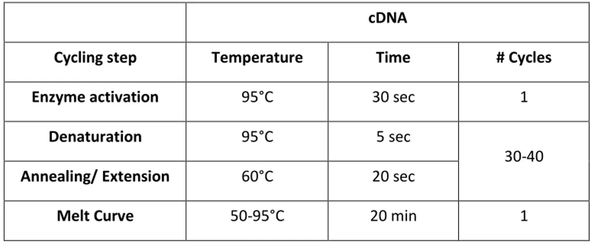
not exceeding 60°C. The sections were exposed to a solution of Safranin-O. Cells cultured in MSCGM alone were used as negative controls. For the analysis of the expression of genes related to stemness, total RNA was isolated from cells using Master Script RT-PCR System (5 PRIME, Hamburg, Germany) and quantified at Nanodrop instrument (Thermo Scientific™ NanoDrop™ instruments 2000/2000c UV-Vis). 1µg of RNA was retrotranscribed in cDNA following the manifacturer’s instructions (GoScript™ Reverse Transcription System, Promega, Italy). The amplification program and the primer sequences are reported respectively in Table 1 and Table 2. All samples were tested in triplicate with the housekeeping genes RPLP0 and GAPDH. GAPDH was the most stable and used for subsequent data normalization. After amplification, melting curves were acquired.
Table 1. Amplification program for RT-PCR
cDNA
Cycling step Temperature Time # Cycles
Enzyme activation 95°C 30 sec 1
Denaturation 95°C 5 sec
30-40
Annealing/ Extension 60°C 20 sec
Melt Curve 50-95°C 20 min 1
Direct detection of PCR products was monitored by measuring the fluorescence produced by SYBR Green I dye (EVA Green PCR Master Mix, Bio-Rad) binding to double strand DNA after every cycle. The parameter threshold cycle (Ct) was defined as the cycle number at which the first detectable increase above the threshold in fluorescence was observed.
The amount of mRNA expression was calculated by the 2−ΔΔCt method. The values of the relative
30
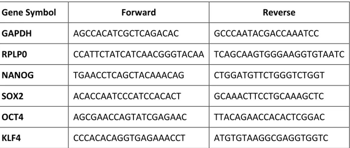
Table 2. Primer sequences related to stemness genes.
Gene Symbol Forward Reverse
GAPDH AGCCACATCGCTCAGACAC GCCCAATACGACCAAATCC
RPLP0 CCATTCTATCATCAACGGGTACAA TCAGCAAGTGGGAAGGTGTAATC
NANOG TGAACCTCAGCTACAAACAG CTGGATGTTCTGGGTCTGGT
SOX2 ACACCAATCCCATCCACACT GCAAACTTCCTGCAAAGCTC
OCT4 AGCGAACCAGTATCGAGAAC TTACAGAACCACACTCGGAC
KLF4 CCCACACAGGTGAGAAACCT ATGTGTAAGGCGAGGTGGTC
3.4. Doubling time
To assess doubling-time, 8 x 10⁴ cells per well were plated using an algorithm available online
(http://www.doubling-time.com): DT = t x lg2 / (lgN t – lgN 0)
where N0 is the number of plated cells (always the same), Nt is the number of harvested cells, and t is culture time in hours. The values are reported as mean ± SD of three independent experiments.
After the characterization, cells from normal myometrium and leiomyomas (study 1) and cell from young and old cervixes (study 2) were subjected to different analyses aimed to confirm the two working hypotheses.
31
3.5. STUDY 1
3.5.1. Immunocytochemistry
For ICC, 1.5 x 10⁴ cells from leiomyoma and myometrium were plated in each well of a 4- well
culture chamber slides (Lab Tek, Nunc, USA) and cultured overnight. Cells were fixed in 4% formalin for 30’ at room temperature, washed in Phosphate Buffer Saline (PBS) 1X and in hydrogen peroxide diluted in distillate water. Then each sample were permeabilized with 0.1% Triton X-100 in PBS 1X for 20’ at room temperature and incubated overnight at 4°C with the following monoclonal antibodies: collagen type I (1:1000 dilution, Sigma-Aldrich), anti-cellular fibronectin (1:400 dilution, Biorbyt) and anti-αSMA (1:400 dilution, Sigma-Aldrich). After washing in PBS 1X, cells were subsequently immunostained using the streptoavidin– biotin– peroxidase technique (LSAB universal peroxidase kit, Dako Cytomation, Milan, Italy). After incubation with 3,3-diaminobenzidine (0.05 diaminobenzidine in 0.05 M Tris buffer, pH 7.6 and 0.01% hydrogen peroxide), the slides were counterstained with Mayer’s hematoxylin, dehydrated and cover slipped with Dako Cytomation Glycergel mounting medium (Dako North America, Inc., Carpinteria, CA, USA). Negative control was performed omitting primary antibodies.
3.5.2. Immunofluorescence
For IIF, 1.5 x 10⁴ cells from leiomyoma and myometrium were plated in each well of a 4- well
culture chamber slides (Lab Tek, Nunc, USA) and cultured overnight. Cells were fixed in 4% formalin for 30’ at room temperature, washed three times with Phosphate Buffer Saline (PBS) 1X and treated with 0.1% of Triton X-100 in PBS 1X for 30’ at room temperature. After other three washes in PBS 1X, cells were blocked with PBS containing 2% bovine serum albumin (BSA) for 15’ at room temperature and incubated with mouse anti-human primary antibody diluted in PBS with 2% BSA anti-collagen type I (1:1000 dilution, Sigma-Aldrich), anti-cellular fibronectin (1:400 dilution, Biorbyt) and anti-αSMA (1:400 dilution, Sigma-Aldrich) overnight at 4°C.
Samples were washed three times and incubated with goat anti-mouse FITC-conjugate antibody (1:1000 dilution, Sigma-Aldrich) for 30’ at room temperature.
Then cells were washed three times and incubated for 5’ at room temperature with Hoechst 33342 (1:1000 dilution, Molecular Probe) in PBS to visualize nuclei and mounted onto a glass slide using Vectashield Vector mounting medium for fluorescence H-1000 (Vector Laboratories,
32
Inc. Burlingame, CA). Fluorescence was detected with Zeiss Axiovert 200M inverted microscope (Carl Zeiss, Jena, Germany) AxioVision. Negative control was performed omitting primary antibodies.
3.5.3. Western blot
For protein extraction from cells derived from myometrium and leiomyoma, pellets were collected and incubated in ice with RIPA buffer (150 mM NaCl, 10 mM Tris, pH 7.2, 0.1 % SDS, 1.0 % Triton X-100, 5 mM EDTA, pH 8.0) containing protease inhibitor cocktail (Roche Applied Science, Indianapolis, IN, USA) for 45 minutes and then centrifuge at 14000 g for 15 minutes at 4°C. Supernatants were aliquoted and stored at -20°C before the use. To determine protein concentration, the Bradford protein assay with Bradford reagent was performed (Bradford reagent, Sigma-Aldrich, Milan, Italy). Different concentrations of BSA (Albumin Bovine Serum) in distilled water were used to draw the standard curve. Total protein extracts (35 µg of protein for each sample) were reduced in DTT (0.5 M) for 10 minutes at 70°C before the electrophoresis. Precast polyacrylamide gels at 4-12% gradient (Invitrogen NuPAGE Bis-Tris protein gels) were chosen for electrophoresis separation. Electroblotting was performed using iBlot® Dry Blotting System (Invitrogen). Membranes were incubated with 5% milk in PBS 1X with 0.05% Tween 20 (T-PBS) to block nonspecific sites for 1 hour. Then, membranes were hybridized with monoclonal mouse antibodies anti-MDR1 (Santa Cruz, 1:400) diluted in 5% milk T-PBS at 4°C overnight. After washing with T-PBS, membranes were incubated with secondary antibody anti-mouse conjugated with horseradish peroxidase (Santa Cruz Biotechnology) at 1:10000 dilution for 1 hour. Detection of antibody binding was performed with the chemiluminescence substrate reagent (ImmunoCruzTM Western blotting Luminol reagent, Santa Cruz Biotechnology) and
images were acquired with UVITEC Alliance Q9 Advanced (UVITEC Cambridge). GAPDH and -Actin were used as housekeeping to normalize. As negative control, Normal Human Lung Fibroblasts (NHLF) were used.
3.5.4. Expression of inflammation-related cytokines by RT-PCR and ELISA
Selected cytokines related to acute and chronic inflammation, IL6, IL12, IFN-γ, TNF-α, IL2, IL4, IL5, IL13, IL10, TGF-β1, IL17A, and GM-CSF, were investigated by RT-PCR (as above reported) and by ELISA (Multi-Analyte ELISArray kit, Qiagen, Milan, Italy).
33
Briefly, medium conditioned for 72 hours by each sample of MSCs derived from myometrium and leiomyoma was collected, concentrated and dispensed into a 96-well microtiter plate from Row B to G. The appropriate Sample Dilution Buffer was added in each well of Row A of the ELISArray plate to set up the negative control, and the final Antigen Standard Cocktail diluted following the protocol was added in the Row H to set up the positive control. The ELISArray plate was incubated for 2 hours at room temperature. After three washes with a Wash Buffer 1X, the diluted Detection Antibodies were transfer to the appropriate rows of the ELISArray plate and incubated for 1 hour. Then, the plate was washed for three times and the Avidin-HRP conjugate, diluted in Assay Buffer, was added into all wells and incubated for 30’ at room temperature in the dark. After four washes, the plate was incubated with a Development Solution for 15’ at room temperature in the dark. Finally, the Stop Solution was added in each well and the absorbance was read at 450 nm and 570 nm using a microtiter plate reader (Multiskangomicroplate reader, Thermo Scientific).
The levels of the cytokines secreted by leiomyoma cells are reported as a percentage of the levels measured in the corresponding myometrial sample. After, mean ± SD from three independent experiments in triplicates was calculated and displayed.
For mRNA analysis, RT-PCR was performed as above mentioned and the primer sequences are summarized in Table 3; the amount of mRNA detected in MSCs from leiomyomas was calculated as X-fold respect to MSCs from myometrium (expressed as 1) by the 2−ΔΔCt method, where ΔCt=Ct
(gene of interest)—Ct (control gene) and Δ (ΔCt) = ΔCt (leiomyomas)—ΔCt (myometrium). X-fold was calculated for the selected genes in all the twelve samples of MSCs. Subsequently, mean ± SD from three independent experiments in triplicates was calculated and displayed.
Since both mRNA levels and ELISA revealed a strong downregulation of Th1/Th17 pathway cytokines, the expression of other Th1/Th17-related soluble factors (IL22, NFKB1, IL23A, STAT3, CCR5, IL17A, IL17RA, CX3CL1, CXCL12, CXCL5) was also assessed by RT-PCR and the amount of mRNA calculated as above described.
34
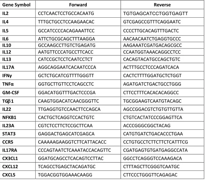
Table 3. Primer sequences related to inflammation pathways: Th1, Th2, Th17.
Gene Symbol Forward Reverse
IL2 CCTCAACTCCTGCCACAATG TGTGAGCATCCTGGTGAGTT
IL4 TTTGCTGCCTCCAAGAACAC GTCGAGCCGTTTCAGGAATC
IL5 GCCATCCCCACAGAAATTCC CCCCTTGCACAGTTTGACTC
IL6 ATTCTGCGCAGCTTTAAGGA AACAACAATCTGAGGTGCCC
IL10 GCCAAGCCTTGTCTGAGATG AAGAAATCGATGACAGCGCC
IL12 AATGTTCCCATGCCTTCACC CCAATGGTAAACAGGCCTCC
IL13 CATCCGCTCCTCAATCCTCT CACAGTACATGCCAGCTGTC
IL17A AGGCAGGAATCACAATCCCA ACTTTGCCTCCCAGATCACA
IFNγ GCTCTGCATCGTTTTGGGTT CACTCTTTTGGATGCTCTGGT
TNFα GGTGCTTGTTCCTCAGCCTC AGATGATCTGACTGCCTGGG
GM-CSF GGACATGGTTTGACTCCCGA CTTCCTTTCACACACAGGCC
TG1 CAAGTGGACATCAACGGGTTC TGCGGAAGTCAATGTACAGC
IL22 TTGAGGTGTCCAACTTCCAGCA AGCCGGACGTCTGTGTTGTTA
NFKB1 CACTGCTCAGGTCCACTGTC CTGTCACTATCCCGGAGTTCA
IL23A CGTCTCCTTCTCCGCTTCAA ACCCGGGCGGCTACAG
STAT3 GAGGACTGAGCATCGAGCA CATGTGATCTGACACCCTGAA
CCR5 CAAAAAGAAGGTCTTCATTACACC CCTGTGCCTCTTCTTCTCATTTCG
IL17RA CCCAGTAATCTCAAATACCACAGTTC CGATGAGTGTGATGAGGCCATA
CX3CL1 GGATGCAGCCTCACAGTCCTTAC GGCCTCAGGGTCCAAAGACA
CXCL12 TCAGCCTGAGCTACAGATGC CTTTAGCTTCGGGTCAATGC
35
3.6 STUDY 2
3.6.1 Proliferation assay
To assess the proliferation rate, XTT Cell Proliferation Assay (Trevigen, Gaithesbrug MD, USA) was performed on cells after 72 hours of culture for the first 6 passages. The data about yC-MSCs are reported as percentages of the values measured in parallel in oC-MSCs (referred as 100%), over three independent experiments.
3.6.2 Senescence associated β-galactosidase assay
Senescence associated β-gal activity was detected with a senescent cell staining kit (Sigma-Aldrich, Milan, Italy) according to the manufacturer’s instructions. 7 x 10⁴ MSCs from old and
young cervixes (o-CMSCs and Y-CMSCs) at passage 6th were seeded in a 6-well plate and
incubated with the staining solution overnight at 37°C without CO2. β- Gal was microscopically
revealed by the presence of a blue, insoluble precipitate within the cell, the percentage of SA- β-gal positive cells was determined by counting at least 500 cells in each sample and calculating the mean.
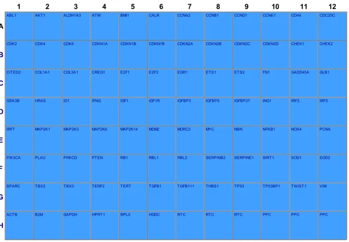
3.6.3. PCR array for the senescence
The expression of 86 genes related to senescence was analysed by PCR array (Qiagen, Milan) in MSCs isolated from cervixes derived from young and old patients.
Total RNA was isolated by using Master Script RT-PCR System (5 PRIME, Hamburg, Germany). cDNA synthesis was performed using SABiosciences RT² First Strand Kits, following the manufacturer’s instruction. mRNA expression was calculated by the 2−ΔΔCt method, where ΔCt =
Ct (gene of interest)—Ct (control gene) and Δ (ΔCt) = ΔCt (yC-MSCs)—ΔCt (oC-MSCs).
3.6.4. Indirect co-culture with HeLa cells
Human cervix epithelioid carcinoma cell line (HeLa, Sigma Aldrich) was cultured in EMEM (EBSS) + 2mM Glutamine + 1% Non-Essential Amino Acids (NEAA) + 10% Foetal Bovine Serum (Sigma Aldrich). HeLa were co-cultured with MSCs isolated from young and old cervixes.
36
In a 6-well plate, 5 x 10⁴ HeLa were seeded at the lower surface of each well and the day after, 5
x 104 MSCs derived from young and old cervixes were individually added in the upper surface of
a polycarbonate transmembrane filter in a transwell filter system (pore size 0.4 μm, BD Falcon). Cells were cocultured for 72 hours. After cocultures, the medium was collected and store at -80°C for ELISA assay and tumor cells were recovered, counted by an automated cell counter and the pellets were freeze down for further analysis. HeLa cells cultured individually served as controls (mock). Data are reported as mean ± SD from three independent experiments.
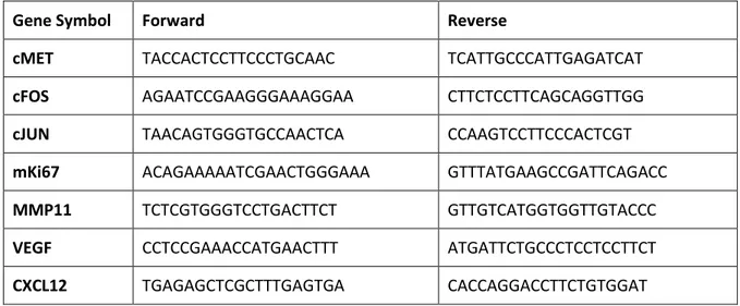
3.6.5. Expression of selected genes in co-cultured HeLa cells by RT-PCR
RT-PCR, performed as above mentioned, was performed to evaluate the expression of genes associated with specific cellular mechanisms, such as oncogenesis (cMET, cFOS, cJUN), proliferation (mKi67), invasion and migration (MMP11) and angiogenesis (VEGF, CXCL12). Quantification of mRNA expression was calculated with the 2−ΔΔCt method, where ΔCt = Ct (gene
of interest) − Ct (control gene) and Δ(ΔCt) = ΔCt (HeLa cells co-cultured with cervix-MSCs) − ΔCt
1 2 3 4 5 6 7 8 9 10 11 12
A
ABL1 AKT1 ALDH1A3 ATM BMI1 CALR CCNA2 CCNB1 CCND1 CCNE1 CD44 CDC25C
B
CDK2 CDK4 CDK6 CDKN1A CDKN1B CDKN1B CDKN2A CDKN2B CDKN2C CDKN2D CHEK1 CHEK2
C
CITED2 COL1A1 COL3A1 CREG1 E2F1 E2F3 EGR1 ETS1 ETS2 FN1 GADD45A GLB1
D
GSK3B HRAS ID1 IFNG IGF1 IGF1R IGFBP3 IGFBP5 IGFBP27 ING1 IRF3 IRF5
E
IRF7 MAP2K1 MAP2K3 MAP2K6 MAP2K14 MDM2 MORC3 MYC NBN NFKB1 NOX4 PCNA
F
PIK3CA PLAU PRKCD PTEN RB1 RBL1 RBL2 SERPINB2 SERPINE1 SIRT1 SOD1 SOD2
G
SPARC TBX2 TBX3 TERF2 TERT TGFB1 TGFB111 THBS1 TP53 TP53BP1 TWIST1 VIM
H
ACTB B2M GAPDH HPRT1 RPL0 HGDC RTC RTC RTC PPC PPC PPC
37
(HeLa cells cultured alone). The values of the relative expression of genes of interest are referred as mean ± DS, over three independent experiments.
The primer sequences are reported in Table 5.
Table 5. Primer sequences of genes related to oncogenesis, proliferation, invasion and migration and angiogenesis.
Gene Symbol Forward Reverse
cMET TACCACTCCTTCCCTGCAAC TCATTGCCCATTGAGATCAT
cFOS AGAATCCGAAGGGAAAGGAA CTTCTCCTTCAGCAGGTTGG
cJUN TAACAGTGGGTGCCAACTCA CCAAGTCCTTCCCACTCGT
mKi67 ACAGAAAAATCGAACTGGGAAA GTTTATGAAGCCGATTCAGACC
MMP11 TCTCGTGGGTCCTGACTTCT GTTGTCATGGTGGTTGTACCC
VEGF CCTCCGAAACCATGAACTTT ATGATTCTGCCCTCCTCCTTCT
CXCL12 TGAGAGCTCGCTTTGAGTGA CACCAGGACCTTCTGTGGAT
3.6.6. Analysis of the effects of age and co-culture in secretion of inflammatory cytokines.
Potential effects related to the age of donors on secretion of cytokines involved in inflammation were tested by ELISA test (Multi-Analyte ELISArray kit, Qiagen, Milan, Italy) as above described. Mean ± SD has been calculated for MSCs derived from cervixes of young and old patients over three independent tests and expressed as pg/ml. Subsequently, MSCs were co-cultured with HeLa cells and the amount of the differentially secreted cytokines was re-evaluated. Levels detected in co-cultured HeLa have been reported as a percentage of the levels measured in HeLa cells cultured alone, and data are presented as mean ± SD over three independent tests.
3.6.7. Statistical Analysis
Statistical analysis was performed by using SPSS 19.0 software (SPSS, Inc., Chicago, IL, USA). All data are presented as mean ± SD. Statistical significance was calculated for two-sample comparisons by Student's t test using SPSS 17.0 software. Statistical significance was analysed for data from at least three independent experiments. P values ≤ 0.05 were defined as statistically significant.