UNIVERSITÀ DEGLI STUDI DI NAPOLI
“FEDERICO II”
Tesi di Dottorato
“New Insights In Veterinary Forensic Medicine and
Pathology”
Candidato Tutor Co-Turor
Dott. Giuseppe Piegari Prof. Gaetano Oliva Prof. Orlando Paciello
Coordinatore
For those who cannot speak for themselves,
For my Dad… How ironic is it that I wasted all these years not listening to you. But now that you’re not here, I’m living life exactly how you told me to
List of Abbreviations 10
List of Figures 12
List of Tables 14
Abstract 15
General Introduction
I Forensic Veterinary Medicine and Pathology 18
II Animal Law 19
III Crime Scene Analysis 24
IV Forensic Necropsy 27
V Post Mortem Interval in Veterinary Medicine 31
V.I Algor Mortis 33
V.II Livor Mortis 34
V.III Rigor Mortis 35 V.IV Dehydration 36
V.V Decomposition 37
V.VI Histological changes 42
V.VII Molecular Methods 43
VI Main Limitations in Veterinary Forensic Pathology 44
VIII References 47
Forensic photography
Chapter 1. Assessment of Google Glass For Photographic Documentation in Veterinary Forensic Pathology
1.1 Introduction 56
1.2 Material and Methods 58
1.3 Results 66
1.4 Discussion 71
1.5 Limitations 75
1.6 Conclusion 76
Forensic Asphyxiology
Chapter 2. Diagnosis of Drowning in Veterinary Forensic Pathology
2.2 Material and Methods 89 2.3 Results 93 2.4 Discussion 100 2.5 Conclusion 104 2.6 References 105 Forensic Thanatology
Chapter 3. Evaluation of Muscular Proteins Degradation to Define Post Mortem Interval (PMI) in Dog
3.1 Introduction 111
3.2 Material and Methods 113
3.3 Results 116
3.4 Discussion 122
3.5 Conclusion 124
3.6 References 125
Forensic Traumatology
Chapter 4. Cardiac laceration following non-penetrating chest trauma in dog and cat
4.1 Introduction 130 4.2 Case reports 131 4.3 Discussion 135 4.4 Conclusion 138 4.5 References 139 Forensic Microbiology
Chapter 5. Contribution of forensic microbiology in sudden and unexpected death cases in young dog
5.1 Introduction 142
5.2 Material and Methods 144
5.3 Results 147
5.4 Discussion 152
Il mio personale percorso di Dottorato mi ha condotto ad investigare vari aspetti della Patologia Forense Veterinaria. Mi sento onorato di aver potuto condurre studi in questo ambito ed è per questo, che è per me un piacere ringraziare il mio Tutor, Prof. Gaetano Oliva ed il mio co-tutor, Prof. Orlando Paciello, per avermi dato l’opportunità di entrare a contatto con il mondo della ricerca. Inoltre, esprimo la mia riconoscenza per avermi guidato nel mio percorso con preziosi consigli. Un ringraziamento anche per aver potuto fare affidamento su di loro nei momenti di gioia o di sconforto del percorso di Dottorato.
Un ringraziamento speciale è rivolto al mio collega ed amico Dott. Davide De Biase con cui abbiamo condiviso gioie e dolori di questo percorso. Senza di lui, forse, questa tesi si sarebbe presentata con un assetto totalmente diverso. Il suo “rigore” scientifico è stato, di fatto, un esempio da seguire… Vorrei inoltre esprimere la mia immensa gratitudine ai miei colleghi di lavoro, Dott. Francesco Prisco, Dott.ssa Arianna Ilsami, Dott.ssa Ilaria D’aquino, Alessio Sardiello, Lello Ilsami, che, nel corso di questi tre anni, hanno condiviso con me gioie e dolori, sia in laboratorio che in sala settoria.
Desidero ringraziare, altresì, mia madre Patrizia, mia sorella Maria Cristina e la mia compagnia Giulia. Grazie per essereci sempre stati, anche quando non lo meritavo…
Infine, mi piacerebbe ringraziare me stesso per la tenacia dimostrata, ma sarebbe una falsità … la verità è che tutti i sopracitati nomi, insieme, mi hanno danno la forza di terminare questo percorso. Complessivamente, Grazie !
PMI Post Mortem Interval IHC Immunohistochemistry ASA Animal Sexual Abuse DSLR Digital single-lens reflex
ICC Intraclass Correlation Coefficient HE Hematoxylin and Eosin
SUID Sudden and Unexpected infant death SIDS Sudden infancy death Syndrom CPV-2 Canine Parvovirus Type 2 PCR Polymerase Chain Reaction PMR Proportional Mortality Ratio
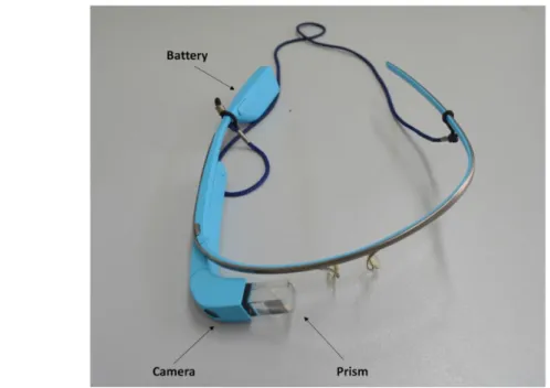
1.1 Google Glass Device.
1.2 The forensic pathologist wears Google Glass and takes pictures of small anatomical details in a hands-free manner.
2.1. Representative H&E –stained sections from lung tissues of animals of the groups A, B and C.
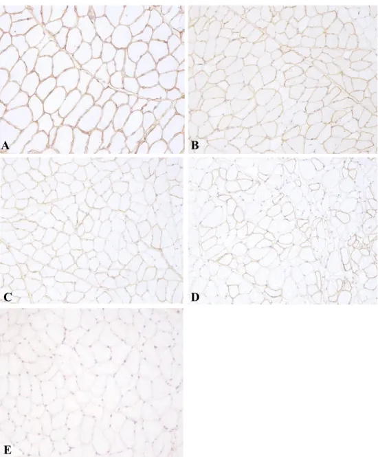
2.2 The most common diatoms identified in animals of the Group A. 3.1 Representative H&E –stained sections from muscle tissues at different time elapsed since death.
3.2 Changes produced in immunohistochemical labelling for dystrophin in cross-sections of dog muscles during post mortem storage.
3.4 Changes produced in immunohistochemical labelling for desmin in cross-sections of dog muscles during post mortem storage.
4.1 Cardiac laceration; tear on the lateral-inferior portion of right ventricle near inter-ventricular septum.
4.2 Macroscopic examination: (A) pericardium filled with water shows a large vertical tear on its upper third (B) little fragment of the right ventricle within the pericardial sac.
4.3 Macroscopic examination: (A) clotted blood in the thoracic cavity (B) laceration of size 0.6 cm x 0.4 cm over the right auricle of the heart
(arrow). Histopathological examination: (C) section from margin of tear; hemorrhagic laceration of the right auricle of the heart (arrow); H&E stain (original magnification 10x), (D) aorta: peri-aortic hemorrhage (arrow); H&E stain (original magnification 4x).
I Main steps of the crime scene analysis. II Forensic necropsy protocol.
III Post-mortem histological changes. 1.1 Scoring system for image quality.
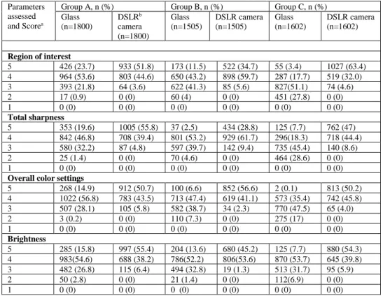
1.2 Frequencies and percentages of evaluations given by 5 pathologists for images taken during forensic necropsies with Google Glass and Nikon D3200 reflex camera stratified for each group.
1.3 Unpaired rank sum, 2-sided Mann-Whitney U (Cronbach alpha=.05) for ratings of images taken with Google Glass and Nikon D3200 reflex camera for each group for each of the 4 assessed parameters.
1.4 Unpaired rank sum, 2-sided Mann-Whitney U (Cronbach alpha=.05) for ratings of the images of the 3 groups taken with Google Glass. 1.5 Descriptive statistics of loss of battery consumption during forensic necropsy stratified by the device used to acquire the images: a Nikon D3200 reflex camera and the Google Glass device.
2.1 Diatom test protocol and internal protocol used to avoid diatom contamination.
2.2 Macroscopic Lesions of Drowning observed in study animals stratified by group.
2.3 Mann–Whitney rank sum test between control and drowned animals. 2.4 No. of diatoms per 300 µl recovered from animal tissue samples. 2.5 No. Diatoms per 300 µl recovered from animal tissue samples. 2.6 Mann–Whitney rank sum test of lung diatom test results. 3.1 Median and p-values for each post-mortem interval.
5.1 frequency and percentage of unexpected and expected deaths and the frequency of total deaths stratified by age classes.
5.2 Viruses or bacteria detected.
Over the last years, the knowledge on the veterinary forensic medicine and pathology has experienced a rapid increase as evidenced by number of peer-reviewed publications, textbooks and inclusion of the topic in many veterinary medical conferences. However, most of the information in veterinary forensic medicine is still acquired by human forensic literature. This lack of information is currently considered a serious problem in veterinary forensic medicine. Indeed, although it is undeniably true that the mechanisms of forensic injuries as well as the post-mortem cadaveric changes are similar between humans and animals, the different morphology, weight and tissue resistance of animals compared to human anatomy and other species-specific factors make the information validated in human forensic medicine not always applicable in the veterinary forensic field. In addition, in human forensic medicine, the macroscopic examination associated with the histological analysis is often not sufficient to determine the victim’s cause and manner of death “beyond all reasonable doubt". Therefore, in human medicine, a range of ancillary tests have been proposed to confirm specific causes of death such as the diatom test for the diagnosis of drowning or a seminal fluid test for the diagnosis of Animal Sexual Abuse (ASA). Although the application of some of these tests has been sporadically described in veterinary medicine, their use in veterinary forensics practice requires additional rigorous validation studies. Therefore, the purpose of this thesis work is to summarize the studies carried out throughout the PhD scholarship, which were based on development of new methodological approaches in veterinary forensic medicine and pathology. Attention was paid to these sub-fields of the veterinary forensic medicine:
1) Forensic photography 2) Forensic traumatology 3) Post mortem
interval 4) Diagnosis of drowning and 5) Forensic microbiology.
As regard the forensic photography, we assessed the suitability of “Google Glass device” in veterinary forensic photographic documentation; furthermore, in forensic traumatology field, we reported the first case of cardiac rupture following non-penetrating chest trauma (NCT) in a cat. Moreover, we presented unusual cases of cardiac rupture with NCT in two dogs. As regard the post mortem interval, we investigated the correlation between time since death and post mortem muscle proteins degradation in dog. In addition, as regard the diagnosis of drowning, we evaluated: 1) the macroscopic and microscopic findings in drowned animals and the contribution of necropsy and histological examination to determine the cause of death in drowning cases in veterinary forensic pathology 2) the differences in the number and location of diatoms between animals who died in drowning and nondrowning conditions and 3) the correlation between the time of permanence in water and the number and location of diatoms in animals dead for causes other than drowning and subsequently used for experimental drowning in standard conditions. Finally, in forensic microbiology field, we assessed the contribution of post-mortem microbiology in establishing a cause of death in young dogs who died of sudden and unexpected death. The results of my thesis showed that Google Glasses were usable in the veterinary forensic pathology of pet animals allowing a reduction in the mean execution time of necropsy and the acquisition of images useful for forensic documentation purposes. Furthermore, as regard the post-mortem modification of the muscles, we
observed a time depend post-mortem degradation of the muscle proteins such as desmin and dystrophin. In addition, as regard the diagnosis of drowning, we reported a statistically higher diatom number in the tissues of drowned animals than in the tissue of nondrowned animals and experimentally drowned cadavers. In contrast, similar macroscopic and histological injuries were observed in both nondrowned and drowned animals. Finally, as regard the forensic microbiology, we observed a high frequency of viruses and bacteria detected in cases of animals who died of sudden and unexpected death such as the following: Canine Parvovirus type 2, Clostrifium Perfrigens and Canine Distemper Virus. Together, these findings will provide useful tools to increase the knowledge in veterinary forensic medicine by reducing the acquisition of information from the human medical literature which, although very complete, does not provide information that are perfectly applicable to the species of veterinary interest.
I. Forensic veterinary medicine and pathology
The word “forensic” is defined in the Concise Oxford English Dictionary as meaning “relating to, used in, or connected with a court of law”. Thus, “forensic veterinary medicine” can be defined the “branch of veterinary medicine that studies the applications of veterinary medical science to legal settings”. The veterinary forensic pathology can be considered a subfield of the forensic medicine that deals with death investigations (Brooks, 2018). It is based on a transverse and multiorgan approach that includes necropsy, histological examination, immunohistochemistry (IHC) and collateral examinations such as laboratory analysis and diagnostic imaging to resolve obscure fatalities (Piegari et al. 2018). The duties of a veterinary forensic pathologist are similar to those of a human forensic pathologist and include: crime scene investigation, evaluation of the clinic history of the animals, forensic necropsy, forensic histopathology, forensic photographic documentation and writing of the necropsy report. The ultimate goal of this branch of veterinary medicine is to determine the cause of the death of the animal, whether natural or violent, the manner of death, the time since death (post-mortem interval) and to examine and preserve any physical evidence that might produce useful information for identifying and charging those guilty of crimes against animals (Brooks, 2018).
II. Animal law
Forensic Veterinary Medicine has its reason to exist, thanks to the laws that protect animals and make those who commit crimes against them liable to penalties or sanctions. In Italy, the most significant animal protection laws include:
- Legislative Decree 157/92 Rules for the protection of homeothermal wild animals and for hunting purposes.
- Legislative Decree 281/91 low on pets and the prevention of straying.
- Legislative Decree 150/92 Regulation of offences relating to the application in Italy of the Convention on the international trade of endangered animal and plant species.
- Legislative Decree 151/2007 Penalty provisions for violating the provisions of the Regulations (EC) no. 1/20055.
- Legislative Decree 189/2004 Provisions concerning the prohibition of animal abuse, as well as the use of animals in clandestine fighting or unauthorised competitions.
- Regulation (ec) 1523/2007 of the European Parliament and Council of 11 December 2007 banning the placing on the market and the import to, or export from, the Community of cat and dog fur, as well as products containing such fur.
- Ministerial Ordinance of 18 December 2008 and its subsequent amendments: Guidelines on the prohibition of the use and keeping of poisoned baits.
- Law 727 of the Italian Criminal Code punishes the abandonment of animals.
Among these, Law 189 introduced in 2004, is one of the most important in the field of the veterinary forensic medicine. This law changed the legal basis for the protection of animals, which until then had been governed only by Article 727 of the Criminal Code. With the amendment of this law, animal sentiment is now being harmed and no longer just the "human morality".
With Art. 1"Amendments to the Criminal Code" of the aforementioned Law, the legislator introduced (after title IX) the title IX bis, entitled "Crimes
against animal sentiment" into the Criminal Code. Following this change,
animal abuse by just one offence becomes a crime.
This change involves:
1) an aggravation of penalties (from fines to imprisonment); 2) the impossibility of overturning the offence by means of an
oblation;
3) the lengthening of the limitation period;
4) the necessity of willful misconduct, also in the form of the so-called " potential " misconduct (negligent misconduct is excluded from the regulations, except for the offence referred to in Article 727 of the Criminal Code).
The articles introduced by this law are: -
- 544 bis: killing of animals; - 544 ter: animal abuse;
- 544 quater: prohibited shows or events using animals; - 544 quinquies: prohibition of animal fighting.
In particular, the articles of greatest interest for the forensic veterinary pathologist are 544 bis and 544 ter. Art. 544 ter states the following:
Par. 1 “Anyone who, for cruelty or without necessity, causes injury to an
animal or subjects it to torture, hard labour or behaviour, or to unbearable work due to its ethological characteristics, shall be punished with imprisonment from 3 months to 1 year, or given a fine ranging from €3,000 to €15,000”
Par. 2 “The same punishment is applicable to anyone who administers
prohibited or narcotic substances to animals or subjects them to treatments that cause damage to their health”
Par. 3 “The penalty is increased by half if the facts referred to in the first
paragraph results in the death of the animal”
From this article, it can be deduced that the action perpetrated constitutes a crime when the conscience and will to cause damage to the animal is present.
This damage, according to what has been mentioned above, may be of different types: physical injury, torture, hard labour or behaviour, or unbearable work due to its ethological characteristics, administration of drugs or prohibited treatments that cause damage to the health of the animal. The third paragraph of Article 544 provides for an aggravating circumstance, which materializes if the conduct referred to the first paragraph results in the death of the animal. Such an aggravating circumstance only exists if the death of the animal is an unintended consequence of the abuse. If this is not the case, the crime of killing animals is then committed as provided for in Article 544 bis of the Italian Criminal Code, pursuant to which:
"Whoever, whether cruelly or without necessity, causes the death of an animal shall be punished by imprisonment from three to eighteen months".
This low does not provide for:
- a distinction between owned or stray animal;
- the specific methods used to cause the death of the animal; both action and negligence, resulting in death, are punishable.
Finally, article 727c.c., reports the following:
Par. 1 "Whoever abandons pets or animals that have acquired captive
habits is liable to imprisonment for up to one year or a fine between €1,000 and €10,000.”
Par. 2 “The same punishment is applicable to anyone keeps animals in
conditions incompatible with their nature and producing severe suffering."
Here, we have two types of punishment: the abandonment of animals and their keeping in conditions that conflict with their nature and cause suffering. The concept of abandonment can be traced back to carelessness or neglect of the animal and not to cruelty to the animal or the infliction of gratuitous suffering, attitudes that are punished with the crime of animal abuse (art. 544 ter).
Abandonment, in any case, is not just a matter of abandoning the animal, but must be understood in the more general intention of no longer taking care of it. Furthermore, as regard the keeping of animals in conditions incompatible with their nature, it is not to be understood as a necessarily intentional offence, as it can be committed through negligence alone. Therefore, the keeper of animals in conditions incompatible with their nature or in a state of abandonment is criminally liable even through negligence alone. With regard to "serious suffering", the Court of Cassation has specified that, “while it is undeniably true that the concept of the gravity of the suffering necessary to fulfill the conduct described in art. 727 of the Italian Criminal Code is indeed different from the concept of serious damage to the animal's health provided for in art. 544 ter of the Italian Criminal Code, it is nevertheless essential that the suffering to which poorly kept animals should be subjected reach a level such as to make the condition in which they are kept absolutely irreconcilable with the proper conditions for the animal to be in a situation of well-being”. This opinion should be expressed with reference to contingent situations, it being clear that a
temporary situation of distress for the animal cannot be confused with the “contra legem” situation set forth in paragraph 2 of art. 727.
III. Crime scene analysis
The crime scene can be divided in primary crime scene, secondary crime scene and disposal site. The primary crime scene is the place where the most of the crime act was committed or, more in detail, “the place where offender engaged in the majority of his or her attack or assault upon the victim or victims" (Turvey, 1999; Savino et al. 2011). In contrast, the secondary crime scene is “any place where there are evidence of interaction between criminal and victim outside the primary scene” (Turvey,1999; Savino et al. 2005). Finally, disposal site is “the place where the body of the victim is found” (Savino et al. 2011). As a whole, a crime scene can be compared to a canvas of the expressionist period because of the set of disjointed information that a forensic pathologist must be able to acquire and subsequently relate to each other in order to reconstruct the initial situation which, exactly as in a canvas, produced the effects that then need to be analyzed. The complexity of a crime scene is inextricably linked to the unpredictability of human actions, which do not always follow a definite logic. The information obtained from the crime scene analysis is important not only for correctly interpreting the injuries observed during the forensic post-mortem examination but also to establish specific relationship between a suspect and the crime scene or victim (Gardner, 2011; Touroo and Fitch; 2016). Indeed, according to the Locard's exchange principle “Every contact leaves a trace". In particular, when two objects come into contact, there is
always a mutual exchange of matter between them. Therefore, matter from the crime scene may be carried away on the criminal and, at the same time, criminal may live matter at the crime scene (Reddy, 2011). For these reasons, the work of the forensic pathologists should always begin with the analysis of the crime scene. Unfortunately, in Italy, the veterinary forensic pathologist hardly ever visits the crime scene. Although the pathologist may request photographs of the crime scene and law enforcement reports, the information that can obtain by examining the crime scene directly is irreplaceable. Indeed, in most cases, the law enforcement does not have specialist skills. Therefore, it is possible that important findings from a medical-legal point of view are not detected and consequently not photographed and collected. In general, the intervention of a pathologist on the crime scene is all the more useful the more promptly it is conducted. Indeed, the biological materials, such as traces of blood or semen, deteriorate rapidly. Furthermore, with the increase of the time elapsed since death, post-mortem modifications are more influenced by environmental variables, such as temperature, weather and presence or absence of shade. In general, all evidence obtained from a crime scene can be divided in 3 categories: testimonial, physical and situational.
Testimonial evidence can be defined as “a written or oral statement made by witness or suspect to the law enforcement” (Touroo and Fitch, 2016). The physical evidence is “any object that could be used to determine whether a crime has been committed or not, that could provide or disprove a link between a crime and victim or a link between a crime and a suspect”
(Touroo and Fitch,2016). Finally, the situational evidence is “any
conditions, sounds and temperatures” (Touroo and Fitch, 2016). Since the statement made by witness or suspect can be easily altered, testimonial evidence should always be supported by specific physical or situational evidence. Finally, the evidence obtained during the evaluation of the crime scene should then be matched with the findings obtained from the forensic anatomopathological examination, histological analysis and collateral examinations. According to Toouro and Fitch (2016), we report in textbox 1 the main steps of the crime scene analysis in veterinary forensic medicine.
Textbox 1. Main steps of the crime scene analysis
1. photographic documentation of the crime scene upon arrive
2. photographic documentation of the environments and living conditions of the animal
3. evaluation of the presence of animals other than victim, if present, photographic documentation of the animal condition, execution of a brief forensic clinical examinations, identification and preservation of the evidences and rapid transport to the hospital
4. photographic documentation of the victim using a photomacrographic scale (ABFO No. 2 Standard Reference Scale, i.e.)
5. external examination of the victim, photographic documentation of the evidence on the surface of the body, preservation of the evidences.
6. estimate of the time since death
7. support to law enforcement for the identification and preservation of medical and nonmedical evidences (anabolic steroids or dogfighting paraphernalia, i.e.)
IV. Forensic necropsy
Necropsy, literally meaning “observation of the corpse”, is carried out for clinical (diagnostic or “clinical” necropsy) as well as for medico-legal purposes (forensic necropsy).
Diagnostic necropsy is performed to establish the nature of the disease that has caused the death of the animals in cases in which the ante-mortem diagnosis has failed. Furthermore, clinical necropsy can be carry out to epidemiological purposes, to check and verify the diagnoses formulated in life or to study specific disease even when the cause of death is known (Mason et al, 2007, Tariq et. al 2008, Kuker et al 2018, Brooks, 2018, Nwoha and Onoja; 2016) .
In contrast, the aims and objectives of forensic necropsy are very broad and include not only to determine the cause of death, but also to establish the identity of the deceased, evaluate the manner of death, estimate time since death (post-mortem interval) and examine and preserve any physical evidence observed during the necropsy (Brooks, 2018, Brooks, 2016, Touroo and Fitch; 2016).
Although the forensic necropsy technique is similar to that performed during a diagnostic necropsy, there are important differences in the approach and pathologist’s training (Brooks, 2018). Indeed, since the information obtained from the forensic necropsy is used for legal purposes, this procedure should be performed using previously established standard guidelines to avoid possible disputes during the process. Currently, the National protocols and the published international guidelines as well as the textbook of veterinary forensic pathology advocate a schematic, rigorous
and multidisciplinary approach to the investigation of all cases of veterinary forensic interest (Brooks et al. 2016, Brooks, 2018). In particular, the necropsy should consist of 2 components, specifically an “outer” and an “inner” necropsy (Piegari et al. 2018). The outer necropsy is the external examination of the body, the inner necropsy is the internal examination of the body and starts with the skinning of the cadaver (Piegari et al. 2018). However, the “outer” necropsy should not begin before a careful examination of the modalities and the conditions in which the cadaver has been received (Brooks, 2018). In particular, in the event that the cadaver is delivered inside a package, a radiographic examination should be performed before removing its from the packaging (Brooks, 2018). This procedure is useful to detect visible radiographically evidence (such as metal) that might be contained within the packaging. After this procedure, packaging and any physical evidence should be collected and preserved for any further laboratory test. Once removed from the package, the body should be radiographed to assess the presence of bone fractures, metallic objects in the animal body or other radiographically visible evidence (Brooks, 2018; Brooks et al. 2016). After radiographic examination, the body should be photographed from multiple angles using a photomacrographic scale (Brooks, 2018; Piegari et al., 2018). During this step, any evidence present on the surface of the body should be documented, photographed and subsequently sampled before any further handling of the cadaver (Brooks, 2018). The pathologist should document species, breed and sex of the animals, estimate the time since death and, if possible, estimate the age of the victim. (Brooks, 2018; Piegari et al., 2018). Subsequently, after a general evaluation of the body, a systematic evaluation of body parts of the animals
should be performed. For this purpose, the head, mouth, mucous membranes, thorax, abdomen, perianal region, outer genitalia, hair coat, tail, as well as the front and hind limbs should be surveyed. After this, a full skilling of the animals, inspection of the muscles and sub cutis as well as the opening of all body cavities (skull, chest, abdomen, pelvis) should be performed. Finally, all organs should be examined and dissected (Piegari et al. 2018). Each of these phases requires a careful and complete photographic documentation. Since necropsy is an unrepeatable procedure, the forensic photographic documentation should be accurate and detailed, but also produce a minimum delay in the execution time of the necropsy (Piegari et al. 2018). Forensic photography should be used to document both the presence of injuries (positive photograph) and the absence (negative photographs) of injuries (Dolinak et al. 2005). In addiction, during images acquisition, a photomacrographic scale (ABFO No. 2 Standard Reference Scale, i.e.) placed near the injuries should be used to provide a geometrical reference in the forensic photographic documentation of the evidences (Brooks, 2018; Piegari et al., 2018). At the end of necropsy, representative tissue samples should be collected and fixed in 10% neutral buffer formalin for further histopathological examinations. In human forensic pathology, the National Association of Medical Examiners recommends to perform the histological examination on all forensic cases where the cause of death is not determined after the forensic macroscopic examination (Peterson and Clark; 2006). The histological examination of tissues can in fact provide a wide range of additional information that can help the pathologist to better interpret the injuries found during the necropsy. It may help to exclude other possible causes of death, confirm or deny the diagnosis formulated on the
macroscopic examination or allow the detection of specific injuries that cannot be observed on the forensic macroscopic examination (such as myocarditis and neurodegenerative diseases) (Brooks, 2018). According to Piegari et al. (2018), we report in textbox 2 the main steps of the forensic necropsy in veterinary forensic pathology.
texbox 2. Forensic necropsy protocol.
1. Victim identification procedures
2. Evaluation of thanatological aspects and estimation of the time elapsed since death 3. External examination of the body (state of nutrition, mucous membranes, body
orifices, general conformation, superficial lesions, hair coat, external parasites, and teeth)
4. Skinning with evaluation of subcutis and muscles
5. Opening and evaluation of body cavities (skull, thorax, abdomen, and pelvis) 6. Extraction and general macroscopic evaluation of organs
7. Dissection of all organs
8. Specific evaluation of wounds or injuries
9. Complete photographic documentation of external appearance of the animals, body cavities, organs, and injuries
V. Postmortem interval in veterinary medicine
In common language and, more generally, for the usual clinical purposes, clinical death is defined as a permanent cessation of main vital cardiovascular, respiratory and neurological functions, which constitute the so-called "Bichat’s tripode" (Kanstenbaum, 2006; Steffers and Credner; 2012). In contrast, biological death begins with the cessation of physiological processes that maintain the integrity and function of the cell (Steffers and Credner; 2012). In general, almost immediately after death, an irreversible and progressive sequence of physical and biochemical changes occurs in the body of cadavers. Normally, these changes have a well-defined order of progression; however, the rate of their onset and development is influenced by a wide range of variables that are both environmental and intrinsic to the subject itself, such as weight, age, size, amount of adipose tissue, previous diseases and state of health of the animal before death, environment temperature, humidity and air flow. A knowledge of the rate of development of postmortem changes is useful not only to better interpret the macroscopic and histological findings at necropsy but also to estimate the time since death, also called the “postmortem interval (PMI)” (Brooks; 2018). The postmortem interval is important in human forensic pathology and is equally important in veterinary forensic pathology. Determining the PMI can in fact provide law enforcement officials with useful information to include or exclude particular individuals in a group of suspects or possibly validate the testimony of a witness (Brooks; 2016). Unfortunately, although studies on the postmortem interval are one of the most popular topics of research in human forensic medicine, in veterinary medicine, published data
on postmortem changes and their rate of development are scarce (Brooks; 2018). A veterinary pathologist could try to estimate the PMI using data and parameters applied in human medicine. However, it is important to consider that most of these parameters have not been validated in the veterinary field. Therefore, they can not be use to estimate the post-mortem interval in animals of the veterinary forensic interest. Furthermore, it is important to bear in mind that the accuracy of the PMI estimate decreases appreciably with increasing time since death. Moreover, despite decades of research, no additional techniques have been introduced into human medicine to significantly increase the accuracy of a postmortem interval (Brooks, 2018; Perper; 2006; Swift; 2010). Thus, to estimate the PMI accurately, a wide range of post-mortem modifications should be evaluated, and the data obtained should be interpreted by analysing a wide range of variables both environmental and intrinsic to the subject itself. Here, we report the main postmortem changes evaluated in the forensic field for estimating the post mortem interval in both human and animals.
V.I Algor mortis
Algor mortis, letteral meaning “Coldness of death”, is a normal and progressive cooling of the body that ends when the temperature of the cadavers equilibrates with environment temperature. This cooling is due to the cessation of the normal metabolic processes that maintain a baseline temperature during the life (Brooks, 2018). Currently, post-mortem temperature decay model is one of the most frequently used methods to estimate the PMI in human forensic pathology (Brooks, 2018). Indeed, many studies have been conducted in human medicine to obtain reference ranges useful for estimating the post-mortem interval. In contrast, only few studies have investigate the correlation between body temperature and time since death in veterinary forensic pathology (Kaliszar et al.; 2005; Kaliszar et al. 2009; Proctor et al. 2009; Erlandson, 2007). A veterinary pathologist could try to estimate the PMI using data and parameters applied in human medicine. However, the different size, weights and basal temperatures of humans compared to animals of veterinary interest make human temperature decay ranges of limited use in forensic veterinary field (Munro R and Munro HM, 2013; Munro R and Munro HM; 2008). Proctor et al. (2009), in a recent study conducted on 16 dead dogs stored for 32 h at temperature of 21.5 °C (range 20.7–22.7C, SD 0.55C) with relative humidity of 45% and mean air flow of 0.1 m, showed a reduction in rectal temperature of 0,5 degrees per hour and a reduction in liver temperature of 0.4 degrees per hour. In this study, the cooling was inversely proportional to the Body weight and body volume. In contrast, the rate of post-mortem temperature reduction was not affected by sex, body mass and hair coat density. These data should be
applied to estimate the time since death in veterinary forensic medicine; however, considering the limited sample size used for the experiment, caution should be used in their practical application.
V.II Livor mortis
Livor mortis, letteral meaning “bluish color of death”, is a post mortem gravity depend accumulation of the blood in the lower portions of the body (gravity depend area) causing a purplish red discoloration of the soft tissues, skin and internal organ (Brooks, 2016). This post mortem modification is not only important to estimate the PMI, but also to obtain valuable information about the position taken by the body after death and, in some cases, information about the cause of death. Indeed, although hypostasis normally presents a purplish red color, in some cases, it can show a deviation from the normal color related to the victim’s cause of death, such as blue in asphyxiation or cherry red in deaths caused by carbon monoxide inhalation (Sorg, 1996 ). As regard the estimation of the time since death, few studies have investigated the rate of the development of hypostasis in animals of veterinary forensic interest (Erlandsson and Munro, 2007). In contrast, in human forensic pathology, the onset and the evolution of the livor mortis is well established. In general, in human medicine, it commonly develops between 30 minutes and 2 hours after death (Sorg, 1996). In this first phase, repositioning the body, the lividor can disappear completely from the primary site and reappear in the new gravity depend area. In addition, during this step, any object that pressing against the skin with sufficient pressure to
compress small blood vessel can cleared the lividor until the displaced blood is again permitted to flow back into the gravity-dependent areas (Brooks, 2016). This stage is referred as “non-fixed livor mortis”. Subsequently, about 8-12 hours after death, repositioning the body, only a partial migration of the lividor can be observed. Thus, changing the position of the body, lividors can be detected both in the original gravity depend area and in the new gravity depend area. This phase is referred as "partial migration of the livor mortis". Finally, at 24-48 hours post-mortem, hemolysis and decomposition of the blood vessel walls causes leaking of blood into the surrounding tissues. At this stage, lividor no longer disappear in response to mechanical pressure or to the movement of the cadaver. This phase is referred as "fixed livor mortis”
V.III Rigor mortis
Rigor mortis, letteral meaning “the stiffening of death”, is a biochemical reaction that determines a stable complex of adenosine and myosin of the muscle fibers causing a stiffening of the body's voluntary and involuntary muscles (Brooks, 2018). Immediately after the death, muscle fibers continue to consume adenosine triphosphate (ATP). In contrast, its synthesis is stopped (Shkrum and Ramsay, 2007). When the amount of ATP decreases to 85% of its normal level, actin and myosin begin to bind together stably and form a gel (Shepherd , 2003; Saukko, 2016). The maximum intensity and extension of the rigor mortis occurs when the ATP level decreases to 15% of its normal level (Saukko, 2016). Finally, the muscles stiffening
disappear with the onset of autolysis and putrefaction (Brooks, 2016; Brooks, 2018). The progression of rigor mortis is variable; however, in human medicine, precise intervals for the onset and resolution of post-mortem stiffness have been described in literature :
- start of rigor mortis at 1.5-4 hours post mortem - complete rigidity at 12 hours after death
- maximum rigor intensity at 18-36 hours after death - resolution at 24-50 hours after death
In human forensic medicine, the onset and progression of the rigor mortis is usually described according to a sequence of events known as the “Nysten's rule” (Di Luca and Feola, 2017). According to this rule, rigor mortis has a cranial-caudal progression, beginning with the palpebral and mandibular muscles, with subsequent extension first to the muscles of the upper limbs and then to those of the lower limbs. This first step is followed by stabilization phase during which all muscle fibers of the body remaining in a state of permanent stiffening. The last phase, that of resolution, always starts at the palpebral muscles and then extends in a cranio-caudal sense, first to the muscles of the upper limbs and finally to those of the lower limbs (Di Luca and Feola, 2017).
Unfortunately, in veterinary medicine, rates of development of rigor mortis and its resolution are not well-characterized. A study conducted on 11 beagle dogs stored at controlled temperature (range 11-18 °C) for 7 days showed a persistence of rigor mortis up to 7 days post-mortem (Erlandsson and Munro, 2007). However, the limited sample size used in the study,
associated with a lack of statistical analysis to analyze the data, make these findings not usable in forensic practice or during courts proceeding.
V.IV Dehydration
Post-mortem drying begins due to cessation of cardio-circulatory activity and the resultant lack of blood supply to the tissues. This loss of blood supply determines a progressive dehydration of mucous membranes and thinner skin surfaces first, and then of the internal organs. The speed of post-mortem drying of the tissues depends on intrinsic and extrinsic factors such as environmental temperature, humidity and air flow. Furthermore, post mortem drying is also responsible for some ocular modifications which, if opportunely evaluated, can provide useful information about the time elapsed since death. For example, ocular dehydration causes the formation of a horizontal red to brownish-black band on the sclera, called “tache noir”. However, this modification is much more evident in humans than in animals because of the larger exposed sclera of human eye compared to that of animals (Brooks, 2018).
Unfortunately, in veterinary medicine, no study has investigated the rate of dehydration in animals and the ocular changes that result from it.
V.V Decomposition
Vertebrate corpse decomposition is the result of two mechanisms, autolysis
and putrefaction (Dent et al., 2004; Janaway et al., 2009). Autolysis is
defined as a “cellular self-destruction process caused by hydrolytic enzymes contained within the cells” (Shirley et al. 2011). In contrast, putrefaction can be defined as “anaerobic decomposition of organic matter by the action of micro-organisms (bacteria, fungi and protozoa) resulting in the catabolism of tissue into gases, liquids and simple molecules” (Vass, 2001; Dent et al., 2004; Brooks, 2016; Brooks, 2018). Over the years, many scales have been proposed to categorize the phases of decomposition in humans and animals, such as Reed’s four-stage scale, Vass’s four-stage scale (based on degree of decomposition), Galloway’s five-stage scale or Wilson’s six-stage scale (Brooks, 2016). Among these, Galloway’s five general stages scale can be used to better categorize the postmortem process of decomposition in nonburied vertebrate animals or in animals with a longer PMI. In contrast, Wilson’s six-stage scale can be used to better categorize the postmortem process of decomposition in buried animals or in animals with a shorter PMI (Brooks, 2016, Brooks, 2018). According to Galloway et al. (1989), the five-stage scale of decomposition can be summurized as follows:
- Fresh: Visible changes caused by decomposition are limited: no discoloration of the tissues, no insect activity; (0-5 days p.m.)
- Early decomposition: grey to green discoloration of the soft tissues, insect activity, distention of the abdomen, hair loss, rupture of the
skin and internal organs due to the buildup of pressure combined with the loss of tissues integrity (1-21 days p.m.)
- Advanced decomposition: Liquefaction and disintegration of tissues, extensive insect activity, exposure of less than half of the bone surfaces (3 days- 18 months)
- Skeletonization: loss of readily available cadaveric materia; exposure of more than half of the bone surfaces; dry bone (13 days - 3 years)
- Extreme decomposition: skeletonization of the cadaver ( 2 months – 3 years )
Unfortunately, the rate of decomposition is influenced by a broad range of variables. For example, environmental characteristics (temperature, weather, presence or absence of shade), cadavers’ intrinsic variables (open wounds, septicaemia, intracavitary effusion) and the action of micro
and macrofauna can dramatically affect the decomposition process.
Overall, a large number of studies using pig cadavers have investigated the correlation between specific environmental or microenvironmental characteristics and cadaver decomposition rate. Archer (2004), in a study conducted on 20 newborn pig cadavers exposed to a damp forest setting, showed differences in the speed of body decomposition among seasons. In particular, this study showed a greater rate of decomposition in seasons with higher temperatures or in periods of greater rainfall. However, season and
rainfall are not the only environmental parameters that can influence the rate of decomposition. Indeed, within the same season, differences in the decomposition rate can also be observed between buried and nonburied animals. Willson et al. (2006), in a study conducted using pig cadavers buried in three upland habitats (pasture, moorland, and deciduous woodland), showed a relationship between the specific chemical conditions of the burial site and the body decomposition rate. Interestingly, the same study demonstrated that the process of decomposition itself can modify the burial microenvironment in terms of microbiological load, pH, humidity and changes in redox status. Overall, these findings suggested a direct influence of both burial site and cadaver on the body decomposition rate. Furthermore, a number of studies have focused on the influence of sun/shade on the rate of decomposition of a cadaver. In particular, in an experimental study conducted in Canada, a sample of 18 pigs were exposed either in the sun or in the shade in 3 different seasons (Sharanowski et al., 2008). This study showed differences in the rate of decomposition between sun/shade animals only in the spring, with faster decomposition of animals exposed to the sun than those exposed to the shade. Finally, a broad range of nonenvironmental variables can influence the rate of decomposition, such as cause of death,
insect activity, scavenger activity or trauma. A study conducted using
pigs showed a reduction in the rate of decomposition in clothed experimental carcasses compared to unclothed control cadavers (Card et al., 2015). This finding suggests that insect activity is an important variable that significantly influences the rate of decomposition. In addition, the process of putrefaction could be accelerated if there are certain antemortem conditions on the deceased. Sepsis, for example, can increase the bacterial
load in the corpse even prior to invasion of environmental microorganisms (Adams 2009; Zhou and Byard, 2011). In contrast, the relative sterility of newborns allows a slower than normal putrefactive process (Schotsmans et al., 2011). Finally, antemortem trauma was correlated with changes in the rate of decomposition in human forensic pathology studies. However, a study conducted on pigs did not show differences in the rate of decomposition between animals with external body trauma and control animals (Brooks, 2018).
V.VI Histological changes
Few studies have evaluated the correlations between post-mortem histological changes and time elapsed since death in animals of veterinary forensic interest. Overall, this method is considered of little use to estimate the post mortem interval because strongly influenced by a broad range of variables both environmental and subject’s intrinsic. Under these premises, we report in table 1 the post-mortem histological modification assessed on 11 adult beogle dogs maintained at controlled temperature (range 11-18 °C) for 23 days (Erlandsson and Munro, 2007) .
Table 1. Post-mortem histological changes
2 days 3 days 7 days 23 days
Lung limited loss of epithelium // complete loss of epithelium // Heart // // // Loss of myocardial nuclei
Liver // // autolysis of most of
hepatic nulei
Marked autolysis of hepatocytes Thyroid Mild to moderate
autolysis Mild to moderate autolysis disruption of the normal architecture //
Tonsil // Limited loss of the outer layers of the epithelium
Marked loss of the outer layers of the epithelium Marked autolysis of the epithelium and lymphoid tissue
V.VII Molecular methods
After an organism's death, RNA and DNA are degraded by ribonucleases and endonucleases present in the cell as well as by bacteria or other environmental sources. In general, DNA is more stable than RNA. Therefore, its degradation is slower compared to that of RNA. For this reason, the combined evaluation of RNA and DNA could theoretically provide useful information to better estimate the time since death.
Over the last years, many studies have investigate the correlation between time since death and post-mortem molecular degradation in laboratory animals, such as rat and mice (Zhu Y. et al. 2011; Fernanda et al. 2013; Zheng J et al. 2012). In contrast, to the best of our knowledges, only one study has investigated the RNA decay rate in dog. (Abdull-Azzez and Mustapha, 2017). This study, although very interesting and pioneering for the veterinary forensic pathology field, does not provide data applicable to the forensic practice due to the limited sample size (four in total) used in the study. Therefore, although this technique could bring future benefits for estimating the post-mortem interval, currently, this method can be considered of little use in veterinary forensic pathology.
V.VIII Radiographic modification
Few studies have evaluated the correlations between post-mortem radiographic changes and time elapsed since death in animals of veterinary forensic interest (Heng et al., 2009). However, this method is considered of little use to estimate the post mortem interval because strongly influenced by a broad range of variables both environmental and subject’s intrinsic (Brooks, 2016).
VI. Main limitations in Veterinary Forensic Pathology
A broad range of injuries can be observed during forensic necropsy, such as the following:
1) Injuries due to blunt force trauma; 2) Injuries due to penetrating trauma; 3) Thermal injuries;
4) Genital injuries due to animal sexual abuse (ASA); 5) Injuries due to drowning or nondrowning asphyxia; 6) Injuries due to electricity;
7) Injuries due to starvation.
However, most of these injuries as well as the rate of the postmortem changes useful to estimate the time since death of animals have not yet been described in the veterinary literature. Therefore, the information for veterinary forensic medicine has often been acquired from the human forensic literature. This lack of information is currently considered a serious problem in veterinary forensic pathology. Indeed, although it is undeniably true that the mechanisms of lesions or postmortem modifications are similar between humans and animals, the different morphology, weight and tissue resistance of animals compared to human anatomy makes information validated in human forensic pathology not always applicable in the veterinary forensic medicine field. In addition, in human forensic medicine, macroscopic examination associated with histological analysis is often not sufficient to determine a victim’s cause and manner of death “beyond all
reasonable doubt". Therefore, a range of ancillary tests have been proposed to confirm specific causes of death, such as the diatom test for the diagnosis of drowning or a seminal fluid test for the diagnosis of ASA (Brooks, 2018; Brooks, 2016). Although the application of some of these tests has been sporadically described in veterinary medicine, their use in veterinary forensics practice requires additional rigorous validation studies.
Therefore, the purpose of this thesis work is to summarize the studies carried out throughout the PhD scholarship, which were based on development of new methodological approaches in veterinary forensic pathology. Particular attention was pain to these sub-fields of the veterinary forensic medicine:
1. Forensic Photography
2. Forensic Asphyxiology 3. Post Mortem Interval
4. Forensic Traumatology
5. Forensic Microbiology
In particular, as regard forensic photography, we assessed the suitability of “augmented reality” in veterinary forensic photographic documentation; furthermore, in forensic traumatology, we reported and described 3 cases of cardiac rupture following non-penetrating chest trauma in cat and dog. As regard the post mortem interval, we investigated the correlation between time since death and post mortem muscle proteins degradation. In addition, as regard the forensic asphyxiology, we evaluated 1) the macroscopic and microscopic findings in drowned animals and the contribution of necropsy
and histological examination to determine the cause of death in drowning cases in veterinary forensic pathology 2) the differences in the number and location of diatoms between animals who died in drowning and nondrowning conditions and 3) the correlation between the time of permanence in water and the number and location of diatoms in animals dead for causes other than drowning and subsequently used for experimental drowning in standard conditions. Finally, as regard the forensic microbiology, we assessed the contribution of post-mortem microbiology in establishing a cause of death in young dogs who died of sudden and unexpected death.
Abdul-Azeez I, Mustapha M, 2017. Post-Mortem Interval Estimation in Dogs: Hepatic Ribonucleic Acids Purity and Concentration Profile. IMJ, 48-50.
Adams VI, 2009. Legal aspects of autopsy practice. In Handbook of Autopsy Practice, edited by Waters, B.L. Totowa: Humana Press Inc. pp. 137-143.
Archer MS, 2004. Rainfall and temperature effects on the decomposition rate of exposed neonatal remains. Sci Justice, 44: 35–42.
Brooks JW, 2016. Postmortem Changes in Animal Carcasses and Estimation of the Postmortem Interval. Vet Pathol, Sep;53(5):929-40. Brooks JW (ed.), 2018. Veterinary Forensic Pathology, 1st edition MD:Springer Verlag.
Brownlie HW, Munro R, 2016. The Veterinary Forensic Necropsy: A Review of Procedures and Protocols. Vet Pathol, Sep;53(5):919-28. Card A, Cross P, Moffatt C, Simmons T, 2015. The effect of clothing on the rate of decomposition and Diptera colonization on Sus Scrofa
carcasses. J Forensic Sci, 60(4):979–82.
Dent B, Forbes S, Stuart B, 2004. Review of human decomposition processes in soil. Environmental Geology, 45(4): 576-585.
Di Luca MN and Feola T, 2017. Manuale di Medicina Legale. 1th edition. Torino, Md: Minerva Medica.
Dolinak D, Matshes E, Lew E, 2005. Forensic Pathology: Principles and Practice. 1th edition. San Diego, Md: Elsevier.
Erlandsson M, Munro R, 2007. Estimation of the post-mortem interval in beagle dogs. Sci Justice, 47(4):150–154.
Fernanda SS, 1 , Magalhães T, Carvalho F, Dinis-Oliveira RJ, Silvestre R, 2013. Profiling of RNA Degradation for Estimation of Post Morterm Interval. PLoS One, 8(2): e56507.
Galloway A, Birkby WH, Jones AM, Henry TE, Parks BO, 1989. Decay rates of human remains in an arid environment. J Forensic Sc, 34(3):607– 16.
Gardner RM, 2011 Practical Crime Scene Processing and Investigation. 1st edition, Boca Raton, Florida MD: CRC Press.
Gary LM, Dennis JM, 2007. Performing the Field Necropsy Examination. Vet Clin North Am Food Anim Pract, Nov;23(3):503-26
Heng HG, Selvarajah GT, Lim HT, Ong JS, Lim J, Ooi JT, 2009. Serial postmortem thoracic radiographic findings in canine cadavers. Forensic Sci Int, Jul 1;188(1-3):119-24.
Janaway RC, Wilson AS, Díaz GC, Guillen S, 2009. Taphonomic changes to the buried body in arid environments: An experimental case study in Peru. In Criminal and Environmental Soil Forensics, edited by Ritz, K., Dawson, L. & Miller, D. Bradford: Springer Science & Business Media. pp. 341-356
Munro R, Munro HM, 2008. Animal Abuse and Unlawful Killing: Forensic Veterinary Pathology. Philadelphia MD:Saunders/Elsevier. Munro R, Munro HM, 2013. Some challenges in forensic veterinary pathology: a review. J Comp Pathol, 149(1):57–73.
Nwoha RIO, Onoja IR, 2016. Necropsy Findings and Histopathological Changes in Dogs with Conjunct Experimental Trypanosoma congolense.
Br Microbiol Res, 16(2): 1-8,
Perper J, 2006. Time of death and changes after death. In: Spitz WU, Spitz DJ, editors. Medicolegal investigation of death: guidelines for the
application of pathology to crime investigation. 4th ed. Springfield: Charles C. Thomas; p. 87–183.
Peterson GF, Clark SC, 2006. Forensic autopsy—performance standards. Am J Forensic Med Pathol, Sep;27(3):200-25.
Piegari G, Iovane V, Carletti V, Fico R, Costagliola A, De Biase D, Prisco F, Paciello O, 2018. Assessment of Google Glass for Photographic
Documentation in Veterinary Forensic Pathology: Usability Study. JMIR Mhealth Uhealth. 6(9):e180.
Proctor KW, Kelch WJ, New JC, 2009. Estimating the Time of Death in Domestic Canines J Forensic Sci, Nov;54(6):1433-7.
Reddy KS, 2011. The essential of forensic medicine and toxicology. 30th edition, India, MD: K Sugana Devi.
Saukko P, Knight B, 2016. KNIGHT's Forensic Pathology. 4th edition. Boca Raton: Taylor & Francis Group, LLC; pp. 62–6.
Savino JO, Turvey BE, Baeza JJ, 2011. Rape Investigation Handbook. 2nd
edition, MD: Academic Press.
Schotsmans EMJ, Denton J, Dekeirsschieter J, Ivaneanu T, Leentjes S, Janaway, RC, Wilson AS, 2011. Effects of hydrated lime and quicklime on the decay of buried human remains using pig cadavers as human body analogues. FSI, 207(1): 51-59.
Schotsmans EMJ, Denton J, Dekeirsschieter J, Ivaneanu T, Leentjes S, Janaway, RC, Wilson AS, 2011. Effects of hydrated lime and quicklime on the decay of buried human remains using pig cadavers as human body analogues. FSI, 207(1): 51-59.
Simmons T, Cross PA, Adlam RE, Moffatt C, 2010. The influence of insects on decomposition rate in buried and surface remains. J Forensic Sci, 55(4):889–92.
Sorg MH, Haglund WD, 1996. Forensic Taphonomy: The Postmortem Fate of Human Remains. 1st edition, MD: CRC Press.
Steffers G, Credner S, 2012. General Pathology and Internal Medicine for Physical Therapists, 1 ed, TPS.
Swift B, 2010. Methods of time since death estimation within the early post-mortem interval. J Homicide Major Incident Invest, 6(1):97–112. Shkrum MJ, Ramsay DA, 2007. Forensic Pathology of Trauma. 1st edition, Totowa, New Jersey: Humana Press Inc. pp. 24–8.
Sharanowski BJ, Walker EG, Anderson GS, 2008. Insect succession and decomposition patterns on shaded and sunlit carrion in Saskatchewan in three different seasons. Forensic Sci Int, Aug 6;179(2-3):219-40. Shepherd R. Simpson's, 2003. Forensic Medicine. 12th edition, Euston Road, London: Arnold, a Member of the Hodder Headline Group; 2003. pp. 38–9.
Shirley NR, Wilson RJ, Jantz LM, 2011. Cadaver use at the University of Tennessee’s Anthropological Research Facility. Clin Anat, Apr;24(3):372-80.
Simmons T, Cross PA, Adlam RE, Moffatt C, 2010. The influence of insects on decomposition rate in buried and surface remains. J Forensic Sci, Jul;55(4):889-92.
Tariq KA, Chishti MZ, Ahmad F, Shawl AS, 2008. Epidemiology of gastrointestinal nematodes of sheep managed under traditional husbandry system in Kashmir valley. Vet Parasitol, Nov 25;158(1-2):138-43.
Touroo R, Fitch A, 2016. Identification, Collection, and Preservation of Veterinary Forensic Evidence: On Scene and During the Postmortem Examination. Vet Path, Sep;53(5):880-7.
Turvey B, 1999. Criminal Profiling: An Introduction to Behavioral Evidence Analysis, London: Academic Press.
Vass AA, 2001. Beyond the grave-understanding human decomposition. Microbiology Today, 28(1): 190-19.
Kastenbaum, Robert, 2006. "Definitions of Death". Encyclopedia of Death and Dying. Retrieved 27 January 2007.
Kaliszan M, Hauser R, Kaliszan R, Wiczling P, Buczynski J, Penkowski M, 2005. Verification of the exponential model of body temperature decrease after death in pigs. Exp Physiol, sep;90(5):727–738.
Kaliszan M, Hauser R, Kernbach-Wighton G, 2009. Estimation of the time of death based on the assessment of post mortem processes with emphasis on body cooling. Leg Med (Tokyo), May;11(3):111–117.
Küker S, Faverjon C, Furrer L, Berezowski J, Posthaus H, Rinaldi F, Vial F, 2018. The value of necropsy reports for animal health surveillance. BMC Vet Res, Jun 18;14(1):191.
Wilson AS, Janaway RC, Holland AD, Dodson HI, Baran E, Pollard AM, Tobin DJ, 2006. Modelling the buried human body environment in upland climes using three contrasting field sites. Forensic Sci Int, Jun 14;169(1):6-18.
Zheng J, Li X, Shan D, Zhang H,Guan D, 2012. DNA degradation within mouse brain and dental pulp cells 72 hours postmortem. Neural Regen Res, Feb 5; 7(4): 290–294.
Zhou C, Byard RW, 2011. Factors and processes causing accelerated decomposition in human cadavers-An overview. Journal of Forensic and Legal Medicine, Jan;18(1):6-9.
Zhu Y, Dong YC, Liang WB, Zhang L, 2011. [Relationship between RNA degradation and postmortem interval in mice]. Fa Yi Xue Za Zhi,
Forensic Photography
Chapter 1
Assessment of Google Glass For Photographic Documentation
in Veterinary Forensic Pathology
BASED ON: G. Piegari, V. Iovane, V. Carletti, R. Fico, A. Costagliola, D. De Biase, F. Prisco, O. Paciello, Assessment of Google Glass for Photographic documentation in veterinary forensic pathology: Usability study, JMIR mHealth and uHealth (2018), doi:http://dx.doi.org/10.2196/mhealth.9975.
1.1. Background
Google Glass is a device that was released for the first time as Google Glass explorer edition in 2013. It is a head-mounted device designed in the shape of a pair of eyeglasses with a 5.0-megapixel integrated camera; wireless connection; and the ability to take pictures, record a video, and call people with simple voice commands or manually by touching the frame. A small prism placed on the right side of the device allows adisplay of information to the user (Wei et al., 2018; Dougherty et al. 2017). As a whole, the multitasking capabilities of the device provide users a comfortable and multifunctional virtual experience. Although these advantages have not fully met the needs of private consumers, its voice control, wireless transmission capabilities, integrated camera, and app customization have attracted the interest of commercial industries and professionals from various fields, including the health care (Wei et al., 2018; Dougherty et al. 2017). In human medicine, Google Glass has been tested in many non surgical fields suchas on-demand data visualization and real-time analysis (Zhang et al., 2016), clinical simulations (Chaballout et al. 2016), management of diabetes ( Wall et al. 2014), and pediatric cardiopulmonary resuscitation (Siebert et al. 2017). Furthermore, in neuropsychiatry, the usability and acceptability of Google Glass has been tested in children with autism spectrum disorder (Sahin et al. 2018). Similarly, in surgical settings, the multitasking capabilities of the device have allowed Google Glass to be tested in manysurgical subfields such as cardiac surgery (Schaer et al. 2015), neurosurgery (Nakhla et al. 2017), orthopedics (Armstrong et al, 2014), general surgery (Hashimoto et al. 2016) , and plastic surgery (Sinkin et al,. 2016). In these studies, Google Glass has been used as a tool tomonitor
vital signs, as an education instrument, and for telemonitoring and audiovisual recording. In human forensic pathology, Google Glass has been tested as a hands-free image acquisition device to document autopsies and postmortem examinations (Albrecht et al. 2014) . However, to the best of our knowledge, despite the several publications in human medicine, no empirical evidence for using Google Glass in veterinary medicine settingis currently known. The aim of this study was to determine the suitability of Google Glass in veterinary forensics pathologyby assessing (1) the difference in mean duration between necropsies conduced with Google Glass and a digital single-lensreflex (DSLR) camera (Nikon D3200, lens AF-S DX Nikon18-55 mm f/3.5-5.6G VR), (2) the battery consumption during the necropsies, (3) the usability aspects, and (4) the quality ofthe photographic documentation of the Google Glass comparedwith a DSLR camera.