EuropEan Journalof paEdiatric dEntistryvol. 18/4-2017 296
A. Libonati*, D. Montella E. Montemurro, V. Campanella
Department of Clinical and Translational Medicine Tor Vergata University of Rome, Rome, Italy
* Department of Surgical Sciences
Catholic University of Our Lady of Good Counsel of Tirane, Albania
email: [email protected] DOI: 10.23804/ejpd.2017.18.04.06
abstract
Aim External cervical resorption is a form of root
resorption which begins in the cervical region of the tooth and spreads out in the thickness of the dentin in an irregular way; clinically, it may be not visible and, as it is generally asymptomatic before involvement of the pulp, it is often an occasional finding in radiographic examination. Several factors are related to its aetiology. This paper reports a case of external cervical resorption in a mandibular right first molar of a 17-year-old patient; the tooth was extracted and histological analysis was performed.
Methods Radiographic examination showed a
progressive external cervical resorption of the lower right first molar; extension of the lesion and pulp involvement indicated tooth extraction; the sample underwent histological analysis.
Results The histological study confirmed the
presence of vascular connective tissue in the resorptive lacunae, invading the dentin from the external surface of the root, and perforations from the defect into the pulp; it was also noticed the presence of bone-like tissue.
External Cervical
Resorption: a case
report
Keywords Root resorption; External cervical
resorption; External surface of the root; Histological analysis.
Introduction
Root resorption is a condition characterised by the loss of hard dental tissues and it is associated with physiologic and pathologic processes; it is desirable in primary teeth, whereas resorption of permanent ones can be harmful and lead to tooth loss.
Based on the location on the root surface, root resorption can be classified in internal (when it starts within the pulp) or external (when it starts in the periodontium); furthermore, the latter can be classified in surface resorption, external inflammatory resorption, external replacement resorption and external cervical resorption [Patel et al., 2009; Fuk, 2002].
External cervical resorption (ECR), also called invasive cervical resorption (to describe its invasive and aggressive nature), pheripheral inflammatory resorption or extracanal invasive resorption, occurs immediately below the epithelial attachment of the tooth; the location is related to the level of the marginal tissues and the pocket depth [Bergmans et al., 2002].
Several factors have been suggested to play a role in the aetiology of ECR. It is acknowledged that damage to or deficiency of cementum layer resulting in areas of exposed dentin would facilitate its resorption by the action of osteoclasts [Patel and Pitt Ford, 2007]; the deficit of cementum can be due to failed fenestration of Hertwig’s root sheat after dentin begins to form, preventing the differentiation of the cementoblasts induced by dentin, or to external causes. The tendency to develop ECR seems also to be influenced by the anatomic profile of the cementoenamel junction [Neuvald and Consolaro, 2000].
The most important aetiologic factors of ECR include dental trauma, orthodontic treatment [Mummolo et al, 2014] and intracoronal bleaching [Heithersay, 1999a]; other causes might be chronic inflammation of periodontal tissues, periodontal therapy, bruxism or systemic diseases (Paget’s disease, Herpes Zoster infection, hyperparathyroidism, hypothyroidism).
The resoptive process begins at the cervical region of the tooth, spreads across the dentin and it may become quite extensive without involving the root canal so that, at least in the early stages, the pulp preserves its vitality. When the resorption extends coronally, ECR may appear as a pink spot in the crown; the discolouration is due to the presence of highly vascular granulation tissue, which replaces the hard dental tissues and becomes visible through the enamel [Tronstad, 1988]. Without this clinical sign, as ECR is asymptomatic until pulp involvement [Liang et al., 2003], ECR would go unnoticed and, possibly, detected as an occasional finding on routine radiographs. Probing the ECR defect causes bleeding because of the presence of resorptive tissue; after removal of the tissue, the cavity walls feel hard and produce a scraping sound when probed. Teeth with ECR will respond positively to sensitivity test until
Oral paediatric surgery
EuropEan Journalof paEdiatric dEntistryvol. 18/4-2017 297
the pulp is involved [Ahmed et al., 2014].
Radiographically, ECR usually appears as asymmetrical, radiolucent lesions with poorly defined borders in the cervical region of the tooth; in the first stages of
Heithersay classification the outline of root canal is visible while in advanced cases, the lesion spreads out within the root in all directions involving the pulp and holing the enamel [Heithersay, 1999b]. Sometimes, advanced defects have a spotted appearance because of the fibro-osseus nature of the lesions and the deposition of bone-like calcifications.
Treatment depends on the extension of the lesion, in particular it is important to assess the restorability of the tooth and to evaluate the presence of perforations of the root canal. Essentially, the aim of the treatment is to stop resorptive process and restore the root surface; the endodontic treatment is required when the lesion involves the root canal or when an access to the defect is necessary. Otherwise, when lesions are too severe the only viable treatment option is extraction.
Case report
On September 2015, a 17-year-old male patient with a history of orthodontic treatment was seen at the department of Restaurative Dentistry and Endodontics of Policlinico Tor Vergata, Rome to evaluate the position of lower right third molar, and a periapical x ray was taken. Casually, the radiograph revealed a radiolucent lesion in the cervical area of the right first molar (Fig. 1); the tooth was found vital and asymptomatic. In the attempt to save the tooth, an access cavity was prepared but the extension of the lesion (Class 4 Heithersay) was so severe to warrant extraction of the tooth.
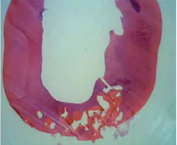
After extraction, the histological study confirmed the origin of resorption on the external surface of the root (Fig. 2) and extension of the lesion to the pulp space (Fig. 3). Histological examination aslo showed the presence of bone-like structures within the invading fibrous tissue (Fig. 4).
Fig. 1 An extended radiolucent lesion was casually detected in the lower right first molar.
Fig. 3 The resorptive process had spread in the thickness of dentin reaching the pulp. Fig. 4 Visible perforations into the pulp and presence of a canalicular structure.
Fig. 2 The lesion started at the external surface of the root.
Libonati a. et aL.
EuropEan Journalof paEdiatric dEntistryvol. 18/4-2017 298
Discussion
External cervical resorption is a rare condition defined as an inflammatory resorption because it is the result of colonisation of dental hard tissues (dentin and cementum) by inflammatory clastic cells (osteoclasts). As previously reported, several factors might be related to the aetiology; the previous fixed orthodontic treatment may have caused excessive orthodontic pressure damaging the layer of cementum thus exposing the dentin to clastic cell resorption.
Treatment and prognosis of the affected teeth are mainly influenced by the extension of the lesion. The ECR defect fell into Class 4 Heithersay, which include large, invasive resorptive processes that have extended beyond the coronal third of the root; in addition, even though a thin layer of predentin generally protects the pulp, perforations of the root canal have occurred. The extension of the lesion and pulp involvement suggested that the resorption was in a very advanced phase making the tooth untreatable, so in this case extraction was the only viable option. Therefore, early diagnosis of this condition is vital.
Histopatologic features of ECR vary with the stage of development of the lesion; at first, the resorption cavity contains granulomatous fibrovascular tissue, which is generally free of inflammatory cells unless colonisation by oral microorganism has occurred. In late stages, deposition of ectopic bone-like calcifications take place; these calcification show a canalicular structure with cellular inclusion and are deposited both within the invading fibrous tissue and directly into the dentin. The finding of bone-like tissue with canalicular
structure confirmed that the lesion was in an advanced stage.
It is possible that the exposed dentin is not recognised by osteoclasts being different by bone, therefore clastic cells begin bone remodeling and, without bone apposition, a resorption lacuna is created. Consequently, the defect is invaded by mesenchymal cells and blood vessels with the final result of deposition of poorly organised bone-like tissue.
References
› Ahmed N, Gopalakrishnan, Mony B, Parthasarthy H. External cervical resorption case report and a brief review of literature. J Nat Sci Biol Med 2014 Jan;5(1):210-4.
› Bergmans L, Van Cleynenbreugel J, Verbeken E, Wevers M, Van Meerbeek B, Lambrechts P. Cervical external root resorption in vital teeth. J Clin Periodontol 2002 Jun;29(6):580-5.
› Fuks AB. Current concepts in vital primary pulp therapy. Eur J Paediatr Dent 2002 Sep;3(3):115-20.
› Heithersay GS. Invasive cervical resorption: An analysis of potential predisposing factors. Quintessence Int 1999 Feb;30(2):83-95.
› Heithersay GS. Clinical, radiologic and histopathologic features of invasive cervical resorption. Quintessence Int 1999 Jan;30(1):27-37.
› Liang H, Burkes EJ, Frederiksen NL. Multiple idiophatic cervical root resorption: systematic review and report of four cases. Dent. Radiol 2003 May;32(3):150-5.
› Mummolo S, Marchetti E, Albani F, Campanella V, Pugliese F, Di Martino S, Tecco S, Marzo G. Comparison between rapid and slow palatal expansion: evaluation of selected periodontal indices. Head Face Med 2014 Aug 15;10:30.
› Neuvald L, Consolaro A. Cementoenamel junction: microscopic analysis and external cervical resorption. J Endod 2000 Sep;26(9):503-8. › Patel S, Kanagasingam S, Pitt Ford T. External Cervical Resorption: a review.
J Endod 2009 May;35(5):616-25.
› Patel S, Pitt Ford T. Is the resorption external or internal? Dent Update 2007 May;34(4):218-20, 222, 224-6, 229.
› Tronstad L. Root resorption-etiology, terminology and clinical manifestations. Endod Dent Traumatol 1988 Dec;4(6):241-52.