3T MR ANGIOGRAPHY AND ASL PERFUSION IN
PRIMARY ANGIITIS OF THE CENTRAL NERVOUS
SYSTEM
Background
Primary angiitis of the central nervous system (PACNS) is a rare and ill-defined disease. Headache and encephalopathy are the most frequent symptoms at onset. Neuroimaging plays an important role in the diagnosis but the pattern of abnormal findings is often not specific and diagnosis requires brain biopsy.
MRA is a reasonable initial modality in the investigation of suspected CNS vasculitis. 3T MRA offers increased signal to noise ratio with better background suppression, leading to better depiction of the intracranial circulation. MR perfusion with Arterial Spin labelling (ASL) technique is not until now performed in patients with PACNS.
We estimate the usefulness of MRA and ASL in the diagnosis of PACNS at 3T. Materials and Methods
We calculated SNR and CNR on TOF angiograms obtained at 1.5T and on TOF and on steady state free precession with in-flow inversion recovery (INHANCE) angiograms at 3T in a group of control subjects. Measurements were performed both in proximal and in distal branches of intracranial circulation.
7 patients with defined PACNS and signs of angiitis at DSA underwent to MRA at 1.5 and 3T. MRAngiograms consisted of a TOF acquisition executed on 1,5T and 3T, and INHANCE acquisition on 3T: intracranial vessels were evaluated for the presence of stenosis, occlusions, pseudoaneurysms, collateral circles with respect to DSA.
MR Perfusion consisted of a pCASL acquisition, with a delay equivalent to 1525 ms. We calculated the cerebral blood flow (CBF) in ACA and MCA territory of supply using 6 ROIs for each region of interest, placed in WM and GM. Two further unique hand shaped ROIs covering respectively the entire MCA and ACA territory of supply were used. Data obtained on PACNS patients were compared with those acquired on controls.
Results
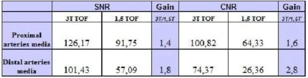
SNR and CNR on 3T TOF angiograms were significantly higher than on 1.5T images in both proximal (SNR=126,17 vs 91,75; CNR=100,82 vs 64,33) and distal (SNR=101,43 vs 57,09; CNR=74,37 vs 26,36) arteries. SNR and CNR ratio decreases in distal branches compared to proximal, larger arteries.
The comparison between 3T TOF and 3T INHANCE revealed that SNR and CNR mean values were higher in the distal branches on TOF compared to INHANCE.
At visual inspection the number of vessel abnormalities detectable with DSA were 640: 73 within proximal and 567 within distal vessels. TOF MRA at 3T depicted 41% of total abnormalities,
INHANCE 3T 19% and TOF MRA at 1,5T 14,2% . TOF MRA at 3T depicted 74% of proximal abnormalities, INHANCE 3T 49% and TOF MRA at 1,5T 52%. TOF MRA at 3T depicted 37% of distal abnormalities, INHANCE 3T 15% and TOF MRA at 1,5T 9,3%.
MR perfusion did not reveal significant differences of CBF values in PACNS patients respect to controls. In one PACNS patient with a diffuse involvement of small vessells seen on DSA, CBF values were lower than the mean CBF values + 2 SD calculated in controls.
Conclusions
MRA at 3T exploiting its higher SNR and CNR improved the sensitivity of MRA in the not invasive preliminary evaluation of patients with PACNS. 3T MRA TOF has higher sensitivity in detecting intracranial circle stenosis than 3T INHANCE and 1,5 MRA TOF. Perfusional pCASL values with delayed 1525 acquisition images don't significantly differ between PACNS patients and controls: further studies with a histogram analysis are needed in order to evaluate the usefullness of ASL in PACNS patients.
INTRODUCTION
PACNS
PACNS is a rare vasculitis confined to vessels of CNS without signs of systemic involvement, which can lead to multiple ischemic and/or hemorrhagic strokes with possible fatal exitus.
The inflammatory process typically affects medium and small arteries of the meninges and cortex, with possible involvement of veins and venules.
Histologically is characterized by multifocal and multisegmental wall vessels infiltration by polyclonal T-lymphocytes with possible variable association with granulomas and fibrinoid necrosis, predominantly represented by T-lymphocytes in association with a variable number of histiocytes, plasma cells, neutrophils and eosinophils. In some cases, beta-A4 amyloid deposits have been found. Further aspecific histologic findings are given by gliosis, mild perivascular mononuclear inflammation, endoluminal thrombosis and vascular occlusion with parenchymal ischemic damage/infarct (1-3).
PACNS affects both genders between the fourth and sixth decades of life (1); literature reports a slight male predominance (3/4 F/M), but Salvarani et al., in a study conducted on 101 PACNS patients, did not found a significant prevalent gender (4). Clinical presentation is variable and often life threatening if an immediate diagnosis is not done. Most common symptoms are insidious and progressive, reflecting a chronic cortical disfunction: headache, weakness, confusion, and cognitive impairment are the most complained disorders. Acute onset of transient ischemic attack, nausea or vomiting, seizure disorder and stroke with multifocal neurological deficit such as brain, cranial nerve, spinal cord, or nerve root lesions are also possible, leading the patient to search for a medical treatment (1).
In order to do an exact diagnosis we must remember that PACNS is a rare disease which accounts for just 1% of the systemic vasculitides. Flogistic blood markers (white cells count, PCR, ESR)
usually altered in systemic vasculitis, are normal in PACNS patients also if an elevation of ESR (30-40 mm/hr) has been described in a 30%-60% of cases (5,6). Markers of systemic vasculitis and infectious diseases, such as antinuclear antibodies, antineutrophil cytoplasmatic antobodies,
complement levels, rheumatoid factor, HIV, HBV and HCV markers are negative. CSF analysis can show only a slight elevation of white blood cell count and total protein level. The possibility of a viral or fungal infection must be excluded with appropriate tests.
In 1988 Calabrese and Mallek proposed the diagnostic criteria for PACNS: 1) the presence of an unexplained neurologic deficit after thorough clinical and laboratory evaluation; 2) documentation by cerebral angiography and/or tissue examination of an arteritic process within the central nervous system; and 3) no evidence of a systemic vasculitides or any other condition to which the
angiographic or pathologic features could be secondary (7). In 2009 Birnbaum and Hellmann proposed a new approach in diagnostic path for a more accurate differential diagnosis between PACNS and reversible vasoconstriction sindrome (RVCS): diagnosis of PACNS is definite if there is confirmation of vasculitis on analysis of a tissue biopsy specimen; and probable if there is no tissue confirmation, in association of high-probability findings on an angiogram with abnormal findings on MRI and a CSF profile consistent with PACNS (1).
The role of radiologic evaluation is crucial in diagnosis, because it permits to exclude other conditions with the same clinical presentation. Cerebral angiography still actually remains the radiologic gold standard study, being able to identify classical signs of flogistic involvement of small vesssel with 250 to 500 mm diameter: multifocal stenoses with interposed regions of normal vessel calibre or sausage-like dilatation, creating the tipical “beading” appearance. Alhalabi et al. (8) described the angiographic pattern in 19 patients affected by PACNS, who were evaluated with serial angiographies, repeated 6 to 9 months after diagnosis. Three patients underwent multiple angiograms prior to initiation of therapy; in all of them new lesions occurred over time. Two patients with negative angiogram were diagnosed PACNS only on the basis of brain biopsy: this fact highlights that can be cases in which affected vessels should have a diameter less than the minimum necessary to be visible on DSA images. Usually distal branches of both anterior and posterior circulation are involved bilaterally. Two types of stenosis are described: segmental and smooth stenosis, measuring between from several millimetres to 3 cm of lengh. The most common finding is represented by bilateral but not symmetrical segmental narrowing. The typical “beading” aspect, which is given by multiple sites of segmental narrowing along a single vessel, is described to appear less often than singly narrowed vessel shrinkage. Smooth narrowing of supraclinoid tract of carotid sinus is a recurrent findings, being possibly associated with secondary collateral flows and retrograde perfusion from posterior circle. Medium size vessel stenoses are also described, being the M1 segment the most affected. Small vessels occlusion, with absence of parenchimography in the corrispective territory of supply, are also possible. Neovascularization and enlargement of preexisting small vessels is a further possible finding on DSA images. These angiographic anomalies are suggestive but not specific for vasculitis. Similar findings can be appreciable in other sistemic vasculitides, atherosclerosis, radiation vasculopathy, drug exposure, infection, and vasospasm (8, 9). Although DSA findings could be suggestive of angiitis, Kadkhodayan et al. (10) in a series of 14 cases, found that none of patients suspected to have PACNS on the basis of clinical and angiographic findings did not have primary angiitis of the CNS
at biopsy. Overall, the sensitivity of angiography in detecting PACNS has been estimated at between 50% and 90% (1, 4, 7, 11).
MRI is an indispensable diagnostic exam in patients with suspected PACNS, being able to reveal abnormalities in 90% to 100% of patients in a morphological study (1, 4, 31).
The most common abnormalities are seen in the subcortical white matter, followed by the deep gray matter, the deep white matter, and the cerebral cortex. In approximately 50% of cases, MRI can demostrate bilateral cortical-subcortical infarcts, which belong to different territory of supply: in most cases, have multiple subcortical infarctions in a small-artery pattern, some have a large-artery distribution, others show a branch-artery distribution (4,12,13). In a group of 18 patients, 15 of whom were diagnosed PACNS, an infratentorial involvement was demonstrated, being however always associated with sopratentorial damage (12). In one-third of cases a contrast enhancement of these lesions is appreciable after contrast administration (4).
In rare cases a leptomeningeal contrast-uptake is also seen. Wilhelm Küker et al. (14) documented vessel wall enhancement of inflammed vessel wall arteries in a cohort of patients affected by cerebral vasculitis, most of which met the Calabrese criteria for the diagnosis of PACNS. In 15% of cases, PACNS can manifest with mass lesions mimicking a malignant neoplasm (37, 38). Less common MRI manifestations include cortical laminar necrosis or confluent white-matter lesions, which can be similar to multiple sclerosis (1).
Both subarachnoid and intraparenchymal hemorrhages are pointed out in approximately 10% of cases (1).
Regarding the territories affected by parenchymal infarctions, the middle cerebral artery is more frequently affected than the anterior and posterior cerebral artery territory of supply (12).
MRI findings in PACNS vasculitis have a differential diagnosis with secondary vasculitides of the CNS which can occur in collagen vascular disorders, viral and bacterial infections, in tumoral lesions and drugs abuse. Haroon et al. (15) describes a case of intravascular lymphoma mimicking a primary vasculitis of the central nervous system, with MRI and DSA findings indicative of a primitive vasculitic process and a diagnosis done with biopsy.
Correlation between MRI and angiographic abnormalities has been demonstrated to be poor. Some studies highlight how in many cases some evident MR lesions do not correspond to extensive vascular involvement at DSA (12, 16, 17, 18, 25). On the other side, there have been described cases of patients that met the criteria of Calabrese and Malleck for the diagnosis of PACNS, who presented with a negative angiographic evaluation and a positive MRI examination (12, 13, 17, 19). An useful general approach in the diagnosis of PACNS is to remind that a negative MR excludes intracranial vasculitis more definitively than does a negative angiogram. The role of MRA in PACNS patients is limited by disadvantages, such as motion artifacts, venetian blind artifacts, drop of signal due to tortuous blood vessels and/or decreased blood flow velocities. Higher field strenght can lead to a spatial resolution improvement, a factor that rapresented a limitation on 1,5T MRA. However advantages of MRA are absence of ionizing radiation, no contrast injection and non-invasiveness. MRA can find applications in the suspected clinical setting of cerebral vasculitis as the first non-invasive angiographical imaging modality.
K. Maclaren (17) argues the possibility that PACNS could manifest with two different patterns: small and middle-sized vessel disease (SVD/MVD). SVD would be angiographically negative,
affecting small arterioles under the minimum angiogram resolution. This condition should correlate with a more subacute-chronic onset (encephalopathy, seizures, headache, altered higher mental function, confusion, deteriorating conscious level), and a worst prognosis for the major incidence of relapses during prolonged periods, causing recurrent, severe and irreversible CNS injury. On the other hand, in MVD PACNS, angiography with MR scan is positive, being able to point out vessel wall abnormalities in intracranial circle, from internal carotid sinus to small peripheral vessels. In this second group of patients clinical onset is noted to be more acute, but with a paucity of relapses during prolonged follow-up. This interpretation could explain the reason why numerous cases of biopsy-proved PACNS reported in literature are negative to DSA (13, 20-25). Further on, also Salvarani et al. (4, 39) and Scolding et al. (36) hypothesizes two different pattern of PACNS based on vessel size involvement, however, in contrast with Maclaren, Salvarani found that patients with small vessel involvement, had a better outcome and prognosis (4). In particular Salvarani and Scolding found a higher incidence of beta-amyloid deposits in those cases with SVD, with respect to MVD, supporting the hypothesis that small vessel angiopathy in PACNS could be a distinct clinicopathological entity, which could be triggered by vascular deposition of Ab in susceptible patients, being part of a more widespread immune reaction to Ab within the CNS.
PURPOUSE
The main goal of our work was to evaluate the sensitivity of 3T MRA with respect to 1.5 MRA in diagnosing PACNS.
The second purpose was to evaluate if MR perfusion with pCASL technique allows to detect brain areas of hypoperfusion in patients with PACNS.
MATERIALS AND METHODS
Patients
From our data base we retrospectively selected 10 patients referred to our hospital for suspected PACNS in which DSA was indicative of cerebral vasculitis. One patient was excluded since the follow up DSA documented a resolution of multiple intracranial stenosis within 4 weeks from admission. Clinical onset and course (26) together with DSA changes leaded to a diagnosis of RCVS. In two cases cerebral vasculitis was established to be of infectious origin (Staphilococceal endocharditis).
Seven patients (2 males and 5 females with age ranging from 35 to 78 years, mean+SD 56.1+14.4) were diagnosed as PACNS. Clinical diagnosis was in line with clinical criteria proposed by Calabrese and Mallek (7).
No patient had history of hypertension, heart diseases, hyperlipidemia, syphilis or other infections, drug-abuse, collagenopathies or any other systemic diseases. Most frequent initial symptoms were headache, lateralized weakness and vigilance impairment; sensitive alterations, speech disturbances, visual field defects, were also observed. Blood cells count and routine laboratory tests were normal.
A mild increase of inflammatory indexes (erythrocyte sedimentation rates and C-reaction proteins) was found in only one patient. In all subjects rheumatoid factor, antinuclear antibodies, anticardiolipin antibodies, antineutrophil cytoplasm antibodies, lupus anticoagulant were negative. CSF examination was performed in all patients and revealed mild increase in leucocyte count and/or total protein concentration in three cases. Combined MRI abnormalities and angiographic findings suggested PACNS.
In particular, MRI was abnormal in all patients. Multiple infarctions associated with hemorrhagic (intraparenchimal and/or subarachnoid hemorrhages) lesions were the most common pattern. In three cases neuroradiologic evaluation pointed out a hemorragic lesion involving deep gray nuclei. In three patients there was parieto-occipital cortico-subcortical hemorrhagic infarctions. Ischemic involvement were found at the level of deep gray nuclei in one patient, and at the level of cortico-subcortical frontal region in another patient. Subaracnoid haemorrages were detectable in four cases. At the moment of diagnosis anomalies were monolateral in six cases, but further lesions appeared also in controlateral emisphere in two cases during relapses. Leptomeningeal enhancement with a bilateral occipito-parietal involvement, associated with stroke in the adjacent brain parenchyma, was pointed out in one case.
Concerning angiography, all patients showed segmental narrowing and irregularities of vessels calibre, with pattern of alternating tracts of focal stenosis and dilatation (“sausage pattern”). Also if pseudoaneurysms are unusual in PACNS patients, being more typical in others sistemic vasculitides, such as polyarteritis nodosa, they were clearly valuable in a patient (Fig.1). In the same patient coexisted abrupt termination of right cerebral middle artery and diffuse irregularities of vessels calibre. One of the patients had biopsy-proven PACNS.
MRA protocol
All patients underwent to standard MR examination on both 1.5 and 3T within 3 months from DSA by using a 1.5 T scanner (Signa HDx, GE Healthcare, Milwaukee) with high performing gradients (gradients strength 50mT/m, maximum slew rate 150T/m/s), equipped with an 8-channel head coil with ASSET-technology and 3T scanner (MR750, GE Healthcare, Milwaukee) with high performing gradients (gradients strength 80 mT/m, maximum slew rate 200 T/m/s), equipped with an 8-channel head coil with ASSET-technology.
MR examination included MRA acquisition with TOF technique at 1.5 T and with TOF and INHANCE techniques at 3T.
Our standard intracranial 1,5 T 3D TOF protocol was performed in all subjects with the following parameters: TR=30 ms, TE=2.70 ms, flip angle=20, thickness=1,4 mm, overlap=0,70 mm, matrix=
4 slabs, RAMP pulse= I>S, MT pulse, flow compensation, EDR, ZIPx2, ZIPx512, ASSET, and imaging time of 6,14 min. A presaturation band, concatenated with the acquisition slab, was applied above the imaging volume to saturate incoming venous blood.
3T 3D TOF protocol was used with the following parameters: TR=23 ms, TE=3,2 ms, flip angle=20, thickness=1 mm, overlap=0,50 mm, matrix= 320 x416, FOV= 20 cm, Phase FOV= 0,81, voxel size= 0,4x0,5x1 mm, voxel volume=0,2 mm³, 5 slabs, RAMP pulse= I>S, MT pulse, flow compensation, EDR, ZIPx2, ASSET, and imaging time of 8:26 min. A presaturation band, concatenated with the acquisition slab, was applied above the imaging volume to saturate incoming venous blood.
3T 3D INHANCE protocol was used with the following parameters: TR= minimum, TE= minimum, flip angle=8, thickness=1,2 mm, matrix= 384x256, FOV= 24 cm, Phase FOV= 1, voxel size=0,6x0,9x1,2 mm, voxel volume= 0,65 mm³, NEX=1, VENC=40 and imaging time of 6:50 min.
The same MRA protocol was acquired on both 1.5 and 3T scanners in a control group of 7 healthy subjects match aged with PACNS patients. All these subjects were voluntaries recruited among personal of radiology department, with negative hystory for CNS disease and with unremarkable neurological examinations. All control subjects give their informed consent to the study that was approved by our local ethical commettee.
ASL protocol
pCASL acquisition has been obtained using gradient echo echoplanar images (EPI). Perfusional analysis with pCASL has been performed using the following parameters: Scan Plane=Axial, Freq FOV=24, slice thickness=4, freq. dir.=R/L, TR=4632 ms, 1 slab =1, TE(s) per scan=1, TE=10,54 ms, flip angle=111, Points=512, Arms=8, Nex=3, Bandwidth=62,5, Bo Maps=off, EDR, Fast, Spiral, auto, FOV=24x31,7mm, post-labeling delay=1525ms; and imaging time 4:29.
Labeling was applied at the level of the cervicomedullary junction.
IMAGES EVALUATION
SNR and CNR
SNR and CNR has been calculated adopting a similar method proposed by Willinek et al. (27). SNR=MVv
/
SDbCNR= MVv – MVbp
/
SDbMVv: mean value intensity in the vessel of interest SDb: standard deviation in the background
MVbp: mean value intensity in the brain parenchyma
MVv was measured by placing a ROI in the largest region of basilar artery, carotid sinus, M1 segment of middle cerebral artery, P1 segment of posterior cerebral aretry, A1 segment of anterior cerebral artery, and distal P3, A3 and M3 branch. MVbp was obtained placing a ROI with the same size at the level of adjacent brain parenchyma.
Because the noise in the background tends to decline centrifugally, MVbp has been calculated doing a media between the higher value of noise (using a ROI placed just outside diagnostic images) and the lesser value of noise ( using a ROI placed distal from diagnostic images) in the background.
We separately calculated the gain in terms of SNR and CNR at the level of proximal and distal vessels, by making a ratio between SNR and CNR values at 3T TOF and at 1,5T TOF. The same has been done for evaluation of gain of SNR and CNR at 3T TOF compared to 3T INHANCE.
DSA
DSA diagnosis of PACNS was supported by using the criterion "more than two stenoses in at least two separate vascular distributions" (11). Antero-posteior and lateral DS Angiograms of intracranial circulations in all PACNS patients were evaluated by an expert neuroradiologist in order to count the number of vessel stenosis, vessels amputation, mircro-aneurysms, collateral circles. Beading was defined as alternating regularly spaced segments of stenosis with normal or dilated intervening segments.
MRA
By using a dedicated workstation (GE Advantage 4.4) MRA acquisitions were reconstructed with a MPVR and volume rendering methods and displayed on an anterior posterior and lateral view in order to mimic the visualization of DSAangiograms. As well as for DSA, MRAngiograms obtained on both 1.5 and 3T were evaluated by a further expert neuroradiologist in order to count the number of vessel stenosis, vessels amputation, mircro-aneurysms, collateral circles for large-medium and small vessels. The vessels alterations were classified on the bases of their distribution in the anterior, medium and posterior circulation if the involvement regarded respectively the anterior, middle an posterior cerebral arteries.
Vessels abnormalities were classified as pertaining to large-medium vessels or small vessels if affecting respectively the vessels of Willis circles or A1-A2, M1-M2, P1-P2 segments or segments beyond A2, M2, P2 of intracranial branches (28).
At 3T MRA were separately evaluated both TOF and INHANCE images. DSA was considered as the gold standard method.
ASL
CBF measurements was performed using a ROI analysis according to the recommendations of earlier studies (29,30). ROIs were placed by one radiologists. ASL CBF values has been estimated by a workstation Advantage 4.5. Once obtained CBF maps, they have been co-registred with an axial T2 acquisition, in order to better discriminate white matter and gray matter. CBF values were calculated placing ROIs in normal appearing white matter (WM), gray matter (GM) and both of them in ACA and MCA teritories of supply. For the evaluation in GM and WM, 6 ROIs with an
area of 316 pixels were positioned in MCA and ACA territory of supply. For the contemporary evaluation of WM and GM in ACA and MCA territory, a unique hand shaped ROI including these two compartements was used for each territory of interest. CBF values of patients and controls were compared using a t-student test.
RESULTS
SNR and CNR
SNR and CNR of TOF angiograms obtained at 3T and 1,5T for each region of interest, are shown in table 1. Mean SNR and CNR of 3T TOF angiograms and 3T INHANCE angiograms for each region of interest, are shown in table 2.
The comparison between 3T TOF and 1,5T TOF demonstrates a general significative difference in terms of SNR and CNR (P<0,05), with higher values at 3T compared to 1,5, both in proximal and distal intracranial vessels.
The comparison between 3T TOF and 3T INHANCE demonstrates a significative increase of SNR and CNR in distal branches by using TOF MRA.
No significant difference has been found between SNR and CNR values of 3T TOF and 3T INHANCE at the level of proximal branches.
SNR and CNR gain of TOF 3T respect to 1,5T angiograms at the level of proximal arteries has been evaluated to be 1,4 (126,17 vs 91,75) and 1,6 (100,82 vs 64,33).
SNR and CNR gain of TOF 3T respect to 1,5T angiograms at the level of distal arteries has been evaluated to be 1,8 (101,43 vs 57,09) and 2,8 (74,37 vs 26,36).
SNR and CNR gain of TOF respect to INHANCE angiograms acquired with 3T at the level of proximal arteries has been evaluated to be 1,02 (126,17 vs 123,15) and 0,88 (100,82 vs 114,24). SNR and CNR gain of TOF respect to INHANCE angiograms acquired with 3T at the level of distal arteries has been evaluated to be 2 (101,43 vs 50,78) and 1,7 (74,37 vs 44,1). The entity of improvement of SNR and CNR values at 3T TOF compared to 1,5T TOF and 3T INHANCE in proximal and distal arteries are shown in tables 3 and 4.
DSA
Table 5 shows the sensitivity and PPV of each diagnostic modality compared to DSA in total circle, proximal branches and distal branches.
In PACNS patients all the DSA studies were abnormal. 640 lesions of intracranial vessels were identified in 7 patients (median 91,43). Lesions were bilateral in 7 / 7 cases, and they were more likely to be distally distributed than proximally (567 / 640 in distal circulation, 73 / 640 in middle and large medium sized vessels).
A total of 6 out of the 7 patients had lesions involving both the anterior and posterior circulation. Lesions were more likely to present a beading aspect, (207) instead singolarly stenotic (125). Rarely lesions were presented as occlusions (n=5) or pseudoaneurysm (n=7). Collateral circles were
appreciable in 4 of 7 patients. Lesions were more frequent in MCA branches. Table 6 shows all the vascular anomalies found with DSA.
3T TOF
All the patients had an abnormal MRA. MRA detected a total number of 262 stenosis compared to 640 total stenosis seen in the conventional angiography studies yielding a sensitivity of 41%. Total false positive findings were 29, pointing out a positive predictive value of 90%. Proximal stenosis detectable at 3T TOF were 54, with respect to 73 stenosis seen at DSA, assessing a sensitivity of 74%. Proximal false positive findings were 3, pointing out a positive predictive value of 95%. Distal stenosis detectable at 3T TOF were 208, with respect to 567 seen at DSA, assessing a sensitivity of 37 %. Distal false positive findings were 26, pointing out a positive predictive value of 89%.
3T INHANCE
All the patients out of the 7 had an abnormal MRA. MRA detected a total number of 119 stenosis compared to 640 total stenosis seen in the conventional angiography studies yielding a sensitivity of 19% . Total false positive findings were 115, pointing out a positive predictive value of 51%. Proximal stenosis detectable at 3T INHANCE were 36, with respect to 73 seen at DSA, assessing a sensitivity of 49 %. Proximal false positive findings were 9, pointing out a positive predictive value of 80%.
Distal stenosis detectable at 3T INHANCE were 83, with respect to 567 seen at DSA, assessing a sensitivity of 15 % . Distal false positive findings were 161, pointing out a positive predictive value of 44%.
1,5 T TOF
6 patients out of the 7 had an abnormal MRA. With respect to 3T TOF MRA, 1,5T TOF sensitivity for patients was 86%, the specificity was 100%, the PPV was 43% and the NPV was 58%. 1,5 TOF MRA detected a total number of 91 stenosis compared to 640 total stenosis seen in the conventional angiography studies yielding a sensitivity of 14,2%. Total false positive findings were 169, pointing out a positive predictive value of 35%. Proximal stenosis detectable at 1,5T TOF were 38, with respect to 73 seen at DSA, assessing a sensitivity of 52%. Proximal false positive findings were 8, pointing out a positive predictive value of 82,6%. Distal stenosis detectable at 1,5T TOF were 53, with respect to 567 seen at DSA, assessing a sensitivity of 9,3%. Distal false positive findings were 161, pointing out a positive predictive value of 25%. A false positive and a false negative example
on 1,5T TOF are showed in Fig. 2 and 3. A false positive on 3T TOF MRI is appreciable in Fig.4. The case with a negative angiogram at 1,5T is reported in Fig 5.
ASL
CBF values in PACNS patients and controls are shown in table 7.
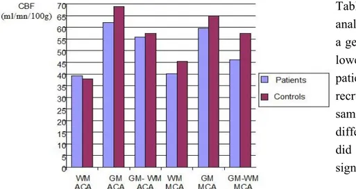
ROIs analysis pointed out slight lower CBF values on patients with respect to controls. However the statistical evaluation did not find a significant difference between patients and controls in normal appearing WM, normal appearing GM, and in normal appearing GM+WM both in ACA and MCA territory of supply (P > 0,05). Mean CBF values in patients are: 39,28 in WM; 62,18 in GM; and 55,97 in WM+GM ml/mn/100g in ACA territory; 40,19 in WM, 59,65 in GM; 46,15 in WM+GM ml/mn/100g in MCA territory. Mean CBF values in controls are: 37,99 in WM; 68,9 in GM; and 57,47 in WM+GM ml/mn/100g in ACA territory; 45,5 in WM, 65,01 in GM; 57,53 in WM+GM ml/mn/100g in MCA territory.
Only one patient presented CBF values lower than the mean value of the control group, added with two standard deviations. The patient showed the following mean values: 23,71 in WM; 37,6 in GM; and 36,31 in WM+GM ml/mn/100g in ACA territory; 24,8 in
WM, 33,33 in GM; 28,05 in WM+GM ml/mn/100g in MCA territory. This patient presented a positive DSA, where a massive bilateral involvement of small distal vessel of entire intracranial circle, with sparing of proximal arteries, was pointed out.
Table 8 shows that ROIs analysis pointed out that there is a general trend characterized by lower CBF values in PACNS patients than controls, the latter recruited on the basis of the same age and sex. The difference of values however did not reach the level of significance with t-student test.
DISCUSSION
SNR and CNR
By our experience SNR of TOF angiograms at 3T is respectively 1,4 and 1,8 higher than that obtained at 1,5 T for proximal and distal intracranial vessels. These values slightly differ from those reported in literature, being lower with respect to those calculated by Willinek on the basilar artery in human brains (27) and by Bernstein on phantoms (32), who reported a doubling of SNR at standard 3T. A similar value has been calculated for the CNR increase at 3T TOF compared to 1,5T TOF, attested at 1,6 in proximal arteries. The major improvement has been evaluated for CNR in distal arteries, which documented a gain of 2,8 at 3T TOF. The difference of our results compared to those reported in literaure can be explained with the fact that different technical parameters have been used. In particular Willinek (27) used a matrix of 336x212 with a volume voxel of 0,92 while we used a matrix of 320x416 with volume voxel of 0,2 for the TOF imaging at 3T. Willinek evaluated SNR and CNR gain on 3T by applicating two TOF sequences, the standard TOF with the same voxel size of 1,5T TOF, which demostrated a doubling SNR and CNR values on 3T, and the high resolution TOF, which demostrated a evident drop of SNR and CNR values on 3T, in relationship with its lower voxel volume. We have to consider that we used a TOF acquisition used in clinical practice, wich was characterized by optimized spatial resolution, in order to obtain a better depiction of vessel morphology.
SNR and CNR mean values calculated in proximal arteries at 3T TOF and 3T INHANCE do not show evident differences, respectively estimating a ratio of 1,02 and 0,88. In particular INHANCE demostrates to have a little higher contrast quality in proximal circle.
At the level of distal branches SNR and CNR INHANCE values decrease, attesting a gain at 3T TOF respect to INHANCE of 2 and 1,7 respectively. These data can be explained with the fact that INHANCE is a flow-related method, able to represents vessels on the basis of the velocity on inflowing blood protons (33). Higher VENC factors lead to a better evaluation of proximal vessels,
lower VENC factors better depict distal vessels. As we used a VENC factor of 40 cm/sec, the representation of proximal vessells was favoured than distal vessels.
Comparison between DSA and MRA
Differently from 1,5T 3D TOF, the higher SNR and CNR ratio at 3T, 3D TOF is able to visualize distal branch arteries, with a good quality up to the third-fourth division. In our experience 3D TOF at 3T has demonstrated a better sensitivity than 3D TOF at 1,5T, both in proximal and distal branches. Previous studies which evaluated the capacity to detect complete occlusion in steno-occlusive disease , demostrated a 3T TOF MRA sensitivity of 100%, specificity of 99%, PPV of 87% and NPV of 100%. Further on, for detecting high-grade stenosis in atherosclerotic disease, 3D TOF-MRA at 3T had been calculated to have a sensitivity of 78%–85%, a specificity of 95%, a PPV of 75%– 79%, and a NPV of 95%–97% (35). S. Bash et al (34) found at 1,5T TOF a sensibility of 87%, specificity of 98%, PPV of 59%, and a NPV of 99,5% using DSA as the reference standard in atherosclerotic disease. As regard intracranial vasculitic disease, Pomper et al (4) found, with a field strenght of 1,5T, an agreement between TOF MRA and DSA of 44%, detecting 30 of 68 stenosis on TOF, using DSA as reference standard. Salvarani et al. (23) in a review of 101 patients, detected a vasculitic pattern on MRA in 59% of cases. Our data are similar to those reported in literature, as regard 1,5T TOF, which demonstrated to have a sensitivity and PPV of 52% and 82,6% in proximal vessels; and 9,3% and 25% in distal vessels. Global sensitivity and PPV have been calculated to be 14,2% and 35% at 1,5T. We calculated a 86% of sensitivity at 1,5T TOF angiograms calculated for patients, a higher value than that reported by Salvarani et al. On the other hand, in our experience, 3T TOF MRA resulted to be more effective than 1,5T TOF MRA in detecting vascular stenoses, attesting a sensitivity at the level of proximal segments of 74%. This value is however lower than those calculated in previous studies performed at 3T in steno-occlusive disease (35). We think that this difference depends on the fact that a different class of vessels has been considered: Choi (35) focuses on distal internal carotid artery, M1 and M2 segments of middle cerebral artery and on vertebro-basilar artery, while we considered as proximal arteries the first and second division of each vessel, thus including A1-A2, and P1-P2 segments. By our knowledge, distal stenosis evaluation has never been reported in literature, and we found low sensitivity values and quite high PPV values. We must add that, differently from atherosclerotic disease, which predominantly affects proximal vessells, vasculitic abnormalities involve the entire intracranial circle. This consideration was the reason why we evaluated both proximal and distal vessels, leading to a predictable reduction of sensitivity values.
3T INHANCE demostrated a lower sensitivity and PPV values than 3T TOF. This fact is mostly due to the lower capacity to delineate vessel morphology in proximal circle and to the reducted number of distal vessel visualizable with this sequence. As the intensity of vessels depends on the velocity of inflowing blood protons, the drop of signal is particularly appreciable in distal vessels, burdening a correct interpretation of the images.
Further on, while both 3T TOF and 3T INHANCE detected in all cases at least one stenosis, in one patient, who presented only distal stenoses, 1,5T TOF was negative. The disponibility of higher field strenght can be exploited in order to obtain a higher spatial resolution on MRA images. In
particular, the better depiction of proximal and distal vessels on 3T MRA angiograms, due to the higher SNR and CNR, should allow the radiologist to take more confidence in the evaluation of vessel anomalies.
ASL
In our experience no significant CBF difference has been found between PACNS patients and controls, using a ROIs analysis of pCASL images acquired with a delayed interval of 1525 ms. Possible causes can be related to the fact that we used a ROI-based analysis, which is not able to evaluate the entire brain parenchyma. Further histogram-based analysis would be useful for a more accurate assessement of CBF values in patients and controls.
In vasculitic disease, ASL can be used for a qualitative and quantitative evaluation of hypoperfused regions in cases of proximal stenosis. Fig 6 shows a case of a signal drop at the level of the territory supplied by right MCA, in a patient with a severe stenosis of M1-M2 segment Morphologic evaluation was negative in cortical-subcortical region in the same territory.
The role of ASL in cases with prominent distal vessel involvement is more questionable.
Previous studies (4, 17, 23) proposed that PACNS could be distinguished in two different clinical patterns: patients with medium-large vessels involvement and patients with small vessels involvement. This hypothesis is supported by the hystopatologic reply of beta-amyloid deposits in those cases of PACNS with an essential small vessel and leptomeningeal involvement, in contrast
with cases of PACNS with proximal vessel involvement in which this protein was not found (36). These findings support the hypothesis that two different pattern could exist in PACNS patients (4, 36, 39).
In the cathegory of patients characterized by a small vessel involvement DSA can fail in detecting vessel wall abnormalities, as affected vessels can be under the minimum resolution of the method. In this situation a perfusional analysis could be crucial, being able to visualize the health status of capillary network. In particular we found that, in a patient with massive small vessel involvement in the entire intracranial circle (95 stenosis), an ASL perfusional evaluation pointed out a significative drop of values ( 23,71; 37,6 and 36,3 in WM, GM and WM+GM in ACA territory, and 24,8; 33,33 and 28,5 in WM, GM and WM+GM in MCA territory), which was below two standard deviations from the mean of the control group. This fact could let the neuroradiologist to have more confidence in suspecting PACNS, also in front of a negative DSA or a DSA with a pattern of a diffuse small vessel involvement. Obviously diagnosis of PACNS cannot forget Calabrese and Mallek criteria, given by clinical signs indicative for a vasculitic process, in absence of other sistemic causes. In addiction, besides the technical problems related to the sequence, there must be excluded other clinical causes of apparent diffuse hypoperfusion: age, arterial transit delay, loss of cardiac output, decreased brain volume, vasospasm, and exogenous drugs may result in decreased perfusion. A limit of our analysis is that our patients were recruited on the basis of a positive angiogram, so we had no patients with a negative DSA with a brain-biopsy proved PACNS. However, the only patient with proved reducted perfusional data previously underwent a brain biopsy, which was diagnostic for PACNS.
CONCLUSION
In conclusion MRA can be applied as a not invasive tool both in the diagnosis and follow-up of PACNS patients, being able do give a better depiction of vascular architecture compared to 1,5T MRA. However, in PACNS MRA still requires further confirmatory diagnostic testing such as DSA and/or brain biopsy. MR perfusion with pCASL have to be tested with histogram analysis in a larges sample of PACNS patients, in order to establish its role in the clinical settings.
REFERENCES
1- Birnbaum J, Hellmann D. Primary Angiitis of the Central Nervous System. Arch Neurol. 2009;66(6):704-709 2 - Stock K W , Radue E W, Jacob A L, Bao X S and Steinbrich W. Intracranial arteries: prospective blinded comparative study of MR angiography and DSA in 50 patients. Radiology. 1995 May;195(2):451-6
3 - Miller DV, Salvarani C, Hunder GG, Brown RD, Parisi JE, Christianson TJ, Giannini C. Biopsy findings in primary angiitis of the central nervous system. Am J Surg Path. 2009 Jan;33(1):35-43
Hunder G. Primary Central Nervous System Vasculitis: Analysis of 101 Patients. Ann Neurol 2007;62:442–451 5 - Graeme J. Hankey. Isolated Angiitis/Angiopathy of the Central Nervous System. Cerebrovasc Dis 1991;1:2-15 6 – Nadeau S, Diagnostic approach to central and peripheral nervous system vasculitis. Neurologic Clinics, Volume 15, Issue 4, Pages 759-777
7 - Calabrese LH, Mallek JA. Primary angiitis of the central nervous system: report of 8 new cases, review of the literature, and proposal for diagnostic criteria. Medicine (Baltimore). 1988;67(1):20-39
8- M.Alhalabi, P. Moore. Serial angiography in isolated angiitis of the central nervous system. Neurology 1994;44;1221 9 – Aviv R.I, BenselerS.M. , DeVeber G., Silverman E.D. , Tyrrell P.N,. Tsang L.M, Armstrong D. Angiography of Primary Central Nervous System Angiitis of Childhood: Conventional Angiography versus Magnetic Resonance Angiography at presentation. AJNR Am J Neuroradiol 28:9 –15, Jan 2007
10 – Kadkhodayan Y, Alreshaid A, Moran C, Cross DW, Powers W, Derdeyn C. Primary Angiitis of the Central Nervous System at Conventional Angiography. Radiology 2004; 233:878–882
11 – Demaerel P, Ruyter N, Maes F, Velghe B, Wilms G. Magnetic resonance angiography in suspected cerebral vasculitis. Eur Radiol (2004) 14:1005–1012
12 – Pomper M, Miller T, Stone J, Tidmore W, and Hellmann D. CNS Vasculitis in Autoimmune Disease: MR Imaging Findings and Correlation with Angiography AJNR Am J Neuroradiol 20:75–85, January 1999
13 - Greenan TJ, Grossman RI, Goldberg HI. Cerebral vasculitis: MR imaging and angiographic correlation. Radiology 1992;182:65-72
14 - Küker W, Gaertner S, Nägele T, Dopfer C, Schöning M, Fiehler J, Rothwell P, Herrlinger U. Vessel Wall Contrast Enhancement: A Diagnostic Sign of Cerebral Vasculitis Cerebrovasc Dis 2008;26:23–29
15 – Haroon AM, Molloy E, Farrel M, and Shaffeq. Central Nervous System Vasculitis: All That Glitters Is Not Gold J Rheumatol 2012;39;662-663
16- Lefond CA. Catheter angiogram in central nervous system vasculitis: still first among equals Developmental Medicine & Child Neurology 2010, 52: 788–793
17 – Maclaren K, Gillespie J, Shrestha S, Neary D and Ballardie F.W. Primary angiitis of the central nervous system: emerging variants. Q J Med 2005; 98:643–654
18 – Lie J.T. . Primary (granulomatous) angiitis of the central nervous system: A clinicopathologic analysis of 15 new cases and a review of the literature Review Article Human Pathology, Volume 23, Issue 2, February 1992, Pages 164-171
19 - Harris KG, Tran DD, Sickels WJ, Cornell SH, Yuh WTC. Diagnosing intracranial vasculitis: the roles of MR and angiography. AJNR Am J Neuroradiol 1994;15:317-330
20 - Duna GF, Calabrese LH. Limitations of invasive modalities in the diagnosis of primary angiitis of the central nervous system. J Rheumatol 1995;22:662-667
21 - Calabese LH, Gragg LA, Furlan AJ. Benign angiopathy: a distinct subset of angiographically defined primary angiitis of the central nervous system. J Rheumatol 1993;20:2046–50
22 - Koo EH, Massey EN. Granulomatous angiitis of the central nervous system: protean manifestations and response to treatment. J Neurol Neurosurg Psychiatry 1988; 51:1126-1133
23 - Scully R. Massachusetts General Hospital Case Records, case 8-1989: weekly clinicopathological exercises. N Engl J Med 1989; 320:514-524
24 - Younger DS, Hays AP, BrustJCM, Rowland LP. Granulomatous angiitis of the brain: an inflammatory reaction of diverse etiology. Arch Neurol 1988; 45:514-518
25 - Clifford-Jones RE, Love S. , Gurusinghe N. Granulomatous angiitis of the central nervous system: a case with recurrent intracerebral hemorrhage. J Neurol Neurosurg Psychiatry 1985; 48:1054-1056
26 – Gerretsen P, Kern R. Reversible Cerebral Vasoconstriction Syndrome or Primary Angiitis of the Central Nervous System? Can. J. Neurol. Sci. 2007; 34: 467-477
27 – Willinek W, Born M, Simon B, Tschampa M, Krautmacher C, Gieseke J, Urbach H, Textor H, Schild H. Time-of-Flight MR Angiography: Comparison of 3.0-T Imaging and 1.5-T Imaging—Initial Experience. Radiology 2003; 229:913–920
28 – Benseler S, Silverman E, Aviv R, Schneider R, Armstrong D, Tyrrell P, and De Veber G. Primary Central Nervous System Vasculitis in Children . Arthritis and Rheumatism . American College of Rheumatology, Voll. 54, No. 4, April 2006, pp 1291–1297
29 – Weber M A , Zoubaa S, Schlieter M, Juttler E, Huttner H B , Geletneky K, Ittrich C , Lichy M P , Kroll A , Debus J, Giesel F L, Hartmann M , Essig M . Diagnostic performance of spectroscopic and perfusion MRI for distinction of brain tumors. Cancer Imaging (2006) 6, S32–S41
30 - Law M, Cha S, Knopp EA, Johnson G, Arnett J, Litt AW. High-grade gliomas and solitary metastases: differentiation by using perfusion and proton spectroscopic MR imaging. Radiology. 2002;222:715–21
31 –Catarsi E, Pelliccia V, Cosottini, Pesaresi I, Puglioli M, Moretti P, Tavoni A. Primary angiitis of the central nervous system: report of eight cases from a single Italian center. Journal of the Neurological Sciences (2011) Volume: 307, Issue: 1-2, Pages: 69-73
32 – Bernstein M, Huston J, Lin C, Gibbs G, Jfelmlee J. High-resolution intracranial and cervical MRA at 3.0T: Technical considerations and initial experience. Magnetic Resonance in Medicine Volume 46, Issue 5, pages 955–962 33 - Marks MP, Pelc MJ, Ross MR, Enzmann DR. Determination of cerebral blood flow with a phasecontrast cine MR imaging technique: Evaluation of normal subjects and patients with arteriovenous malformation. Radiology.
1992;182:467-476
34 – Bash s, Villablanca JP, Jahan R, Duckwiler G, Tillis M, Kidwell C, Saver J, and Sayre J. Intracranial Vascular Stenosis and Occlusive Disease: Evaluation with CT Angiography, MR Angiography, and Digital Subtraction Angiography. AJNR Am J Neuroradiol 26:1012–1021, May 2005
35 – Choi C.G, Lee D.H., Lee J.H. , Pyun H.W, Kang D.W. , Kwon S.U., Kim J.K., Kim S.J., Suh D.C.Detection of Intracranial Atherosclerotic Steno- Occlusive Disease with 3D Time-of-Flight Magnetic Resonance Angiography with
sensitivity Encoding at 3T. AJNR Am J Neuroradiol 28:439–46 Mar 2007
36 – Scolding N, Joseph F, Kirby P, Mazanti I, Gray F, Mikol J, Ellison D, Hilton D, Williams T, MacKenzie J, Xuereb J, Love S. Ab-related angiitis: primary angiitis of the central nervous system associated with cerebral amyloid angiopathy. Brain (2005), 128, 500–515
37 – Lee Y, Kim J, Kim E, Park S, Yim Y, Sohn C, Hyun Chang K. Tumor-mimicking primary angiitis of the central nervous system: initial and follow-up MR features. Neuroradiology 2009 Oct;51(10):651-9.
38 - Johnson M, Maciunas R, Dutt P. Clinton ME, Collins R. Granulomatous angiitis masquerading as a mass lesion. Surg Neurol 1989; 31 :49-53
39 – Salvarani C, Brown R, Calamia K, Christianson T, Huston J, Meschia J, Giannini C, Miller D, and Hunder1 G. Primary Central Nervous System Vasculitis With Prominent Leptomeningeal Enhancement A Subset With a Benign Outcome . Arthritis & Rheumatism Vol. 58, No. 2, February 2008, pp 595–603