ORIGINAL ARTICLE
Transorbital endoscopic approaches to the skull base: a systematic
literature review and anatomical description
Alperen Vural
1,2 &Andrea Luigi Camillo Carobbio
1,3,4 &Marco Ferrari
2,5 &Vittorio Rampinelli
2 &Alberto Schreiber
2 &Davide Mattavelli
2 &Francesco Doglietto
6 &Barbara Buffoli
7 &Luigi Fabrizio Rodella
7 &Stefano Taboni
2,5 &Michele Tomasoni
2 &Tommaso Gualtieri
2 &Alberto Deganello
2 &Lena Hirtler
8 &Piero Nicolai
5Received: 4 August 2020 / Revised: 9 December 2020 / Accepted: 29 December 2020 # The Author(s) 2021
Abstract
Transorbital endoscopic approaches are increasing in popularity as they provide corridors to reach various areas of the ventral
skull base through the orbit. They can be used either alone or in combination with different approaches when dealing with the
pathologies of the skull base. The objective of the current study is to evaluate the surgical anatomy of transorbital endoscopic
approaches by cadaver dissections as well as providing objective clinical data on their actual employment and morbidity through
a systematic review of the current literature. Four cadaveric specimens were dissected, and step-by-step dissection of each
endoscopic transorbital approach was performed to identify the main anatomic landmarks and corridors. A systematic review
with pooled analysis of the current literature from January 2000 to April 2020 was performed and the related studies were
analyzed. Main anatomical landmarks are presented based on the anatomical study and systematic review of the literature. With
emphasis on the specific transorbital approach used, indications, surgical technique, and complications are reviewed through the
systematic review of 42 studies (19 in vivo and 23 anatomical dissections) including 193 patients. In conclusion, transorbital
endoscopic approaches are promising and appear as feasible techniques for the surgical treatment of skull base lesions. Surgical
anatomy of transorbital endoscopic approaches can be mastered through knowledge of a number of anatomical landmarks. Based
on data available in the literature, transorbital endoscopic approaches represent an important complementary that should be
included in the armamentarium of a skull base team.
Keywords Endoscopy, Neuroendoscopy, Transorbital, Orbit, Skull base
Alperen Vural and Andrea Luigi Camillo Carobbio equally share the first authorship.
Alberto Deganello, Lena Hirtler, and Piero Nicolai equally share the last authorship.
* Alperen Vural
1 Department of Otorhinolaryngology, Erciyes University Faculty of Medicine, 38039 Kayseri, Turkey
2 Unit of Otorhinolaryngology– Head and Neck Surgery, Department of Medical and Surgical Specialties, Radiological Sciences, and Public Health, University of Brescia, Piazzale Spedali Civili 1, 25123 Brescia, Italy
3 IRCCS Ospedale Policlinico San Martino, Genoa, Italy
4
Department of Surgical Sciences and Integrated Diagnostics (DISC), University of Genoa, Genoa, Italy
5 Section of Otorhinolaryngology– Head and Neck Surgery, Department of Neurosciences, University of Padua, Padua, Italy 6
Unit of Neurosurgery, Department of Medical and Surgical Specialties, Radiological Sciences, and Public Health, University of Brescia, Brescia, Italy
7
Section of Anatomy and Physiopathology, Department of Clinical and Experimental Sciences, University of Brescia, Brescia, Italy 8 Division of Anatomy, Center for Anatomy and Cell Biology,
Medical University of Vienna, Vienna, Austria https://doi.org/10.1007/s10143-020-01470-5
Introduction
Surgical approaches to the skull base (SB) significantly
evolved over the last decades. Various meticulous anatomical
studies have improved the understanding of SB anatomy from
the endoscopic perspective, and transnasal endoscopic surgery
has become the preferred approach for most pathologies of the
median anterior SB and is being widely employed for a large
number of lesions of the middle, posterior, and/or non-midline
SB [
42
,
45
,
49
,
54
,
62
]. According to the contemporary
liter-ature, some tumors that were previously thought accessible
only through open approaches are now being resected with a
range of evolving novel techniques that exploit narrow
ana-tomical corridors such as the sinonasal tract and orbit [
47
,
63
].
Transnasal routes can be modified and combined depending
upon the extent of the pathology, yet with the anatomical
con-straints posed by the course of relevant neurovascular structures
[
30
,
62
]. Although modifiable and extremely versatile, transnasal
endoscopic approaches might provide inadequate access to
le-sions with far lateral extension. In these circumstances, the orbit
appears a reliable portal to overcome this limit [
2
]. Transorbital
endoscopic approaches (TEA) have been surmised to provide a
direct toute to the lateral portion of the SB. Consequently, they
have been adopted with increasing frequency to resect SB lesions
over the last decade [
13
,
15
,
19
,
25
,
47
,
48
,
55
,
57
].
While initially limited to the pathologies of the orbit, TEAs
are now used either alone or in combination with transnasal
approaches, allowing to resect a wide range of pathologies of
the SB while avoiding more extended and potentially
disfiguring transfacial/transcranial techniques [
7
]. The term
“transorbital neuroendoscopic surgery” (TONES) describes a
group of endoscopic surgical corridors that may be indicated
for several lesions affecting the anterior and middle cranial
fossae. Understanding the surgical anatomy of TONES
re-quires a certain eclecticism, as it covers areas that are usually
approached by different physicians through other routes, and
needs anatomical landmarks to be identified from the
endo-scopic perspective. This is rewarded with limited morbidity,
neither visible scars nor external craniotomies, and minimal
brain retraction [
47
]. Consequently, potential damage to
adja-cent neurovascular structures is held to a minimum, patient
recovery is rapid, and hospitalization short [
3
,
6
,
47
,
57
]. On
the other hand, the enthusiasm raised by TEAs, which is
witnessed by an increasing number of publications on this
interesting topic, deserves to be weighted based on their
gen-uine clinical indications and morbidity. This need contrasts
with the fact that data on TEAs are heterogenous and
fragmented throughout a number of single-institution
publications.
The aim of the current study is to summarize the surgical
anatomy of TEAs while providing objective clinical data on
their actual employment and morbidity through a systematic
review of the current literature.
Materials and methods
Anatomical study
Anatomical dissections were performed at the Laboratory of
Endoscopic Anatomy of the University of Brescia (Brescia,
Italy) and Division of Anatomy of the Medical University of
Vienna (Vienna, Austria). Four fresh frozen cadaver heads
were used. The specimens originated from voluntary body
donations to the Division of Anatomy of the Medical
University of Vienna (Vienna, Austria) (n = 3) or were
pro-vided by Medcure® (Portland, USA). Approval for the study
by the local ethics commission was obtained (EC Nr.
1277/2016). Specimens were positioned supine, pinned, and
fixed in a Mayfield head holder. Dissections were initiated
with an external incision then continued endoscopically,
fol-lowing the surgical techniques described by Moe et al [
47
].
Endoscopic dissections were performed using a rigid
4-mm-diameter endoscope, 14 cm in length, with 0°, 45°, and 70°
rod lenses. Images and videos were captured using a 4K
dig-ital video system (Olympus®, Japan). A high-speed drill was
used for bone removal.
Review protocol
The study protocol was designed in accordance with the
Preferred Reporting Items for Systematic Reviews and
Meta-Analysis (PRISMA) statement. The database search
in-cluded Pubmed (Medline) and Scopus. Key words searched
were
‘Transorbital’ and ‘Endoscopic Orbital’. Only
English-language articles were included. Articles were screened and
evaluated for eligibility excluding (1) case reports without
anatomical studies; (2) transorbital procedures not including
an endoscopic approach; (3) studies related to solely orbital
pathologies; (4) letters to the editors, commentaries, reviews
without cases or anatomical studies; and (5) other unrelated
studies. The selected studies were included in the qualitative
synthesis. The literature search was performed in April 2020
including only the studies published after 2000. Publications
were reviewed based on title and abstract information to
elim-inate duplicate and irrelevant studies.
RESULTS
Literature review
The literature search with the keyword
“Transorbital”
re-vealed 435 records in Pubmed and 509 records in Scopus
databases. Of those, 393 articles were duplicates. The search
with the keyword
“Endoscopic orbital” revealed 1502 records
in Pubmed and 1631 records in Scopus of which 1208 were
duplicates. After applying the exclusion criteria, a total of 42
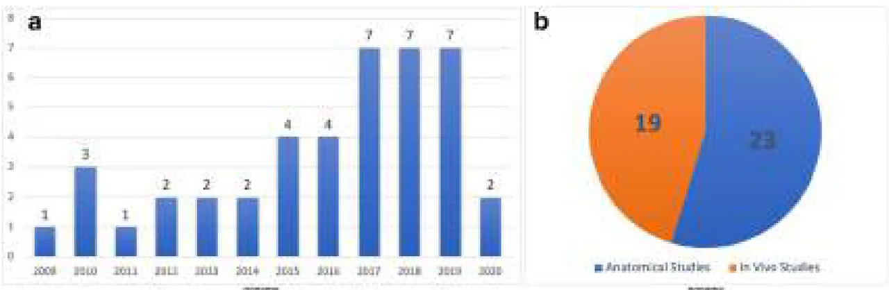
studies were included in qualitative synthesis. Figure
1
pre-sents the PRISMA flowchart summarizing identification,
screening, eligibility, and inclusion criteria. An increasing
trend of publications over the last years was observed (Fig.
2
). Table
1
presents the studies including living patients (or
both case series/report and a cadaver study), whereas Table
2
summarizes purely anatomical studies.
Indications
TEAs refer to a group of surgical corridors that reach or pass
through the orbit without removing any part of the bony
or-bital rim or adjacent structures [
3
,
46
,
47
]. These approaches
may be indicated for the treatment of pathologies located
with-in or adjacent to the orbit [
3
,
31
,
36
,
43
,
47
]. They may also be
Fig. 2 A Chart graph showing the distribution of the number of the articles published throughout the years. B Pie graph showing the distribution of the papers according to study type
Fig. 1 Diagram showing selection process based on Preferred Reporting Items for Systematic Reviews and Meta-Analyses (PRISMA) for the stud-ies related to endoscopic transorbital approaches
Table 1 S tudies which include cases in vivo Au thor Year Origin S tudy type An atomical/ surgica l N° of patients /s pe ci me n Portals (n° of pati ents ) Approach C raniectomy R econstructi on Ta rge t ar ea S u rgic al la ndmark s Park [ 53 ] 2020 Sout h K orea CSt S x 2 4 p at ient s (11 TO) T O (vs minip terional approach) (11) S L C D rill ing o f G S W F as ci a la ta or All o Derm, w ith TachoSil, abdominal fat Sphenoid w ing S OF, O C, IOF, MLA, GSW, MOB, T M Gerges [ 27 ] 2019 U S A L I + Case An + S x 4 cadaver heads + 1 pa ti en t TO (1) Inferior eyeli d N one None ITF, P S IOF, GSW, TM, Z R, LPM De Rosa [ 16 ] 2019 Spain /Ital y L I + Case An + S x 3 cadaver heads + 1 pa ti en t TO + (endo scopic extraorbital) (1) SLC + Lat eral canthot omy (for extraorbital corridor) D rill ing o f G S W and L S W Ti sseel , F at graf t F R , FO, S O F SOF, IOF + TM, pterion (for extra o rb ital corridor) Lee [ 34 ] 2019 Sout h K orea CSt S x 2 1 p at ient s (9 T O) TO (7), TO + T N (2) SLC R emoval o f L OW and v ertical crest 2 layers autol ogous fa sc ia or al lo de rm + sut u rin g o f th e dura MC F, C S SOF, M O B , V1, V 2 Gol b in [ 28 ] 2019 Russ ia CSt S x 1 2 p at ient s (6 b iops ies, 6 res ec tions ) TO (9), TO + T N (3) SLC (8 ), retrocaruncul ar (2), late ral retroca n thal (1), upper m edial (1) N o t m enti oned F at g ra ft , fa sc ia lata (3 layer) N/A S OF , IOF , G S W , F R , FO, F S (for S L approach) -A LC, la crim al eth m o id su tu re ,A E A ,P E A , OC (for R C) Lu bbe [ 41 ] 2019 Sout h A frica L I + ca se S x + An 1 p at ient, 1 cadaver he ad TO + T N (1) Contralatera l P C N /A Abdominal fat, DuraGen, NSF L ateral recess o f th e spheno id si nus AE A, S T Kon g [ 31 ] 2018 Sout h K orea CSt S x 1 8 p at ient s T O (16), T O + TN (2) SLC N /A Doubl e layer fasci a la ta or al lod erm and fat if needed N/A N /A Jeon [ 29 ] 2018 Sout h K orea CSt S x 9 pat ients TO (8), TO + T N (1), Sub o cc ip ital cr an io to my (1 ) SLC R emoval o f L OW , v ertical crest o f IO F, an d G S W . Du ra l in cis io n wa s m ad e to reach the te mp o ra l lo b e. İn le sion s limit ed to th e M C, an in terdural app ro ac h reachin g the la teral border o f th e C S TachoSil , d o uble layer au tol ogous fasci a or All o Derm + M edpo r + M inipl ate M C ,T L S O F ,G S W ,I O F , M O B Dal lan [ 15 ] 2018 It aly C St Sx 14 p at ient s TO (10), T O + TN (4) S L C In 2 p at ients w it h in tradural extensio n th rough GSW M u ltilayer, fa scia lata, in tr ad u ral fa t N/A IOF, S OF, G S W , T M , MC F d ura Lu bbe [ 40 ] 2017 Sout h A frica CSt S x 7 pat ients TO + T N (7) LRC D rill ing o f G SW in add ition to removal o f L OW (cranie ctomy in 1p t) Underlay Du ra G en g ra ft Orbi t, S p henoid T M , LOW , G S W Chen [ 8 ] 2015 U S A C ase S x 2 pat ients TO (2) S LC (1 ), SLC extended late rally from lateral canthus (1 ) D rill ing o f G S W b et w een S O F an dI O F (i no n e case addi tion al L O W removal) F ree local tissue g ra ft, dural sealant Hip pocam p u s, am y gdala, entorhinal cortex SOF, IOF Dal lan [ 12 ] 2015 It aly L I + CSt A n + Sx 5 cadaver heads + 4 cases TO + T N (4) SLC + inferio r eyelid crea se B y re mo v al o f G SW and ext ended ITT + Fat IOF, S OF, T M , AE A, PEA
Tabl e 1 (continu ed) Au thor Year Origin S tudy type An atomical/ surgica l N° of patients /s pe ci me n Portals (n° of pati ents ) Approach C raniectomy R econstructi on Ta rge t ar ea S u rgic al la ndmark s in feri o rly if M C F exp o sure is ne ed ed MC F, C S , V 2, V3, ON, ICA, te mporal lobe Ly son [ 43 ] 2014 Pol an d Case Sx 1 p at ient TO Lat er al o rbito tomy N /A T achosi l (for the orbi t) Orbi t T M R aza [ 58 ] 2013 U S A C St Sx 6 p at ients T O + T N (4) P C M in icraniecto m y al ong the superomedial aspe ct o f th e or bi t F as ci a la ta Planum spheno idale (2), AC F (2), cribrif o rm plate, me d ial orbi tal roof AE A, P E A, F E S Kop p e [ 32 ] 2013 France CSt S x 1 0 p at ient s T O S LC Supraorb ital d rill ing Dural sut ure, fa t g raft Se ll a N /A Li m [ 36 ] 2012 U S A C St Sx 13 p at ient s TO PC (4), SLC (9 ) N /A N /A front al sinu s, orbit , ACF N/ A Balakris hnan [ 3 ] 2011 U S A C St Sx 107 patient s T O L RC (50 ), L E (65), P C (55), S LC (17 ) N/ A N /A N /A N /A Moe [ 48 ] 2011 U S A L I + CSt A n + Sx 5 cadaver heads + 1 0 patient s TO SLC + P C (in each cadaver), SLC (5), PC (4), SLC + P C + PS A + LRC (1) N/A F o r supraorbital defects one-layer allo gra ft, in inte rorb ital d efects, 2 layers of al lograft + biog lue + Hadad flap (in some cases) ACF N /A Moe [ 47 ] 2010 U S A L I + CSt A n + Sx 3 cadaver heads + 1 6 patient s TO LRC (1), S LC (6), PC (7), P S (1), LRC + SLC + P C + P S (1) Removal o f G SW N/A OA, sel la, ACF (for PC ), ant temporal lobe, M C F (LRC ), F R (P S) , supraorbital AC F (SLC ) AE A, P E A AC F ant er ior cr ani al fos sa , ACP ante ri or clin oid p ro ce ss, AE A ante ri or ethm oidal artery, AL C ante rio r lacr imal cr est , An an ato m ic al , CS cavernous sinus, CSt clinical st udy, EO A endoscopic o rbital app roac h , FE S fr ontoe thmoid sutur e, FO fo ram en ova le, FR foramen rotund um, FS foramen spinos um, FZ S frontozygomatic suture, GG Gasserian ganglion , GSW greater sphenoidal w ing, ICA int erna l ca rot id art er y, Inf inf eri or, IO F inf er ior or bita l fiss u re , ITF in fr at em pora l fo ss a, KT K aw ase tr ia ngl e, Lat lat era l, LI laboratory investigation, LOW lat er al o rbi tal wa ll, LR C lat era l re troc ant hal, LSW les ser sphenoidal w ing, MC Me cke l`s ca ve, MCF middle cranial fossa, Med medial, MIS middle incisural space, MLA meningolacrimal artery, MMA middle m eningeal artery, MOB meningoorbital b and, MO W medial orbital w all, N/ A not applicable, OA orbital apex, OC optic canal, ON optic n erv e, PC precarun cular, PCF posterior cranial foss a, PE A posterior ethm oidal artery, PS pre sept al low er ey el id , SF supraorbital foramen, SLC superior eyelid crease, SOF superior orbital fissure, Sp sphenoid, ST superior turbinate, Su p superior, Sx surgi ca l, TL temporal lobe, TM te m pora lis mus cle , TN tr ansna sal , TO tr ansor b it al, VC Vidi an ca nal, VN Vidian nerve, ZF zygomatico temporal foramen, ZF B zygomaticofacial bundle, ZTB zygomaticotemp o ral b undle
Table 2 A n ato m ic al st udies A u thor Y ear Or igin N r of spe cimens Portals A pproach Craniectomy R eco nstr uction Target area Surgical landmarks S ar ace n o [ 59 ] 202 0 Italy 5 h eads T O S LC and IL T EA Dril li ng the G S W N /A M C F S O F , IOF , T M Bon-Jo ur L in [ 38 ] 201 9 C h ina 5 h eads T O L ateral canthotomy w ith ca n thol ysi s + p reseptal lo wer eyelid Removal o f G S W , d ri ll ing b etw ee n F R and F O T it an iu m Me sh , Mi n ipla tes F R , F O, PPF, ITF , MC F, MC , GG, LWC S GSW , MOB, ION, SOF, TM, IOF La le v a [ 33 ] 201 9 B u lgaria 3 heads T O S LC ext ending th rough th e zygoma By removal o f L OW and sphenoi d ridge N /A A nteromedi al : ACP, op tic canal, ON, ICA; P o st eromedial : LWCS; P osteri or: M C, petrous apex; In fe ri o r: ITF, pteryg oid fossa ZF, SF, TM, F ZS, sphenoi d rid g e, MOB, AC P, LSW , SOF, GS W , FR, VC, F O Bon-Jo ur L in [ 37 ] 201 9 C h ina 4 h eads T O S LC + L ateral cantho tomy and ca n thol ysi s Large bone dril li ng of the G SW, L OW, S O W to reach to ACF and MC F dura N /A M IS , tent o rium, M C , int erpeduncular ci stern, prepon tin e cis tern MLA, MOB, SOF, OC, IOF, anterior cli noi d, M 1 o f MC A No iphi tak [ 52 ] 201 8 U SA 7 h eads T O + (endos copic ex traorbit al co rr id o r) Ext ended incis ion from the lateral canthu s towards late ral + cantho tomy R emoval o f L OW, d rilling from IOF to SO F. D u ra li n ci si o n w as ma d e to re ac h the temporal lobe N /A M IS , tent o rium, M C , int erpeduncular ci stern, prepon tin e cis tern MLA, MOB, SOF, OC, IO F , A C P, M 1 o f MC A No iphi tak [ 51 ] 201 8 U SA 5 h eads T O S LC R emoval o f S OW, laterally from th e S O F to FS , removal o f L O W from S OF to TM and fro m S OF to IOF N one ACF, MCF, ICA , ACA, Chiasm, M CA SOF, IO F , AEA , PEA, OC, MOF No iphi tak [ 50 ] 201 8 U SA 5 h eads T O + (endos copic ex traorbit al co rr id o r) + an te ri o r transpetrosal Ext ended incis ion from the lateral canth us tow ar d s ca n thot omy N/A N /A Infratento rial region , PF, C N IV, V , V II, VIII, mos t anterosu perior, anteroinferior an d poste rosuperior accesible p o ints of the b rains tem LO W, TM, G WS , Temporal du ra , G SPN, LSPN, M M A , C NV 1-3, Kawase tri angle, IAC, tentoriu m cerebelli, C N IV Di S o mma [ 20 ] 201 8 Italy 5 h eads T O + Supraorbi tal SLC (+ ey eb ro w incis ion) Init iall y p erforme d th rough zygo m a (temporal foss a) and cont inued though GSW N/A P ar asellar an d lateral M C F (i.e. Sylvia n fissu re ), MCA, most in ferior vi sib le poi nt of CS IOF, SOF , GSW, L S W, MOB, AC P, IC A Di S o mma [ 21 ] 201 8 Italy 5 h eads T O + Supraorbi tal SLC Init iall y performe d th rough zygo m a (temporal foss a) and cont inued though GSW N /A P et rous bon e, Cerebello ponti n e angle sp ac e, M IS, Ventral b rainstem space SOF, TM, IOF, M M A , FS, FO, M O B , G SPN, pICA, GG, tent orium Di S o mma [ 19 ] 201 7 Italy 10 heads T O S LC 4 types p ro posed: 1 ) lateral corridor to M C F, 2 ) lateral corrido r to A C F , 3 ) com b ined late ral to M C F an d A C F w ith L S W rem ova l an d 4 ) m ed ial corridor to o p ti co ca rot id regio n N /A A CF , M CF GS W, S O F , LS W, TM , IOF, M M A, MOB, MC A Al me id a [ 1 ] 201 7 U SA 4 h eads T O + (TN ) SLC V ia dri lli ng the o rbi tal roof and G SW. TM is the lateral limit for craniecto m y. 2 -layer temporal fa sc ia g raf t S y lvian fissu re , M C A , A L surface of ins u la, IC A, crural and ambiens cistern M L A , S O F , IO F, T M , ACP, MCA Pri d dy [ 56 ] 201 7 U SA 9 h eads T O S LC Dril li ng of GS W an d L S W N /A M C SOF, M O B, LOW , G S W Dal lan [ 14 ] 201 7 Italy 5 h eads T O S LC Dril li ng of GS W N /A CS S O F , M L A, GS W , M O B Di S o mma [ 21 ] 201 7 Italy 5 h eads T O + (TN ) SLC U nt il opt ic chiasm by removal o f A CP N /A ON, OC SOF, OC, P EA, ICA Ci por en [ 11 ] 201 7 U SA 3 h eads T ransnasal (cl ival) co mp ar ed w it h TN + T O PC Transnasa l transclival N /A P o sterio r cerebral v essels (BA – proxi m al to its apex –,P C A , S C A ,a n d A IC A ) AE A, P E A, lamina papyracea Ci por en [ 10 ] 201 6 U SA 8 h eads T ransnasal (cl ival) co mp ar ed w it h TN+T O PC Transnas al transcl ival N /A Cavernou s ICA AE A, P E A, lamina papyracea Mat su o [ 44 ] 201 6 U SA 7 o rbits T ransla te ra l o rbit Translateral orbital w all approach (orbit ozygomati c approach) L O W, GS W o st eo to m y N/ A S u p er io r. and lateral surfa ce s o f the o rb it, OC, S OF, and CS (aft er dri lli ng the G SW an d A C P MC F, KT could b e reached) T M , L O W ,G S W ,S O F Ferrari [ 25 ] 201 6 Italy 7 h eads T O Inferolat er al o rbital ri m Triang le between SOF and IOF expos ing TM, IT F , and MCF N /A 4 corrid o rs: M C corridor (GG , SP S ), carotid foram en (ET , ICA), petro u s corridor (GS P N, E A ), transdural MCF corridor ZFB, ZTB, IOF , S OF, MOB, MLA, FO, V 2, V3, MMA, FS, F R
Tab le 2 (continued) A u thor Y ear Or igin N r of spe cimens Portals A pproach Craniectomy R eco nstr uction Target area Surgical landmarks (m ed ial surface o f TL, temporal horn o f lateral v ent ricle, tent o rium) Al qahtani [ 2 ] 201 5 Italy 5 h eads T O + TN Transp al pebral (trans verse suprat ar sa l skin incisi on) By removal o f su p erior o rbi tal wall M u ltil ayer with synth et ic g raft, m u co pe rio ste al se ptal graft AC F, MC F (i.e. ON, IC A, sellar/suprasellar structures) AE A, P E A, ON, SOF Bl y [ 6 ] 201 4 U SA 5 h eads + comput er --aided modell ing TO LRC G SW rem o val b etween IOF and S O F N /A Lateral C S IOF, S OF , ION Bl y [ 4 ] 201 2 U SA 4 h eads + 14 CT scans (comput er --aided modell ing ) 2T N , 8T O T N , L R C , T C ,P C , S L C , N /A N /A P re-p o st ch ia sm at ic ci st er n ,C S , M C .S O F , Third v en tricle , b asal cistern, clivus N/ A Ci por en [ 9 ] 201 0 U SA 5 h eads T O + TN + Su p rao rb it al P C (plu s T N and sup ra o rbital minicraniotomie s) N/A N /A P G , O C, cavernous ICA, clivu s, A E A , P E A , F E S Du z [ 24 ] 200 9 T urkey 5 heads T O + (TN + keyhole) 1) Inferolateral orbit o tomy-E OA, 2) endoscopi c en donasal m edial orbit al approach, and 3) transcrani al keyhole endoscopi c o rbit al ap proach N/A N /A Orbit T M, AEA , PEA ACF an te ri or cr ani al fos sa , ACP anterior clinoid p roces s, AE A anterior ethmoidal artery, ALC an te ri or la cr im al cr es t,C S cavernous sinus ,E O A endoscopic o rb ital approach, FE S fr ont oethmoid sutur e, FO for am en o va le , FR foramen rotundum, FS foramen spinosum, FZ S frontozygomatic suture, GG Gasserian ganglion, GS W g reater sphenoidal w ing, ICA in ter n al ca ro tid ar te ry, ILT E A in fe rol ate ral tr ans o rbit al appr oac h , IO F inferior orbital fissure, IT F infr at empora l fo ssa , KT K aw ase tr ian g le, LO W la ter al o rb ita l w all , LRC la ter al retr o ca nthal , LSW lesser sphenoidal w ing , LW C S la te ral w all o f the cavernous sinus, MC M ecke l` s ca ve, MCF middle cranial foss a, MIS middle incis ural space, ML A mening olacrimal artery, MMA middle m eningeal artery, MOB meningoorbital b and, MOW medial orbital wa ll, N/ A not applicable, OA orbital apex, OC optic canal, ON optic nerve, PC precaruncular, PC F pos terior cranial fossa, PE A posterior eth m oid al artery, PS pr ese p tal low er eye lid, SF supraorbital for am en, SL C superior eyelid crease, SOF superior orbital fissure, ST su perior turbinate, TL temporal lobe, TM te mpora lis musc le , TN tr ansna sal , TO tr an so rb it al , VC Vidian canal, VN Vidian nerve, ZF zy gomaticotemporal fo ramen, ZFB zygomaticofacial bundle, ZT B zygomaticotemporal bund
used to target distant anatomical regions by using the orbital
cavity as a corridor [
1
,
2
,
4
,
5
,
8
,
10
,
14
–
16
,
18
,
19
,
21
,
25
,
27
,
29
,
31
,
33
,
34
,
37
,
38
,
40
,
41
,
47
,
48
,
56
,
57
]. They can be
used either as a uniportal route [
3
,
5
,
8
,
14
,
18
,
27
,
33
,
36
,
38
,
47
,
48
,
50
,
56
] or may be combined with transnasal,
transmaxillary, or supraorbital paths [
1
,
2
,
4
,
9
–
12
,
16
,
18
,
20
,
24
,
28
,
29
,
31
,
34
,
41
,
44
,
46
,
50
–
52
,
58
]. The decision
making regarding the approach must be done considering the
critical (neurovascular) structures involved by or adjacent to
the pathology, the space needed for insertion of instruments,
capability to reach the target from the approach angle,
possi-bility to perform a reconstruction, corridor-related morbidity,
and experience of the surgical team. The patients’ preference
must also be taken into consideration [
3
,
8
,
11
,
14
,
16
,
18
,
25
–
27
,
29
,
34
,
46
,
50
,
52
,
56
,
57
].
Surgical techniques
Transorbital endoscopic surgery is based on 4
pillar-approaches through orbital quadrants: the superior eyelid
crease (SLC), precaruncular (PC), the lateral retrocanthal
(LRC), and the preseptal lower eyelid (PS), which cross the
superior, medial, lateral, and inferior orbital quadrants,
respec-tively (Figs.
3
,
4
,
5
,
6
,
7
,
8
, and
9
) [
47
]. Several variants of
these pillar approaches have been described over the last
de-cade in clinical and anatomical studies, each aiming to
facili-tate surgical goals [
8
,
9
,
20
,
24
,
25
,
29
,
33
,
44
,
50
–
52
,
59
]. A
thorough understanding of the anatomy of the eyelid is
essen-tial in each approach, and possible need for a reconstruction
and additional corridors (i.e., multiportal approach) must also
be precisely planned before surgery.
Superior eyelid crease approach
With the superior eyelid crease approach (Fig.
5
), also named
as upper eyelid approach, the superior orbit, frontal sinus,
Fig. 3 Scheme depicting the extension and reach of transorbital endoscopic approaches (TO) with respect to transnasal endoscopic (TN) and most relevant open skull base approaches.A Anterior approaches (e.g., subfrontal); AL1, paramedian anterolateral approaches (e.g., supra-orbital); AL2, anterolateral approaches (e.g., pterional, frontotemporal, orbito-zygomatic, frontotemporal-orbitozygomatic); L, lateral ap-proaches (e.g., transpetrous, subtemporal middle cranial fossa, infratemporal); PL, posterolateral (e.g., trans-sigmoid, retrosigmoid); P1, paramedian posterolateral approaches (e.g., far lateral); P2, posterior approaches (e.g., suboccipital) [17]
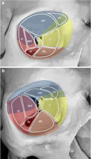
Fig. 4 A, B Schemas presenting the relations of transorbital approaches with different anatomical sites. Right orbit of a dry skull. ACF, anterior cranial fossa, E, ethmoids; FS, frontal sinus; ITF, infratemporal fossa; MCF, middle cranial fossa; MS, maxillary sinus; PS, TF, temporal fossa. The colors indicate the transorbital surgical approaches as blue, superior eyelid; yellow, precaruncular; red, lateral retrocanthal; orange, inferior eyelid
supraorbital and posterior-central portions of the anterior
cra-nial fossa (ACF) and anterior skull base (ASB), and lateral
portion of the middle cranial fossa (MCF) can be reached.
This is the commonest approach used in transorbital
endo-scopic surgery [
2
–
4
,
8
,
12
,
15
,
16
,
18
–
21
,
28
,
29
,
31
,
32
,
34
,
36
,
38
,
47
,
48
,
51
,
53
,
56
,
59
]. The skin incision is made in the
supratarsal fold as done in an upper blepharoplasty and can be
tailored according to the path-to-target analysis [
26
,
47
]. Deep
to the preseptal orbicularis oculi muscle, the orbital septum is
identified, through which the prelevator fat can be seen.
Dissection is continued raising the deep surface of the
orbicularis muscle towards the superior orbital rim. This is
crucial to avoid the aperture of the orbital septum and
periorbit, which causes fat to prolapse into the surgical
corri-dor. After the identification of the orbital rim, the periosteum
is incised, and dissection is further progressed in a
subperiosteal plane through the orbital roof. In the posterior
portion of the orbit, the orbital end of the optic canal is
iden-tified, and medially the ethmoidal foramina can be visualized.
According to the target, the dissection can be extended as far
Fig. 5 Superior eyelid andprecaruncular approaches.A The superior eyelid crease approach starts with a skin incision performed at level of the supratarsal fold (black dashed line).B Superior tarsus (Ta) and levator palpebrae superioris mus-cle (LPSM) are identified.C The superior orbital rim is detached from the periorbit (Pe).D The subperiosteal dissection is contin-ued along the orbital roof (OR). The anterior (AEF), medial— when present—(MEF), and pos-terior ethmoidal foramina (PEF) are identified in the medial aspect of the surgical corridor. The optic canal (OC) and the superior or-bital fissure (SOF) are identified in the posterior portion of the or-bit.E Both the precaruncular (PC) and lateral retrocanthal approach (LRC) display an overlap as regards the superior eyelid crease corridor. The trajectory of the precaruncular approach (white arrow, PC) lies at the medial as-pect of the orbital cavity and re-quires sequential cut of the eth-moidal bundles.F The lateral retrocanthal approach (white arrow, LRC) is located in the lat-eral aspect of the orbital cavity and, similarly to the superior eye-lid crease approach, offers direct exposure of the inferior orbital fissure (IOF), inferiorly, zygo-matic bone (ZB) and greater sphenoidal wing (GW) laterally, and SOF superiorly. AEA, ante-rior ethmoidal artery; Ost, optic strut
laterally as necessary. Identification of the superior orbital
fissure (SOF) is paramount to achieve adequate orientation
in the most posterior and lateral aspect of the corridor. If a
more lateral corridor is needed, detachment of lateral canthal
ligament can be done [
26
]. Diamond burr or a chisel is used to
perform the craniectomy, which is sized and located
depend-ing on the targeted area [
16
,
37
,
56
]. The dura of the ACF and
MCF can be elevated and/or incised based on the situation of
the disease.
Precaruncular approach
This approach via the medial quadrant provides a direct
and avascular access to medial orbital roof, lamina
papyracea, ethmoidal arteries, cavernous sinus, parasellar
and paraclinoid tracts of the internal carotid artery, optic
nerve, and the central corridor towards the anterior skull
base (ASB) [
3
,
4
,
9
,
10
,
26
,
36
,
46
–
48
,
58
].
An incision between the caruncle and skin is made
through the conjunctiva at the apex of the medial
can-thus. The avascular plane is entered deep to the
Horner’s muscle and to the posterior limb of the medial
canthal tendon, and the periorbit is incised at crista
lacrimalis (i.e., the posterior border of the lacrimal
fos-sa). Dissection is performed from anterior to posterior
between the periorbit and the medial orbital wall. The
level of the ASB can be estimated by the ethmoidal
bundles which are found along the fronto-ethmoidal
su-ture and can be cauterized and cut. Reaching the
poste-rior ethmoidal artery warns the surgeon that the optic
nerve is close (i.e., around 7 mm posterior to the
pos-terior ethmoidal foramen) and attention must be paid
not to damage it (Fig.
5
). After that level, dissection
through the medial orbital wall makes the surgeon reach
the orbital apex and then the bony removal is performed
depending on the anatomical site of the target [
26
,
47
].
Lateral retrocanthal approach
Via LRC approach, access to the deep lateral orbit,
lat-eral aspect of the ACF, MCF, and infratemporal and
temporal fossa is possible [
3
,
4
,
28
,
40
,
47
]. LRC
over-comes morbidities like scarring and disruption of eyelid
support caused by cutaneous or canthotomy incisions. A
conjunctival incision is performed immediately posterior
to the insertion of the lateral canthus (Fig.
6
). The
subperiosteal dissection is performed along the lateral
orbital wall, from the inferior orbital fissure (IOF) to
the orbital roof. This maneuver exposes the greater
sphe-noidal wing (GSW) (located below the SOF, above and
lateral to the IOF, and posterior to the zygomatic bone),
whose removal provides access to the temporal fossa,
infratemporal fossa, and MCF [
47
]. The optic nerve is
not at risk through this corridor as it stands medially in
the orbit and is separated from the lateral wall by the
contents of the SOF. At the superior aspect of the lateral
orbital wall lies the sphenofrontal suture by which a
su-perior craniectomy yields to lateral frontal fossa, while
an inferior craniectomy guides to the MCF [
47
].
Preseptal approach
The PS approach is useful to access to the inferior orbit
[
38
,
47
]. It can be combined with LRC or PC to
in-crease maneuverability and exposure of the lateral and
medial orbital quadrants, respectively (Fig.
6
). It gives a
Fig. 6 Lateral retrocanthal and preseptal lower eyelid approaches.A The lateral retrocanthal (LRC) approach starts with a conjunctival incision (black dashed line) of the palpebral conjunctiva located on the lateral aspect of the orbital rim and passing posterior to the lateral canthal tendon (LCT). With the aim of increasing maneuverability and exposure through the inferolateral orbital quadrant, the lateral retrocanthal approach can be combined with a preseptal lower eyelid approach (PS), which is also started with a conjunctival incision (black dotted line) on the inner surface of the lower eyelid.B The preseptal lower eyelid approach (white arrow, PS) exposes the orbital floor (OrF) and early stops at the inferior orbital fissure (IOF), which needs to be cut (black dashed line) to extend expo-sure to the greater sphenoidal wing (GW) while merging the inferior quadrant corridor with the lateral quadrant corridor (i.e. inferolateral transorbital endoscopic approach).C The lateral retrocanthal shares the potential to expose the greater sphenoidal wing and adjacent structures with the superior eyelid crease (SLC) approach (white arrow, SLC).D The removal of the coronal portion of the greater sphenoidal wing pro-vides access to masticatory space, inferiorly, and middle cranial fossa dura (MCFD), superiorly.E The dissection can be continued along the extracranial aspect of the horizontal portion of greater sphenoidal wing by dissecting lateral pterygoid muscle (LPM) off the skull base. This maneu-ver provides exposure of the foramen ovale (FOv) and the extracranial tract of the mandibular nerve (V3) in the infratemporal fossa.F Epidural dissection along the anterior portion of the middle cranial fossa exposes the intracranial segments of maxillary (V2) and mandibular nerves.G Posterior and lateral to the mandibular nerve, the middle meningeal artery (MMA) runs from the foramen spinosum with a medial-to-lateral direc-tion and provides vascular supply to the dura mater of this anatomical region.H After completing the removal of the bony contour of foramina ovale and spinosum and sectioning the middle meningeal artery, the bony-cartilaginous junction of the eustachian tube (ET) is identified. The eustachian tube crosses the mandibular nerve posteriorly and runs from superolateral to inferomedial.I The petrous segment of the internal carotid artery (peICA) is located posteriorly to the bony-cartilaginous junction of the eustachian tube.J After removing the eustachian tube and removing the anterior contour of the carotid canal, the vertical (v) and the horizontal (h) subtracts of the petrous portion of the internal carotid artery are visualized. GSPN, greater superficial petrosal nerve; LPM, lateral pterygoid muscle; LPP, lateral pterygoid plate; MPM, me-dial pterygoid muscle; MCF, middle cranial fossa; V1, ophthalmic nerve; Pe, periorbit; SOF, superior orbital fissure; TM, temporalis muscle; ZB, zygomatic bonepath through orbital floor, maxillary sinus, IOF, and
foramen rotundum. The conjunctival incision for a
preseptal approach is made 2 mm inferior to the tarsus
on the conjunctival surface of the lower eyelid (6 mm
inferior to the eyelid margin). The orbicularis oculi
muscle is identified, and the dissection is carried out
on its posterior face, which is anterior to the inferior
orbital septum. The orbicularis muscle is followed
through the inferior orbital rim, where the periosteum
is incised and lifted off the orbital floor. Dissection
may proceed further by sectioning the infraorbital
bun-dle and IOF [
47
].
Extended and combined transorbital endoscopic approaches
Along with the 4 pillar TEAs, extended or combined
ap-proaches can be applied in selected cases [
2
,
4
,
5
,
8
–
12
,
16
,
18
,
20
,
24
,
25
,
28
,
29
,
31
,
34
,
40
,
41
,
44
,
50
–
52
]. In the
literature, TEAs are mostly used in combination with
transnasal endoscopic procedures [
1
,
2
,
4
,
9
–
12
,
22
,
24
,
28
,
29
,
34
,
40
,
41
]. Other than transnasal corridors, different
ad-ditional extraorbital corridors, such as supraorbital, lateral
or-bital, and pterional, have also been investigated and described
in combination with transorbital routes (Figs.
7
,
8
, and
9
) [
9
,
16
,
20
,
44
,
51
,
52
]. Transorbital extended approaches, in
Fig. 7 Transorbital exposure of the lateral anterior skull base.A Theremoval of the orbital roof provides exposure of the anterior cranial fossa dura (ACFD), which can be resected or incised to expose the inferior aspect of the frontal lobe and related neurovascular structures. Among transorbital endoscopic approaches, superior eyelid crease and precaruncular provide the best exposure of this portion of the cranial base and adjacent structures. The orbital beak (OBe) is the line located above ethmoidal foramina where the anterior cranial base turns from horizontal to cranially-convex (i.e., from the ethmoidal roof to the orbital roof, respectively). The lateral orbital wall (LOW) can be used as land-mark to define the lateral limit of the craniectomy. B Posterior craniectomy can include the anterior clinoid process, medially, and lesser
sphenoidal wing, laterally. This provides exposure of the intracanalicular portion of the optic nerve (ON), anterior clinoid process dura (ACPD), and meningo-orbital fold (MOF), which is the area where the dura of anterior and middle cranial fossae turns into periorbit.C Focusing on the posteromedial portion of the surgical corridor, the optic strut (OSt) between the optic nerve and the paraclinoid tract of the internal carotid artery (pcICA).D In the most medial and anterior portion of the surgical corridor, bone removal of the superomedial orbital wall provide access to the frontal sinus (FS), frontoethmoidal region, and anterior ethmoid (AE). AEF, anterior ethmoidal foramen; Pe, periorbit; PSp, planum sphenoidale; PEF, posteror ethmoidal foramen; Tr with white dashed line, position of the trochlea
which more than one orbital quadrant was used, have also
been presented [
25
,
59
].
Endoscopic transorbital craniectomy and bony landmarks
The term craniectomy describes the removal of the bone cover
on the dura with no bone repositiong at the end of the
proce-dure [
26
]. The type of craniectomy is determined by the
anatomo-surgical target for the underlying pathology. A drill,
ultrasonic dissector, or chisel can be used to remove the bone
at the desired site [
47
]. Surgeon should be familiar with the
bony landmarks when performing a craniectomy. The
thick-ness of bone varies throughout the orbital walls and different
landmarks need to be exposed to complete the bone removal
safely and accurately. In the 33 studies in which the
craniectomy was mentioned in the text, SOF was the most
common landmark (75.8%), followed by the IOF (54.5%)
and GSW (39.3%). The other anatomical structures that were
pointed as landmarks were temporalis muscle (TM), lateral
orbital wall (LOW), medial orbital wall (MOW), anterior
eth-moidal artery (AEA), posterior etheth-moidal artery (PEA), lesser
sphenoidal wing (LSW), meningo-orbital band (MOB),
frontozygomatic suture (FZS), anterior clinoid process
(ACP), optic nerve (ON), optic canal (OC), internal carotid
artery (ICA), foramen ovale (FO), foramen rotundum (FR),
foramen spinosum (FS), vidian canal (VC), vidian nerve
(VN), zygomaticotemporal foramen (ZF), supraorbital
fora-men (SF), frontoethmoidal suture (FES), zygomaticofacial
bundle (ZFB), zygomaticotemporal bundle (ZTB), orbital
a p e x ( O A ) , a n t e r i o r l a c r i m a l c r e s t ( A L C ) , a n d
meningolacrimal artery (MLA) [
1
,
2
,
4
,
5
,
8
–
16
,
18
–
20
,
24
,
25
,
27
–
29
,
32
–
34
,
37
,
38
,
40
,
41
,
44
,
47
,
51
–
53
,
56
,
58
].
Reconstruction
As in general in SB surgery, reconstruction may be necessary
to achieve a safe separation between different compartments
(i.e., sinonasal tract, orbit, intracranial space). Postoperative
cerebrospinal fluid (CSF) leak is a common concern related
to TEAs and the surgeon must be familiar with different
re-constructive techniques. In case of small defects with limited
CSF leak, reconstruction is deemed unnecessary thanks to the
support of the orbital structures [
1
,
14
,
56
]. For larger defects,
reconstruction is indeed required, and the technique of
recon-struction has to be tailored according to pathology, type of
approach, and craniectomy size and site. When the defect is
extended beyond the limits of the orbit (e.g., frontal sinus,
cribriform plate, planum sphenoidale, lateral recess of the
sphenoid), watertight closure should be achieved following
the principles of transnasal endoscopic SB reconstruction [
3
,
41
,
47
,
48
,
58
]. Various materials such as autologous grafts
(e.g., fascia lata, temporalis fascia, iliotibial tract, abdominal
fat, septal mucoperichondrium) or synthetic materials (e.g.,
TachoSil®, AlloDerm®, DuraGen®) have been used either
alone or in combination [
8
,
12
,
14
,
16
,
28
,
29
,
31
,
32
,
34
,
38
,
40
,
41
,
43
,
48
,
53
,
58
]. In the majority of the studies, a
mul-tilayer reconstruction was conducted. A total of 74 patients
required dural reconstruction, and postoperative CSF leak
was reported in 3 (4.1%) [
8
,
12
,
15
,
16
,
28
,
29
,
31
,
32
,
34
,
40
,
41
,
48
,
53
,
58
]. Even after the reconstruction of large
defects, the risk of CSF leak appears to be low as the orbital
contents keep the reconstruction in place and tight [
1
,
12
,
14
,
46
,
48
,
56
]. Pulsation of the globe may be noted for 1 to 2
weeks postoperatively, which generally resolves
spontaneous-ly [
8
,
56
,
60
]. In addition, in order to prevent postoperative
enophthalmos, Medpor can be placed over the dural defect as
a buttress [
29
]. When the lateral orbital rim was removed in
extended TOA, the orbital rim can be reconstructed by a mini
plate or a titanium mesh [
29
,
38
].
Complications
TEAs provide the chance to avoid some complications related
to open craniotomies. However, by nature, they also have
risks of complications. A total of 60 complications were
re-ported in 193 cases presented (Tables
3
and
4
) [
3
,
8
,
12
,
15
,
16
,
28
,
29
,
31
,
34
,
53
,
58
]. The majority of the complications
reported were transient. Balakrishnan et al. [
3
] reported 3
per-sistent vision loss after surgery (3% of cases), Golbin et al.
[
28
] reported one persistent abducens nerve paresis, and Lee
et al. [
34
] reported postoperative facial numbness in 38.1% of
their patients which did not resolve. When the complications
were evaluated in terms of Clavien-Dindo classification, all
except one were Grade 1 and 2 [
23
]. The only complication
requiring surgical intervention (Grade 3b) was an orbital
pseudomeningocele which resolved after shunt insertion [
8
].
Table
5
shows the Clavien-Dindo classification for grading
complications [
23
].
Discussion
TEAs, initially described as ancillary alternatives to traditional
transcranial/transnasal routes, have been evolving to the state
of well-established surgical methods, which are intended to
overcome the limits of conventional procedures for selected
pathologies of the SB, either alone or in combination with
other techniques [
12
,
39
,
47
]. For this reason, they raised a
progressively increasing interest, as evident from the number
of publications in a relatively short timeframe (Fig.
2
). They
have become relevant tools in SB surgery, facilitating access
to a number of sites deemed challenging to reach, yet with
relatively low morbidity [
39
,
41
,
47
].
In order to achieve the surgical goal, the surgical approach
must be precisely selected, which majorly depends on the
quadrant of the orbit that is involved by or is the most
forth-right route to the target pathology [
46
,
47
]. The basic
require-ment to safely harvest a transorbital surgical corridor and
properly manipulate the lesion is sound knowledge of surgical
anatomy. As learnt from transnasal endoscopic surgery, this
should ground on the understanding of anatomical landmarks,
which are basic anatomical relationships through which the
surgeon can maintain orientation even in an intricate surgical
corridor. The present paper systematically summarizes the
main landmarks that were emphasized throughout available
publications (Tables
1
and
2
; Figs.
3
,
4
,
5
,
6
,
7
, and
8
). The
created pathway through the target needs to give the surgeon
optimal visualization and provide space for the use of
endo-scopic instrumentation. This is crucial both for the
manipula-tion of the target and SB reconstrucmanipula-tion. By TEAs, this short
and direct path yielding to the needed comfortable working
space can be frequently achieved, but one should keep in mind
the spectrum of potential surgical alternatives to offer the best
treatment to patients. The flexibility that the SB surgeon/team
has in hand, which is the ability to extend or combine various
approaches, provides a significant comfort and success in
“complex” procedures [
26
,
39
,
46
,
47
]. The surgeon can
benefit from the possible better exposure provided by a
com-bination of approaches. In a recent study, it was indicated that
the endoscopic transnasal approach better exposes the
inferomedial 1/3 of the SOF and the cavernous sinus while
the TEA yields a better exposure of the superolateral 2/3s of
the SOF. A combination of the two approaches can make it
feasible to access the entire SOF endoscopically [
35
]. In
an-other study, the combined transnasal-transorbital approach to
the petrous apex was investigated in cadavers in order to
quan-tify the amount of bone removal that can be obtained via each
pathway, and the authors concluded that with a combined
approach, 97% of the bone removal can be performed [
61
].
The application of these procedures is best set by close
collaboration between surgeons from different specialties
such as otorhinolaryngology, neurosurgery, and
ophthalmol-ogy. The SB surgeon can be unfamiliar with transorbital
pro-cedures. There doubtlessly exists a learning curve which can
best be surpassed by proper anatomical training and teamwork
[
2
,
39
]. The proper instrument setting is also important for
performing this type of surgery which has to include an
oculoplastic set with retractors, corneal protectors, lacrimal
probes, a SB endoscopic set with high-quality endoscopes
(0, 30, 45 and 70 degrees), drill, endoscopic Doppler probe.
and surgical navigation system [
46
]. Dedicated instruments
specifically designed for this type of surgery are worth
design-ing and testdesign-ing in order to overcome pitfalls like orbital fat
blocking the view. The surgical team always needs to keep in
mind that there may be need to intraoperatively shift the
ap-proach from endoscopic to an open one, and the preoperative
planning must include every possible scenario on which the
patient should be clearly informed and consented [
26
,
39
].
The TEAs have been applied to various anatomical sites of
the SB in several clinical studies. Orbital cavity and adjacent
portions of the ASB and MSB are the most frequently targeted
areas. Other potential targets have been recently analyzed and
discussed. Gerges et al.[
27
] investigated the application of
TEA for the ITF and PPS in anatomical specimens. They also
presented a case with a recurrent glioblastoma of the ITF,
which was approached through TEA for a biopsy. The authors
conclude that TEA can be a safe alternative with less
morbid-ity in this area, adding that pathologies extending inferiorly to
the masticator space and neck may be a contraindication but
those that extend to the MCF and spread anteriorly with
or-bital extension are more susceptible to this approach. Lee et al.
[
34
] indicated that access to the CS via TEA may be
consid-ered more practical than the endoscopic transnasal approach.
The reason of this provision is explained by the shorter access
route, ability to perform interdural dissection through the CS,
ability of facilitating exploration through the anteromedial
(between V1 and V2) and anterolateral (between V2 and
V3) corridors, and avoidance of morbidity caused by
Fig. 8 Transorbital exposure of the lateral middle skull base, parasellar area, and Sylvian fissure.A The middle cranial fossa dura (MCFD) can be exposed through a craniectomy in the area between the superior orbital fissure (SOF) and inferior orbital fissure (IOF). This portion of the skull base can be exposed through both the superior eyelid crease approach and lateral retrocanthal approach. The former provides a slightly descending trajectory towards the middle cranial fossa (MCF), whereas the latter route is parallel to the plane of the horizontal portion of the greater sphe-noidal wing (GW).B The meningo-orbital fold (MOF) is identified as the line where the dura of the middle and anterior cranial fosse merge with the periorbit.C Epidural dissection along the middle cranial fossa allows exposure of the oftalmic (V1) and the maxillary (V2) branches of the trigeminal nerve, which run towards the superior orbital fissure and fora-men rotundum (FRo), respectively.D Interdural dissection above the trigeminal branches provides access to the parasellar area and allows identification of the mandibular branch of the trigeminal nerve (V3) and foramen ovale (FOv). The cavernous sinus (CS) is identified above the ophthalmic nerve (i.e., infratrochlear or Parkinson’s triangle) and in the space between the ophthalmic and maxillary nerves (i.e., anteromedial or Mullan’s triangle) and the parasellar tract of the internal carotid artery (sICA), abducens nerve (VI), and trochlear nerve (IV) are exposed.E Further posterior interdural dissection exposes the Gasserian ganglion (GG) and Meckel’s cave (MeC). F The oculomotor nerve (III), cavernous sinus roof (CSR), posterior wall of the cavernous sinus (PWCS), and paraclival portion of the internal carotid artery (pICA) can be identified by further elevating the dura propria of the parasellar area.G The dura propria of the parasellar area and lateral middle cranial base is incised (black dotted line) to access the intradural compartment Sylvian fissure (SyF).H The first tract of the middle cerebral artery (M1), early frontal branch (EFB), and temporal polar arteries (TPA) are identified between the frontal (FL) and temporal lobe (TL). BaP, base of the pterygoid process; OR, orbital roof; Pe, periorbit; SyB, sympathetic branch of the abducens nerve; SpB, sphenoid body; TF, Temporal fossatransnasal and/or open approaches. The authors however
in-dicate that the TEA is a challenging route for the posterolateral
triangle (corridor between the V3 and the petrous apex). They
presented a case in which TEA was insufficient for
ap-proaching a dumbbell-shaped schwannoma involving the
MCF and PCF who later required additional retrosigmoid
surgery. Chen et al. [
8
] performed surgeries via TEA in two
patients for lesions (gliosis) in the temporal lobe. They
expressed that the TEA minimized risks related to
convention-al approaches to the temporconvention-al lobe like cognitive deficits,
hemiparesis, cranial nerve deficits, and visual field loss by
providing a direct path towards the temporal lobe and
allowing earlier visualization of the target. It is also
men-tioned that by the TEA, the surgeon achieves an early
visualization of the cranial nerves which may facilitate
the avoidance of an injury. Studies indicate that the
TEAs provided optimal exposure and surgical freedom
for adequate handling of lesions of the ACF and MCF
[
3
,
16
,
47
]. If needed, removal of the superior orbital
wall is possible, exposing the entire ACF from the
mid-line to its most lateral point. Suchlike, the removal of the
lateral orbital wall may help for an adequate view of
MCF, from the lateral wall of the cavernous sinus to
the lateral aspect of the temporal lobe [
51
].
TEAs have been successfully applied in various
pa-thologies ranging from CSF leak to SB tumors
(Table
6
) [
3
,
8
,
12
,
16
,
27
,
28
,
32
,
36
,
40
,
43
,
47
,
53
,
58
]. Among tumors, with 67 (65.6%) presented
cases, meningioma was the most common pathology
followed by trigeminal schwannomas (9.8%). In the
102 tumor cases presented, 50 (49.0%) gross-total
resec-tions, 9 (8.8%) near-total resecresec-tions, 30 (29.4%)
sub-total resections, and 6 (5.9%) partial-resections were
re-ported [
12
,
15
,
16
,
27
,
29
,
31
,
32
,
34
,
53
,
58
]. In 7
(6.9%) patients, biopsies were obtained through TEAs
[
27
,
28
]. Radiation therapy was performed with different
techniques (stereotactic radiotherapy, Gamma Knife
ra-diosurgery, proton beam radiotherapy) in 19 cases in
whom noncomplete resection was achieved [
28
,
31
,
34
]. Inflammatory pathologies like epidural abscesses,
frontoorbital mucoceles, and cavernous sinus thrombosis
were also managed by TEAs either alone or in
combi-nation with transnasal approach [
3
,
28
,
36
,
47
].
With either small or invisible incisions, TEAs provide a
pleasant cosmetic outcome. However, the absence of a
surgi-cal scar may give a misperception that the procedure is a
minor surgery. The complications related to the globe, as well
as neurovascular structures and eyelid apparatus, should
al-ways be kept in mind and the risks should be counseled with
the patient (Tables
3
and
4
). Care must be taken to protect the
cornea and the globe intraoperatively as continuous orbital
retraction may cause an increase of orbital pressure. This
may also result in cardiac arrhythmias, thus, frequent
moni-toring of pupil size and blood pressure, along with
electrocar-diography, is required [
29
]. Intermittent relaxation of the
eye-ball every 20–30 min during the procedure and keeping
tis-sues dislocation less than 10 mm is recommended [
5
,
48
,
51
].
Overall, TEAs have relatively less morbidity than
tradition-al SB procedures. The reports indicate mostly minor
morbid-ities, rapid postoperative healing, minimal pain, and short time
of hospitalization [
26
,
39
,
46
,
47
]. Despite these relatively low
morbidity rates, it is the authors’ opinion that it is still too
emphatic to name them as minimal invasive approaches, as
complications do occur in a non-negligible rate of patients
(31.1%) [
3
,
14
,
34
]. Indeed, the classification employed in this
systematic review rates complications such as diplopia and
upper eyelid necrosis as minor events, whereas one should
take into consideration the dismal impact of these occurrences
on patient’s quality of life. In fact, morbidity should not be
considered as a secondary issue, as most patients receiving a
TEA are affected by benign disease with good tumor-related
prognosis and long-life expectancy.
The findings of the current study, aiming to summarize the
surgical anatomy and objective clinical data on TEAs through
a systematic review of the current literature, have to be seen in
light of some limitations. Although it shows the anatomical
approaches in detail with dissections, it does not include a
quantitative analysis and comparison of each approach. The
literature contains clinical studies with heterogenity both in
terms of pathologies and approaches as well as patient
num-bers due to the relatively new growing nature of these
ap-proaches which makes the generalization of the findings
difficult.
Fig. 9 Extended transorbital transdural approach to the internal carotid artery bifurcation and adjacent structures.A The corridors through the dura of the anterior (ACFD) and middle cranial fossa (MCFD) can be merged by sectioning the meningo-orbital fold (black dashed line) in the lateral portion of the superior orbital fissure (SOF), which, as opposed to the medial portion, does not contain relevant neurovascular structures other than the superior ophthalmic vein (SOV) and meningo-orbital/-lac-rimal or recurrent meningeal branch of the middle meningeal artery.B After sectioning the meningo-orbital fold (black dotted line), the full exposure of the dura of the posterolateral anterior cranial fossa, parasellar area, and lateral middle cranial fossa is achieved. Anterior clinoid process dura (ACPD), optic nerve (ON) and the paraclinoid tract of the internal carotid artery (pcICA) are identified. Among other potential transdural targets, the intracranial tract of the internal carotid artery (iICA) and its bifurcation can be exposed by incising the anterior cranial fossa and anterior clinoid process dura (black dashed line). This maneuver would be more difficult if the meningo-orbital fold is not sectioned.C Once the dura of the anterior clinoid process is incised, the following structures are encountered: oculomotor nerve (III), frontal lobe (FL), intracranial tract of the internal carotid artery, optic nerve, tentorium cerebri (Te), and tem-poral lobe (TL). Thanks to its tangential trajectory as respect to the ante-rior cranial fossa dural plane, the transorbital perspective provides the exposure of both the intradural and extradural portion of several neurovascular structures. Particularly, the figure shows both the intracra-nial and paraclinoid tract of the internal carotid artery as well as intradural and intracanalicular portions of the optic nerve.D The bifurcation of the internal carotid artery into the precommunicating tract of the anterior cerebral artery (A1) and proximal tract of the middle cerebral artery (M1) can be identified by moving the scope forward through the transdural window.E, F The free edge of the tentorium cerebri can be used as landmark to follow the oculomotor nerve (III) towards the ambiens cistern and interpeduncular fossa. The Liliequist’s membrane (LiM), postcommunicating tract of the posterior cerebral artery (P2), pos-terior communicating artery (PCoA), and superior cerebellar artery (SCA) can be also identified. CS, cavernous sinus; IOF, inferior ophthal-mic veinTable 3 Complications of transorbital approaches in which surgical procedures were presented (numbers of complications) (superscripts in the first row indicate the reference numbers)
Author Number of TO cases Complications
Jeon [29] 9 Complete ptosis improved in 6 months (1), mild ptosis (3) De Rosa [16] 1 Proptosis which resolved in 6 months (1)
Lee [34] 9 Decrease in visual acuity (1), CN V neuropathy (2), CN VI neuropathy (2), ptosis (3) keratitis (2) Golbin [28] 12 Transient CNV1hypoesthesia (2), transient ptosis (1)
Kong [31] 18 CSF leaks (2), transient lateral rectus muscle paresis (2), transient ptosis (3) Dallan [15] 14 Upper eyelid necrosis (1), diplopia (3), CNV2hypoesthesia (3), CNV1
hypoesthesia (1), palpebral edema (3) (of which 1 persistent)
Chen [8] 2 Orbital pseudomeningocele (1)
Dallan [12] 4 Superior eyelid edema (2)
Balakrishnan [3] 107 Diplopia (14), persistent vision change (3)
Park [53] 11 CSF leak (1), diplopia (1), ptosis (1)
Raza [58] 6 Diplopia (1)
Total Number 193 60 (31.1%)
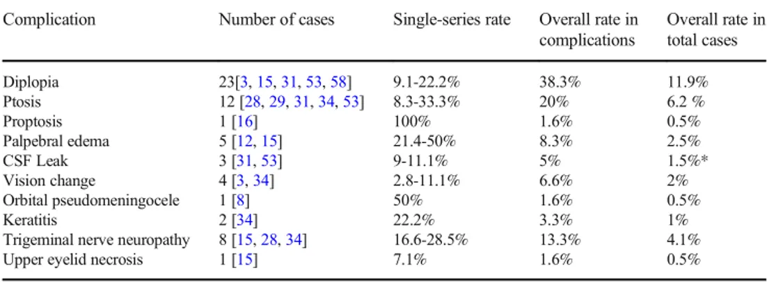
Table 4 Rates of complications. * The rate of CSF leak in patients undergoing dural defect reconstruction is 4.1% (superscripts in the second row indicate the reference numbers)
Complication Number of cases Single-series rate Overall rate in complications Overall rate in total cases Diplopia 23[3,15,31,53,58] 9.1-22.2% 38.3% 11.9% Ptosis 12 [28,29,31,34,53] 8.3-33.3% 20% 6.2 % Proptosis 1 [16] 100% 1.6% 0.5% Palpebral edema 5 [12,15] 21.4-50% 8.3% 2.5% CSF Leak 3 [31,53] 9-11.1% 5% 1.5%* Vision change 4 [3,34] 2.8-11.1% 6.6% 2% Orbital pseudomeningocele 1 [8] 50% 1.6% 0.5% Keratitis 2 [34] 22.2% 3.3% 1%
Trigeminal nerve neuropathy 8 [15,28,34] 16.6-28.5% 13.3% 4.1%
Upper eyelid necrosis 1 [15] 7.1% 1.6% 0.5%
Table 5 The Clavien-Dindo Classification of Surgical Complications [56]
Grade Description
Grade 1 Any deviation from the normal postoperative course not requiring surgical, endoscopic or radiological intervention. (Allowed therapeutic regimens are: drugs as antiemetics, antipyretics, analgesics, diuretics, electrolytes, and physiotherapy. This grade also includes wound infections opened at the bedside) Grade 2 Requiring pharmacological treatment with drugs other than such allowed for grade I complications Blood
transfusions and total parenteral nutrition are also included
Grade 3 Complications requiring surgical, endoscopic or radiological intervention Grade 3a—intervention not under general anesthetic
Grade 3b—intervention under general anesthetic
Grade 4 Life-threatening complications; this includes central nervous system complications which require intensive care
Grade 4a—single-organ dysfunction (including dialysis) Grade 4b—multi-organ dysfunction
Conclusion
This study aimed to display each method of transorbital
endo-scopic surgery with anatomical dissections and to condense
the data regarding this subject by making a systematic review
of the current literature. It would be precise to comment that
TEAs are important bricks in the wall of the endoscopic
ap-proaches to the SB. Data clustered so far indicate that these
approaches provide important advantages reaching different
pathologies and target areas through the SB. These versatile
approaches allow the surgeon to avoid extra soft tissue
dissec-tion and provide a relatively short and direct corridor. They are
not proposed to replace the transnasal or external approaches
but are useful and important complementaries that should be
in the armamentarium of a SB team.
Supplementary Information The online version contains supplementary material available athttps://doi.org/10.1007/s10143-020-01470-5. Acknowledgments Olympus Europa SE & Co. KG is acknowledged for supporting the present study through provision of 4K endoscopic camera and endoscopes. The Center for Anatomy and Cell Biology (Division of Anatomy) of the Medical University of Vienna is acknowledged for pro-viding the laboratory and instrumentation for anatomical dissection.
Authors’ contributions AV, MF, VR, AS and FD: Design of the study; AV, ALCC, MF, VR, DM, AS, FD, BB, ST, MT and TG: Cadaver dissections, collecting of data; AV, ALCC: Literature re-view; AD, LH, PN: Supervision of the project; AV, ALCC, MF, VR: Writing the study with input from all authors; MF, VR, TG, ST, MT: Praparation of figures; MF, FD, AS, DM, LFR, AD, LH, PN: Final editing of the study; All authors provided critical feed-back and helped shape the manuscript. The authors confirm that they have reviewed and approved the manuscript.
Funding Open Access funding provided by Università degli Studi di Brescia.
Data availability Not applicable.
Compliance with ethical standards
Conflict of interest The authors declare that they have no conflict of interest.
Ethical approval Approval for the study by the local ethics commission was obtained (EC Nr. 1277/2016)
Consent to participate Not applicable. Consent for publication Not applicable. Table 6 Pathologies and clinical
conditions for which transorbital endoscopic surgeries were applied in the literature
Pathologies and clinical conditions Number of cases References
Meningioma 67 (45.0%) [12,15,16,28,29,31,32,34,40,53] Schwannoma 10 (6.7%) [12,29,31,34] Dermoid cyst 2 (1.3%) [29,34] Chondrosarcoma 2 (1.3%) [29,34] Osteoblastoma 1 (0.7%) [28] Osteosarcoma 1 (0.7%) [31] Gliosis 2 (1.3%) [8] Inflammation/infection/abscess 17 (11.4%) [3,36,47] CSF Leak 23 (15.4%) [3,41,47,48,58] Plasmocytoma 1 (0.7%) [31] Teratoma 1 (0.7%) [31] Glioblastoma 1 (0.7%) [27] Metastatic tumor 2 (1.3%) [28,29] Mucocele 7 (4.7%) [26,36,47] Hemangioma 1 (0.7%) [43]
Cavernous sinus thrombosis 1 (0.7%) [36]
Sebaceus gland carcinoma 1 (0.7%) [31]
Malignant peripheral nerve sheat tumor 1 (0.7%) [28]
Pituitary adenoma 1 (0.7%) [32]
Adenoid cystic carcinoma 1 (0.7%) [47]
Juvenile nasopharyngeal angiofibroma 1 (0.7%) [58]
Olfactory neuroblastoma 1 (0.7%) [58]
Paget disease 1 (0.7%) [58]
Pseudotumor 3 (0.7%) [28]