Teenagers’ perceptions of their scoliotic curves.
An observational study of comparison between
sports people and non- sports people
A. Notarnicola
1,2, G. Farì
1,2, G. Maccagnano
1,2, A. Riondino
1, I. Covelli
2,
F. P. Bianchi
3, S. Tafuri
1,3, A. Piazzolla
2, B. Moretti
1,21 Course of Motor and Sports Sciences, Department of Medical Sciences of Basis, Neurosciences and Organs of Sense, School of Medicine, Aldo Moro University of Bari, Bari, Italy
2 Orthopedics Section, Department of Medical Sciences of Basis, Neurosciences and Organs of Sense, Faculty of Medicine and Surgery, Aldo Moro University of Bari, Bari, Italy
3 Department of Biomedical Sciences and Human Oncology, Aldo Moro University of Bari, Bari, Italy
CORRESPONDING AUTHOR:
Angela Notarnicola
Course of Motor and Sports Sciences, Department of Medical Sciences of Basis, Neurosciences and Organs of Sense, School of Medicine,
Aldo Moro University of Bari, Bari, Italy E-mail: [email protected] DOI: 10.32098/mltj.02.2019.11 LEVEL OF EVIDENCE: 3b SUMMARY
Background. Idiopathic scoliosis is a disease caused by a deformity of the spine and is
frequently found during adolescence. Sports, and so the training of motor skills and proprioception, could have a positive influence on the perception of the body schema and scoliotic deformity. Methods. We designed an observational study in which we recruited
young patients with scoliosis. Analysis of the scoliotic radiographic curve was conducted with specific radiograph examination. The giving of the spine silhouette to the patient allowed the patient to quantify his/her perception of the scoliotic curve. Results. We
recruited 106 young patients with scoliosis (mean age: 14.1 ± 2.2 years; age range: 8-18 years), with at least one scoliotic curve. 37.7% of the sample performed one or more of the following sporting activities: swimming, dance, gym, football, volleyball, martial arts, tennis and athletics. 34.8% of the sedentary patients reported that they did not sense their existing scoliotic curve compared to 17.5% of those who did sports (p=0.05). Conclusion.
There exists a circular relationship between physical activity, physical self-concept and motorial abilities. However, there did not emerge a greater perception of young athletes compared to sedentary adolescent regarding the characteristics of their own column.
KEY WORDS
adolescents; athletes; perception; sedentary; spine image
BACKGROUND
Idiopathic scoliosis is a deformity associated with struc-tural deformity of the spine and is characterized by lateral curvature and vertebral rotation which push the ribs on the convex side of the curve (1). It develops during puberty and affects 1–3% of the population aged 10–16 years (2). Scoli-osis causes multiple trunk deformations that can affect a person’s perception of their body (3). Cosmetic impairment is noteworthy in persons with idiopathic scoliosis (IS). The perception of one’s body is influenced not just by postures, visual, vestibular and proprioceptive inputs, but also by cognitive, behavioural, emotional factors. The body schema is the sense-perception mental representation of one’s body,
whereas the image of the body is the integration of the body schema in the cognitive socio-emotional context.
Body image is the mental image formed from the size and shape of one’s own body (4). It is a multidimensional construct that encompasses feelings, thoughts and behaviour concerning physical attributes (5,6). Anatomically the body schema is situated in the cortex of the right parietal lobe and it is in a functional connection with the areas dedicat-ed to vision and spatial recognition and with the motor and sensory function areas. The organization of the body sche-ma is therefore influenced by proprioceptive and extero-ceptive activities. The structuring of the body schema takes place thanks to movement and delineates a solid
relation-ship between the body image and the coordination of action, i.e. the body and the movement. In this way the body image reflects a dynamic representation that changes according to the actions, emotions and feelings of the individual. Conse-quently, perceived body image is an important factor when assessing health-related quality of life of the individual (7). Positive body image perception leads to a sense of efficacy, self-determination, personal acceptance and self-acceptance, which are linked to the development of self-esteem.
In general, the severity of scoliosis is indicated by some deformity parameters. However, this is not often the patient’s primary concern (8). Patients as well as their parents and peers visualize the scoliosis deformity as a body image disturbance. The location of the main curve apex and the number of curves may affect the patients’ self-image score (9). Adolescents with scoliosis are more likely to be dissatisfied with their appearance and fear that their bodies are developing abnormally with respect to adolescents with-out scoliosis (10). The negative effect of spinal deformity on perceived self-image and appearance appears to be the predominant clinical symptom and cause for treatment in adolescent IS (AIS). In addition to the structural deformity of the spine, also therapeutic approaches, such as the need to wear an orthosis for many hours in a day, can have a nega-tive effect on the aesthetic appearance of the body, leading to physical and psychological discomfort (11).
Research has demonstrated that body disfigurement, which can be seen in AIS, can have a consistent negative effect on the development of an individual’s body image. This, in turn, can additionally result in decreased self-es-teem and social confidence along with increased anxiety, depression and stress (12).
The critical moments of postural genesis, in which a variety of deformations appear, are the so-called periods of rapid growth. In these stages, the child’s lifestyle changes. There are also modifications in the psyche and personality. Growth is a particularly delicate phase of life and dissatisfaction with one’s body may occur (13-16). Childhood and adoles-cence are basic steps for the development of one’s personal identity (17), which also involves the construction of one’s own body image. The physical and psychological changes that occur in the growth phase can have a negative effect on the perception of body image (18). The central nervous system must adapt to the rapid growth of the skeleton and, in some cases, muscle receptor and adaptive systems may fail (19). Children with physical disorders do not accept their appearance. The onset and evolution of scoliosis at this phase of one’s life makes scoliosis patients more vulnera-ble to developing a distorted perception of their own patho-logical spine. SOSORT (the International Scientific Society on Scoliosis Orthopaedic and Rehabilitation Treatment)
experts proposed aesthetics as the first morphological goal of treatment (20). The visual correction of external defor-mation is an important issue in proprioception and in the rehabilitation of idiopathic scoliosis (21).
In a previous study it was found that children suffering from scoliosis have a distorted perception of their spine, espe-cially when the curve is in a thoraco-lumbar region, with angular values greater than 15 degrees, in the presence of a double curve, in patients with lumbar pain, with familiar-ity for scoliosis and a lower Risser score (22). In addition to use of orthosis, sports activities are recommended in the treatment of scoliosis since they can help young patients to become aware of their own proprioceptive component (23). The aim of this study is to analyze if sports activities can improve the perception of the image of one’s own column in young people suffering from scoliosis.
MATERIALS AND METHODS
We designed a prospective observational study that involved young patients evaluated at our Orthopedic Unit in the period between January 2017 and December 2017. Individuals and their parents, if they were underage, were instructed regarding the study procedures and gave their informed consent. The project was approved by the local ethics committee (study n. 5762; code n. 0084705). The research was conducted ethically according to international standards and as required by the journal (24).
Patients on that list were only recruited if they met the following inclusion criteria:
a) Age between 8 - 18 years. We chose 18 years as the cut-off value because it is usually the age required to include patients in “adult” scoliosis registries;
b) Idiopathic scoliosis. Scoliosis, defined as a lateral spinal curvature between 10° and 45°. This cut-off value of 45° was chosen because at this magnitude, surgical treatment is usually recommended (25);
c) Informed consent form signed by the patient or legal guardian (in the case of minors).
Patients who met the following criteria were excluded from the study:
a) Prior surgical procedure (any);
b) Indication for the surgical treatment of scoliosis; c) Delayed neuro-psychomotor development; d) Failure to attend the research interview;
e) Presence of other neurological or orthopedic conditions involving the vertebral column or limbs.
All patients involved in this study underwent a treatment program according to the Italian guidelines, which are in
agreement with current International guidelines (20,26). Type, intensity and duration of treatment were individu-ally defined according to the AIS severity. All the patients included in the study underwent a medical examination, including objective examination and radiographic evalua-tion and received a quesevalua-tionnaire to evaluate the percepevalua-tion of their scoliotic curves, adapting a spine silhouette chart already used in a previous work published in literature22. Section 1: collection of demographic information consisting of these parameters: sex (male/female), age, menarche (yes/ no), age of menarche, sport practice (yes/no), and, in the case of a positive answer to sports practice which sport, how many months, for how many hours a week.
Section 2: analysis of scoliotic curves, by studying a full radiograph of the column, considering the scoliotic curva-ture (from the most cranial to the caudal), the seat (dorsal, dorsal-lumbar, lumbar) of each curve, the side of the convexity (right/left), the degrees of Cobb angle (27). To measure these angles, the physician drew lines manually onto a hardcopy radiographic film of the end vertebrae and used a protractor to measure the angles (28). Intraobserv-er reliability was good to excellent for detIntraobserv-ermining the end vertebra (kappaa = 0.69-0.88) with 83.5% intraobserver agreement., while interobserver reliability was poor (kappaa = 0.26-0.39) for each vertebral level, with interobserver
agreement for only 48.7% of end vertebra (29). The litera-ture attributes excellent intra- and inter-observer reliability with Intraclass Correlation Coefficient >0.75, also in rela-tion to new technical procedures (digital computer-assisted, automatic, and smartphone app techniques (30).
The spine silhouette chart was used to estimate one’s own spine image perception (figure 1). This chart was proposed
by Picelli et al. in a previous paper (22)and we adapted their spine silhoulette images.The chart consists of 63 images showing a series of scoliotic curves involving three segments of the spine (1st row: scoliosis present in the dorsal seat; 2nd row: scoliosis present in the lumbar region; 3rd row: scoli-otic curve present in the lumbar region). Each line enclos-es 21 silhouettenclos-es in the rear view. At the center of the line there is shown a column without scoliosis, while moving to the right and left are progressively shown spines with curves with a increasing homolateral convexity (with a gap of 5° between two successive figures) up to a maximum of 50° Cobb. On the day of the visit the individuals marked one or more silhouettes that were most similar to their current spine image. The assessment took place individually under the supervision of the same trained research assistant. All study individuals received the same verbal instructions and explanation to reduce misunderstanding. Eventual doubts and questions were clarified before the questionnaires were
Figure 1. The spine silhouette (scale) chart, containing 63 images, that was shown to the subjects of the study. In the first line
the seat of the scoliotic curve is dorsal, in the second line it is lumbar, in the third line it is double (dorsal and lumbar). Each line enclose 21 silhouettes: at the center of the line is showed a column not having scoliosis, while moving to the right and left are progressively showed spines with curves with a increasing homolateral convexity. The image was reprinted of figure 1 in adapted form from Picelli A, Negrini S, Zenorini A, Iosa M, Paolucci S, Smania N. Do adolescents with idiopathic scoliosis have body schema disorders? A cross-sectional study. J Back Musculoskelet Rehabil. 2016;29(1):89-96. doi: 10.3233/BMR-150602, with permission from IOS Press and the Authors. The publication is available at IOS Press through http://dx.doi.org/10.3233/ BMR-150602.
filled out. The instruments also contained written instruc-tions on how to use them. During the application of the instruments, there was no communication between individ-uals or of the patients’ parents and there was no time limit imposed. The researcher who measured x-rays was unaware of the spine silhouette chart.
The completed forms were placed on a database created with Office Excel software and analyzed with Office Excel and Stata SE14 Software. Continuous variables were expressed as mean, standard deviation and range, categorical variables as proportions. The normality of continuous variables was evaluated and, for those not distributed normally, a normal-ization model was performed using the logarithmic function. To compare the averages by gender and playing sports (yes/ no), the student t-test (parametric) and the Wilcoxon Ranks (non-parametric) test were used. For the comparison of the proportions between gender and sport playing, the k-square test and the Fisher test were used. The K test of agreement was used to evaluate the accordance between the actual site and the perceived site of the curve convexity and between the actual convexity and the perceived convexity of each scoliotic curve. Univariate logistic analysis was used to determine the relationship between each of the following outcomes: correct patient perception of the seat of the first scoliotic curve, correct patient perception of the Cobb angle of the first scoli-otic curve, correct patient perception of the convexity of the first scoliotic curve, patient’s perception of a curve that does not exist, non-perception of the patient of a curve that exists and respectively age, gender, sport playing (yes/no), months of sport activities practiced and hours in a week spent prac-ticing that sport. The ORs (Odds Ratio) with IC 95% and the test z score were calculated. A multivariate logistic regression model was constructed, using as outcomes: correct patient perception of the seat of the first scoliotic curve, correct patient perception of the Cobb angle of the first scoliotic curve, correct patient perception of the convexity of the first scoliotic curve, perception of the patient of a curve that does not exist, no perception of the patient of a curve that exists and respectively age, gender, sport (yes/no), months of prac-ticing a sport, hours/week playing. The aOR (adjusted Odds Ratio) with IC 95% and the test z score were calculated. ROC curves were used to describe the role of the practice of a sport activity as predictor of the correct perception of the seat, the convexity and the Cobb angle of the first scoliotic curve; the area under ROC curve were evaluated.
Univariate linear regression was used to determine the rela-tionship between the difference between the Cobb angle of the first perceived curve and the Cobb angle of the first real curve, stratified according to sport and gender. The relation coefficient was calculated, with IC 95% and the student t test. A multivariate linear regression model was constructed,
using as outcome the difference between the Cobb angle of the first real curve and the Cobb angle of the first perceived curve and stratified according to age, gender, sport (yes/ no), how many months of playing and hours per week. The correlation coefficient was calculated, with IC 95% and the test t student. We performed a Principal component analy-sis using as variables the correct perception of seat, curve of Cobb angle of first and second scoliotic curve.
For all the tests used, a value of p <0.05 was considered significant.
RESULTS
The study sample consists of 106 patients, of which 18/106 are male (17%) and 88/106 are female (83%); the mean age of the sample is 14.1 ± 2.2 years (range: 8-18). The mean age of the menarche is 12±1.4 years (range: 9-16). 91/106 patients (85.9%) are in orthotic treatment with corrective brace. 37.7% of the sample (n=40/106) carries out sports activities. The sports performed are the following: 32.5% swimming, 20% dance, 12.5% fitness, 12.5% football, 10% volleyball, 7.5% martial arts, 2.5% tennis, 2.5% athletics. On average, patients who have been playing sports for 42.8 ± 36.3 months (range: 1-120) and play 4.3±2.8 hours/ week (range: 1-12). No statistically significant differences were observed in the comparison of the variable (sport vs. non-sporting) between gender (t = 1.2; p = 0.244).
Analysis of the first cranial scoliotic curve
Radiographic examination showed 70/106 patients (66%) with a scoliotic curve at dorsal level, 20/106 patients (18.9%) at lumbar level and 16/0106 patients (15.1%) at dorso-lum-bar level; the mean Cobb angle value of the first curve was 21.3±7.8 degrees (range: 10-45), convex more frequently to the right (n=67/106, 63.2%). There were not statistical-ly significant differences between sportsman/non-sportsman in the distribution of the proportions of the site (X2=2.2; p=0.336), in relation to the Cobb angle (t=1.2; p=0.249) and in the distribution of the proportions of patients with right or left convexity (X2=0.9; p=0.343). For 105/106 patients (99.1%) the perception of the characteristics of the first scoli-otic curve was known: 75/105 (70.8%) perceived it on the dorsal level, 18/105 (17%) on the dorsal-lumbar level and 12/105 (11.2%) at the lumbar level; the perceived average Cobb degree was 33.4±11.3 (range: 5-50) and the perceived convexity was more frequently on the left (n=53/105; 50.5%). The agreement k test showed a statistically significant discrep-ancy of 37.1% between the actual site and the perceived seat of the first scoliotic curve (k=0.2; z=3.1; p=0.001) and of 36.2% between real convexity and perceived convexity of the first
curve scoliotic (kappa=0.3; z=3; p=0.002). Table I describes
the distribution of patients for whom the perception of their scoliosis was known (n=105), for correct / incorrect percep-tion of the seat, Cobb angle and convexity of the first scoliotic curve; we observed 64.8% of subjects with correct percep-tion of curve seat, 13.3% with correct perceppercep-tion of Cobb angle and 61.0% of curve convexity. No statistically signifi-cant differences were observed in the comparison of the seat of the curve, the value of the Cobb angle and the side of the convexity between sportsmen and non-athletes (curve seat in sports group = 65.0% vs. non-sports group = 64.6%; curve angle in sports group = 20.0% vs. non-sports group = 9.2%; curve convexity in sports group = 67.5% vs. non-sports group = 56.9%; p> 0.05) (table II). 4/106 patients (3.8%) perceived
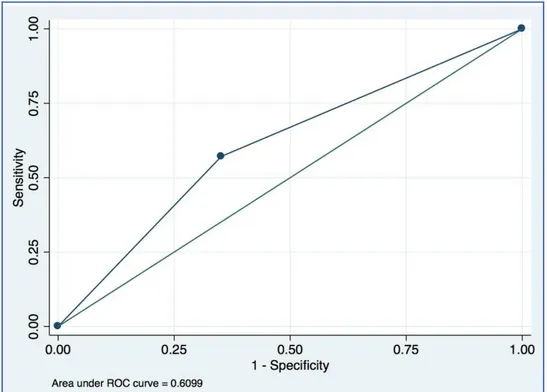
a non-existent scoliotic curve. 30/106 patients (28.3%) did not perceive an actually existing scoliotic curve; there were no statistically significant differences in the distribution of proportions between gender (females=29.6%, n=26/87 vs. males=22.2%, n=4/18, X2=0.4, p=0.775). However, there were differences close to statistical significance regarding the practice of a sport (sports=17.5%; n=7/40 vs. non-practic-ing sport= 34.8%; n=23/66; X2=3.7; p=0.055). The univari-ate analysis does not show an association between the correct perception of the seat of the first scoliotic curve and the prac-tice of a sports activity (OR=1.1; 95% CI=0.4-2.3; z=0.0; p=0.968; figure 2) and the gender (OR=0.6; 95% CI=0.2-1.8;
z=0.9; p=0.372).
Table I. Division of the population between correct and
incorrect perception of the first scoliotic curve.
Variable
Correct
perception perceptionUncorrect Total
n % n % n %
Curve seat 68 64.8 37 35.2 105 100.0
Cobb angle 14 13.3 91 86.7 105 100.0
Convexity 64 61.0 41 39.0 105 100.0
Distribution of the patients whose perception of their scoliosis is known (n = 105), for correct perception of the seat, Cobb angle and convexity of the first scoliotic curve. The data are expressed as number and percentage.
Table II. Subdivision of the population with correct
perception of the first scoliotic curve between athletes and non-athletes.
Variable Sportsmen (n=40) No sportsmen (n=66) X
2 p
n % n %
Curve seat 26 65.0 42 64.6 0.0 0.968
Cobb angle 8 20.0 6 9.2 2.5 0.115
Convexity 27 67.5 37 56.9 1.2 0.281
Proportion of patients with scoliosis with the correct perception of the seat, angle of Cobb and convexity of the first scoliotic curve, for the practice of a sport. The data are expressed as number and percentage. The values of X2 and p are expressed. The statistical significance is set for p <0.05.
Figure 2. ROC curve analysis.
ROC curve of practice of a sport activity as predictor of the correct perception of the seat of the first scoliotic curve.
From the analysis of the variables (gender, age, sport, sports practice time, training hours/week) regarding the correct perception of the seat of the first scoliotic curve, we observed a statistically significant association between the outcome and the age expressed in years (aOR=0.6; z=2.3; p=0.024); no other variable seemed to be associated with the outcome (p> 0.05). The univariate logistic analysis did not show an association between the correct perception of the convexity of the first scoliotic curve and the practice of a sport (OR=1.6; 95% CI=0.7-3.6; z=1.1; p=0.282; figure 3) and the gender (OR=0.6; 95% CI=0.2-1.6; z=1; p=0.299). From the analysis of the variables (gender, age, sport, sports practice time, training hours/week) regarding the correct perception of the convexity of the first scoliotic curve, no variable was associated with the outcome (p> 0.05). The univariate logistic analysis did not show an association between the correct perception of the Cobb angle of the first scoliotic curve and the sport (OR=2.5; 95% CI=0.8-7.7; z=1.5; p=0.123; figure 4) and the gender (OR=2.2; 95%
CI=0.6-8; z=1.2; p=0.232).
From the analysis of the variables (gender, age, sport, sports practice time, training hours/week) regarding the correct perception of the Cobb angle of the first scoliot-ic curve no variable was associated with the outcome (p> 0.05). The ROC curve of practice of a sport activity (Yes/ No) as predictor of the correct perception of the seat of the first scoliotic curve (Yes/No) is described in figure 2.
The univariate logistic analysis did not show an associa-tion between the percepassocia-tion of a curve that did not exist and the practicing of a sport (OR = 0.5; 95% CI=0.1-5.3; z=0.5; p=0.598) and gender (OR=1.7; 95% CI=0.2-17; z=0.4; p=0.666). The univariate logistic analysis did not show an association between the non-perception of an existing curve and the sport activities (OR=0.4; 95% CI=0.2-2.2; z=1.9; p=0.059) and gender (OR=0.7; 95% CI=0.2-2.3; z=0.6; p=0.531). From the analysis of the vari-ables (gender, age, sport, sports practice time, training hours/week) regarding the non-perception of a curve that exists, no variable was associated with the outcome (p> 0.05). The ROC curve of practice of a sport activity (Yes/ No) as predictor of the correct perception of the convexity of the first scoliotic curve (Yes/No) is described in figure 3. Univariate linear analysis did not show any association
with the “difference between the real and perceived Cobb angle of the first curve and practicing of a sport 0.5; 95% CI=-5.5-4.5; t=0.2; p=0.835) and gender (coef.=-1; IC 95%=-7.4-5.4; t=0.3; p=0.762). From the analysis of the variables (gender, age, sport, sports practice time, train-ing hours week) regardtrain-ing the difference between the real and perceived Cobb angle of the first curve no variable was associated with the outcome (p> 0.05). The ROC curve of practice of a sport activity (Yes/No) as predictor of the correct perception of the Cobb angle of the first scoliotic curve (Yes/No) is described in figure 4.
Figure 3. ROC curve analysis.
ROC curve of practice of a sport activity as predictor of the correct perception of the convexity of the first scoliotic curve.
Figure 4. ROC curve analysis.
ROC curve of practice of a sport activity as predictor of the correct perception of the Cobb angle of the first scoliotic curve.
2. Second (if any) scoliotic curve (caudal in relation to the first)
For 48/106 patients (45.3%) the presence of a second curve was found; for 41/48 patients (85.4%) the second curve was in the lumbar region, for 5/48 (10.4%) in the dorsal-lumbar site and for 2/48 (4.2%) in the dorsal site; the mean value of Cobb angle of the second curve was 21.1 ± 7.8 degrees (range: 6-44), convex mainly on the left (n=37/48, 77.1%). For 21/106 patients (19.8%) the perception of the characteristics of a second scoliotic curve was known and 13/21 patients (61.9%) felt it in the lumbar seat and 8/21 (38.1%) in the dorsal-lumbar site; the perceived average Cobb degree was 34.8 ± 11.6 (range: 5-50) and the convexity was felt mainly on the left (n=11/21; 52.4%). There was no statistically significant difference between the real and perceived seat of the second scoli-otic curve (agreement=52.9%; kappa=0.1; z=0.3; p=0.62), real convexity and perceived convexity of the first scoliot-ic curve (agreement=64.7%; kappa=0.3; z=1.3; p 0.102).
Table III describes the proportion of patients with the
correct perception of the seat, Cobb angle and convexity of the second scoliotic curve (n=17/106; 16%); we observed 58.8% of subjects with correct perception of curve seat, 11.8% with correct perception of Cobb angle and 58.8% of curve convexity. No statistically significant differenc-es were seen in the comparison between different gender
about the correct perception of the seat (females=53.3%; n=8/15 vs. males=100%; n=2/2; X2=1.6; p=0.485), about Cobb angle of the convexity (females=13.3%; n=2/15 vs. males=100%; n=2/2; X2=0.3; p= 1) and about the convex-ity (females=66.7%; n=10/15 vs. males=0.0%, n=0/2, X2=3.2, p=0.154). Table IV describes the proportion of
patients with correct perception of their second scoliot-ic curve, among those who played sports; no statistscoliot-ically significant differences were observed in the comparison of any of the variables under analysis between athletes and non-athletes (curve seat, sportsmen = 85.7% vs. no sportsmen = 40.0%; curve angle, sportsmen = 0.0% vs. no
Table III. Division of the population between correct and
incorrect perception of the second scoliotic curve.
Variable perceptionCorrect perceptionWrong Total
n % n % n %
Curve seat 10 58.8 7 41.2 17 100.0
Cobb angle 2 11.8 15 88.2 17 100.0
Convexity 10 58.8 7 41.2 17 100.0
Scoliotic proportions with correct perception of the seat, Cobb angle and convexity of the second scoliotic curve. The data are expressed as number and percentage.
sportsmen = 20.0%; curve convexity, sportsmen = 57.1% vs. no sportsmen = 60.0%; p> 0.05). No statistically signif-icant differences were found between the real and the perceived Cobb angle, in the gender comparison (t=0.0; p=0.967), and in the comparison between sportsmen and non-athletes (t=0.7; p=0.478).
3. Principal component analysis.
The scree plot (Figure 4) of eigenvalues after principal
component analysis of correct perception of the characteris-tics of first and second curve (Table V), shows that the most
significant contribution to the system comes from the first and second component, while the other components give very little additional information.
DISCUSSION
The results of this study show that young patients suffer-ing from scoliosis do not have a correct perception of their spine, in relation to the characteristics of the seat of the curve (dorsal, dorso-lumbar, lumbar), the side of the convexity (right/left) and the value of the Cobb degree. The analysis of the effect of epidemiological variables on their
Table V. Principal component analysis of correct perception of the characteristics of first and second curve.
Variable Comp1 Comp2 Comp3 Comp4 Comp5 Unexplained
First curve seat* - - - - -
-First curve convexity 0.5 0.5 -0.1 0.2 -0.7 0 First curve Cobb angle -0.2 0.6 -0.4 -0.6 0.2 0 Second curve seat -0.5 0.3 -0.2 0.7 0.2 0 Second curve convexity 0.6 0.3 0.2 0.2 0.7 0 Second curve Cobb angle -0.3 0.4 0.9 -0.1 -0.1 0
*dropped because of zero variance
Table IV. Subdivision of the population with correct
perception of the second scoliotic curve between athletes and non-athletes.
Variable Sportsmen No sportsmen X2 p n % n %
Curve seat 6 85.7 4 40.0 3.6 0.134
Cobb angle 0 0.0 2 20.0 1.6 0.485
Convexity 4 57.1 6 60.0 0.0 1.000
Proportion of patients with the correct perception of the seat, angle of Cobb and convexity of the second scoliotic curve, regarding those who plays sports. The data are expressed as number and percentage. The values of X2 and p are expressed. The statistical significance is set for p <0.05.
perception revealed that younger patients are better at iden-tifying the seat of the curve. In addition, the boys who prac-ticed sport had better perception of their scoliotic curve. Proprioceptive information is acquired through kinesthet-ic, visual, articular, muscular sources (31). Moreover, the vestibular and balance functions contribute to structuring the body schema and, starting from this, there develops the sense of posture and limb movement, contributing to the creation of the sensory consciousness of oneself (32). Thus, enhancing motor skills can be an important support to improve the perception of one’s body schema (33). The data we found, that the entire amount of young scoli-otics was wrong to identify the main
characteristics of its scoliotic column, is cohesive with other studies in the liter-ature, in which it was shown that these patients tend to overestimate the severity of their scoliosis, triggering prob-lems regarding psycho-physical behaviours (eg, anorexia, bulimia, depression, etc) (11,34,35). Furthermore, during growth, the young patients have a negative perception of their body, due to their inexperience in controlling senso-rial perceptions and developing motor skills, resulting in a distorted view of their body image. In the analyzed group we found a better knowledge of the trait of the scoliotic curve among the younger patients; we can explain this datum with the theory of sensitive phases of Martin: in puberty there is a peak in the acquisition of motor skills. This acquisition tends to decline, throughout the rest of adolescence (36,37). The perception of one’s scoliosis could follow the trend of the enhancement of motor skills, with the alternation of turgor phase and proceritas phase. The knowledge of this trend can be useful to the clinician, the physiotherapist, sports teacher and sports coach to be more focused during these “critical phases”, to the education of the perception of one’s own body image.
We found that young sportspeople were less wrong in iden-tifying their scoliotic curve. The literature confirms that physical activity plays an important role in physical self-con-cept and body self-esteem (38,44). There is a close
connec-tion between sports practicing and the percepconnec-tion of one’s own body (18,45,46). The researchers assumes that there is a relationship between physical activity, physical self-concept and motor abilities(47,48). Sport enhances the motor skills of strength, endurance, coordination, and flexibility. Each sport involves specific motor skills that require the comple-tion of particular postures and movements(49,51). Further-more, sport allows a person to strengthen those alternative channels, such as touch and hearing, which are the basis for a more precise mental image. As a consequence, a more positive physical self-concept develops. Several studies have proven that, already in pre-adolescence, those who have a good physical fitness show a higher physical self-concept than those who do not (52,53). In the same way, those who regularly practice sport have a higher self-concept (54,55), particularly regarding physical self-concept (57). On the other hand, the results of our study did not show a great-er ability of sportspeople than non-sportspeople in identi-fying the specific traits of their own scoliotic curves, such as seat, side of the convexity and value of the Cobb angle. The sportsperson, like the non-sportsperson, even though he knows he has a scoliotic curve, is wrong in its location and overemphasizes magnitude. At this point, we could assume that sport has not proved to be an advantage. If, on the one hand, sport exerts proprioception, determining a greater knowledge of the body in the young, on the other hand, emphasizing the role of the physical aspect, it can be responsible for a greater critical sense of the dismorphism of one’s own column. In sport, body shape and aesthetic presentation may influence outcomes (56,57). The presence of a scoliosis is believed a discomfort and a physical disad-vantage for the achievement of good sports performance. In conclusion, the presence of scoliosis can lead to body dissat-isfaction for young people who practice sports (56-58). Having said that, our study has some weaknesses. First-ly, the patients recruited for the study practiced different disciplines, with different training programmes and with a variable physic effort. Secondly, the self-report of spine silhouette chart is problematic because measurement errors may exist. However, for the variables evaluated in this study, there were no other validated assessment methods. Thirdly, we did not use any scales to study the column’s aesthetic deformity, such as the Walter Reed Assessment Scale (WRVAS), the Trunk Appearance Perception Scale (TAPS), the SRS-22 Patient Questionnaire Self-Image scale or the Body Image Disturbance Questionnaire -Scolio-sis version (BIQD-S). On the other hand, we chose to use the thorn silhouette chart, proposed by Picelli et al. (22), because we shared the perplexity of these authors regard-ing the other scales i.e. they do not allow one to focus on the perception of deformity of the spine and the advantage of
a greater attention to the asymmetric body caused by scoli-osis. Subsequent studies, with the administration of these scales, could integrate the perception of the deformity of the column and the asymmetry of the body. However, we cannot provide error, precision, validity of the spine silhou-ette chart because Picelli et al., in their paper (22), did not take them into consideration. But, in agreement with Trunk Appearance Perception Scale (TAPS) the Authors postu-lated a good reliability (ICC value=0.92)(59).Fourthly, the analyzed population, depending on the severity of their scoliosis, was treated with different therapeutic approach-es: wait and watch, rehabilitation, and orthotic treatment. Subsequent studies could select a population that receives the same treatment method. Moreover, it is important to highlight that other variables not measured in this study may interfere with body perception. In addition, we must remember that reliability of the measures has its limita-tions. Test-retest is missing. During the same visit it would have generated confusion in the patient request to perform the same procedure twice a few minutes later, while after some time it was not easy, being some patients coming from distant towns. Despite these limitations, this study is the first to analyze the influence that sport may have in a young sportspeople on the perception of the dysmorphism of their own column. Two implications emerge from this study: first, the need to promote educational programs for one’s own body structure among young scoliotic patients; second, the importance that the knowledge of one’s own deformity could have in order to activate corrective postur-al strategies.
In conclusion, sport can be a valuable instrument to improve the knowledge that a young people have of their own dysmorphism and how to manage it. In fact, physi-cal activity, exercising proprioception, the control of one’s body and the surrounding space, can provide the vehicle to identify the problem, overcome physical and psychological discomfort and correct the body dysmorphic disorder. On the other hand, in our experience, we have found that young scoliotic patients have difficulty to describe and to recognize the main trait of their column deformity. We believe that until now, not enough attention has been paid to empower the young sportsperson with scoliosis in dealing with their pathology and to perform specific jobs in the sports. We suggest a greater effort by health professionals and coaches to inform and explain the main traits of dysmorphism and to consciously propose strategies for postural correction of related paramorphism.
Conflict of Interest
Author Contributions
According to literature the Author Contributions are as follows: AN and GS drafted the article; GF and IC gave substantial contributions to interpretation of data for the study; AR, FPB and AP gave substantial contributions to the acquisition, analysis, and interpretation of data for the study; ST revised the article critically for important intellec-tual content; BM finally approved the version of the article to be submitted (61).
Reference: Padulo J, De Giorgio A, Oliva F, Frizziero A, Maffulli N. I performed experiments and I have results. Wow, and now? Muscles Ligaments Tendons J. 2018 Jan 10;7(3):403-410.
REFERENCES
1. Nachemson AL, Lonstein JE, Weinstein SL. Report of the prevalence and natural history committee of the Scoliosis Research Society. Denver: Scoliosis Research Society 1982. 2. Negrini S, Minozzi S, Bettany-Saltikov J, et al. Braces for
idiopathic scoliosis in adolescents. Spine (Phila Pa 1976). 2010;35:1285–1293.
3. Carrasco MI, Ruiz MC. Perceived self-image in adolescent idiopathic scoliosis: an integrative review of the literature. Rev Esc Enferm USP. 2014;48(4):748-58.
4. Slade PD. What is body image? Behav Res Ther. 1994;32(5): 497-502.
5. Pinheiro AP, Giugliani ER. Body dissatisfaction in Brazilian schoolchildren: prevalence and associated factors. Rev. Saúde Públ. 2006;40(3): 489-96.
6. Muth JL, Cash TF. Body-image attitudes: what difference does gender make? J Appl Soc Psychol. 1997;27(16):1438-52. 7. Tones M, Moss N, Polly DW Jr: A review of quality of life and
psychosocial issues in scoliosis. Spine 2006; 31(26):3027–3038. 8. D’Andrea LP, Betz RR, Lenke LG, et al. Do radiographic parameters correlate with clinical outcomes in adolescent idio-pathic scoliosis. Spine (Phila Pa 1976) 2000;25:1795–802. 9. Wang L, Wang YP, Yu B, Zhang JG, Shen JX, Qiu GX, Li
Y. Relation between self-image score of SRS-22 with deformi-ty measures in female adolescent idiopathic scoliosis patients. Orthop Traumatol Surg Res. 2014;100(7):797-801.
10. Payne 3rd WK, Ogilvie JW, Resnick MD, Kane RL, Trans-feldt EE, Blum RW. Does scoliosis have a psychological impact and does gender make a difference. Spine (Phila Pa 1976) 1997;22:1380–4.
11. Schwieger T, Campo S, Weinstein SL, Dolan LA, Ashida S, Steuber KR. Body Image and Quality-of-Life in Untreated Versus Brace-Treated Females With Adolescent Idiopathic Scoliosis. Spine (Phila Pa 1976) 2016;41(4):311-9.
12. Weiss HR, Reichel D, Schanz J, Zimmermann-Gudd S. Defor-mity related stress in adolescents with AIS. Stud Health Tech-nol Inform. 2006;123: 347–51.
13. De Gracia M, Marcó M, Trujano P. Factors associated with eating behavior in pre-adolescents. Psicothema 2007;19: 646–653.
14. Jensen CD, Steele RG. Brief report: body dissatisfaction, weight criticism, and self-reported physical activity in preado-lescent children. J. Pediatr. Psychol. 2009;34:822–826. 15. Schore AN. Affect Regulation and the Origin of the Self: The
Neurobiology of Emotional Development. London: Routledge 2015.
16. Blakely-McClure SJ, Ostrov JM. Relational aggression, victim-ization and self-concept: testing pathways from middle child-hood to adolescence. J. Youth Adolesc. 2016;45:376–390. 17. Mancilla A, Vázquez R, Mancilla JM, Amaya A, Álvarez G.
Body dissatisfaction in children and preadolescents: a system-atic review. Rev. Mex. Trastor. Aliment. 2012;3:62–79. 18. Contreras OR, Fernández-Bustos JG, García LM, Palou
P, Ponseti J. Relationship in adolescents between physical self-concept and participating in sport. Rev. Psicol. Deporte 2010;19:23–39.
19. Burwell RG, Freeman BJ, Dangerfield PH, Aujla RK, Cole AA, Kirby AS, et al. Etiologic theories of idiopathic scolio-sis: neurodevelopmental concept of maturational delay of the CNS body schema (“body-in-the-brain”). Stud Health Tech-nol Inform. 2006;123:72-9.
20. Negrini S, Aulisa AG, Aulisa L, et al. 2011 SOSORT guide-lines: Orthopaedic and Rehabilitation treatment of idiopathic scoliosis during growth. Scoliosis 2012;7(1):3.
21. Picelli A, Negrini S, Zenorini A, Iosa M, Paolucci S, Smania N. Do adolescents with idiopathic scoliosis have body schema disorders? A cross-sectional study. J Back Musculoskelet Reha-bil. 2016;29(1):89-96.
22. Negrini S, Aulisa AG, Aulisa L, et al. 2011 SOSORT guide-lines: Orthopaedic and Rehabilitation treatment of idiopathic scoliosis during growth. Scoliosis Spinal Disorders 2012;7. 23. Padulo J, Oliva F, Frizziero A, Maffulli N. Basic principles and
recommendations in clinical and field science research: 2018 update. MLTJ 2’18; 8 (3): 305-307 .
24. Scoliosis Research Society. Patient & family [Internet] [Cited 2018 August 13]. Available from: http://www .srs.org/patient_ and_family /scoliosis/idiopathic /adolescents / observation.htm 25. Schiller JR, Thakur NA, Eberson CP. Brace management in adolescent idiopathic scoliosis. Clin Orthop Relat Res. 2010 Mar;468(3):670-8.
26. Negrini S, Aulisa L, Ferraro C, Fraschini P, Masiero S, Simonazzi P, et al. Italian guidelines on rehabilitation treat-ment of adolescents with scoliosis or other spinal deformities. Eura Medicophys. 2005;41(2):183-201.
27. Langensiepen S, Semler O, Sobottke R, Fricke O, Franklin J, Schönau E, et al. Measuring procedures to determine the Cobb angle in idiopathic scoliosis: A systematic review. Eur Spine J. 2013;22(11):2360-71.
28. Cobb JR. Outline for the study of scoliosis. Am Acad Orthop Surg Instr Course Lect. 1948;5:261–275.
29. Potter BK, Rosner MK, Lehman RA Jr, Polly DW Jr, Schroed-er TM, Kuklo TR. Reliability of end, neutral, and stable vSchroed-erte- verte-brae identification in adolescent idiopathic scoliosis. Spine (Phila Pa 1976). 2005 15;30(14):1658-63.
30. Langensiepen S, Semler O, Sobottke R, Fricke O, Franklin J, Schönau E, Eysel P.Measuring procedures to determine the Cobb angle in idiopathic scoliosis: a systematic review. Eur Spine J. Nov;22(11):2360-71.
31. Arber S. Motor circuits in action: specification, connectivity, and function. Neuron. 2012;74(6):975-89.
32. Allum JH, Adkin AL, Carpenter MG, Held-Ziolkowska M, Honegger F, Pierchala K. Trunk sway measures of postur-al stability during clinicpostur-al bpostur-alance tests: effects of a unilaterpostur-al vestibular deficit. Gait Posture 2001;14(3):227-37.
33. Galgon AK, Shewokis PA, Tucker CA. Changes in standing postural control during acquisition of a sequential reaching task. Gait Posture 2010;31(2):265-71.
34. Schwieger T, Campo S, Weinstein SL, Dolan LA, Ashida S, Steuber KR. Body Image and Quality of Life and Brace Wear Adherence in Females With Adolescent Idiopathic Scoliosis. J Pediatr Orthop. 2017;37(8):e519-e523.
35. Olafsson Y, Saraste H, Ahlgren RM. Does bracing affect self-image? A prospective study on 54 patients with adolescent idiopathic scoliosis. Eur Spine J. 1999;8(5):402-5.
36. Singh MM, Ashok L, Binu VS, Parsekar SS, Bhumika TV. Adolescents and Body Image: A Cross Sectional Study. Indian J Pediatr. 2015;82(12):1107-11.
37. van der Fels IM, Te Wierike SC, Hartman E, Elferink-Gemser MT, Smith J, Visscher C. The relationship between motor skills and cognitive skills in 4-16 year old typically developing chil-dren: A systematic review. J Sci Med Sport. 2015;18(6):697-703. 38. Campbell A, Hausenblas HA. Effects of exercise interventions on body image: a meta-analysis. J Health Psychol. 2011;14:780–93. 39. Kruger J, Lee CD, Ainsworth BE, Macera CA. Body size
satis-faction and physical activity levels among men and women. Obesity. 2008;16(8):1976–9.
40. Coelho CG, Giatti L, Molina MD, Nunes MA, Barreto SM. Body image andnutritional status are associated with physical activity in men and women the ELSA-Brazil study. Int J Envi-ron Res Public Health. 2015;12(6):6179–96.
41. Schmalz DL, Deane GD, Birch LL, Davison KK. A longi-tudinal assessment of the links between physical activity and self-esteem in early adolescent non- Hispanic females. J Adolesc Health. 2007;41(6):559–65.
42. Korn L, Gonen E, Shaked Y, Golan M. Health perceptions, self and body image, physical activity and nutrition among under-graduate students in Israel. PLoS One. 2013;8(3):e58543. 43. Fox KR. Physical education and the development of children’s
self esteem. In: Amstrong N, eds. New directions in physical education.2. Towards a National Curriculum. Champaign, IL: Human Kinetics, 1992;33–54.
44. Gruber J. Physical activity and self esteem development in children: a metaanalysis. In: Stull G, Eckern H, eds. Effects of physical activity on children. Champaign, IL: Human Kinetics, 1986;330–48.
45. Homan KJ, Tylka TL. Appearance-based exercise motivation moderates the relationship between exercise frequency and positive body image. Body Image 2014;11:101–108.
46. Rodríguez DF, Alvis KM. Overview of the body image and its implications in sport. Rev. Fac. Med. 2015;63:279–287. 47. Wienke B, Jekauc D. A Qualitative Analysis of Emotional
Facilitators in Exercise. Frontiers in Psychology 2016 . 48. Jekauc D. Enjoyment during Exercise Mediates the Effects
of an Intervention on Exercise Adherence. Psychology 2015;6:48-54.
49. Hrysomallis C. Balance ability and athletic performance. Sport Med. 2011;41(3):221–232.
50. Maurer C, Mergner T, Peterka RJ. Multisensory control of human upright stance. Exp Brain Res. 2006;171(2):231–250. 51. Paillard T. Plasticity of the postural function to sport and/or
motor experience. Neurosci Biobehav Rev. 2017;72:129–152. 52. Mitchell NG, Moore JB, Bibeau WS, Rudasill KM. Cardio-vascular fitness moderates the relations between estimates of obesity and physical self-perceptions in rural elementary school students. J. Phys. Act. Health 2012;9:288–294. 53. García A, Burgueño R, López D, Ortega F. Physical fitness,
adiposity and self-concept in adolescents. A pilot study. Rev. Saúde Públ. 2013;22:453–461.
54. Pastor Y, Balaguer IY, García-Merita ML. Relaciones entre el autoconcepto y el estilo de vida saludable en la adolescencia media: un modelo exploratorio. Psicothema 2006; 18: 18–24. 55. Reigal R, Videra A, Parra JL, Juárez R. Physical sports activity,
physical self-concept and psychological wellbeing in adoles-cence. RETOS. Nuevas Tendencias Educ. Fís. Deporte y Recreación 2012;22: 19–23.
56. Esnaola I, Revuelta L. Relations between the physical activ-ity, physical self-concept, expectations, perceived value and perceived difficulty. Acción Psicol. 2009;6:31–43.
57. Werner A, Thiel A, Schneider S, Mayer J, Giel KE, Zipfel S. Weight-control behaviour and weight-concerns in young elite athletes - a systematic review. J Eat Disord. 2013;1:18 58. Fernández JG, Contreras OR, García LM, González S.
Phys-ical Self-concept depending on the kind of physPhys-ical activ-ity practised and motivation to it. Rev. Latinoam. Psicol. 2010;42:251–263.
59. Bago J, Sanchez-Raya J, Perez-Grueso FJ, Climent JM.The Trunk Appearance Perception Scale (TAPS): a new tool to evaluate subjective impression of trunk deformity in patients with idiopathic scoliosis. Scoliosis. 2010 Mar 25;5:6.
60. Padulo J, De Giorgio A, Oliva F, Frizziero A, Maffulli N. I performed experiments and I have results. Wow, and now? Muscles Ligaments Tendons J. 2018 Jan 10;7(3):403-410.