Acta orthop. scand. 54, 574-579, 1983
METAL DETERMINATION IN ORGANIC FLUIDS OF PATIENTS
WITH STAINLESS STEEL HIP ARTHROPLASTY
UGO E. PAZZAGLIA*, CLAUDIO MINOIA**, LUCIANO CECILIANI* & CARLO RICCARDI*
*Orthopaedic Clinic and **Foundation of Occupational Health of Pavia, Research Centre of Pathophysiology and Occupational Security, University of Pavia, Italy
In 20 stainless steel Charnley hip arthroplasties (with a follow-up of 10-13 years) nickel, chromium and manganese levels were measured in blood, plasma and urine by atomic absorption spectrophotometry. Skin patch tests for these metals, and clinical and roentgenographic results of arthroplasty were also assessed.
Metal levels in organic fluids were plotted against a control population homogene-
ous for age, residence and anamnestic conditions with the first, but which had never undergone a prosthesis or other metallic implant surgical procedure.
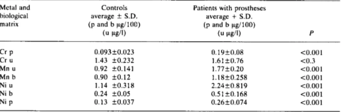
Nickel levels in blood, plasma and urine, manganese levels in blood and urine and chromium levels in plasma were significantly higher in the hip prostheses popula- tion.
Metal ion release from stainless steel prostheses is discussed with regard to implant failure, metal sensitivity and carcinogenesis.
Key words: metal ions; metal sensitivity Accepted 1S.i.83
An increased concentration of alloy metals in the blood and urine of patients with total hip pros- theses with both metallic stem and cup has been observed. Metals are released by wear from the surface of the prostheses in the form of small particles and by corrosion in the form of soluble ions (Coleman et al. 1973, Dobbs & Minsky 1980, Jones et al. 1975).
There are no data in the literature on blood and urine concentrations of metal in patients with metal-plastic prostheses, but in vitro experiments show a very small release of metals from these implants (Rae 1979).
The study of release of metal ions in the body by implants is supported both by observations of carcinogenic properties of metals in experimental animals (Daniel et al. 1963, Heath et a]. 1971, Oppenheimer et al. 1956), though there have
This paper was supported by grant no. CT 810012604 of Italian National Research Council.
been no reports of cases of tumor induction by implants in man, and by metal sensitivity as a cause of bone necrosis and loosening of the prosthesis (Benson et al. 1975, Brown et al. 1977, Deutman et al. 1977, Elves et al. 1975, Evans et al. 1974, Jones et al. 1975, Rooker & Wilkinson 1980).
Sensitivity has been observed chiefly in all-metal prostheses; it apparently does not occur in metal-plastic implants (Deutman et al. 1977, Rooker & Wilkinson 1980). On the other hand in animal studies it has been observed that the in- troduction of metal implants (which are also more resistant to corrosion) entails an ionization of the surface of the implant and a release of the alloy metals, even if in very small quantities (Clarke & Hickmann 1953, Ferguson et al. 1962, Ferguson et al. 1960, Laing et al. 1959, Perkins 1973).
On the basis of these observations and consid- ering the possibility that metal accumulates in the body following long presence of the implant,
Acta Orthop Downloaded from informahealthcare.com by 93.62.227.228 on 01/26/12
575
similar to what is expected for articular pros- theses, we decided to study the sensitivity to alloy metals and their blood, plasma and urine con- centrations in patients with a Charnley metal- plastic prosthesis functioning for more than 10 years, together with the clinical and roentgeno- graphic results of the arthroplasty.
MATERIALS AND METHODS
The studied population consists of 20 patients who un-
derwent a total hip arthroplasty 30-13 years ago at the Orthopaedic Clinic of the University of Pavia. In all the cases the same Charnley hip prosthesis was applied with an EN58J stainless steel stem provided by Thackray Ltd (model MKlS) and RCH 1000 polyethylene cup.
The population includes males and females, from 60
to 82 years of age, with an average age of 68.8 years. All the patients come from Pavia and its surrounding area (which has a predominantly agricultural economy) and for the last 10 years they have remained in this area.
Because they may have a great influence on metal concentration in the organic fluids, the patients’ jobs and the location of industries near the place of resi- dence have been carefully investigated in the anam- nesis.
For the same reason alcohol and tobacco consump- tion and drugs taken in the last 2 years were recorded.
The following pathologies were specifically re- searched:
hemophilia liver diseases thalassemia kidney diseases Wilson’s disease heart disease diabetes
The patients underwent general and local physical examination with a hip roentgenogram (both frontal and lateral views). Blood was drawn using a polyethylene syringe and a 5-ml moplen test tube with sodium heparin.
For every patient a whole blood test tube was refrig- erated at + 5 T , while another was centrifuged at about 2500 r.p.m. at room temperature and the plasma pipetted and stored at -20°C till 6 h before beginning the analysis.
Urine samples were taken using plastic containers decontaminated with a water solution of 10 per cent nitric acid and repeatedly washed with bidistillated wa- ter.
Analytical determinations were performed with two atomic absorption spectrophotometers (Perkin Elmer model 5000 and 603 equipped respectively with HGA
500 and HGA 76(b)).
Chromium determination in plasma and urine was performed following the method of Guthrie et al. (1978), partially modified for the utilized equipment.
Manganese determination in blood and urine was performed utilizing both the method of Ross & Gon-
zales (1974) and the extractive procedure with NaDDC/MiBK (Minoia & Cavalleri 1982).
Nickel determination in plasma, blood and urine was performed after mineralization of the specimen and extraction of the Ni-DMG complex in methylisobutyl- ketone (Zachariassen et al. 1975). The accuracy of the methods employed, the limits of relative revealability and the data of accuracy are reported in Table 1.
The research of skin sensibility to the alloy metals was performed utilizing as aptene a 2 per cent solution
of FeCl,, a 0.5 per cent solution of K,Cr,O,, a 2 per cent solution of MnCl,, a 3 per cent solution of NiSO, .7H,O. The skin test was read after 48 h by a trained dermatologist.
The control population consists of 20 individuals, male and female, with no record of prostheses or other
Table 1. Accuracy, precision and detection limits of the methods employed
Element Biological Detection Precision* Recovery
matrix limit C. V** per cent per cent
(CLg/l) (in the series)
Cr Cr Mn Mn Ni Ni Ni urine plasma urine blood urine plasma blood 0.02 0.03 0.02 0.02 0.02 0.02 0.02 9.7 7.8 8.7 8.2 8.9 8.1 8.2 89.9 95.1 94.8 94.7 87.4 92.7 90.7
**
Coefficient of variation.the data refer to physiological levels of the metal.
Acta Orthop Downloaded from informahealthcare.com by 93.62.227.228 on 01/26/12
576 U . E. PAZZAGLIA ET AL.
metallic implants; this population is similar to the first in age, residence and anamnestic conditions.
RESULTS
The clinical results of the population with pros- theses after 10-13 years have been satisfactory, as assessed by interview and physical examination of the patients.
Roentgenograms were considered according to the following parameters:
- radiolucent line between bone and cement - incomplete envelope of cement around the
stem
- loosening between cement and stem - fracture of the cement
- air bubbles inside cement.
The results are reported in Table 2 .
Chromium, nickel and manganese average levels in blood, plasma and urine of prostheses and control population are reported in Table 3.
Skin sensitivity tests to chromium, nickel, man- ganese and iron were negative in 16 patients; in four it was not possible to read the skin test be- cause they failed to return 2 days later.
DISCUSSION
According to the results it is possible to assume that the significantly higher levels of chromium, nickel and manganese in blood and urine of the patients with hip arthroplasty arise from metal ions released from the femoral stem.
The ion release from a metal implant into a saline solution takes place exclusively from this
Table 2 . Roentgenographic valuation of hip arthroplusties (10-13-year follow-up) ~ ~~
1 2 3 4 5 6 7 8 9 10 11 12 13 14 15 16 17 18 19 20
x x x x x x x x x -
Radiolucent line between bone and cement
x
Incomplete cement envelope x x x X x x x x x -
Loosening cement-metal x x x x x -
Cement fracture x x X X X
Air bubbles inside the cement
- - X
- Radiolucent cement was used.
Table 3. Chromium, nickel and manganese levels in biological fluids* of patients with prostheses and controls Metal and
biological matrix
Controls Patients with prostheses average f S.D. (p and b pg/lOO) average
+
S.D. (p and b pg/lOO) ( u pg/l) ( u p g 4 P Cr P Cr u Mn u Mn b Ni u Ni b Ni p 0.093f0.023 1.43 f0.232 0.92 f0.141 0.90 50.12 1.14 f0.318 0.24 f 0 . 0 5 0.13 50.037 0.19k0.08 <0.001 1.6 1 f 0.76 4 . 3 1.77f0.20 <0.001 1.18f0.258 <0.001 2.24f0.819 <0.001 0.51t0.168 <0.001 0.26k0.074 <0.001*
p, plasma; b, blood; u, urine.Acta Orthop Downloaded from informahealthcare.com by 93.62.227.228 on 01/26/12
surface and in hip arthroplasties fixed with acrylic cement only the head and the neck of the femoral prosthesis are exposed to the body interstitial fluids, while the shaft (about two-thirds of the whole metal prosthesis) is separated from them by the cement envelope.
However, in nine of the 20 cases this condition was not satisfied from the moment of prosthesis application, because there was an incomplete en- velope of cement around the stem.
In these cases the increased extension of the exposed surface is not as relevant as the presence of a border-limit between the metal surface ex- posed to body fluids and that covered by the ce- ment; indeed micromovements between the ce- ment and the metal surface may damage the thin oxide film of the metal on these borders and prime a corrosion process similar to that observed between metal plates and screws, known as “cre- vice corrosion”.
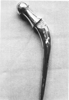
We have observed corrosion pits on the stem of a prosthesis (not included in this series) removed after 5 years for aseptic loosening (Figure 1).
On the other hand the relationships between bone, cement and stem undergo, with time, other modifications, which increase the exposed metal- lic surface and can also prime the corrosion pro- cess. Bone resorption around the cement (observed in 10 cases) involves a deeper settle- ment of the acrylic cement envelope inside a larger bone bed; it may be observed in roentgenograms as sinking of the whole pros- thesis and cement envelope inside the femoral diaphysis or as a cement fracture and loosening between the cement and the stem. These observations indicate that there are micromove- ments and therefore favorable conditions to prime the corrosion processes.
In this series, on the whole, 12 prostheses show a stem exposure for one or both the considered factors (Table 2).
The metal ions released from the implant sur- face have been documented in the periprosthetic tissues in experimental animals both by spec- trochemical methods (Ferguson et al. 1960, 1962, Laing et al. 1959), and by X-ray spectro- graphy (Emneus et al. 1960, Emneus & Stenram 1960). From this tissue they spread through the extra-cellular fluids (Maroudas 1973, Perkins
577
Figure I. Corrosion pits on the stem of a prosthesis re- moved after 5 years.
1973) and can reach the various target organs and tissues.
Therefore in man and in still working pros- theses the only reasonable measurement of metal ions may be performed in the biological fluids: blood andlor plasma and urine.
Chromium determination was performed only on plasma on account of the different metabolism of this metal ion in the human body according to its valence, indeed, since the hexavalent chromium is selectively transferred inside the erythrocytes, where it is reduced to the trivalent form and released in the plasma, the latter seems to be the more stable form of the ion in the body. Therefore whatever the valence of the chromium ions released from the prosthesis surface, the quantity in plasma appears to be the most sig- nificant index of the metal in the human body. Nickel ions were determined quantitatively both in the plasma and the whole blood with a good concordance of the results.
Acta Orthop Downloaded from informahealthcare.com by 93.62.227.228 on 01/26/12
578 U. E. PAZZAGLIA ET AL Although the increased levels of chromium,
nickel and manganese in the prostheses popula- tion are highly significant (P
<
0.001, except chromium in urine), the absolute quantity of metal is very low and well below the threshold of metal toxicity. This indicates that stainless steel prostheses articulating with polyethylene sockets also release metal ions through corrosion, but over a period of 10 years the quantity of metal released in the body is very low. It also corrobo- rates the physical findings, which do not show any particular symptomatology of metal poisoning.A second concern of increased metal in the body is the risk of tumor induction. In experi- mental animals the carcinogenic properties of chromium, nickel and manganese ions have been proved and also in workers professionally ex- posed to these elements a carcinogenetic risk has been demonstrated (Kazantzis 1981, Norseth 1981, Sunderman 1981).
Although results from animal experiments cannot be applied to human tissues and the pro- fessional exposure entails completely different quantities and assumptions, not comparable to the considered case, this risk must still be kept in mind, especially over very long periods of expo- sure.
Experience with articular prostheses now cov- ers a period of 20 years and up to now cases of tumor induction or higher tumor incidence have not been reported in these patients. Nevertheless, because there is a tendency to extend the indica- tion for prostheses to younger people, this possi- bility deserves careful consideration.
Finally, a third aspect which deserves discus- sion is sensitivity to metals.
Cases of aseptic loosening have been reported and have been ascribed to bone necrosis around the implant caused by an allergic reaction to the metals (Benson et al. 1975, Evans et a]. 1974, Uchida et al. 1980). These reports generally con- cern cobalt-chromium all metal prostheses, al- though skin sensitivity to metal has also been observed in some metal-plastic prostheses (Deutman 1977). Moreover many papers raise doubts about a direct relation between skin sen- sitivity tests and allergic reaction in periprosthetic tissues with consequent bone necrosis and loosening (Brown et al. 1977, Elves et al. 1975,
Langlais et al. 1980, Rooker & Wilkinson 1980). In metal-plastic prostheses in particular these phenomena seem to have no relevance.
In the studied prostheses population no cases of skin sensitivity were observed; it may be that chromium, nickel and manganese have less capacity to stimulate immunity than cobalt ions, or that the quantity of metal released by stainless steel alloy metal-plastic prostheses is too low to stimulate immunity.
CONCLUSIONS
Patients with a 10-13-year-old stainless steel hip pros- thesis articulating with polyethylene sockets have higher levels of chromium, nickel and manganese in
blood and urine than in a control group.
The increased level of metal ions is nevertheless well
below the threshold of metal toxicity for each of the various metals.
The release of metal ions by stainless steel met- al-plastic prostheses does not seem to cause sensitiza- tion even after a prolonged period of time.
The monitoring of metals in biological fluids may be a useful non-invasive method to investigate the corrosion of the metal component of the prosthesis in the body.
REFERENCES
Benson, M. K. D., Goodwin, P. G . & Brostoff, J.
(1975) Metal sensitivity in patients with joint re- placement arthroplasties. Er. Med. J . 4, 374-375. Brown, G. C., Locksin, M. D., Salvati, E. A. & Bul-
lough, P. G. (1977) Sensitivity to metal as a possible cause of sterile loosening after cobalt-chromium total hip replacement arthroplasty. J . Bone J o i n r Surg. Clarke, E. G . C. & Hickmann, J. (1953) An investiga- tion into the correlation between the electrical po- tentials of metals and their behaviour in biological fluids. J . Bone J o i n t Surg. 35-B, 467-473.
Coleman, R. F., Herrington, J. & Scales, J . T. (1973) Concentration of wear products in hair, blood and urine after total hip arthroplasty. Er. Med. J . 1, Daniel, M., Dingle, J. T., Webb, M. & Heath, J. C. (1963) The biological action of cobalt and other metals. I. The effect of cobalt on the morphology and metabolism of rat fibroblasts in vitro. Er. J . Exp. Path. 44, 163-1 76.
Deutman, R., Mulder, T. J., Brian, R. & Nater, J. P. (1977) Metal sensitivity before and after total hip arthroplasty. J . Bone J o i n t Surg. 59-A, 862-865. 59-A, 164-169.
5 27-5 29.
Acta Orthop Downloaded from informahealthcare.com by 93.62.227.228 on 01/26/12
579 Dobbs, H. S. & Minsky, M. J. (1980) Metal ions release
after total hip replacement. Biomaterials 1, 193-198. Elves, M. R., Wilson, J. N., Scales, J. T. & Kemp, H. B.
S. (1975) Incidence of metal sensitivity in patients with total joint replacement. Br. Med. J. 4,376-378. Emneus, H. & Stenram, V. (1960) Reaction of tissues
to alloys used in osteosynthesis. Acta Orthop. Scand. Emneus, H., Stenram, V. & Baecklund, J. (1960) A n
X-ray spectrographic investigation of the soft tissue around titanium and cobalt alloy implants. Acta Orthop. Scand. 30, 226-236.
Evans, E. M., Freeman, M. A. R., Miller, A. J. & Ver- non-Roberts, B. (1974) Metal sensitivity as a cause of bone necrosis and loosening of the prosthesis in total joint replacement. 1. Bone Joint Surg. 56-B, 626-642.
Ferguson, A , B., Jr., Akahoshi, Y., Laing, P. G. &
Hodge E. S. (1962) Characteristics of trace ions re- leased from embedded metal implants in the rabbit.
J. Bone Joint Surg. 44-A, 323-336.
Ferguson, A. B., Jr., Laing, P. G. & Hodge, E. S. (1960) The ionization of metal implants in living tissues. J. Bone Joint Surg. 42-A, 77-90.
Guthrie, B. E., Wolf, W. R. & Veillon, C. (1978) Background correction and related problems in the determination of chromium in urine by graphite fur- nace atomic absorption spectrometry. Anal. Chem. 50, 1900-1905.
Heath, J. C., Freeman, M. A. R. & Swanson, S. A . V. (1971) Carcinogenic properties of wear particles from prostheses made in Co-Cr alloy. Lancet 1, Kazantzis, G. (1981) Role of cobalt, iron, lead, man-
ganese, mercury, platinum, selenium and titanium in carcinogenesis. Environ. Health Perspecr. 40, Jones, D. A,, Lucas, H. K., O'Driscoll, M., Price, C. &
Wibberleg, B. (1975) Cobalt toxicity after McKee total hip arthroplasty. J . Bone Joint Surg. 57-B,
29,315-330.
564-566.
143-161.
2 8 9-296.
Laing, P. G., Ferguson, A. B. Jr. & Hodge, E. S. (1959) Spectrochemical determination of trace metals in normal striated muscle in the rabbit. J . Bone Joint Surg. 41-A, 137-744.
Langlais, F., Postel, M., Berry, J. P., Le Carpentier, Y.
& Weill, B. J. (1980) L'intolerance aux debris d'us- ure des protheses. Bilan immunologique et anatomopathologique de 30 cas. Int. Orthop. 4, Maroudas, A. (1973) Distribution of metallic ions be- tween tissue and extracellular fluids. J . Bone Joinf Surg. 56-8, 424.
Minoia, C. & Cavalleri, A. (1983) Determinazione dei metalli in tracce nel laboratorio clinico e tossicologico. La Goliardica Pavese, Pavia.
Norseth, T. (1981) The carcinogenicity of chromium. Environ. Health ferspect. 40, 121-130.
Oppenheimer, B. S., Oppenheimer, F. T., Danishefsky, I. & Stout, A. P. (1956) Carcinogenic effect of metals in rodents. Cancer Res. 16, 439-441.
Perkins, D. T. (1973) Transport, deposition and excre- tion of trivalent metal ions in the body. J . Bone Joint Surg. 55-B, 423-424.
Rae, T. (1979) Comparative laboratory studies on the production of soluble and particulate metal by total prostheses. Arch. Orthop. Traum. Surg. 95, 71-79. Rooker, G. D. & Wilkinson, J. D. (1980) Metal sen-
sitivity in patients undergoing hip replacement. J . Bone Joint Surg. 62-B, 502-505.
Roos, R. T. & Gonzales, J . G . (1974) Direct determi- nation of trace quantities of manganese in blood and serum samples using selective volatization and graphite tube reservoir. Bull. Environ. Contam. Toxicol. 12, 470.
Sunderman, F. W. Jr. (1981) Recent research on nickel carcinogenesis. Environ. Hlth Perspecr. 40, 13 1-1 41. Uchida, S., Yoshino, S., Doi, M. & Kudo, H. (1980) Side-effects of prosthetic materials on the human body. Int. Orthop. 3, 285-291.
Zachariassen, H. (1975) Technique for determining nickel in blood by flameless atomic absorption spec- trophotometry. Clin. Chem. 21, 562-567.
145-1 53.
Correspondence to: Dr. Ugo E. Pazzaglia, Clinica Ortopedica dell'Universita, Via Taramelli, 1-27 100 Pavia, Italy.
Acta Orthop Downloaded from informahealthcare.com by 93.62.227.228 on 01/26/12