For Review Only
A REVIEW ON SKELETAL ANOMALIES IN REARED EUROPEAN
FISH LARVAE AND JUVENILES. PART 1: NORMAL AND
ANOMALOUS SKELETOGENIC PROCESSES
Journal: Reviews in Aquaculture Manuscript ID: RAQ-06-12-0023.R1 Manuscript Type: Special Issue Date Submitted by the Author: n/a
Complete List of Authors: Boglione, Clara; University of Rome 'Tor Vergata', Biology Gavaia, Paulo; University of Algarve, CCMAR
Koumoundouros, Giorgos; University of Crete, Biology Gisbert, Enric; IRTA,
Moren, Mari; NIFES,
Fontagné, Stephanie; INRA, Unité de Nutrition, Métabolisme et Aquaculture NuMéA Pôle d'Hydrobiologie
Witten, P; University of Ghent, Biology
Keywords: bone tissue, cartilage, juveniles, larvae, skeletal anomalies, ossification
For Review Only
A REVIEW ON SKELETAL ANOMALIES IN REARED EUROPEAN FISH LARVAE AND JUVENILES. PART 1: NORMAL AND ANOMALOUS SKELETOGENIC PROCESSES
C. Boglione1, P. Gavaia2, G. Koumoundouros3, E. Gisbert4, M. Moren5, S. Fontagné6, P.
E. Witten7
1
Laboratory of Experimental Ecology and Aquaculture – Department of Biology – University of Rome Tor Vergata - Via della Ricerca Scientifica s.n.c., 00133 Rome (Italy) – [email protected] - fax +39 0672595965
2
University of Algarve –CCMAR. Faro, Portugal 3
Biology Department, University of Crete, Heraklio, Crete, Greece 4
IRTA-SCR, Crta, Sant Carles de la Rapita (Spain) 5
NIFES, Bergen, Norway 6
INRA, Saint Pée-sur-Nivelle, France 7
Ghent University, Department of Biology, 9000 Ghent, Belgium
SHORT RUNNING TITLE: Normal and anomalous skeletogenesis in reared fish 7 8 9 10 11 12 13 14 15 16 17 18 19 20 21 22 23 24 25 26 27 28 29 30 31 32 33 34 35 36 37 38 39 40 41 42 43 44 45 46 47 48 49 50 51 52 53 54 55
For Review Only
Table of contents
Table of contents ... 2
Abstract ... 3
1. Introduction ... 4
2. Plasticity, ontogenesis, remodelling and resorption of skeletal elements in teleost fish ... 8
3. Teleost skeletal tissues ... 10
4. The notochord ... 13
5. Regulatory mechanisms of skeletal tissues in fish ... 1514
6. Bone formation and the replacement of the cartilaginous anlage ... 16
6.1 Endochondral ossification ... 1716
6.2 Perichondral ossification ... 18
6.3 Parachondral ossification ... 2019
6.4 Intramembranous ossification ... 2120
7. Modulation and Transformation ... 2523
7.1 Dedifferentiation, Transdifferentiation and Metaplasia ... 2524
8. Late events in teleost skeletal tissue modelling and remodelling ... 2827
9. Bone resorption and remodelling ... 3029
9.1 Osteocytic osteolysis... 3231
10. Main gaps in the scientific knowledge and further research needs ... 3331
11. Acknowledgments ... 3634 12. References ... 3735 Figure legends ... 7265 7 8 9 10 11 12 13 14 15 16 17 18 19 20 21 22 23 24 25 26 27 28 29 30 31 32 33 34 35 36 37 38 39 40 41 42 43 44 45 46 47 48 49 50 51 52 53 54
For Review Only
Abstract
This critical review summarises the knowledge about fish skeletal tissues and inherent normal and anomalous development. Particular emphasis is given to existing literature on reared European fishes. The aim was to identify the main gaps of knowledge that require to be fulfilledfilled, in order to precociously identify anomalous developmental patterns that lead to skeletal anomalies in reared finfish larvae and juveniles. The review also aims at to extendingextend our knowledge about the factors that are possibly be involved in the onset of skeletal anomalies. The long periodfinal goal is the optimization of the morphological quality of farmed juvenile fish.
Key words: bone tissue, cartilage, juveniles, larvae, mineralization, ossification,
skeletal anomalies, skeletogenesis 7 8 9 10 11 12 13 14 15 16 17 18 19 20 21 22 23 24 25 26 27 28 29 30 31 32 33 34 35 36 37 38 39 40 41 42 43 44 45 46 47 48 49 50 51 52 53 54 55
For Review Only
1. Introduction
The presence of skeletal anomalies in farmed teleosts is a constant world-wide problem in aquaculture; it entails economical, biological and animal welfare issues. Deformed fishes have to be manually and repeatedly removed from production and products from these fish are often downgraded to filets or fish meal (flour) with loss of profit (Hilomen-Garcia 1997; Koumoundouros et al. 1997a, b; Boglione et al. 2001, 2003, 2009; Cahu et al. 2003; Matsuoka 2003; Georgakopoulou et al. 2007a; Lall & Lewis-McCrea et al.2007; Le Vay et al. 2007; Castro et al. 2008; Lijalad & Powell 2009). Even filet processing is impaired by the presence of skeletal (particularly vertebral) anomalies as machines are designed for normal shaped fish, and more manual processing and extra trimming are necessary (Branson & Turnbull 2008). The prevalence of skeletal deformitiesanomalies in farmed fish suggests that we still need to improve our knowledge about genetic and epigenetic factors that can cause skeletal
malformationsanomalies under rearing conditions.
The fact that an accurate study of skeletogenesis during larval development is of uttermost importance for the recognition and identification of abnormalities in skeletal structures has been long suggested by the scientific community long ago, as can be seen in the statement of McMurrich (1883)): “From the rather peculiar
arrangement of the mandibular skeleton at this stage no little difficulty would no doubt be experienced in determining the homologies of the cartilages from a single specimen, since it is only by tracing their development that one can be certain of the signification of abnormalities.” 7 8 9 10 11 12 13 14 15 16 17 18 19 20 21 22 23 24 25 26 27 28 29 30 31 32 33 34 35 36 37 38 39 40 41 42 43 44 45 46 47 48 49 50 51 52 53 54
For Review Only
First publications about skeletal anomalies in reared fishes appeared in the early 70sand the. The species object of earlythat have been studied and subsequent publications reflect the development of modern aquaculture. Rainbow trout (Oncorhychus mykiss) was the first species to be reported to show anomalies under farming conditions (Aulstad & Kittelsen 1971). Next, skeletal malformations were reported in gilthead seabream (Sparus aurata) (Paperna et al. 1977), European seabass (Dicentrarchus labrax) (Barahona-Fernandes 1978) and flatfish (Plecoglossus altivelis) (Komada 1980).
The first record about the cause of a specific skeletal deformityanomaly concerned the failure of swim bladder inflation and the resulting lordosis in European seabass and gilthead seabream (Chatain 1994). Since then, improvements of abiotic (Polo et al. 1991; Divanach et al. 1997; Sfakianakis et al. 2006; Georgakopoulou et al. 2007b, 2010) and nutritional (Lewis-McCrea & Lall 2010, Izquierdo et al. 2010) conditions have been made in order to lower the incidence of skeletal malformationsanomalies in many species. The problem is still topical and in the last decade many case studies and reviews have been published concerning skeletal deformitiesanomalies in some reared species (Atlantic halibut, Hippoglossus hippoglossus: Hamre et al. 2005; Atlantic salmon, Salmo salar: Witten et al. 2005,2005aand b, 2006, 2009, Fjelldal et al. 2012; red porgy, Pagrus pagrus: Izquierdo et al. 2010; European marine fishes: Koumoundouros 2010; Sparidae: Boglione & Costa 2011), for particular anomalies (uninflated swim bladder: Woolley & Qin 2010), and about possible causative factors (Fig. 1): inflammation (Gil Martens 2010), unsaturated essential fatty acid requirements (Izquierdo 1996), inappropriate microdiet formulation (Takeuchi 2001), 7 8 9 10 11 12 13 14 15 16 17 18 19 20 21 22 23 24 25 26 27 28 29 30 31 32 33 34 35 36 37 38 39 40 41 42 43 44 45 46 47 48 49 50 51 52 53 54 55
For Review Only
nutritional deficiency (Cahu et al. 2003; Lall & Lewis-McCrea 2007; Fjelldal et al. 2010; Lewis-McCrea & Lall 2010), phosphorus deficiency (Sugiura et al. 2004, Fjelldal et al. 2009; 2010), administered dietary phosphoglyceride classes (Tocher et al. 2008); inappropriate levels of vitamin D (Lock et al. 2010), vitamin A (Fernández & Gisbert 2011; Georga et al. 2011; Fernández et al. 2012), vitamin D and vitamin C (Darias et al. 2011); presence of toxic waterbone metals (Jezierska et al. 2009); inappropriate light regimes and light spectrum (Fjelldal et al. 2004, Blanco-Vives et al. 2010) and tank colour (Cobcroft & Battaglene 2009). However, the problem persists and many hypotheses for the causes of skeletal malformations are still being discussed today, because different causative factors can have a common symptomatology and frequently act synergistically. The present difficulties to separate causes regarding the many multilevel genetic and non genetic factors that interact in aquatic organisms remain an open problem. Consequently, in marine fish farming a frequency of about 20% of severely deformed fish at the end of the hatchery phase is considered to be a good, but quite rare, result.The scenario is complicated by the following observations: (a) different epigenetic factors can induce the same anomaly in different species; (b) the same causative factor can induce different malformations in different fish species (Boglione & Costa 2011); (c) deformations are induced by different factors in different cohorts of the same species (Kause et al. 2007); (d) fish sensitivity to a causative factor may change dramatically during ontogeny (Mazurais et al. 2009); (e) the action of a single causative factor can be compensated by the action of a different factor (Sfakianakis et al. 2006); (f) particular factors show a high correlation with anomalies in a particular body region 7 8 9 10 11 12 13 14 15 16 17 18 19 20 21 22 23 24 25 26 27 28 29 30 31 32 33 34 35 36 37 38 39 40 41 42 43 44 45 46 47 48 49 50 51 52 53 54
For Review Only
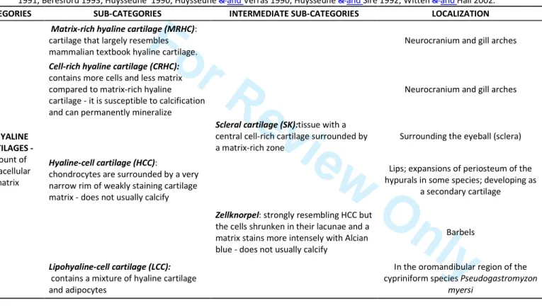
in some species, but not in other species (Koumoundouros 2010); (g) the same causative factor may provoke higher incidence of anomalies in some skeletal elements but not in others with the same bone type and ossification, in the same individual (Fernández & Gisbert 2011).Mammalian skeletal tissues are often categorised as either bone or cartilage but fish skeletal tissues include several types of bone (Fig. 2), many different types of cartilage (Tab.1) and many tissues types that are intermediate between connective tissue and bone, and between bone and cartilage (Hall & Witten 20082007, Witten et al.
2010b2010) (Fig. 3). Since the pioneering studies of Melvin Moss (Moss 19611961a, 1962, 1965) about fish skeletal tissues, a lot of progress has been made regarding the scientific knowledge about biomolecular, genetic and physiological mechanisms that underlie bone differentiation, modelling and remodelling in fish (for reviews see: Fowler 1970,; Meunier 1989, 2002; Meunier & Huysseune 1992; Huysseune & Sire 1998,; Huysseune 2000; Meunier 2002; Akimenko et al. 2003; Sire & Huysseune 2003,;
Sire & Akimenko 2004; Witten & Huysseune 2007, 2009; Hall & Witten 2007; Witten & Huysseune 2009; et al.et al.et al.Schilling et al. 2010; Spoorendonk et al. 2010; Apschner et al. 2011; et al.Duran et al. 2011; Meunier 2011; Dean & Shaharet
al.Shahar 2012; Harris 2012; see also references in Witten et al. (eds) 2010a and 2012). Unfortunately, and despite the many publications about special characters of the fish skeleton, it is still common to view fish skeletal tissues from a "human textbook perspective" (Witten & Huysseune 2010). As a consequence, teleost bone metabolism is often still misunderstood as being primarily calcium driven (as it is the case in humans but not in fish) and the specific change of vertebral bone tissues in fishes 7 8 9 10 11 12 13 14 15 16 17 18 19 20 21 22 23 24 25 26 27 28 29 30 31 32 33 34 35 36 37 38 39 40 41 42 43 44 45 46 47 48 49 50 51 52 53 54 55
For Review Only
reared under different conditions are often misinterpreted.The presence of deformed fish concerns also ethical issues: fish with deformed mouth, fins or vertebral axis show impaired feeding and swimming performances, with consequent lower feeding rates, slower growth rates, and a higher susceptibility to stress and pathogens than healthy undeformednondeformed individuals. These deformed fish cannot be considered to be in proper welfare conditions.
The aim of this review is to provide a synthetic but comprehensive picture of the actual knowledge on bone and cartilage development in larvae and juveniles of European farmed fish; to identify the main gaps of knowledge that require to be filled, in order to identify anomalous developmental pattern leading to skeletal anomalies in reared finfish larvae and juveniles. Moreover, we aim to extend knowledge on the factors that are possibly involved in the onset of skeletal anomalies. The long-term goal is the optimization of the morphological quality, welfare and health status of farmed juvenile fish.
In the present review, all the information on skeletonskeletal tissues, cells and processes are drawn from the comparative analysis of the available documentation onliterature about vertebrates, integrated with recent information on whatthat can be
ascertained up to now inabout reared finfish species, whenif available.
2. Plasticity, ontogenesis, remodelling and resorption of skeletal
elements in teleost fish
There are several differences between mammals and teleosts regarding skeletal tissue types and their differentiation, as well as remodelling and resorption of skeletal tissues 7 8 9 10 11 12 13 14 15 16 17 18 19 20 21 22 23 24 25 26 27 28 29 30 31 32 33 34 35 36 37 38 39 40 41 42 43 44 45 46 47 48 49 50 51 52 53 54
For Review Only
(Huxley 1859; Huysseune & Sire 1992, GillesGillis et al. 2006, Witten & Huysseune 2009; Ren & Winkler 2010; Apschner et al. 2011; Dewit et al. 2010, 2011, 2012). In tetrapods, the skeleton is tightly integrated into the animals' daily calcium homeostasis whereas in most teleosts, calcium from the skeleton is only mobilized in case of extreme calcium deficiency conditions. This is because fish can obtain and release calcium from and into the water via their gills (Lewis-McCrea & Lall 2010). The bone marrow in tetrapods contains haematopoietic tissue, from where the cells of theosteoclastic lineage differentiates.differentiate. Osteoclasts are bone resorbing,
multinucleated giant cells. In teleosts, the bone marrow is filled with adipose tissue, besides nerves and blood vessels and some connective tissue cells, and the head kidney is the haematopoietic organ (Witten & Huysseune 2009). Early stages of all teleosts and also leterlater stages of advanced teleosts typically do not show multinucleated osteoclasts. The majority of osteoclasts areis small and mononucleated. These cells can perform an alternative mode of bone resorption, without generating typical resorption lacunae (Witten & Huysseune 2010). Another characteristic of bone in advanced teleosts is the lack of osteocytes (cells inside the bone) (Moss 1961a, b; Parenti 1986, Meunier & Huysseune 1992). In mammals, osteocytes are responsible for maintenance of the bone matrix and serve as receptors for mechanical loads, transducing the physiological responses to these forces (Bonucci 2009). The lack of osteocytes (acellular bone) in teleosts implies that bone remodelling in response to mechanical load, as shown by Huysseune et al. (1994), is triggered by
other cell types than osteocytes.
TeleostsMany teleost species never stop growing and therefore growth-related (and 7 8 9 10 11 12 13 14 15 16 17 18 19 20 21 22 23 24 25 26 27 28 29 30 31 32 33 34 35 36 37 38 39 40 41 42 43 44 45 46 47 48 49 50 51 52 53 54 55
For Review Only
not only metabolism-related) skeletal modelling continues throughout life (Witten & Huysseune 2009). Unlike sharks, that are not able to repair their endoskeleton (Ashhurst 2004), teleosts can repair their skeleton (e.g. fracture repair) (Moss 1962,Dean & Shahar 2012). Dermal skeletal elements (teeth, scales, fin rays) have a large regenerative capacity (Akimenko et al. 2003, Huysseune et al. 2007) whereas in most cases, and in contrast to amphibians and basal actinopterygian fish, the endoskeleton can only be repaired but cannot regenerate (Kirschbaum & Meunier 1981;
CuevoCuervo et al. 2012). Other differences concerning skeletal system among terrestrial vertebrate and fish are furnished below.
3. Teleost skeletal tissues
It is generally considered that four classes of mineralized tissues can be identified in vertebrates: bone, cartilage, dentine and enamel/enameloid. These main categories and the tissue-related cells (chondroblasts, chondrocytes, osteoblasts, bone lining cells,
osteocytes, osteoclasts, odontoblasts, ameloblasts; Fig. 3) are conserved in teleosts,
but different cell morphologies and intermediate tissue types occur. Teleost fish display a large range of intermediate skeletal tissues as part of their mature – non-pathological, non-regenerating – skeleton (Beresford 1981; Benjamin 1990; Hall & Witten et al. 2007) (Figs. 2 and 3, and Tab. 1). As many as 15 diverse types and subtypes of cartilage have been identified in teleosts (Benjamin, 1988a, b, 1989, 1990; Benjamin et al. 1992). In addition, many permanent skeletal tissues found in
Teleostteleost are intermediate forms between any of the above mentioned tissues (Beresford et al. 1981) representing a continuum (or perhaps continua), and not 7 8 9 10 11 12 13 14 15 16 17 18 19 20 21 22 23 24 25 26 27 28 29 30 31 32 33 34 35 36 37 38 39 40 41 42 43 44 45 46 47 48 49 50 51 52 53 54
For Review Only
discrete skeletal categories (Hall & Witten 2007). Some intermediate tissues are characteristic for pathological alterations of the teleost skeleton, as described below. Skeletal anomalies in reared fish can affect all skeletal tissues, but from a production related viewpoint, alterations of the notochord, cartilage and bone abnormalities arethe most important. Anomalies of dermal skeletal elements, such as teeth, scales (and fin rays), are possibly indicative for the skeletal heath status of the animal (Persson et
al. 1997, 2000); however anomalies affecting teeth and scales are rarely studied. Bone is a specialized mesenchymal tissue, an aerobic vascularised tissue with high
oxygen consumption, supporting skeletal tissue. Bone tissue consists of cells (osteoblasts, osteocytes, and bone lining cells), a mineral phase (mainly composed of calcium phosphate forming hydroxyapatite crystals) and an organic, mineralized,
extracellular matrix. Collagen type I is referred as the major organic component of
bone but in teleost fish bone can also contain collagen type II (Benjamin & Ralphs 1991). The degree of bone matrix mineralisation is variable and seems to depend on the type of bone (acellular vs cellular bone), life style (active swimmers vs poor swimmers) and on the nature of the aquatic environment that the fish inhabit (seawater vs freshwater) (Meunier & Huysseune 1992, Danos & Staab 2010, Sfakianakis et al. 2011, Dean & Shahar 2012, Fiaz et al. 2012). As fish have no haematopoietic tissue inside the bone marrow, bone marrow spaces are filled with adipose tissue,; blood vessels can be also present (Huysseune 2000; Witten & Huysseune, 2009; Witten et al. 2001; Apschner et al. 2011).
Structurally, teleost bone first develops as woven bone, followed by parallel-fibered and lamellar bone in more mature individuals. In larger fish, lamellar bone can also 7 8 9 10 11 12 13 14 15 16 17 18 19 20 21 22 23 24 25 26 27 28 29 30 31 32 33 34 35 36 37 38 39 40 41 42 43 44 45 46 47 48 49 50 51 52 53 54 55
For Review Only
form osteons (Moss 1961a; Smith-Vaniz et al. 1995; Witten & Hall 2002, 2003; Meunier 2002). The various terms that are being used to describe the same bone element can be confusing. Different terms are, however, not synonymous. They relate to the location of the bone (a), to the origin of the bone (b), to its mode of development (c), to its structural properties (d) or to phylogenetic assets (e). The terms are sometimes mixed up in the literature. Terms like endo- and exoskeleton refer to the location (a) of the skeleton. Using the term dermal skeleton instead of exoskeleton, refers to the fact that these exoskeletal elements (teeth, scales, dermal bones, fin rays) originate (b) from the interaction between the epidermis (ectoderm) and the underlying mesenchyme. The endoskeleton originates (b) from sclerotome-derived mesenchyme. The mode of development (c) of the dermal skeleton is usually intramembranous, mesenchymal precursor cells develop directly into bone. Cartilage and chondroid bone as part of the dermal skeleton are secondary tissues (e). The default mode of development (c) of endoskeletal bone elements is trough endochondral ossification, where a cartilaginous scaffold is replaced by bone. However, several endoskeletal elements are developeddevelop through intramembranous bone formation. Structurally (d),) both, dermal skeletal elements and endoskeletal elements, can consist of woven or of lamellar bone. Phylogenetically (e), the bony elements of the dermal skeleton are older than bone of the endoskeleton (for reviews see Smith & Hall 1990; Huysseune & Sire 1998; Donoghue & Sansom 2002; Sire & Huysseune 2003; Hall & Witten 2007, Witten & Huysseune 2007).The different skeletal tissues, cells and extracellular matrices in fish are described in details by Kölliker 1859, 1873, 1959a; Moss 1961a, b, 1962, 1964, 1965; Meunier 1983; 7 8 9 10 11 12 13 14 15 16 17 18 19 20 21 22 23 24 25 26 27 28 29 30 31 32 33 34 35 36 37 38 39 40 41 42 43 44 45 46 47 48 49 50 51 52 53 54
For Review Only
Huysseune 1985, 1989; Huysseune et al.,. 1989; Francillon-Vieillot et al. 1990; Benjamin 1989, 1990; Benjamin &and Ralphs 1991; Takagi &and Yamada 1991, 1992; Meunier &and Huysseune 1992; Nishimoto et al. 1992; Takagi &and Yamada 1992, 1993; Beresford 1993; Hamada et al. 1995; Persson et al. 1995; Smith-Vaniz et al. 1995; Witten 1997; Witten and Villwock 1997; Ramzu 1998; Lehane et al. 1999; Wittenet al. 1999, 2000, 2001, 20052005a,b, 2006, 2009, 2010; Huysseune 2000; Pinto et al. 2001; Weiss Sachdev et al. 2001; Witten &and Hall 2002, 2003; Diekwisch et al. 2002; Kemp 2002; Pinto et al. 2003; Smits &and Lefebvre 2003; Takenaka et al. 2003; Cole
&and Hall 2004; Kang et al. 2004; Kawasaki et al. 2004; Gil-Martins Martens et al. 2005; Hall 2005; Kranenbarg et al. 2005b; Nordvik et al. 2005; Redruello et al. 2005; Franz-Odendall et al. 2006; Gavaia et al. 2006; Gillis et al. 2006;Hell et al. 2006; Hall
&and Witten 2007; Roy &and Lall 2007; Witten &and Huysseune 2007, 2009, 2010; Kang et al. 2008; Rotllant et al. 2008; Zylberberg &and Meunier 2008; Horton &and
Summers 2009; Renn &and Winkler 2010; Apschner et al. 2011; Estêvão et al. 2011; Lie
&and Moren 2011; Meunier 2011; To et al. 2012.
4. The Notochordnotochord
The notochord is an essential tissue that plays both structural and patterning roles: early notochord signals influence cell-fate and patterning in the central nervous systemspinal cord and in somites (Fleming et al. 2001, Anderson et al. 2007, de Azevedo et al. 2012). Structurally, the notochord is the sole skeletal support tissue in the embryo and in early life stages post-hatching, a stiffened rod against which muscular contraction can drive motility.
7 8 9 10 11 12 13 14 15 16 17 18 19 20 21 22 23 24 25 26 27 28 29 30 31 32 33 34 35 36 37 38 39 40 41 42 43 44 45 46 47 48 49 50 51 52 53 54 55
For Review Only
Along with development, the teleost notochord stiffens throughout secretion of fibrous collagens (mostly collagen II) and cells then vacuolate and differentiate into epithelial cells, called notochordoblasts (Fishelson 1966; Yan et al. 1995; Nordvik et al. 2005). These produce fibrous sheath surrounding the notochord. New evidences allow hypothesizing that the rigidity given by collagen encasing sheath constrains expanding notochord cells forfrom inflation of their vacuoles, sothus generating a hydrostatic pressure that drives the elongation, stiffening and straightening of the notochord (Anderson et al. 2007). Different from other vertebrates, teleost vertebral centra form without a cartilaginous anlage. (primordium). In teleosts, the mineralisation of the notochord sheath, not cartilage and not bone formation, establishes the identity of vertebral bodies. Formation of skelerotome-derived bone is only the second step of teleost vertebral body formation (Huxeley Huxley 1859; Kölliker 1859b1859; Bensimon-Brito et al. 2012; de Azevedo et al. 2012). Consequently, anomalies/mutations affecting specification of early notochord cells provoke profound defects on vertebral body patterning in teleost fish (Fleming et al 2001, 2004; Moren-Kensicki et al. 2002; Crotwell & Mabee 2007, Willems et al. 2012). After vertebral bodies have fully developed, notochord tissue in the intervertebral spaces can transform into cartilage under pathological conditions (Witten et al. 20052005a). Studies about the structure of the notochord in basal actinopterygians and in teleost fish (Fig. 4) are available for shortnose sturgeon, Acipenser brevirostratus (Schmitz 1998), medaka Oryzia latipes (Ekanayake & Hall 1991), yellow perch Perca fluvescens flavescens (Schmitz 1995), Atlantic salmon (Grotmol et al. 2006), and zebrafish Daniorerio (Inohaya et al. 2007).
Formatted: Font: 12 pt 7 8 9 10 11 12 13 14 15 16 17 18 19 20 21 22 23 24 25 26 27 28 29 30 31 32 33 34 35 36 37 38 39 40 41 42 43 44 45 46 47 48 49 50 51 52 53 54
For Review Only
5. Regulatory mechanisms of skeletal tissues in fish
The assembly of the structures composing the skeleton is the net result of two processes acting at two different time scalescales: a phylogenetic process (over millions of years) and an ontogenetic process (over life span of the individual) (Prendergast 2002). In general, according to Hall (2005), the skeletal development is modular at levels of:
a) individual skeletal systems (axial, appendicular, cranio-facial, ‘extraskeletal’ elements);
b) individual skeletal elements c) cellular condensations d) gene networks e) epigenetic control.
As far as epigenetic (i.e., non-genetic) control is concerned, mechanical forces isare by far the main studied factor, due to their implication for human health (e.g., osteoporosis, fracture healing). External mechanical forces are recognized to regulate genetic pathways of both cartilage and bone development in all vertebrates (Danos & Staab 2010), giving to). This gives skeletal tissues the capability to adapt its structuretheir structures, shape and mechanical features through adaptation in response to altered loading conditions in teleost fish (Huysseune et al. 1994; Meyer 1987, Kranenbarg 2005a,b; Fiaz et al 2012) and in mammals (Vahdati & Rouhi 2009). At present, a large body of studies dealdeals on mechano-regulated tissue differentiation models developed on Pauwels’s theory (Pauwels, 1980) that the 7 8 9 10 11 12 13 14 15 16 17 18 19 20 21 22 23 24 25 26 27 28 29 30 31 32 33 34 35 36 37 38 39 40 41 42 43 44 45 46 47 48 49 50 51 52 53 54 55
For Review Only
mechanical environment in the medium determines tissue phenotype. phenotypes. It is well known from studies on mammals that bone responds preferentially to dynamic stimuli rather than static and that only short loading durations are necessary to induce an adaptive response, allowing bone cells to adapt to specific mechanical loading environments (Warden 2006). A series of different mechanobiological models introduced a growing number of mechanical and biological theoretical factors acting and interacting during mesenchymal cells differentiation, fracture healing, intramembraneous bone formation, distraction osteogenesis, bone-implant reaction, osteochondral defect repair in tetrapods and then tested in vivo. Deeper insight on different mechano-biological modelling are achievable in Gómez-Benito et al. (2005) (strain-based), Carter and Beaupré (2001) (hydrostatic stress/deformation), Prendergast (2002), Kelly and Prendergast (2005) (byophisical) and Vahdati and Rouhi (2009) (semi-mechanistic).6. Bone formation and the replacement of the cartilaginous
anlage
Bone in mammals is formed by replacement of a cartilaginous template (Fig. 5) by bone (endochondral bone) or by intramembranous ossification, originating as dermal bones. In fish, three bone formation mechanisms, according to the considered species and skeletal elements, have been described: endochondral, perichondral and
intramembranous (or direct or dermal) ossification. Here belowBelow, a synthetic characterization of the bone formation processes in Vertebratevertebrate is
furnishedprovided: some highlights on availablefrom the literature on reared 7 8 9 10 11 12 13 14 15 16 17 18 19 20 21 22 23 24 25 26 27 28 29 30 31 32 33 34 35 36 37 38 39 40 41 42 43 44 45 46 47 48 49 50 51 52 53 54
For Review Only
Teleostteleost species isare given, whenif available.
6.1 Endochondral ossification
According to Hall and Witten (2007), not all the bony material of endochondral bone originated by endochondral ossification in vertebrates, but i) for primary ossification of the cartilaginous anlage occurring subperiosteally around the shaft by perichondral bone apposition; ii) for a secondary endochondral replacement of cartilage of the shaft metaphysis; iii) for the later extension of the skeletal element by appositional bone (Zuwachsknochen), in a process similar to intramembranous bone formation. And even, endochondralEndochondral ossification can replace marrow, tendon or ligament
tissue, without any cartilaginous template. SoThus, the use of replacement bone and
indirect ossification should be preferredrestricted to endochondral bone and endochondral ossification, respectively (Hall and& Witten, 2007).
Most of the bones that ossify endochondrally have their origin inoriginate from
embryonic mesoderm (Hall 2005). It involves a cartilaginous template, which is replaced by, or remodelled into, bone by several co-ordinated sequential steps. Endochondral bone formation is one of the main ossification process in mammals but the typical process is often lacking in teleosts, especially in small size species: replacement of cartilage by spongiosa (endochondral bone formation) can be observed in large (e.g., carp Cyprininus carpio, Atlantic salmon) but not in small (medaka and zebrafish) teleost species (Witten et al. 2000, 2001, 2010; Verreijdt et al. 2002; Witten et al. 2000; Witten et al. 2001; Witten & Huysseune 2007, 2009; Zylberberg & Meunier 2008; Witten et al. 2010; ; Apschner et al. 2011). In smaller 7 8 9 10 11 12 13 14 15 16 17 18 19 20 21 22 23 24 25 26 27 28 29 30 31 32 33 34 35 36 37 38 39 40 41 42 43 44 45 46 47 48 49 50 51 52 53 54 55
For Review Only
teleosts, such as medaka and zebrafish, where endochondral bone formation is uncommon (but does occur in vertebral arches, hypurals, pterygiophores, according to Bird & Mabee 2003; Bird & Hernandez 2009; Gavaia et al. 2006). Typically,typically a persisting cartilage rod remains inside the bone shaft and if cartilage is removed, it is replaced by adipose tissue. Young fish (larvae) generally are considered to not have endochondral bone formation (Witten et al. 2001, 2010), but Estêvão et al. (2011) described it as occurring in hypurals, vertebral arches, frontal bone, coracoid, sclera and dentary during gilthead seabream osteogenesis. Whilst vertebrae bodies are formed throughoutthrough endochondral ossification of cartilage templates in tetrapods, vertebral centra ossify without any cartilaginous anlage in all teleosts. Although no cartilage contributes to the initial formation of Teleostteleost vertebral bodies (Huxley 1859, Nordvik et al. 2005; Witten & Villwock 1997), in older individuals cartilage at the base of the arches undergoes endochondral ossification, and bone that derives from this process becomes part of the vertebral body (Zylberberg & Meunier 2008; Apschner et al. 2011).Growth of endochondral bone depends on maintaining the growth of a primary cartilaginous model that, on its turn, requires functional stimuli, such as mechanical stress. Consequently, the continuation of the deposition of endochondral bone depends secondarily upon biomechanical stimuli (Hall 2005).
6.2 Perichondral ossification
Perichondral bone formation is the most common mode of bone ossification in fish, if. If a cartilaginous precursor is present, andit usually starts with the transformation of a 7 8 9 10 11 12 13 14 15 16 17 18 19 20 21 22 23 24 25 26 27 28 29 30 31 32 33 34 35 36 37 38 39 40 41 42 43 44 45 46 47 48 49 50 51 52 53 54
For Review Only
perichondrium into a periosteum. It typically occurs in the teleost fin endoskeleton (Fig. 6). Young fish (larvae) essentially only have perichondral bone formation and no endochondral bone formation. Differently from mammals, it is often not linked to endochondral bone formation (Hall 1998; Huysseune 2000; Witten & Villwock 1997; Apschner et al. 2011). In perichondral ossification, the bone develops in connective tissue within the perichondrium, and thus immediately around the cartilage. SuchPerichondral an ossification produces the bone that surrounds the cartilage of the gill arches in mosquitofish Gambusia affinis, the pharyngeal jaws of Astatotilapia elegans (now Haplochromis elegans) (Benjamin 1989), the hypural cartilage in Nile tilapia (Oreochromis nilotichusniloticus) and desert pupfish (Cyprinodon macularis) (Witten & Huysseune 2007), in dorsal and anal proximal pterygiophores in desert pupfish (Witten & Huysseune 2007), in pectoral, pelvic and caudal fins of rainbow trout (Ferreira et al. 1999), in dorsal and anal proximal pterygiophores, initial pectoral girdle ossification, secondary gill arches ossification and pectoral radials in Teleostteleost
(Witten & Huysseune 2007). Parahypural (a ventral support of caudal fin) is reported by Witten and Huysseune (2007) as undergoing perichondral ossification, whilst Fernández et al. (2008) report that it is forming by endochondral ossification.
Cartilage plays no role in either perichondral or parachondral ossification: i.e., theThe
hyaline-cell cartilage (HCC) attached to the basioccipital in adult of black molly (Poecilia
sphenops) only develops after perichondral bone has appeared (Benjamin 1989). As reviewed by Witten and Huysseune (2007) in teleost fish, perichondral bone formation is the basic process of ossification of the fin endoskeleton. Perichondral bone is laid down at the immediate contact of the cartilaginous template by cells that were 7 8 9 10 11 12 13 14 15 16 17 18 19 20 21 22 23 24 25 26 27 28 29 30 31 32 33 34 35 36 37 38 39 40 41 42 43 44 45 46 47 48 49 50 51 52 53 54 55
For Review Only
formerly part of the perichondrium, but have now taken up the characteristics of osteoblasts and secrete the bone matrix. Nevertheless, an admixture of cartilage and bone matrix is not excluded (Huysseune and Sire 1992; Huysseune 2000; Verreijdt etal. 2002).
In smaller species, such as zebrafish and medaka, cartilage that is also enclosed by perichondral bone and chondrocytes can hypertrophy, but the cartilage is removed without being replaced by bone. For (for example, in the splanchnocranium, the). The
result is a bone collar with cartilage at one or at both ends (enabling further growth) and adipose tissue inside (Witten et al. 2010).
In the endoskeleton, membranous apolamellae can form from osteoblasts of perichondral bone, a process that resembles intramembranous bone formation (Witten & Huysseune 20012007).
At our knowledge no data on regulatory mechanisms underlying perichondral ossification are available in literature. Prestinicola et al. (2012) found that, conversely to intramembranously and endochondrally ossified bones, perichondral bones development is not influenced by variation of environmental conditions during gilthead seabream larval rearing.
6.3 Parachondral ossification
Blanc (1953) cited the formation of bone around Meckel's cartilage in Salmo salarAtlantic salmon as an example of parachondral ossification. Such bone develops around cartilage, but is separated from it by a layer of ordinary connective tissue (Benjamin 1989). 7 8 9 10 11 12 13 14 15 16 17 18 19 20 21 22 23 24 25 26 27 28 29 30 31 32 33 34 35 36 37 38 39 40 41 42 43 44 45 46 47 48 49 50 51 52 53 54
For Review Only
6.4 Intramembranous ossification
Most of theCranial bones that develop intramembranously have a neural crest origin (Hall 2005). This mode of bone development has been described in many teleosts and it consists of mesenchymal. Mesenchymal cells differentiationdifferentiate into osteoblasts formingand form bone without a cartilaginous modeltemplate
(Franz-Odendaal et al. 2006). The bones formed in this formway are designated dermal or membrane bones. Sire and Huysseune (2003) described categories of odontode derived dermal skeletal elements in vertebrates teleosts: : teeth, denticles, cranial dermal bones, scutes, postcranial dermal plates, ganoid scales of polypterids, ganoid scales of lepisosteids, elasmoid scales and fin rays. With the exception of ganoid scales of polypterids and of lepisosteids, these elements are present in teleosts. Cranial dermal bones are frontal, infraorbitals, lacrimal, nasal, parasphenoid, parietal, vomer, ectopterygoid, endopterygoid, dentary, maxillary, premaxillary, interopercle, opercle, preopercle, subopercle, branchiostegal rays, and urohyal. All these dermal skeleton elements form in the mesenchyme or in the dermis, without a cartilaginous precursor and below a multilayered epithelium o epidermis (Sire & Huysseune 2003). As far as reared species are concerned, dermal ossification has been described in vertebrae bodies, fin rays, and opercular plate in gilthead seabream (Estêvão et al. 2011)and.
During the early development of vertebral bodies in salmon, Nordvik et al. (2005) described the importance thatof the notochord sheath tissues has eontissue for the initial mineralization. of the vertebral body. These authors described how, after the initial notochord segmentation and formationexpression of a segmentalsegmented
alkaline phosphatase activity pattern withinby the chordoblast layer (Grotmol et al. 7 8 9 10 11 12 13 14 15 16 17 18 19 20 21 22 23 24 25 26 27 28 29 30 31 32 33 34 35 36 37 38 39 40 41 42 43 44 45 46 47 48 49 50 51 52 53 54 55
For Review Only
2005), the formation of a chordacentrum by direct mineralization of the external layers of the notochord occurs. The chordacentrum forms as mineralized ring within the preformed notochordal sheath through mineralization of specific subpopulations of chordoblasts, and are located in the external half of the notochordal sheath, bellow the elastic membrane (Nordvik et al. (Nordvik et al. 2005).Prior the beginning of dermal ossification in, the dentary, it anlage consists of densely packed mesenchymal cells with a barely visible ECM,extracellular matrix (here after called ECM), while in the frontal bone, that develops later in ontogeny than the
dentary, cells are sparse amidst a collagenous matrix (Sire & Huysseune, 2003). In fin developmentsdevelopment, mesenchymal cells populate the area between the two epitheliumepithelial layers constitutingthat constitute the embryonic fin fold. They progressively increase their number, so forming a dense core separated by the basal lamina by, an acellular layer with collagen randomly depositedarranged collagen (Sire & Huysseune 2003).
The anlagen of cranial dermal bones and fin rays in teleosts are poorly defined papillae (or osteogenic papillae), located deeply in the mesenchyme or nearclose to a forming
cartilage anlage. No indicationsindication of epithelial-mesenchymal interactions
havehas been individuated and matrix deposition is not polarised, occurringit occurs
on both surfaces (Sire & Huysseune 2003). The resulting tissue is fairly identifiable as bone, with an inner part composed of woven-fibred matrix and a peripheral onepart
composed of parallel-fibred matrix, deposed after the initial fast growing of the inner part.
In dentary and frontal bones, the papillae merge with perichondral cell population very 7 8 9 10 11 12 13 14 15 16 17 18 19 20 21 22 23 24 25 26 27 28 29 30 31 32 33 34 35 36 37 38 39 40 41 42 43 44 45 46 47 48 49 50 51 52 53 54
For Review Only
close to the cartilage of taenia marginalis (frontal) or Meckel’s cartilage (dentary). The start of ossification is characterized by a local thickening of the stratum compactum along with an accumulation of flattened fibroblast-like cells and tubby pre-osteoblasts (mesenchymal cells agglomeration and condensation). Chondroblasts/cytes are at this phase PCNA- (Proliferating Cell Nuclear Antigen)-positive in gilthead seabream (Estêvão et al. 2011).Subsequent phase is characterized by the synthesis of the different matrices constituting the different skeletal dermal elements and, so, it differs in different skeletal elements: odontodes, teeth, denticles and fin rays maintain a close relation with the epithelial cells, whilst dermal cranial bones do not maintain or establish no relation with epithelial cells.
Fin raysray dermal ossification has some peculiar features: the anlagen are osteogenetic papillae but differentiation takes place immediately below the epithelial-mesenchymal boundary, thus suggesting the presence of some relationship with the epithelial cover. At the beginning of fin raysray ossification, a multilayered epithelium thickens in the vicinity of the developing lepidotrichium. The epithelial cells show features of differentiation, indicative of epithelial-mesenchymal interactions, but they do not directly participate in the matrix production (Sire & Huysseune 2003). The rays’ anlage is Fin ray’ anlagen are composed of a woven-fibred matrix, acellular and not penetrated by cellular processes, forming. The anlagen form a continuous
subepidermalsub epidermal sheet located between the epithelial basal cells and the mesenchymal cells. Only theCollagen type I of collagen and Osnosteonectin (here after called OSN) are secreted and mesenchymal PCNA-positive cells are detectable around 7 8 9 10 11 12 13 14 15 16 17 18 19 20 21 22 23 24 25 26 27 28 29 30 31 32 33 34 35 36 37 38 39 40 41 42 43 44 45 46 47 48 49 50 51 52 53 54 55
For Review Only
the bony structures in gilthead seabream (Estêvão et al. 2011). The matrix mineralises rapidly, from the central region of the subepidermal sheet toward the exterior. With growth, a single layer of mesenchymal cells infiltrated the epithelial-mesenchymal interface and progressively separates the ray matrix from the epithelial surface.DifferentlyDifferent from fin rays, the cranial dermal bonesbone ossification process
does not takehas no relation with epithelial cells, and their anlagen are initially not sharply defined from the surrounding tissues. The anlage is covered by osteoblasts in a single layer, surmounted by a mesenchymal space, at the side facing the epithelium. At the opposite side, the anlage is separated from the underlying cartilage by not clearly delimited by osteoblasts. In jewelfish, (Hemichromis bimaculatus), the space between
the osteoblast monolayer and the epithelium is fulfilledfilled with a woven-fibred, acellular network, embedded in a fine granular, electron-dense, background substance
that. This substance mineralises soon after its deposition (periosteal ossification). Later, a parallel-fibred matrix is deposited on both bone surfaces.
TRAP activity appears in association with cells at the periphery of calcified structures. (Sire & Huysseune, 2003).
Deposition of membrane bones is far less dependent on mechanical factors than endochondral bone growth (Hall 2005) but transdifferentiation in chondroid bone is reported occurring in compressed and fused salmon vertebrae. (Witten et al. 2005a, 2006).
In conclusion, intramembranous ossification has some features that are different for different dermal skeleton elements: however, all need for the presence of a support, a well-structured mesenchyme or a bone, cartilage or another support, but 7 8 9 10 11 12 13 14 15 16 17 18 19 20 21 22 23 24 25 26 27 28 29 30 31 32 33 34 35 36 37 38 39 40 41 42 43 44 45 46 47 48 49 50 51 52 53 54
For Review Only
epithelial-mesenchymal interactions are not always necessary. The fulfilment of this need modulates the timing of ossification of different dermal bones.7. Modulation and Transformation
The differentiation and ossification processes of different skeletal elements foresee some individual processes that can deviate from the differentiative pathways, induced by alteration ofcan be altered by microenvironment conditions (Hall 2005). Modulation (of cellular activity) is a physiological response to altered environmental conditions: it is characterized by a temporary change in cell behaviour, structure and/or the type of ECM products. The maintenance of the ‘new’ state is depending on the enduring of the environmental stimulus, so it is a reversible status. An example is given by the cell switching from the synthesis of collagen type I to the synthesis of collagen type II.
Transformation (of cell identity) is generally a permanent change, even when the
stimulus is not present anymore. It creates a permanent intermediate tissue (chondroid or chondroid bone) (Hall & Witten, 2007).
7.1 Dedifferentiation, Transdifferentiation and Metaplasia
In a dedifferentiation process, a differentiated cell loses its specific phenotypic characteristics, transforming and transforms into an undifferentiated mesenchymal cell.
Transdifferentiation is a transformation of one differentiated cell type into another cell
type (Okada 1991): i.e., chondroid bone type I can arise via incomplete endochondral ossification (forming chondroid bone II) or transdifferentiation of skeletal cells (from 7 8 9 10 11 12 13 14 15 16 17 18 19 20 21 22 23 24 25 26 27 28 29 30 31 32 33 34 35 36 37 38 39 40 41 42 43 44 45 46 47 48 49 50 51 52 53 54 55
For Review Only
osteoblast to chondroblast) within multipotential periostea (thus forming chondroid bone type I as in dentary tip of salmons) (Gillis et al. 2006; Witten et al. 2010& Hall2002). It can be either a pathologic or a normal process: the transdifferentiation from osteogenic to chondrogenic cells of the vertebral growth zone in compressed and fused vertebrae of Atlantic salmon, Senegalese sole, (Solea senegalensis), European seabass and gilthead seabream is a pathological condition in response to a compressive mechanical environment (Beresford 1981; Hall 2005; Kranenbarg et al. 2005b; Witten et al. 20052005a, 2009; Roberto 2006; Fiaz et al. 2010, Cardeira et al. 2012). This is in accordance with Pauwels’s mechano-regulated theory of tissue differentiation, which states that compression is the specific stimulus for the development of cartilaginous tissue (Prendergast 1997). Transdifferentiation, however, occurs also during normal development in other skeletal elements of Atlantic salmon and in other teleost species (Kranenbarg et al. 2005a; Gillis et al. 2006; Witten & Hall 2002, 2003, 2009; Hall & Witten 2007; Witten & Huysseune 2007; Fiaz et al. 2010). Chordoblasts and intervertebral ligament cells likely show a mechanically induced transdifferentiation into a cartilaginous phenotype (Fiaz et al. 2010). In some cases, transdifferentiation can be accompanied by cell division, whereas in others it is not (Beresford 1990). Transdifferentiation can occur either directly (the cells possess characteristics of both cell types simultaneously during the transition period) or indirectly (implies a dedifferentiation phase in which the phenotypic characteristics of the cell first disappear before a new phenotype is established) with respect to the timeframe of the phenotypic transformation (Dewit et al. 2011).
Although transdifferentiation of cartilage into fibrous tissue may be rare, many other 7 8 9 10 11 12 13 14 15 16 17 18 19 20 21 22 23 24 25 26 27 28 29 30 31 32 33 34 35 36 37 38 39 40 41 42 43 44 45 46 47 48 49 50 51 52 53 54
For Review Only
types of transdifferentiation in connective tissues have been described: cartilage and chondroid bone into bone, bone into chondroid bone, bone into cartilage, fibroblastic tissue into bone or cartilage, hyaline- into fibro-cartilage, periosteal chondrogenesis, perichondral osteogenesis, fat into bone, and muscle into bone (see Dewit et al. 2011 for details). This scenario evidences that the different phenotypes of connective tissues are neither fixed nor terminal, but rather form a continuous spectrum in which differentiation can be modulated by a variety of factors, and that phenotypic plasticity can play an important role in various developmental and homeostatic processes. (Hall & Witten 2007).Metaplasia is the normal transformation of tissue from one type to another, as in the
ossification of cartilage to form bone. Differently from mammals, in teleost normal skeletogenic processes, cartilage elements may arise also by multiple subdivisions of an existing cartilage, through dedifferentiation of chondrocytes into fibroblasts. During this process, the surrounding matrix loses its cartilaginous character (Witten et al. 2010). This kind of metaplasia occurs during the development of the endoskeleton of teleost pectoral, dorsal and anal fins (Grandel & Schulte-Merker 1998; Witten & Huysseune 2007; Dewit et al. 2010; Witten et al. 2010b2010). The absence of signs of apoptosis or resorption during subdivision of the cartilage larval pectoral plate into radials in zebrafish suggests that the separation of cartilage elements may involve metaplasia which occurs through dedifferentiation of cartilage cells and their redifferentiation into noncartilaginous connective tissue (Witten & Huysseune 2009; Dewit et al. 2011). So, bone elements can arise via metaplasia, a process that intervenes when matrix changes as the result of trapped chondroblasts and
Formatted: Font: Not Italic 7 8 9 10 11 12 13 14 15 16 17 18 19 20 21 22 23 24 25 26 27 28 29 30 31 32 33 34 35 36 37 38 39 40 41 42 43 44 45 46 47 48 49 50 51 52 53 54 55
For Review Only
chondrocytes assuming osteoblastic activity, and modifying the ECM toward an osseous tissue (Hall 2005).Unlike in intramembranous ossification, cells resembling osteoblasts are generally absent during metaplasic ossification (Sire et al. 2009).
The chondro-bone metaplasia is a progressive transformation of cartilage into bone, without any previous destruction of pre-existing tissue, conversely to neoplasia which is the substitution of a tissue (i.e. cartilage) with another one (i.e. bone) (Meunier et al. 2008). In fish, it may occur in both normal and in pathological development: from a histopathological point of view, both vertebrae compression and fusions involve metaplasic changes of bone forming cells (osteoblasts) that differentiate into cartilage forming cells (chondroblasts); then, in the growth zone of vertebral bodies, developing heterotopic cartilage mineralizes and it is remodelled into bone tissue (Witten et al.
20052005b, 2006; Gil- Martens 2010). Compressed vertebrae in short-tail Atlantic salmons, i.e., are the results of metaplasic chondrogenesis (metaplasic synchondrosis) as a skeletogenic response late in ontogeny (Witten et al. 20052005a).
8. Late events in teleost skeletal tissue modelling and
remodelling
Teleost fishMany teleost species never stop growing and thus all skeletal tissues may continue to differentiate and to transform through metaplasia, mineralisation, or remodelling throughout life (Witten & Huysseune 2009). Modelling occurs when the
old bone resorption and new bone formation processes are disacoupled, then the resulting bone shape could be altered. Conversely, inshape of a bone needs to be
7 8 9 10 11 12 13 14 15 16 17 18 19 20 21 22 23 24 25 26 27 28 29 30 31 32 33 34 35 36 37 38 39 40 41 42 43 44 45 46 47 48 49 50 51 52 53 54
For Review Only
altered. In bone remodelling (bone turnover) resorption is followed by new bone formation (either through intramembranous or endochondral ossification), without any change in shape. Osteoblasts and osteoclasts are the bone remodelling units. The remodellingRemodelling in teleosts occurs by resorption and de novo formation, but also by transdifferentiation (metaplasia) of skeletal tissuetissues (Beresford 1981; Witten & Hall 2002; Hall 2005; Gillis et al. 2006; Hall & Witten 2007; Witten & Huysseune 2009). So the replacing one tissue type by another could be included also in skeletal remodelling.
Some mechanisms of skeletal remodelling are more prominent and/or more commonly observed in teleosts than in mammals and are thus recognised as regular processes that shape the skeleton in the course of development and growth (Sire et al. 1990; Huysseune 2000; Witten et al. 2001, 2003; Hall & Witten 2007; Witten & Huysseune 2007), bone repair processes (Gil-Martens 2012) and the growth of the
kype in mature male salmon (Witten & Hall 2003; Witten & Huysseune 2009). Furthermore, lordosis, scoliosis, kyphosis and fusion of vertebral bodies must involve bone resorption and bone remodelling, as a primary pathology or in response to altered mechanical load (Kranenbarg et al. 20052005b; Witten et al. 2006 et al.).).
There are some reports of bone resorption connected to bone deformitiesanomalies, mainly caused by phosphorus deficiency (in haddock Melanogrammus aeglefinus: Roy
et al. 2002; in farmed Atlantic salmon: Roberts 2001).
An extreme case of remodelling is the complete pathological fusion of vertebral bodies in Atlantic salmon (Witten et al. 2006) and in advanced teleosts with acellular bone (Sawada et al. 2006a,b2006). So compression and fusion of vertebrae bodies involve 7 8 9 10 11 12 13 14 15 16 17 18 19 20 21 22 23 24 25 26 27 28 29 30 31 32 33 34 35 36 37 38 39 40 41 42 43 44 45 46 47 48 49 50 51 52 53 54 55
For Review Only
the metaplasic transformation of bone-forming cells in the vertebral growth zone into cells that produce cartilaginous tissue instead of bone. Later this cartilage is mineralized and remodelled into bone (Witten et al. 2005a, 2006). Remnants of notochord tissue in the intervertebral space are also remodelled.Hypermineralized vertebral bodies constitute another pathology that involves modelling, that can be caused by heterotopic cartilage occupying bone marrow spaces (Helland et al. 2006). The observed resorption of such cartilage suggests that it is not a permanent structure.
Also infectious diseases can trigger skeletal resorption in teleosts: the parasite
Myxobolus cerebralis causes skeletal deformities and lysis of cranial cartilage (Halliday
1973; Garden 1992; Kelley et al. 2004).
9. Bone resorption and remodelling
Differently from cartilage that is reshaped by chondroclasts, changes in bone structure can only occur through remodelling, a process in which resorbing cells (osteoclasts) remove existing bone and osteoblasts form new bone.
Basically, bone remodelling in fish is required for teeth replacingtooth replacement, allometric growth and for removing temporary skeletal elements (e.g., kype in male Atlantic salmon), or it occurs as adaptation to mechanical load (Hall & Witten 2007). Bone remodelling intervenes also in bone repair processes in teleosts that however evidence a higher regenerative capacity of dermal (rays and scales) than endo- skeleton (Witten & Huysseune 2009).
Some differencesDifferences between aquatic and terrestrial vertebrates have been outlined by Witten and Huysseune (2009). In particular, some others than 7 8 9 10 11 12 13 14 15 16 17 18 19 20 21 22 23 24 25 26 27 28 29 30 31 32 33 34 35 36 37 38 39 40 41 42 43 44 45 46 47 48 49 50 51 52 53 54
For Review Only
morphological differences between small, mononucleated osteoclast-like cells in advanced teleosts, and giant, multinucleated osteoclast of tetrapods and basal teleosts, have been found: fish do not show any intimate spatial relationship between bone resorbing cells and haematopoietic cells (as in teleosts the bone marrow is filled with adipose tissue); in advanced teleosts osteoclast-like cells can perform resorption without generating typical resorption Howships’s lacunae; whilst in mammals endochondral ossification is a prime cause of skeletal resorption and remodelling, in fish it occurs rarely..All teleosts have mono- and multinucleated osteoclasts, but the first are the main bone-resorbing cells observed in advanced teleosts. Remodelling processes with intervention of osteoclasts have been observed onlyare prominent in basal teleosts, e.g. cyprinids and salmon that have cellular bone, where it starts only laterlate in the
development (around 60 DPH in zebrafish) (Witten et al. 2001, Witten & Huysseune 2010). In advanced teleosts, with acellular bone, metabolic driven bone remodelling is considered to be limited or absent (an there are reports in literature about the absence of the process (see Witten & Huysseune 20072009 for references). Regular resorption and rebuilding of scales and bony skeletal elements is well documented for Atlantic salmon (Kacem et al., 1998; Persson et al. 2000, 2007; Witten and Hall, 2002, 2003; Persson et al. 2000, 2007). A comparative review on skeletal remodelling in teleostean fish has been recently published (see Witten & Huysseune 2009).
Studies carried out on zebrafish (Witten et al. 2001) evidenced heterochronic shifts in the appearance of bone-resorbing cells: in early stages, bone is resorbed by mononucleated osteoclasts, while multinucleated osteoclasts (as mammalian’s onesin
7 8 9 10 11 12 13 14 15 16 17 18 19 20 21 22 23 24 25 26 27 28 29 30 31 32 33 34 35 36 37 38 39 40 41 42 43 44 45 46 47 48 49 50 51 52 53 54 55
For Review Only
mammals) appear only later, when the bone switches from acellular to cellular. Bone resorption by mononucleated osteoclast was found to coincide with the dominance of acellular bone (Witten & Huysseune 2010) but in all teleosts whose early skeletogenesis hasthat have been studied so far, independently from the bone cellularity or acellularity, mononucleated are the dominating resorbing cells. in early stages, when the bone is still acellular. The presence of osteoclast key enzymes, transcription factors and receptors (H+-ATPase and tartrate-resistant acid phosphatise,
TRAP, RANK, RANKL, Cathepsin K) in mononucleated osteoblasts prove their capability of bone resorption inas shown in advanced teleost such as Nile tilapia (an advanced Teleost), but in some (dentary, opercular, preopercular shaft, neural arches)Oreochromis niloticus), and not in other (vertebral centra, hypurals) skeletal elementsmedaka (Oryzias latipes) (Witten 1997, Witten & Villwock 1997, To et al. 2012). Lysis of cranial cartilage and increased skeletal resorption resulting from bacterial infection has been reported for salmonids infected with Flexibacter
psychrophilus (Ostland et al. 1997; Witten & Huysseune 2009).
Despite the many morphological differences between mammalian multinucleated osteoclast and teleost mononucleated ones, the molecular mechanisms underlying bone resorption regulation are considered to be identical (consult Witten & Huysseune 2009; GilsMartinsGil Martens 2012; To et al. 2012).
9.1 Osteocytic osteolysis
Osteocytes interconnected cell processes function as stress sensors in cellular bone, so activating bone deposition and resorption processes carried out by the osteocytes 7 8 9 10 11 12 13 14 15 16 17 18 19 20 21 22 23 24 25 26 27 28 29 30 31 32 33 34 35 36 37 38 39 40 41 42 43 44 45 46 47 48 49 50 51 52 53 54
For Review Only
themselves (Witten & Huysseune 2010): e.g., the enhanced demand for calcium in pregnant and lactating bats is satisfied by osteocytic osteolysis and not by osteoclasts resorption. Osteocytic osteolysis has been described only in teleosts from different groups, such as eels (Anguilla anguilla) (Lopez et al. 1980; Sbaihi et al .. 2007), salmonids (Hughes et al. 1994b1994; Kacem & Meunier 20002009) and cyprinids (Cyprinus carpio) (Witten et al. 2000).10. Main gaps in the scientific knowledge and further research
needs
What reported up to now evidenced thatStudying the available knowledge on higher vertebrates amniotsabout amniot (i.e., birds and mammals, incl. humans) skeletogenic processes can be a promising tool for deeper insight in what happens in the same processes occurring in teleost fishes but also that a direct application to fish is . However, data from the amniote skeleton are not always applicable. Main gaps to fish skeleton. Gaps of knowledge are up to now evident in what concern endochondral ossification in different fish, particularly during larval species and juveniledifferent life
stages, and the available literature presents some evident contradictions: the bone composing gilthead seabream skeleton is acellular, with the exception of the gill arches that have recently been described as chondroid bone (Estêvão et al., 2011). However thefish. For example, studies by Benjamin (1989), Benjamin et al. (1992) and Witten (2010) have described the gill arches as being composed of hialinehyaline cell cartilage (HCC) or chondroid but not as Chondroidchondroid bone (for review see Witten et al.
2010b2010), while another specific type of cartilage, Zellknorpel, is found supporting 7 8 9 10 11 12 13 14 15 16 17 18 19 20 21 22 23 24 25 26 27 28 29 30 31 32 33 34 35 36 37 38 39 40 41 42 43 44 45 46 47 48 49 50 51 52 53 54 55
For Review Only
the gill filaments. Some controversy still persists in the classification of the skeletal tissues of teleosts, particularly the many forms of cartilage and on the transitory forms between cartilage and bone that coexist in teleost skeletons. Some effort should be undertaken by the scientific community towards a more precise classification of the skeletal tissues and the mechanisms of ossification that they undergo, and to create a uniformed classification system for the most relevant species/families of fish produced in aquaculture. Even the same ossification modality (e.g., intramembranous ossification) may show differences among the considered skeletal elements in fish. Consequently, the necessity of a deeper insight on the current molecular literature achieved on model fishes (zebrafish and medaka, e.g.) and comparison with the mammalian literature resulted to be one of the main gaps, in order to fully establish what is specific for fish, what is specific to mammals and what is common in fish and mammals. In this scenario, this review represents a starting basis for deeperfurtherstudies.
Other more specific gaps and need in scientific knowledge may be synthetically resumed as follows:
• cartilage development is a tightly regulated morphogenetic event where much has been studied on gene regulation by different types of signalling molecules, but less is known of their downstream regulations, even at vertebratesthe mammalian level. In particular, the regulatory mechanisms that control the synthesis of the noncollagenous elements of the cartilage remain unknown. Also, the role of ECM during mesenchymal condensation still remains unclear and needs further investigation (Quintana et al. 2009). Time- and 7 8 9 10 11 12 13 14 15 16 17 18 19 20 21 22 23 24 25 26 27 28 29 30 31 32 33 34 35 36 37 38 39 40 41 42 43 44 45 46 47 48 49 50 51 52 53 54
For Review Only
space-dependent expression of transcription factors that regulates the first step of chondrogenesis are quite common to all vertebrates but the large variety of cartilaginous tissues found in fish need forrequires deeper studiesonabout regulatory and differentiation processes. Further, according to Hall (2005) the most critical event in skeletal patterning is arguably the formation of the condensations that prefigure theprefigures skeletal elements but too little is known about what determines their size, shape and number, particularly in fish;
• perichondral ossification and chondral bone needs further characterization
onto enhance our understanding about the (ultra) mechanisms, and(ultra)
structural, and chemical knowledge and complete understanding processes of what happenhappens in different fish species;
• further insights into the external (non genetic factors) and internal (genetic, microenvironmental) factors modulatingthat modulate morphogenesis of
different skeletal tissues morphogenesis and mineralization in phylogenetic
primivite (basal) and more advanced teleosts, as well into are required. Also,
the ontogenetic steps of different skeletal anomalies (with the identification of timing windows) should be necessary and integrated, towardsstudied in a comprehensive way of manipulation ofand comparative context, in order to be able to manipulate biotic and abiotic factors to improvefor improving larval development and promotepromoting a “better” skeletal quality. For instance, the ontogenic pathway of many skeletal anomalies is rather unclear as well the changes in the vertebral architecture during growth in different reared species; 7 8 9 10 11 12 13 14 15 16 17 18 19 20 21 22 23 24 25 26 27 28 29 30 31 32 33 34 35 36 37 38 39 40 41 42 43 44 45 46 47 48 49 50 51 52 53 54 55