1
A
A
l
l
m
m
a
a
M
M
a
a
t
t
e
e
r
r
S
S
t
t
u
u
d
d
i
i
o
o
r
r
u
u
m
m
–
–
U
U
n
n
i
i
v
v
e
e
r
r
s
s
i
i
t
t
à
à
d
d
i
i
B
B
o
o
l
l
o
o
g
g
n
n
a
a
DOTTORATO DI RICERCA IN
SCIENZE FARMACEUTICHE
Ciclo XXIII
Settore scientifico disciplinare: CHIM/01
BIOANALYTICAL APPLICATIONS OF
MULTICOLOUR BIOLUMINESCENCE IMAGING:
NEW TOOLS FOR DRUG DISCOVERY AND
DEVELOPMENT
Tesi di dottorato di:
LAURA MEZZANOTTE
Coordinatore Dottorato
Relatore
Chiar.mo Prof. Maurizio Recanatini Chiar.mo Prof. Aldo Roda
Responsabile Scientifico
Chiar.mo Prof. Aldo Roda
3
Contents
Abstract
Chapter 1 Introduction
Chapter 2 In vivo bioluminescence imaging of murine xenograft cancer models with a red-shifted thermostable luciferase
Chapter 3 A new gastric-emptying mouse model based on in vivo non-invasive bioluminescence imaging
Chapter 4 Sensitive dual color in vivo bioluminescence imaging using a new red codon optimized firefly luciferase and a green click beetle luciferase Chapter 5 Spectral-resolved gene technology for multiplexed bioluminescence and high-content screening.
Chapter 6 Combining intracellular and secreted bioluminescent reporter proteins for multicolor cell-based assays.
Chapter 7 General conclusion
List of publication
5
Abstract
The subject of this thesis is multicolor bioluminescence analysis and how it can provide new tools for drug discovery and development. Different applications of bioluminescence imaging using multicolor luciferases are defined in the first part of the thesis while in the second part the development of multicolor cell based assay is shown. Both in vivo and in vitro methods are useful in pharmacological research: cell-based assay are usually employed in high-throughput screening while bioluminescence based mouse models are useful both for target discovery and validation and for preclinical studies of drug efficiency, drug release and biodistribution. The advantage of using luciferases with different emission spectrum, so that the bioluminescence emission peak can extend range between the yellow to the red red region of the UV/visible spectrum, is that multiple analysis can be performed and information can be obtained in one analytical session. Moreover small animal bioluminescence imaging (BLI) is a technique that allows the collection of data with no need for animal sacrifices and permit to perform longitudinal studies on the same animal.
In Chapter 2 the potential of red-emitting firefly luciferase mutants to enhance bioluminescence imaging experiments is demonstrated. By establishing two mouse cancer models employing the Ppy-RE-TS mutant ( max =620nm) or WT luciferase (commonly used in bioluminescence imaging studies in vivo), the superior characteristics of the red enzyme for in vivo imaging have been pointed out. That is because the optimal window for in vivo imaging is after 600nm where the tissue absorption (mostly due to haemoglobin) is minimal.
In Chapter 3 an optimized version of the red luciferase named above, Ppy Re8, has been shown to be useful in imaging gastric emptying in mouse models. Non -pathogenic bacterial expressing the luciferases were administered by gavages to the animals: the bacteria acted as luminescent beads and mixed to the gastric contents. Images of the mice were taken at different time points and used to derive curves for the gastric emptying process. This model demonstrated to be useful for the evaluation of the physiological process and can be employed for the development of new drugs acting of gastric motility and in particular for the study of side effects of drugs. Moreover mice in different pathological conditions can be investigated for physiopathological studies of the gastric emptying process in drug efficacy studies.
6 In Chapter 4 Ppy-RE8 enzyme, which is codon optimized for mammalian expression in combination with the green click beetle luciferase, CBG99 for in vitro and in vivo dual color imaging applications has been investigated. The comparison of the spectral characteristics of this dual expression strategy, using a single substrate D-luciferin, in human embryonic kidney cells (HEK293) utilizing different proportions of cells expressing each luciferase, showed the potential of this strategy to follow two distinct events in real time and in vivo.
In Chapter 5 and 6. A new luciferase isolated from L. italica and thermostable red- and green-emitting mutants obtained by random and site-directed mutagenesis. were tested for their suitability for multicolor assays. A mammalian triple-color reporter model system was then developed using a green-emitting wild-type P. pyralis luciferase, a red thermostable mutant of L. italica luciferase and a secreted Gaussia
princeps luciferase (GLuc) to monitor the two main pathways of bile acid
biosynthesis. The two firefly luciferases were used to monitor cholesterol 7-α hydroxylase and sterol 27- hydroxylase, while secreted constitutively expressed GLuc was used as an internal vitality control. By treating the cells with chenodeoxycholic acid it was possible to obtain dose-dependent inhibitions of the two specific signals together with a constant production of GLuc, which allowed for a dynamic evaluation of the metabolic activity of the cells. The reported assays were the first triple-color mammalian reporter assay that combines secreted and non-secreted luciferases requiring different substrates, thus avoiding reciprocal interference between different BL signals. This approach is demonstrated to suitable for high content analysis of gene transcription in living cells to shorten the time for screening assays, increasing throughput and cost-effectiveness. Multiple assays can be developed using this strategy fuelling the drug discovery process.
In vivo Bioluminescence imaging has known a rapid progress since its first application no more than 15 years ago. It is becoming an indispensable tool in pharmacological research. Nowadays researchers put a lot of efforts into improvements of instrumentation for light detection and analysis and into the improvements of the bioluminescent probes available. The application of the technology is booming and the author of this thesis is sure that multicolor bioluminescence imaging will boom and improve as well leading to new discoveries in life science, medicine and pharmacological research.
7
Chapter 1
Introduction
Adapted from : “Luminescent Probes and Visualization of Bioluminescence” in Bioluminescence E. Michelini, L. Cevenini, L. Mezzanotte, A. Roda
Rich, Preston B.; Douillet, Christelle (Eds.) Humana press 2009
“Analytical and biotechnological use of bioluminescent proteins” in “Luciferases and Fluorescent Proteins: Principles and Advances in Biotechnology and Bioimaging”
E. Michelini, Guardigli M, L. Cevenini, L. Mezzanotte, A. Roda
V. R. Viviani and Y. Ohmiya (Eds.), Transworld research network 2007 “Biomolecular interactions” in Chemiluminescence and Bioluminescence E. Michelini, L. Cevenini, L. Mezzanotte, A.Coppa, A. Roda
A.Roda Editor, RCS Publishing 2010 “Cell based assay: fuellig drug discovery”
E. Michelini, L. Cevenini, L. Mezzanotte, A.Coppa, A. Roda Anal Bioanal Chem 2010; 398 (1):227-38 Review
8
Introduction
Bioluminescence
The term Bioluminescence is generally used for the definition every kind of Chemiluminescence produced by a specific biochemical reaction occuring in a living organism. Bioluminescence reactions involve an enzyme “luciferase” and a substrate “luciferin” and present high quantum yield. The latter is a very important parameter: in theory, every molecule that reacts could produce a photon but only a fraction of them brings to the release of photons. The quantum yield of a bio/chemiluminescence reaction is defined by following the equation:
bio/chemiluminescence = emitted photons/ n° reacting molecules
For the luciferase/luciferin reaction from Photinus pyralis firefly the value is higher compared to those of chemiluminescence reaction and it is 0.441.
One of the unique features of bioluminescence is that, unlike other forms of light, it is “cold light”. In fact, bioluminescent light is produced with very little heat radiation. The modern study of bioluminescence beagan when Dubois demonstrated the first example of a luciferin/luciferase reaction in 1885 and he concluded that an enzyme and a specific, relatively heat stable substance, which he designated "luciferine" were necessary for the light-emitting reaction. Following the discovery the person who coniated the term “bioluminescence” and made important studies of the phenomenon was E. Newton Harvey (1887-1959) of Princeton University. Harvey traveled widely and studied the bioluminescence of a great variety of luminous organisms, producing over 300 publications. His book Bioluminescence published in 1952 is considered the bible of bioluminescence. In the 20th century many other scientists gave outstanding contribution to the description and the application of bioluminescence in life sciences and medicine: among them Stanley P.E., Kricka L, Mc Capra F. ,Hastings J. W., Mc Elroy W. and Shimomura O. Nowadays the book “Bioluminescence” published in 2008 by the nobel prize O.Shimomura is considered the new bible2.
Bioluminescence can serve more purposes within a single organism, both offensive such as to lure, confuse or illuminate prey and defensive, such as sturtle, burglar alarm or misdirection of predators as well as mate attraction and recognition. In the
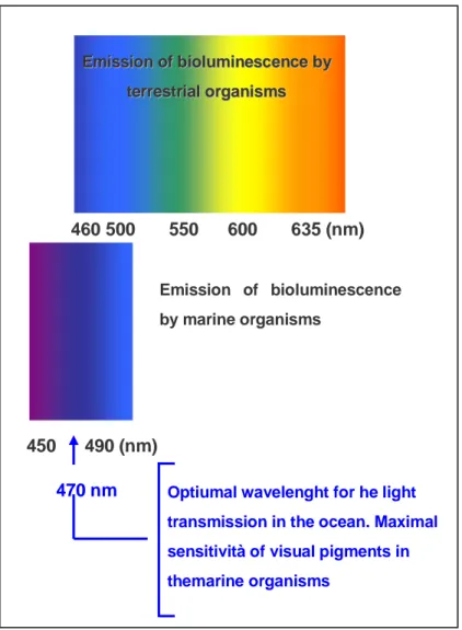
9 marine environment the predominant bioluminescence colour is blue (around 470nm) because light at this wavelength transmits furthest in water so many organism evolved to be sensitive only to blue light, lacking visual pigments for longer or shorter wavelengths. In the terrestrial environment, green is the predominant bioluminescence colour as a result of an ecological adaptation of bioluminescence to the photic environment 3.
Figure 1 Wavelenghts emission of tereestrial and marine organisms.
E Emmiissssiioonnooffbbiioolluummiinneesscceenncceebbyy t teerrrreessttrriiaalloorrggaanniissmmss 460 500 550 600 635 (nm) 450 490 (nm) Emission of bioluminescence by marine organisms
Optiumal wavelenght for he light transmission in the ocean. Maximal sensitività of visual pigments in themarine organisms
10
Bioluminescent systems
Firefly luciferases and mutants
Luciferases from fireflies family are the most studied and the most commonly used. The luciferin-luciferase reaction of fireflies was first demonstrated by Harvey (1917), although the light observed was weak and shortlasting. Thirty years after Harvey's discovery, McElroy (1947) made a crucial breakthrough in the study of firefly bioluminescence. He found that the light-emitting reaction requires ATP as a cofactor. In 1949, McElroy and Strehler found that the luminescence reaction requires Mg2+ in addition to luciferin, luciferase and ATP. The active luciferin was found to be in the D-form while the L-form is practically inactive. Luciferase from Photinus pyralis belongs to the Lampyridae family that are carachterized by sexual dimorfism. Though the females of some species are similar in appearance to males, larviform females are found in many other firefly species. These females can often be distinguished from the larvae only because they have compound eyes. In the major part of the species male are 1cm long and have wings. The organ for light emission is in the terminal part of the abdomen. In some species this organ is bigger in male individuals but generally both sexes can emit light.
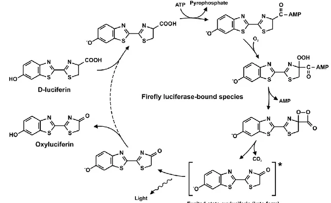
11 The reaction mechanism is not fully understood yet and it consists in the rapid conversion of the substrate luciferin in luciferyl-adenilate linked to the enzyme in presence of Mg2+ and ATP. Pyrophospate is released in this phase then the combination with oxygen lead to the formation of the luciferase-oxyluciferin- adenilate complex and to the elimination of CO2. The complex is in an “excited state” and it
returns to the fundamental state by emitting photons. At the end the complex is dissociated in luciferase, AMP and oxyluciferin (Figure 3)
Figure 3: Mechanism of reaction of firefly luciferase and D- luciferin
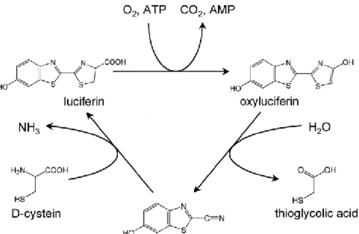
Since luciferase demonstrated a greater affinity for the oxyluciferin compared to reduced luciferin, high concentration of substrate may inhibit the reaction with a competitive mechanism so that the last step of the reaction result to be the limiting one4. Recent studies have demonstrated that in nature Luciferase regenerating enzyme (LRE), peptides of 38 kDa, can convert oxyluciferin in a form that in presence of D-cystein can be converted in luciferin (Figure 4)5,6 . The reactivation of the enzyme luciferase, instead, is operated by the coenzyme A that removes the adenil-oxyluciferin from the surface of the enzyme7.
12
Figure 4. Proposed mechanism of luciferin regeneration
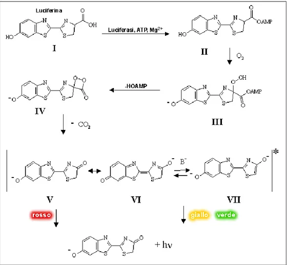
Luciferase from P. pyralis is an enzyme of 62 kDa and with a maximum of emission at 562nm at 25°C and pH 7.8. The enzyme presents two polypeptididic subunit with identical amminoacidic sequences but only one is responsible for bioluminescence activity. The active site create san hydrophobic enviroment with two sulfidric groups essential for the catalytic activity8. In the mechanism proposed by Branchini et al. the red emission (max 615 nm), observed at pH 6.0 is produced by the chetonic form while the yellow-green emission (max 560 nm) at pH 8.0 is produced by the enolate anion form of the excited oxyluciferin9 (Figure 5,6).
Figure 5. Keto-enolTautomerization of D-luciferin
In nature, beetle luciferases have different emission colors: one of the proposed hypotesis is that the charge delocalization in the excited state cause the different wavelenght emissions. Recently other hypotesis have been proposed and the real cause of the different emission colors is still controversial1,10. In order to improve characteristics for analytical application luciferase from Photinus pyralis underwent random and site specific mutagenesis. The group of Prof. B. Branchini generated two variants: Ppy GR-TS and Ppy RE-TS that have a maximum emission wavelength respectively 546nm and 610nm. The former is mutated on the residues Val241Ile,
N S O -S N O* N S S N *O -O
(Eq. 5)
13 Gly246Ala and Phe250 Ser, while the latter on the residue Ser284Thr, which is responsible for the red emission, and Thr214Ala, Ala215Leu, Ile232Ala, Phe295Leu and Glu354Lys for increasing stability at 37°C11. Moreover additional changes in the amminoacidic sequence have been apported : Arg330Gly e Glu354Lys generated the Ppy RE8 form, with a maximum emission wavelength at 617nm. Such a red shift causes a loss of thermostability and activity of the enzyme so that mutations in the amminoacidic residues Phe465Arg e Ile351Val were necessary. Moreover the CDS sequence of Ppy RE8 enzyme has been codon optimized for mammalian expression, by eliminating repeats, local hairpin and criptic splicing sites. Ppy RE8 has 8 different amminoacid compared to the wild type enzyme and its thermostability rends it useful for cell based assay and in vivo imaging12.
14
Click beetle and railroad worm
Click beetle luciferases
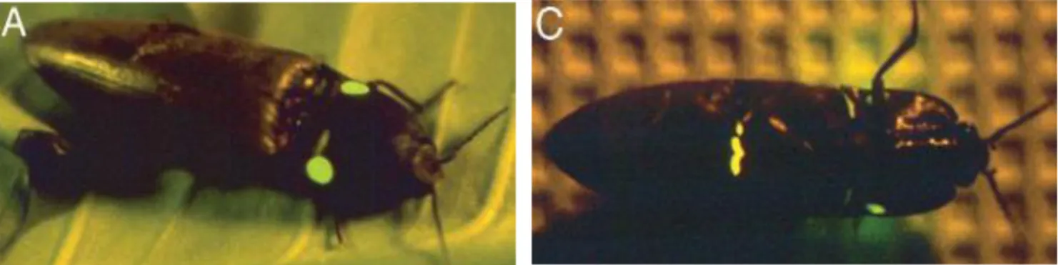
The genus Pyrophorus belongs to the Eleteridae family and includes 26 bioluminescent species amongst Caribbeans, Central anb south America. The biolumienscence reaction is similar to that of coleoptera and requires the same substrate and cofactors even is the ventral light emission is continuos13. They generally eat pollen and little insects like aphids. They lay eggs that are luminescent as larvae that growth up slowly (even years) Pyrophorus plagiophthalamus, the lamaica click beetle present a strong polymorphism in the color of light emission It has two different light organs: a ventral one that emits light in the yellow green (558-562nm) and orange (591-595nm) and two dorsal organs that emit green (544-548nm) and yellow-grren (557-562nm) (figure 7).
Figura 7: Pyrophorus plagiophthalamus emits green light form the dorsal organ (A) and
orange from the ventral ones (B)
Four genes were cloned and mutagenized from this insect that differs one another for emission spectrum and intensity porperties.
15 Thermostable mutants emits in the red region of the Uv/visible spectrum as CBRLuc or in the green region as CBG68luc and CBG99luc. In every sequence the peroxisome targeting sequence has been removed to obtain a citoplasmatic localization of the espresse enzyme14,15. CBG99Luc has a peak of emission at 537 nm while CBRluc at 615 nm at the temperature of 25°C. The most important characteristics is that the emission spectrum of these enzyme is not influenced by variation in the pH like fireflies enzymes(Figure 8).
Railroad worm
The railroad worm Phrixothrix is well known for displaying two different colors of luminescence from a single organism. This genus is widely distributed in Central and South America and belongs to the family of Phengodidae (Figure 9).The larva of
Phrixothrix (and also the adult female) emits a greenish yellow light (max 535-565 nm) from 11 pairs of luminous organs on the posterior lateral margins of the second to the ninth segment, and a red light (max 600-620 nm) from the luminous area on the head. The adult male is a typical beetle and does not show a noticeable luminescence. The bioluminescence systems of these phengodids were essentially the same as that of the fireflies, involving the same luciferin (firefly luciferin), ATP and Mg +. Their emission maxima of luminescence from the lateral and head organs are in the ranges of 535-592nm and 562-638 nm, respectively. The color differences are probably due to the presence of luciferase isoenzymes (Mr about 60,000) according to the authors16. Viviani et al. (1999) cloned the luciferases from the lateral light organs of Phrixothrix vivianii (emission Xmax 542 nm) and the head light organs of
Phrixothrix hirtus (emission Amax 628 nm). Recently, the latter enzyme has been
mutated for improving stability and activity17.
16
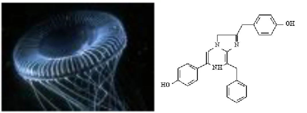
Renilla and Gaussia lucifarases
Luciferase from Renilla reniformis
Renilla reniformis, is a marine coral belonging to the family of Renilladae, class of Anthozoa (Figure 10). Renilla luciferase is a monomeric photprotein with a molecular wheight of 36Kd and shares the conserved catalytic triad of residues employed by the dehalogenases, as confirmed by its crystallographic data18. The enzymatic reaction does not require ATP and its substrate coelenterazine has a good penetration through cell membrane. The use of Renilla luciferase as reporter gene involves its application in combination with fireflies luciferase for dual reporter assay. Coelenterazine, shown in figure 10, is the common substrate for Renilla luciferase, apoequorin, gaussia and metridia luciferases that have been recently cloned. The luciferase/coelenterazine reaction produces mostly blue light: peak of emission of Renilla luciferase is 480nm. In 2007 A.Loening et al. generated by random and site directed mutagenesis Renilla variants with different emission spectrum and increased activity and stability19.Rluc8 shows an emission peak at 535nm has an half life of 50h and an activity of 1.4 fold higher than the native enzyme. Consequently Rluc8 improved the sensitivity of the detection in both in vitro and in vivo applications. Moreover they generated a new red shifted mutant Rluc7-535 with an half life comparable to the one of the native enzyme (6.5H) in order to achieve sensitivity in transient gene expression analysis20. Renilla luciferase has been widely applied as luminescent donor in bioluminescence resonance Enrgy Transfer method for studing protein-protein interaction21.
17
Luciferase from Gaussia princeps
Gaussia luciferase (Gluc) is a novel secreted luciferase, cloned from the copepod Gaussia princeps, that catalyzes the oxidation of the small molecule coelenterazine to produce light (Figure 11). Unlike the firefly luciferase systems, these coelenterazine-utilizing luciferases do not require accessory high-energy molecules such as ATP, simplifying their use in a number of reporter applications22,23. This luciferase catalyzes the oxidation of the substrate coelenterazine in a reaction that emits light (λem = 470 nm),and has considerable advantages over other reporter genes. Thanks to its pH resistance in a range from 3 to 11 with an optimum at pH 7.8, and its good thermostability (up to 60°C), GLuc is an attractive tool in report gene assays or as an enzyme label for bioanalytical applications, particularly for the development of bioluminescence (BL) cell based assays and for BL imaging animal models. GLuc is the smallest luciferase isolated to date (19.9 KDa) and this small size is a crucial factor for the construction of fusion proteins to avoid steric hindrance. Moreover since Gluc, when expressed into mammalian cells, is secreted into the culture medium the BL measurements are performed by simply addition of coelenterazine in culture medium, without the need for cell lysis.
18
Bioanalytical application of bioluminescence
The typical bioanalytical applications of bioluminescence (BL) proteins include the investigation of protein–protein interactions, protein conformational changes, protein phosphorylation, second-messengers expression, and, in general, the study of gene expression and gene regulation in vitro and in vivo24,25. The expression of a BL protein can be put under the control of tissue-specific regulatory elements allowing non-invasive imaging of physiological and pathological processes like differentiation, apoptosis, tumor progression, and inflammation, even in a 3D fashion by means of BL tomography, which allows 3D BL source reconstruction26. Since BL proteins can be detected down to very low levels, they allow ultrasensitive detection of the target analytes and monitoring of the physiological phenomena under investigation. These BL features, associated to instrumental and technical advancements in miniaturization, enable the analysis of small-volume samples, which leads to the development of miniaturized and high-throughput assays.
Bioluminescence imaging in vivo
Bioluminescence in vivo imaging The commercial availability of ultrasensitive imaging systems based on charge-coupled device (CCD) technology together with the high number of BL probes greatly expanded the use of BL in a variety of imaging formats and techniques, spanning from Petri dishes to microtiter plates and to whole-animal imaging. Usually the spatial resolution of the BL signal is in the order of 100–200 µm and may reach 0.4 µm when in combination with optical microscopy, thus similar to that achieved with conventional light imaging.27 Optical imaging makes it possible to reveal cellular and molecular events in real time, thus tracking biological processes in living animals with a significant reduction in the number of animals needed. Diverse imaging strategies have been developed and successfully employed to study tumour progression and metastasis, infectious pathways of viruses, gene expression patterns, graft-versus-host diseases.28-31 Thanks to high sensitivity and the availability of new red and BL proteins emitting in the near infra-red (whose emission is scarcely absorbed by animal tissues) in vivo BL molecular imaging is emerging as one of the leading imaging technologies in the areas of cancer biology, cell biology, gene therapy and stem cell research. Moreover, the advancements in molecular biology allowed to obtain organisms in which the expression of a BL protein is under the control of tissue-specific regulatory elements, allowing non-invasive imaging of
19 selected physiological or pathological processes. Furthermore, BL tomography enables 3D light source reconstruction to obtain, for example, tumor shape in small animals. However, many factors have to be considered when a BL molecular imaging experiment has to be settled up, for instance optical properties (scattering and absorption properties) and thickness of the tissue through which photons are travelling. Haemoglobin is the main chromophore within tissues absorbing light in the visible spectrum (400 to 760 nm) but, if the animal is pigmented, also melanin contributes to light absorption. Haemoglobin light absorption is significantly lower at wavelengths longer than 600 nm. Unfortunately, wild-type BL proteins usually emit in the blue-green spectral region and, therefore, much effort has been recently put to develop of red and near-infrared emitting BL proteins for in vivo imaging applications. Bacterial, firefly and Renilla luciferases are the most used BL reporter proteins in whole-body imaging. Bacterial luciferase is the only reporter protein that allows the construction of self-luminescent engineered organisms through the introduction in the cell of the whole lux gene cassette (luxCDABE), which contains the genes encoding both for luciferase and for the enzymes able to synthesize its BL substrate. Genetically engineered BL bacteria (Escherichia coli, Vibrio cholerae, Salmonella
typhimurium) based on the luxCDABE system have been employed to localize
tumors and metastasis in living animals. Such bacteria take advantage of the anaerobic microenvironment and the nutrient-rich growth conditions, being able to replicate in the necrotic central area of tumors.32 Besides tumor and metastasis localization, self-luminescent bacteria may have others promising applications, for example monitoring of bacteria-mediated gene product delivery systems for therapy of solid tumors. Firefly luciferase is by far the most employed BL protein in several bioanalytical applications. Mutant luciferases with different spectral properties have been developed by random and site-directed mutagenesis of P. pyralis wild-type luciferase,33 and a single amino acidic residue (Ser284) appeared to be the most promising for developing mutants with altered emission properties.34 Besides emission color, also the thermostability of BL reporters is an important factor for in vivo imaging applications. Indeed, BL measurements performed in cell cultures and other in vitro assays are usually taken at room temperature, on the contrary in vivo imaging has to be performed at body temperature (37°C). A systematic study to investigate the thermostability of commercially available luciferases (FLuc+, CBGr68, CBRed, and hRLuc) was recently performed by Zhao et al, who reported a 34-nm
20 spectral red shift for the the firefly enzyme.35 The study showed that for all the investigated luciferases the activity increased with temperature, and the luciferases with the highest increments were CBGr68 (6.4-fold) and CBRed (7.8-fold). Spectral profiles are also affected by temperature and the most evident change was observed for firefly luciferase, whose emission maximum shifted from 578 nm at 25°C to 612 nm at 37°C. A long-term continuous delivery system based on implanted micro-osmotic pumps was also developed to overcome one of the main pitfalls of firefly luciferase, the need for the BL substrate D-luciferin.36 This system did not require repetitive injections of the BL substrate that, together with substrate pharmacokinetics, put constraints on intervals between image acquisition.
BLI- based mouse models for drug discovery and development
BLI has become a routine modality for use in cancer biology particularly suited for assessing tumor burden and metastatic spread. The most common use of BLI in cancer has been to assess mass and location of xenografted cells constitutively expressing luciferase, providing a robust strategy to monitor effectiveness of anti-tumor drugs in vivo. Whole body BLI using firefly luciferase allows semi-quantitative measurements of tumor load and progression, metastasis and treatment response. Due to the sensitivity of BLI luciferase-expressing tumor cells can be transplanted to at any orthotopic site within a mouse or rat and subsequent tumor development, progression, and possible metastasis can be monitored in a rapid and time-sensitive manner. Also BLI has proven very useful for the early detection of micro-metastases and minimal residual disease states in animal37.38. Apart from preclinical studies on cancer BLI-based mouse models are routinely used by pharmaceutic companies since they provide information on where a drug or compound takes part in a specific regulatory pathway related to the disease. A growing number of luciferase expressing animal models are (commercially) available for drug metabolism and toxicology, disease areas like oncology/angiogenesis, metabolic -and neurodegenerative diseases and inflammation39-45. That is consequently to the development of the methodology for producing reporter animals by efficiently integrate luciferase gene in the mouse genome under the control of a specific promoter. Luciferase expressing mouse models can highlight the mechanism of many regulatory sequences. BLI can be used to monitor inflammation by driving luciferase with inflammation-specific
21 regulatory sequences. For instance, Carlsen et al. generated mice expressing luciferase under the control of the regulatory sequences of the NFκB gene, which in its turn is under the direct control of TNFα, a key cytokine produced during inflammation. Using NFκB-luciferase mice, Carlsen et al.46
monitored osteoarthritic inflammation induced by injection of bacterial lipopolysacharide, and quantified the therapeutic potential of dexamethasone treatment of the arthritic lesion. Moreover luciferase expressing animal models can be employed for studying drug availability and distribution47.
Cell based assay in drug discovery and development
Cell-based assays include a variety of assays that measure cell proliferation, toxicity, production of markers,motility, activation of specific signalling pathways and changes in morphology.Many of these assays rely on reporter gene technology.
Due to signal amplification of cell-signalling cascades, reporter gene assays are very sensitive, and thus ideal forminiaturization; they have been widely applied in HTS formats48,49. Despite these advantages, such assays are based on signal-transduction events that occur downstream of receptor activation and require gene expression. This causes long response times, which span from hours to days for the whole analysis time, and the possibility of interference from other intracellular pathways. For these reasons, alternative approaches relying on the monitoring of the first activation step, e.g., receptor dimerization, for example fluorescence and bioluminescence resonance energy transfer (FRET and BRET) and split complementation assays have also been proposed to satisfy the demands of HT drug discovery47-50. Recent improvements in optical imaging instrumentation and the wide choice of bioluminescent and fluorescent probes fuelled the implementation of cell-based assays for high-content screening (HCS). Because a very comprehensive overview has recently been published by Zanella et al. this review will not deal with HCS51. Cellular screening still presents a variety of challenges, and key aspects of improving this early phase of the drug-discovery process seem to be predictability, automation, miniaturization, cost-effectiveness, high-speed, and multiplexing.The state of the art, and challenges and future directions,will be discussed in this review, together with an up-to-date overview of recent ameliorations and trends in cell-based assays for drug discovery.
22 The implementation of reporter gene assays in thedrug-discovery process enables in vitro investigation of ADMET (absorption, distribution, metabolism, elimination,
and toxicity) properties well in advance of animal studies. The main ADMET-related genes that have beentargeted are those encoding for drug metabolizing enzymes, for example the cytochrome P450 family(e.g., CYP3A4, CYP2B6, CYP2C9, and CYP2C19) anddrug transporters (e.g., MDR1, MRP2, MRP4, BSEP,BCRP, and NTCP) whose expression is regulated at thetranscriptional level by many of nuclear receptors (e.g., PXR, CAR, GR, and PPARα)52. The key advantages of reporter assays are highsensitivity, reliability, convenience, dynamic range, and adaptability to high throughput-screening. The major weakness encountered in their use is the high variability of cell response, mainly caused by sample-aspecificeffects on cell vitality. To improve robustness, an internalor external reference signal must be introduced in order to correct the analytical response and separate the specific signal from nonspecific interferences. This can be easily achieved by introduction of a second reporter gene which is constitutively expressed and whose activity thus parallels cell vitality. Commercial dual reporter assays with bioluminescence detection were introduced commercially to address this issue but, being based on the measurement of firefly and Renilla luciferase in the same sample, they require addition of a reagent to stop one reaction before adding the second substrate. This inevitably increases assay cost and time. More interestingly, use of new BL and fluorescent proteins with altered emission properties enabled simultaneous monitoring of more reporters in the same cell.The use of reporter proteins emitting at different wavelengths facilitates separation of the analytical and the control signals and expands the applicability of these reporters to multiplexed cell-based assays.The major pitfall of reporter gene assays is the possible disengagement between changes in enzyme activity and its corresponding mRNA levels. For example Lim et al. reported that antibiotic rifampicin and the natural furanocoumarin bergamottin (from grapefruit juice) both activate CYP3A4 gene transcription, but enzyme activity is increased by rifampicin and reduced by bergamottin, which is able to covalently inactivate the enzyme53.This disconnection can be hindered by reporter gene assays, which could thus be combined with other drug screeningassays to increase their predictability.
23
Multicolor luciferases for multiplexed analysis
HTS reporter gene cell-based assays for lead identification are usually performed in serial or parallel mode and few multiplexed assays have been reported to date. Although the concept of multiplexed screening is well appreciated for image-based technologies, it is emerging as a more feasible option for plate-based, homogeneous assays, because implementation is simple, accessible, and cost effective. An efficient drug-discovery workflow needs the development of reliable information-rich assays. Recently, a cell-based transactivation high-throughput luciferase reporter assay has been developed to identify potential cytochrome P-450 3A4 (drug-metabolizing enzyme) inducers. This 384-well multiplexed homogeneous assay was developed and validated for simultaneous detection of PXR transactivation and HepG2 cell cytotoxicity by combining as fluorescence and bioluminescence readouts. When its analytical performance was compared with that obtained with the conventional singleplex PXR transactivation assay (with separate toxicity assay), using four well-known PXR inducers, the reported EC50 values were not statistically different in either the singleplex or multiplex formats. The authors reported that switching from singleplex to multiplex reduced the overall number of cells by 29% and the consumable costs by 38%. Furthermore, use of cryopreserved cells with multiplexing and automation enabled elimination of a total of 92 processing steps (42% reduction)54. Some multiplexed reporter assays have been developed. For example, Nakajima et al. proposed a novel reporter assay system in which three luciferases that emit at different wavelengths (green, orange, and red) in the presence of the same substrate are used as reporter proteins. By using longpass filters and applying a signal processing algorithm, a cell-based monitoring system was developed for simultaneous evaluation of the expression of three different target genes within a cell, achieving a dynamic range of three orders of magnitude55. As an alternative, we proposed the combined use of secreted BL reporter proteins, for example Gaussia princeps luciferase, and intracellular proteins (the green-emitting P. pyralis luciferase and a red-emitting thermostable mutant of L. italica luciferase). We developed a multicolour cell-based assay for CYP7A1 and CYP27A1, the two main enzymes responsible for bile acid biosynthesis, and the secreted Gaussia luciferase was used as vitality control under the regulation of a constitutive promoter. The use of a secreted luciferase makes measurement of its activity straightforward, because its expression is measured simply by taking small aliquots of cell culture medium.
24 Besides, the combination of secreted and intracellular reporters in the same cell-based assay has the advantage of complete absence of interference between the two luminescence signals, which are measured in separate wells of high-throughput microplate formats56. This concept could be easily applied in the drug screening workflow to improve consistency of cell-based assay results and reduce cost and time.
Protein-protein interaction studies
In the postgenome era, the analysis of protein expression, protein structure and protein-protein interaction is a much harder task since a global perspective is necessary to understand the complex network of interactions involving proteins, nucleic acids, co-factors and other unknown actors that participate to biochemical and pathological processes. Diverse technologies, ranging from protein affinity chromatography to library-based methods (e.g., phage display, two-hybrid system), clustered together under the term “proteomics”, have been developed to investigate protein expression and function in cells and organisms. Among these, elegant approaches relying on Bioluminescence Resonance Energy Transfer (BRET) and split reporter protein complementation and reconstitution strategies have been employed to investigate protein-protein interactions and explore biological pathways57. Bioluminescence Resonance Energy Transfer is a nonradiative resonance energy transfer occurring between a light-emitting luciferase donor and an acceptor fluorescent protein. When donor and acceptor are brought into close proximity (1-10 nm) to one another and are properly oriented each other, the former transfers its energy to the latter, which then emits. Since light emission from the donor takes place at a different wavelength than that from the acceptor, the energy transfer can be easily detected by measuring the ratio of the acceptor to the donor emission intensities. Such ratiometric output allows to compensate for well-to-well aspecific signal variations (e.g., due to different cell numbers in each well or signal decay across the plate). Because BRET strictly depends on the molecular proximity between donor and acceptor, it is suitable for monitoring the activation state of any protein (e.g., receptor or transcription factor) that undergoes association or conformational changes as a consequence of ligand binding. For example, to evaluate receptor dimerization the two subunits of the receptor are genetically tagged
25 either with the donor (e.g., Renilla or firefly luciferase) or the acceptor (e.g., a green fluorescent protein variant). When the activation of the receptor brings the donor and acceptor in a favourable position, BRET will occur. This phenomenon can be either observed in vitro using purified proteins or directly within the cells where the fusion proteins were produced58. Differently from Fluorescence Resonance Energy Transfer (FRET), BRET does not require a light source, therefore it is not affected by photobleaching, light scattering or autofluorescence, and direct excitation of the acceptor cannot take place. The intrinsic low background of BRET should allow either detection of weak interactions and performing experiments with low concentrated proteins. As of today, several combinations of BRET formats and reagents are available for proteomics applications, including receptor research and mapping of signal transduction pathways59. The first application of BRET methodology goes back to 1999 and regarded the study of the dimerization of cyanobacteria circadian clock proteins60. Bioluminescence Resonance Energy Transfer has been successfully applied in the study of receptor dimerization, like the insulin receptor61, and to the evaluation of the homo- and hetero-dimerization of opioid receptors in live cells62. Estrogen receptor homodimerization was studied with BRET and BRET2, a BRET variant in which the energy transfer occurs between
Renilla luciferase and a green fluorescent protein mutant (GFP2)63,64. Extended BRET (eBRET) is also gaining popularity as a technique that allows the monitoring of protein-protein interactions in real time for many hours65. Split reporter-based BL imaging is a newly developed strategy for studying protein-protein and, more generally, intracellular interactions. It relies on the complementation-reconstitution concept, i.e., the spontaneous assembling (in some circumstances) of active/functional proteins from one or more polypeptide fragments. In the split reporter strategy, a BL protein is cleaved into N-terminal and C-terminal fragments and each fragment is linked to one of the target interacting proteins. When protein-protein interaction brings the two fragments close each other, the complete functionality of the BL protein is recovered (Figure 11). This approach works either via protein segment complementation assays through a non-covalent assembly or via intein-mediated reconstitution assays based on covalent binding. A number of reporter proteins have been used for split protein strategies, for example firefly luciferase, Renilla luciferase, GFP, β-galactosidase66-69
26
Figure 12: Schematic view of the split reporter-based complementation strategy. The
N-terminal and C-N-terminal halves of a reporter protein (e.g., luciferase) are genetically fused to a recognition element (receptor, ligand binding domain) through a short peptide linker. The interaction with the target analyte causes a conformational change that recovers the reporter protein activity through the protein complementation of split N- and C-terminal fragments.
Recently a rapid screening assay based on a genetically encoded BL biosensor was developed to assess the androgenic effect of ligands by detecting the intramolecular association of the androgen receptor ligand binding domain (AR LBD)70. The authors demonstrated for the first time the possibility to use a single molecule-format BL probe to monitor cellular signalling steps. The AR LBD and the N-terminal domain of AR (AR NTD) where sandwiched between the dissected fragments of firefly luciferase. The association of AR LBD and AR NTD in the presence of androgens causes the complementation of the N- and C-terminal fragments of the luciferase, which partially recover its activity. Different chemicals known to possess agonist or antagonistic androgenic activity were assayed with this test, which was able to detect in a short time (20 minutes) as low as 10-5M dihydroxytestosterone (DHT). Recently, Kim and colleagues also validated a single-molecule-format complementation system of split click beetle luciferase (CBLuc) to investigate protein-protein interactions. The ligand binding domain of the androgen receptor was connected to a functional peptide sequence via a flexible linker. This fusion protein was sandwiched between the dissected N- and C-terminal fragments of CBLuc. In the presence of androgens, the association between AR LBD and a functional pepdide causes the complementation of N- and C-terminal fragments of CBLuc. After 20-min stimulation
27 of the MCF-7 cells carrying the fusion construct with 10-5M DHT luminescence intensities 29 times higher than a control were produced671 A combinatorial screening approach was used to identify novel firefly luciferase split sites with improved characteristics over the previously published ones72. A total of 115 different combinations were screened and the fragments were characterized with five different interacting proteins and an intramolecular folding strategy. A novel firefly luciferase split reporter showing increased sensitivity and complementation was identified, with potential application for the study of protein-protein and other interactions in cells and animal models. An intramolecular luciferase complementation probe for the detection of specific RNAs vas developed by constructing a peptide-inserted firefly luciferase containing short RNA-binding peptide sequences73. The same principle was applied by Kanno et al. to investigate the release of proteins from mitochondria toward cytosol74. For this purpose, a target mitochondrial protein was fused to a N-terminal fragment of Renilla luciferase and a N-terminal fragment of DnaE intein and expressed in the mitochondria of mammalian cells. If, for some reasons, the genetically modified target protein is released from the mitochondria, it interacts with a C-terminal fragment of DnaE interin fused to a C-terminal Renilla luciferase present in the cytosol, thus reconstituting an active Rluc. This method allowed high throughput screening of chemicals able to increase or inhibit the release of mitochondrial proteins in living cells and small animals.
28
Bibliography
1. Nakatsu T, Ichiyama S, Hiratake J, Saldanha A, Kobashi N et al. (2006)
Nature 440, 372-376
2. Shimomura O “Introduction” in Bioluminescence: chemical principles and methods O Shimomura (Ed.) World Scientific, Singapore 2006
3. Viviani VR. (2006) Cell. Mol. Life Sci. 59: 1833-50
4. White E., Branchini B.R.:(1975) J Am Chem Soc.5;97(5):1243-5.
5. Gomi K. e Kajiyama N.:(2001) The journal of biological chemistry, Sep 28;276(39):36508-13
6. Day J. C. e Bailey M. J. (2003) Insect Molecular Biology,
7. Airth R.L., Rhodes W.C., McElroy W.D.(1958) The function of coenzyme A in
luminescence, Biochim Biophys Acta
8. DeLuca M. (1969) Hydrophobic nature of the active site of firefly luciferase,
Biochemistry, 1969.
9. Branchini B.R., Magyar R.A., Murtiashaw M.H., Portier N.C. (2001) Biochemistry
10. Navizet I, Liu YJ, Ferré N, Xiao HY, Fang WH, Lindh R. (2010) J Am Chem
Soc;132(2):706-12
11. Branchini BR, Southworth TL, Khattak NF, Michelini E, Roda A. (2005) Anal
Biochem. 345(1):140-8
12. Branchini B.R., Ablamsky D.M., Davis A.L., Southworth T.L., Butler B et al. (2009) Anal Biochem. 396(2):290-7
13. Feder J. L. e Velez S. (2009) Evolution 63(5):1203-16
14. Stolz U., Velez S., Wood K.V., Wood M., e Feder J.L (2003) PNAS
100(25):14955-9
15. Almond B., Hawkins E., Stecha P., Garvin D., Paguio A. et al.(2003) Promega Notes 85
16. Viviani VR, Bechara EJ, Ohmiya Y. (1999) Biochemistry. Jun 29;38(26):8271-9.
17. Li X, Nakajima Y, Niwa K, Viviani VR, Ohmiya Y. (2010) Protein Sci. 19(1):26-33.
18. Stepanyuk GA, Liu ZJ, Markova SS, Frank LA, Lee J et al (2008) Photochem
29 19. Loening AM, Wu AM, Gambhir SS. (2007) Nat. Methods 4: 641–643.
20. Loening AM, Dragulescu-Andrasi A, Gambhir SS. (2010) Nat Methods. 7(1):5-6.
21. De A, Ray P, Loening AM, Gambhir SS. (2009) FASEB J 23(8):2702-9.
22. Tannous BA, Kim DE, Fernandez JL, Weissleder R, Breakefield XO. (2005)
Mol. Ther. 11:435-443
23. Maguire CA, Deliolanis NC, Pike L, Niers JM, Tjon-Kon-Fat LA et al. (2009) Anal Chem. 81(16):7102-6.
24. Roda, A., Pasini, P., Mirasoli, M., Michelini, E., Guardagli, M. (2004) Trends.
Biotechnol. 22, 295-303.
25. Roda, A., Guardagli, M., Pasini, P., Mirasoli, M. (2003) Anal. Bioanal. Chem. 377, 826-33.
26. Sato, A., Klaunberg, B., Tolwani, R. (2004) Comp. Med. 54, 631-4.
27. Roda A, Guardigli M, Michelini E, Mirasoli M, Pasini P. (2003) Anal
Chem;75:463-70.
28. Asokan A, Johnson JS, Li C, Samulski RJ.(2008) Gene Ther doi:10.1038/gt.2008.127.
29. Rogers KL, Martin JR, Renaud O, Karplus E, Nicola MA, Nguyen M, et al. (2008) J Biomed Opt;13:031211.
30. Kang JH, Chung JK.(2008) J Nucl Med;49(Suppl 2):164-79. 31. Shapiro E, Lu C, Baneyx F. (2005)Protein Eng Des Sel;18:581-7. 32. Pawelek JM, Low KB, Bermudes D. (1997)Cancer Res ;57:4537-44.
33. Branchini BR, Ablamsky DM, Murtiashaw MH, Uzasci L, Fraga H, Southworth TL.(2007) Anal Biochem; 361:253-62.
34. Tafreshi NKh, Sadeghizadeh M, Emamzadeh R, Ranjbar B, Naderi-Manesh H, Hosseinkhani S.(2008) Biochem J;412:27-33.
35. Zhao H, Doyle TC, Coquoz O, Kalish F, Rice BW, Contag CH. (2005) J Biomed Opt;10:41210.
36. Gross S, Abraham U, Prior JL, Herzog ED, Piwnica-Worms D. (2007) Mol
Imaging;6:121-30.
30 38. Edinger M, Cao YA, Hornig YS, Jenkins DE, Verneris MR et al. (2002) Eur. J.
Cancer, 38, 2128.
39. Wetterwald A, van der PG, Que I, Sijmons B, Buijs J et al. Karperien et al. (2002) Am. J. Pathol., 160, 1143.
40. Luo J, Lin AH, Masliah E, Wyss-Coray T, (2006) Proc. Natl. Acad. Sci. U. S. A, 103, 18326.
41. Li Z, Wu JC, Sheikh AY,Kraft D, Cao F, et al. (2007) Circulation, 116, I46. 42. Van der Bogt KE, Sheikh AY, Schrepfer s, Hoyt G, Cao F, Ransohoff KJ et al.
(2008) Circulation,118, S121.
43. Hutchens MA, Luker KE, Sonstein J, Nunez G, Curtis JL, et al.(2008) PLoS.
Pathog.,4, e1000153.
44. Glomski IJ, Piris-Gimenez A., Huerre M, Mock M, Goossens PL et al. (2007)
PLoS. Pathog., 3, e76.
45. Disson O, Grayo S, Huillet E, Nikitas G, Langa-Vives F, et al. (2008) Nature, 455, 1114.
46. Carlsen H, Moskaug JO, Fromm SH,Blomhoff R, (2002) J. Immunol.,, 168, 1441.
47. Mata De Urquiza, A., Solomin, L. & Perlmann, T. (1999) Proc. Natl Acad.Sci.
USA 96, 13270–13275.
48. Paul SM, Mytelka DS, Dunwiddie CT, Persinger CC, Munos BH et al. (2010)
Nat Rev Drug Discov 9:203–214
49. Weigelt J (2009) EMBO 10:941–945
50. Chan JN, Nislow C, Emili A (2010) Trends Pharmacol Sci 31:82–88 51. ShouWZ, Zhang J (2010) Expert Opin DrugMetab Toxicol 6:321–336
52. Kieltyka K, Zhang J, Li S, Vath M, Baglieri C et al. (2009) Rapid Commun
Mass Spectrom 23:1579–1591
53. Banerjee P, Bhunia AK (2009) Trends Biotechnol 27:179–188 54. Wu MH, Huang SB, Lee GB (2010) Lab Chip 10:939–956
55. Nakajima Y, Kimura T, Sugata K, Enomoto T, Asakawa A et al. (2005)
Biotechniques 38:891–894
56. Michelini E, Cevenini L, Mezzanotte L, Ablamsky D, Southworth T et al.(2008)
Anal Chem 80:260–267
57. Massoud TF, Paulmurugan R , De A, Ray P, and Gambhir SS. (2007), Curr.
31 58. Pfleger KD, and Eidne KA (2006), Nat. Methods, 3, 165.
59. Jiang LI, Collins J, Davis R, Lin KM, DeCamp D et al. (2007), J. Biol. Chem., 282, 10576.
60. Xu Y, Piston DW and Johnson, C.H. (1999), Proc. Natl. Acad. Sci. USA, 96, 151.
61. Issad T, Boute N, and Pernet KA. (2002), Biochem. Pharmacol., 64, 813. 62. Wang D, Sun X, Bohn LM, and Sadee W. (2005), Mol. Pharmacol. 67, 2173. 63. Michelini E, Mirasoli M, Karp M, Virta M and Roda, A. (2004), Anal. Chem., 76,
7069.
64. Koterba KL, and Rowan BG. (2006), Nucl. Recept. Signal., 4, 21.
65. Pfleger KD, Dromey JR, Dalrymple MB, Lim EM, Thomas WG, and Eidne KA (2006), Cell Signal., 18, 1664.
66. Wehrman T, Kleaveland B, Her JH, Balint RF, and Blau HM (2002), Proc. Natl.
Acad. Sci. USA, 99, 3469.
67. Kaihara A, Kawai Y, Sato M, Ozawa T, and Umezawa Y. (2003), Anal. Chem., 75, 4176.
68. Paulmurugan R, and Gambhir SS. (2006), Proc. Natl. Acad Sci. USA, 103, 15883.
69. Kim SB, Ozawa T, Watanabe S, and Umezawa Y. (2004), Proc. Natl. Acad.
Sci. USA, 101, 11542.
70. Kim SB, Awais M, Sato M, Umezawa Y, and Tao H. (2007), Anal. Chem., 79 (5), 1874-80
71. Kim SB, Otani Y, Umezawa Y, and Tao H. (2007), Anal. Chem., 79(13):4820-6.
72. Paulmurugan R, and Gambhir SS. (2007), Anal. Chem., 79 , 2346.
73. Endoh T., Mie M, Funabashi H, Sawasaki T, Endo Y et al. (2007), Bioconjug.
Chem., 18(3):956-62
32
Chapter 2
In vivo bioluminescence imaging of murine xenograft cancer models
with a red-shifted thermostable luciferase
Mezzanotte L, Fazzina R, Michelini E, Tonelli R, Pession A, Branchini B, Roda A.
33
Abstract
Purpose. Conventional in vivo bioluminescence imaging using wild-type green-emitting
luciferase is limited by absorption and scattering of the bioluminescent signal through tissues. Imaging methods using a red-shifted thermostable luciferase from P.pyralis were optimized to improve the sensitivity and image resolution. In vivo bioluminescence imaging performance of these red- and green-emitting luciferases were compared in two different xenograft mouse models for cancer.
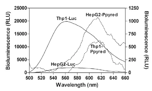
Methods. HepG2 (human hepatoblastoma cell line) and Thp1 (human acute monocytic
leukemia cell line) cells were genetically engineered using retroviral vector technology to stably express the red-shifted or the wild-type green luciferase. A xenograft model of liver cancer was established by subcutaneous injection of the HepG2 engineered cells in the flank regions of mice, and a leukemia model was generated by intravenous injection of the engineered Thp1 cells. The cancer progression was monitored with an ultrasensitive charge-coupled device (CCD) camera. The relative intensities of the green- and red-emitting luciferases were measured and the resulting spatial resolutions of the images were compared. Imaging was performed with both intact and scarified live animals to quantify the absorption effects of the skin and deep tissue.
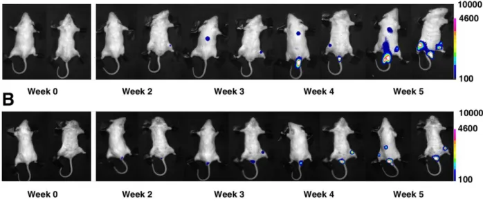
Results. The red-emitting luciferase was found to emit a bioluminescence signal with
improved transmission properties compared to the green-emitting luciferase. By imaging the HepG2 models, which contained tumours just beneath the skin, before and after scarification, the percentage of light absorbed by the skin was calculated. The green bioluminescent signal was 75%±8 absorbed by the skin, whereas the red signal was only 20%±6 absorbed. The Thp1 model, which contains cancer cells within the bones, was likewise imaged before and after scarification to calculate the percentage of light absorbed by all tissue under the skin. This tissue was responsible for 90%±5 absorption of the green signal, but only 65%±6 absorption of the red signal.
Conclusion. Two different bioluminescent mouse cancer models demonstrate the utility of a
new red-shifted thermostable luciferase, Ppy RE-TS, that improved the in vivo imaging performance when compared with wild-type P. Pyralis luciferase. While wild-type luciferase is currently a popular reporter for in vivo imaging methods, this study demonstrates the potential of red-emitting firefly luciferase mutants to enhance bioluminescence imaging experiments.
34
Introduction
Amongst the recent molecular imaging approaches, including Positron Emission Tomography (PET), Single Photon Emission Computed Tomography (SPECT) and Magnetic Resonance Imaging (MRI), Bioluminescence Imaging (BLI) with luciferase reporters is continuously gaining popularity and is largely applied in preclinical cancer research. BLI is often preferred because it provides a simple, sensitive, robust imaging modality with relatively cost-effective instrumentation. BLI and Fluorescence Imaging (FLI) systems are based on the use of bioluminescent reporter genes such as luciferases or fluorescent proteins expressed in cells (e.g., cancer cells, stem cells, bacteria) that are inoculated into small experimental animals and imaged in vivo with the use of ultrasensitive charge-coupled device (CCD) cameras, able to detect light emission even from deep tissues [1,2,3].Throughout the last decade, BLI has been used in xenograft animal cancer models to investigate the factors involved in malignant transformation, invasion and metastasis, and to examine responses to cancer therapy [4]. Such animal models are quite accessible to researchers, because they are easy to make and simple to use. In vivo BLI with these models allows for direct or indirect physiological imaging of specific cellular and molecular events. To initiate the luciferase reaction, luciferin substrate must be intravenously (i.v.) infused or intraperitoneally (i.p.) administered to the animal in sufficient concentration to saturate the reporter enzyme at the anatomical region of interest. Almost all luciferases require the administration of the substrate with the exception of bacterial luciferase, since the lux operon cassette codes not only for the luciferase but also for enzymes to produce the substrate. Unlike fluorescent reporter proteins, which require an excitation source, luciferase reporters generate a highly detectable BL signal with virtually no background noise, resulting in impressive imaging sensitivity. Luciferase from the North American firefly Photinus Pyralis provides a particularly strong signal, due to the high quantum yield (~0.41) of the luciferase/luciferin reaction [5]. The choice of luciferase in a BLI system is a crucial means by which to ameliorate and optimize the imaging technique. Though P. Pyralis luciferase is the most common choice, luciferase from the sea pansy Renilla reniformis, the green- and red-emitting luciferases from the click beetle Pyrophorus plagiophalamus and luciferase from the copepod Gaussia princeps have also been investigated [6,7]. Most of these enzymes emit in the blue/green region of the UV/vis spectrum, where light is strongly absorbed and scattered by tissues. Consequently, the imaging performance suffers
35 from poor sensitivity and spatial resolution. While green light is strongly absorbed by haemoglobin, melanin and other pigmented macromolecules, light greater than 600 nm in wavelength is less strongly absorbed [8], and can travel through living tissue over a much greater distance (1/e absorption length in tissues of 2cm) [9]. Despite the advanced transmission properties of red-emitting luciferases, the red click beetle luciferase recently proposed for molecular imaging was demonstrated to be non-suitable for in vivo imaging due to its poor thermostability [10]. Recently, a
thermostable red-shifted mutant of luciferase from P. pyralis was created by random and rational mutagenesis [11].This luciferase, named Ppy RE-TS, has an emission maximum of 612 nm at pH 7.0, a narrow emission bandwidth and excellent thermostability (the half-life at 37°C is 8.8 hrs, versus 0.26 hrs for wild-type luciferase). We report here the in vivo optical and luminescence properties of
PpyRE-TS, and we present evidence for its potential to enhance BLI systems if used in place of, or in addition to, Photinus pyralis luciferase (WT). These two luciferase reporters were assessed for their applicability in visualizing cancer growth and for 2D localization imaging in two murine cancer models.A liver cancer mouse model, produced for solid tumour monitoring, was developed using HepG2 cells (human hepatoblastoma cell line) stably expressing WT or Ppy RE-TS. The cells were inoculated subcutaneously into the flanks and the upper backs of immunodeficient mice. A leukemia mouse model, produced for monitoring solid metastasizing tumours, was developed using Thp1cells (human acute monocytic leukemia cell line) stably expressing WT or Ppy RE-TS. These cells were intravenously injected into immunodeficient mice.The two luciferase reporters were used to visualize the cancer progression and distribution in both animal models over five weeks, so as to compare green- and red-emitting luciferase performance and to determine the best experimental conditions for in vivo BL imaging.
36
Material and Methods
Construction and generation of lentiviral particles encoding Ppy RE-TS or WT luciferase from P. pyralis
The previously reported pGex plasmid to express Ppy RE-TS (7) was amplified by PCR using the 3‟ atcctcgagatggaagacgccaaAaacat (XhoI) and the 5‟ primer-gctagatctttactttccgcccTTcTTggc (Bglii) and inserted into the pMCSVneo plasmid (Clontech, Palo Alto,CA, USA) to create pMCSVPpyred-ts-neo. The XhoI and BglII restriction sites for used for cloning are shown in italics. For the construction of viral particles encoding the WT luciferase gene, the pMMPLuc-neo vector (kindly provided by Prof. A Kung) was used. Retroviral vector particles encoding WT or Ppy RE-TS were produced in the 293T packaging cell line by transient transfection with jetPEI transfection reagent (Polyplus Transfection, Illkirch, France). The viral stock was collected at 48h and 72h post transfection and filtered with a low-protein-binding 0.45 µM filter. The 293T cells were imaged with the low-light imager system LB981 (Berthold technologies, Bad Wildbad, Germany) after addition of D-luciferin (Beetle Luciferin Potassium Salt, Promega, Madison, WI, USA) at 72h to control the efficiency of transfection.
Generation of luciferase-positive hepatoblastoma and acute monocytic leukemia cell lines
Human hepatoblastoma (Hepg2) cells were cultured in DMEM (Sigma, St. Louis, MO,USA) with 10% fetal bovine serum, 2mM L- glutamine, 0.1mM non-essential amino acids, and 1% vitamin solution (Sigma, St. Louis, MO,USA). Human acute monocytic leukemia (Thp1) cells were cultured in RPMI1640 medium with 10% fetal bovine serum. All cultures were incubated at 37°C with 5% CO2. HepG2 cell lines at
60-70% confluency were transduced by addition of 2 ml viral stock. Thp1 cells were transduced by spinoculation [12]. To facilitate vector penetration of the cells, 8 g/ml of Polybrene (hexadimethrine bromide, Sigma, St. Louis, MO, USA) was added to virus-containing media. Cells were incubated overnight, then washed twice with PBS (phosphate-buffered saline: NaCl 137mM, KCl 2.7mM, NaH2PO4 1.4mM, Na2HPO4
4.3mM, pH 7.2). Cultures were grown for another 24h in the original media described above. Cell clones stably expressing luciferase were selected with 1 g/ml G418 for 14 days. Positive HepG2 and Thp1 cell clones were termed HepG2-Luc or Thp1-Luc
37 when expressing WT luciferase, and HepG2-Ppyred or Thp1-Ppyred when expressing Ppy RE-TS.
In vitro bioluminescence measurements
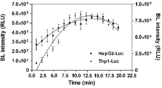
Cell clones showing the highest BL emission were selected by the following intact-cell luciferase assay: 100 l of cell suspension at a density of 106/ml was transferred to a white 96-well cell culture microplate and luminescence signals collected by adding 100 l of the Luciferase Assay System substrate (Promega, Madison, WI, USA) using a spectral scanning multimodal plate reader (Varioskan Flash, Thermo Scientific, Whaltham, MA, USA). The selected luciferase-expressing clones were assayed for bioluminescence emission spectra, as well. A 100 l sample of cell suspension (106/ml) was transferred to a 96 well microplate. After 100 l of Luciferase Assay System substrate was added, emission spectra were collected from 500-670 nm by measuring the light output at 2 nm intervals for 1 s. Luminescence measurements were performed at room temperature (25°C).To prove that photon emission and viable cell count were linearly related, HepG2-Luc, HepG2-Ppyred, Thp1-Luc, and Thp1-Ppyred cells were each plated in triplicate in a series ranging from 1x10⁶ to 4x10³ cells diluted in PBS. After receiving 100 l of Luciferase Assay System substrate, the cells were incubated for 5 minutes and then imaged for 1 minute with the same region of interest (ROI) used to measure luminescent signals of cells in each well.
Mouse cancer models
Animal experiments were approved by the Bioethics Committee of Bologna University in compliance with international guidelines. The liver cancer model was developed using five 14 week-old NOD/SCID mice purchased from Charles River laboratories (Wilmington, MA, USA). Subcutaneous xenografts were established by injection of 5x10⁵ HepG2-Luc and HepG2-Ppyred cells in the upper backs of the mice. For experiments monitoring both red and green bioluminescence in a single mouse, each mouse was injected also with 106 HepG2-Luc cells in the right flank and 2.4x10⁶ HepG2-Ppyred cells in the left flank. The leukemia animal model was developed using ten 6 to 10 week-old NOD/SCID mice. Each mouse was intravenously injected with 5 x10⁶ Thp1-Luc or Thp1-Ppyred cells and the cancer progression was monitored every 7 days for a total of 5 weeks.