Alma Mater Studiorum - Università di
Bologna
Scuola di Dottorato di Ricerca
in Scienze Mediche e Chirurgiche
Dottorato di ricerca in Scienze Biomediche
Progetto formativo in Neurofisiologia
XXIII ciclo
Settore scientifico-disciplinare di afferenza: BIO/09
Tesi di Dottorato
Role of nitric oxide and endocannabinoids in
synaptic plasticity in the perirhinal cortex
and in visual recognition memory
Dott. Francesco Tamagnini
Coordinatore:
Relatore:
Prof. Claudio Galletti
Prof. Giorgio Aicardi
Dipartimento di Fisiologia umana e generale
Esame finale anno 2011
TABLE OF CONTENTS
ABSTRACT ... 5
AKNOWLEDGEMENTS ... 6
LIST OF MAIN ABBREVIATIONS ... 7
1. INTRODUCTION ... 10
1.1. MEMORY AND LEARNING: CLASSIFICATION AND SHORT HISTORICAL REVIEW ... 10
1.2.CELLULAR CORRELATES OF LEARNING AND MEMORY: SYNAPTIC PLASTICITY ... 12
1.2.1.LONG TERM POTENTIATION (LTP): INDUCTION AND EXPRESSION MECHANISMS ... 14
1.2.2.LONG TERM DEPRESSION (LTD): INDUCTION AND EXPRESSION MECHANISMS ... 18
1.2.3.BIOLOGICAL RELEVANCE OF LTP AND LTD ... 21
1.3.PERIRHINAL CORTEX (PRH): ANATOMY AND FUNCTIONS ... 24
1.3.1.PERIRHINAL CORTEX AND VISUAL RECOGNITION MEMORY ... 26
1.3.1.1. Perirhinal cortex and visual recognition memory: behavioural studies ... 29
1.3.1.2. Perirhinal cortex and visual recognition memory: electrophysiological recordings33 1.3.1.3. Perirhinal cortex and visual recognition memory: immunohistochemical studies . 37 1.4.NITRIC OXIDE (NO) ... 38
1.4.1.NITRIC OXIDE SYNTHESIS, RECEPTORS AND DOWNSTREAM SIGNALLING ... 39
1.4.2. TARGETS OF NO SIGNALLING... 48
1.4.3.NO AND SYNAPTIC PLASTICITY ... 53
1.4.4.NO SIGNALLING IN MEMORY AND LEARNING ... 55
1.4.5.ROLE OF NO IN NEUROPATHOLOGY ... 59
1.5.ENDOCANNABINOIDS (ECBS) ... 62
1.5.1.ENDOCANNABINOID-MEDIATED LONG TERM DEPRESSION (ECB-LTD) ... 63
1.5.3.EXPRESSION OF ECB-LTD ... 67
1.5.4. ECBS IN MEMORY AND LEARNING ... 71
1.5.5. ECBS IN NEUROPATHOLOGY ... 73
1.6.ALZHEIMER’S DISEASE (AD): EPIDEMIOLOGY, CLINICAL FEATURES, PATHOGENESIS, AND MOLECULAR PROFILE ... 75
1.6.1.ALZHEIMER’S DISEASE: ANIMAL MODELS ... 83
1.7.AIMS OF THE RESEARCH ... 87
2. MATERIALS AND METHODS ... 88
2.1.LABORATORIES ... 88
2.2.ELECTROPHYSIOLOGY ... 88
2.3.ELECTROPHYSIOLOGICAL RECORDINGS ON TRANSGENIC TG2576 MICE ... 92
2.4.BEHAVIOURAL EXPERIMENTS ... 93
2.4.DRUGS ... 97
3. RESULTS ... 100
3.1.ROLE OF THE NO AND ECBS IN THE SYNAPTIC PLASTICITY IN PRH CORTEX ... 100
3.1.1.ROLE OF THE NOS/SGC/PKG PATHWAY IN THE 5HZ-INDUCED LTD(BOLOGNA) ... 100
3.1.2.ROLE OF THE NOS/SGC/PKG PATHWAY IN THE CCH-INDUCED LTD(BOLOGNA) ... 101
3.1.3.ROLE OF THE NOS/SGC/PKG PATHWAY IN THE CCH-INDUCED LTD(BRISTOL) ... 102
3.1.4.ROLE OF THE NOS IN THE TBS-INDUCED LTP(BRISTOL) ... 103
3.1.5.EFFECT OF THE CANNABINOID RECEPTORS CB1 AND CB2 ACTIVATION ON THE BASAL SYNAPTIC TRANSMISSION (BRISTOL)... 103
3.1.6.ROLE OF THE CB1 ACTIVATION IN 5HZ-INDUCED LTD(BRISTOL) ... 103
3.1.7.ROLE OF THE CB1 ACTIVATION IN CCH-INDUCED LTD(BRISTOL) ... 104
3.1.8.ROLE OF THE CB1 ACTIVATION IN TBS-INDUCED LTP(BRISTOL) ... 104
3.2.ROLE OF THE NO AND ECBS IN VISUAL RECOGNITION MEMORY ACQUISITION ... 106
3.2.1.EFFECT OF THE NEURONAL NOS(NNOS) IN THE ACQUISITION OF VISUAL RECOGNITION
MEMORY (BRISTOL) ... 106
3.2.2.EFFECT OF THE CANNABINOID RECEPTOR 1(CB1) IN THE ACQUISITION OF VISUAL
RECOGNITION MEMORY (BRISTOL) ... 107 3.3.EARLY IMPAIRMENT IN BASAL SYNAPTIC TRANSMISSION AND LTD IN THE PERIRHINAL
CORTEX OF TG2576 MICE ... 108
4. DISCUSSION ... 124
4.1. ROLE OF NOS/SGC/PKG AND CB1 IN RAT PERIRHINAL CORTEX LTD AND LTP INDUCTION
... 124
4.1.1. ROLE OF NOS/SGC/PKG IN LTD AND LTP INDUCTION IN THE RAT PERIRHINAL CORTEX
... 125
4.1.2. ROLE OF CB1LTD AND LTP INDUCTION IN THE RAT PERIRHINAL CORTEX ... 130
4.2.ROLE OF NITRIC OXIDE AND ENDOCANNABINOIDS IN THE ACQUISITION OF VISUAL
RECOGNITION MEMORY ... 133 4.3.EARLY DEFICIT IN THE SYNAPTIC TRANSMISSION AND PLASTICITY IN THE PERIRHINAL
CORTEX OF AN ALZHEIMER’S DISEASE MURINE MODEL ... 137
CONCLUSIONS ... 139
ABSTRACT
Introduction and aims of the research Nitric oxide (NO) and endocannabinoids
(eCBs) are major retrograde messengers, involved in synaptic plasticity (long-term potentiation, LTP, and long-term depression, LTD) in many brain areas (including hippocampus and neocortex), as well as in learning and memory processes. NO is synthesized by NO synthase (NOS) in response to increased cytosolic Ca2+ and mainly exerts its functions through soluble guanylate cyclase (sGC) and cGMP production. The main target of cGMP is the cGMP-dependent protein kinase (PKG). Activity-dependent release of eCBs in the CNS leads to the activation of the Gαi/o-coupled cannabinoid receptor 1 (CB1) at both glutamatergic and inhibitory synapses. The perirhinal cortex (Prh) is a multimodal associative cortex of the temporal lobe, critically involved in visual recognition memory. LTD is proposed to be the cellular correlate underlying this form of memory. Cholinergic neurotransmission has been shown to play a critical role in both visual recognition memory and LTD in Prh. Moreover, visual recognition memory is one of the main cognitive functions impaired in the early stages of Alzheimer’s disease. The main aim of my research was to investigate the role of NO and ECBs in synaptic plasticity in rat Prh and in visual recognition memory. Part of this research was dedicated to the study of synaptic transmission and plasticity in a murine model (Tg2576) of Alzheimer’s disease.
Methods Field potential recordings. Extracellular field potential recordings were
carried out in horizontal Prh slices from Sprague-Dawley or Dark Agouti juvenile (p21-35) rats. LTD was induced with a single train of 3000 pulses delivered at 5 Hz (10 min), or via bath application of carbachol (Cch; 50 µM) for 10 min. LTP was induced by theta-burst stimulation (TBS). In addition, input/output curves and 5Hz-LTD were carried out in Prh slices from 3 month-old Tg2576 mice and littermate controls.
Behavioural experiments. The spontaneous novel object exploration task was performed in intra-Prh bilaterally cannulated adult Dark Agouti rats. Drugs or vehicle (saline) were directly infused into the Prh 15 min before training to verify the role of nNOS and CB1 in visual recognition memory acquisition. Object recognition memory was tested at 20 min and 24h after the end of the training phase.
Results Electrophysiological experiments in Prh slices from juvenile rats showed
that 5Hz-LTD is due to the activation of the NOS/sGC/PKG pathway, whereas Cch-LTD relies on NOS/sGC but not PKG activation. By contrast, NO does not appear to be involved in LTP in this preparation. Furthermore, I found that eCBs are involved in LTP induction, but not in basal synaptic transmission, 5Hz-LTD and Cch-LTD. Behavioural experiments demonstrated that the blockade of nNOS impairs rat visual recognition memory tested at 24 hours, but not at 20 min; however, the blockade of CB1 did not affect visual recognition memory acquisition tested at both time points specified.
In three month-old Tg2576 mice, deficits in basal synaptic transmission and 5Hz-LTD were observed compared to littermate controls.
Conclusions The results obtained in Prh slices from juvenile rats indicate that NO
and CB1 play a role in the induction of LTD and LTP, respectively. These results are confirmed by the observation that nNOS, but not CB1, is involved in visual recognition memory acquisition. The preliminary results obtained in the murine model of Alzheimer’s disease indicate that deficits in synaptic transmission and plasticity occur very early in Prh; further investigations are required to characterize the molecular mechanisms underlying these deficits.
AKNOWLEDGEMENTS
Many thanks to my supervisor Giorgio Aicardi, who is a costant presence in my scientific life since my undergraduate thesis. Thanks to Costanza Burattini for being such a wonderful collegue and loyal friend. And all my gratitude to the guys and girls in ZIB and GLC labs at the MRC Centre for Synaptic Plasticity in Bristol, in particular my supervisor Zafar Bashir and Clea Warburton that believed in this project and gave me the chance to work in the best conditions. Thanks to Garteh Barker, for carrying out the behavioural experiments, and to Alexandra Outram, for being the first one to believe in the role of the nitric oxide (the most amazing molecule ever existed) in visual recognition memory. Also many thanks to Zuner Bortolotto for the precious theoretical and practical support. And of course thanks to all my bristolian collegues and friends, in particular James Wallis and Michael Laing for their human and scientific support.
All my love and gratitude to my mother and father, for being a constant source of inspiration, affection and support.
LIST OF MAIN ABBREVIATIONS
2-AG: 2- arachidonyl glycerol5HT2: Metabotropic receptor for serotonin 2 5Hz-LFS: LFS consisting of 3000 pulses delivered at 5 Hz 5Hz-LTD: LTD induced with 5Hz-LFS application 7-NI: 7-nitroindazolo AA: Arachidonic acid ACEA:
N-(2-Chloroethyl)-5Z,8Z,11Z,14Z-eicosatetraenamide aCSF: Artificial cerebrospinal fluid AD: Alzheimer’s disease
ADAM: Alpha disintegrin metalloproteinase
AEA: Anandamide
ALS: Amyotrophic lateral schlerosis AM251: 1-(2,4-dichlorophenyl)-5-(4- iodophenyl)-4-methyl-N-(1-piperidyl)pyrazole-3-carboxamide AM404 AMPA: α-amino-3-hydroxyl-5-methyl-4-isoxazole-propionate
AMPAR: AMPA receptor AP2: Adaptor protein 2 AP-5: D-(-)-2-amino-5-phosphonovaleric acid ApoE: Apolypoprotein E
APP: Amyloid precursor protein ATP: Adenosine-5'-triphosphate Aβ: Beta amyloid peptide
BACE-1: Beta amyloid cleaving enzyme-1
BDNF: Brain derived neurotrophic factor
BH4: Tetrahydrobiopterin CA1: Cornus ammonis field 1 CaMKII/IV: Calcium-calmodulin protein kinase II/IV
CaMKK: Calcium/calmodulin protein kinase kinase
cAMP: 3'-5'-cyclic adenosine monophosphate
CAPON: Carboxyl-terminal PDZ ligand of neuronal nitric oxide synthase protein
CB1: Cannabinoid receptor type 1 CB2: Cannabinoid receptor type 2
Cch: Carbachol
Cch-LTD: Charbachol bath application mediated LTD cGMP: 3'-5'-cyclic guanosine monophosphate
CNG: Cyclic nucleotide gated channels
CNQX: 6-Cyano-7-nitroquinoxaline-2,3-dione
CNS: Central Nervous System CREB: cAMP responsive element binding protein
C-terminal: Carboxyl-terminal D2: Metabotropic receptor for dopamine 2
DAG: Diacylglycerol DAG: Diacylglycerol
DEA/NO: diethylamine NONOate DEA/NO: Diethylamine NONOate DG: Dentate gyrus DGL: Diacylglycerol lipase DGL: Diacylglycerol lipase DHPG: 3,5-Dihydroxyphenylglycine DHPG-LTD: DHPG application-evoked LTD DSE: Depolarization-dependent suppression of excitation DSI: Depolarization-dependent suppression of inhibition
EC: Entorhinal cortex
eCB-LTD: eCB mediated LTD eCBs: Endocannabinoids
EMT: Endocannabinoid membrane transporter
eNOS: endothelial NOS
EPSP: Excitatory postsynaptic potential
ERK: Extracellular signal related kinase
FAAH: Fatty acid amide hydrolase fEPSP: Field excitatory postsynaptic potential
GABA: γ-amino-butyric acid GluR: AMPAR subunit
Glutamate: L-glutamate
GPCR: G-protein coupled receptor GRAB-1:
HCN: Potassium/sodium
hyperpolarization-activated cyclic nucleotide-gated channel
HD: Huntington disease
HFS: High frequency stimulation Hsp 90: Heat shock protein 90 IEG: Immediat e early gene
I-LTD: LTD of the inhibitory synapses iNOS: inducible NOS
IP3: Inositol 1,4,5-triphosphate IP3: Inositol 1,4,5 trisphosphate KAR: Kainate receptor
KO mouse: Knockout mouse KT5823: ,3,9,10,11,12-hexahydro-10R-methoxy-2,9-dimethyl-1-oxo-9S, 12R-epoxy-1H-diindolo[1,2,3-fg:3’,2’, 1’-kl]pyrrolo[3,4-i][1, 6]benzodiazocine-10-carboxylic acid, methyl ester KT5823: 2,3,9,10,11,12-hexahydro-10R-methoxy-2,9-dimethyl-1-oxo-9S, 12R-epoxy-1H-diindolo[1,2,3-fg:3’,2’, 1’-kl]pyrrolo[3,4-i][1, 6]benzodiazocine-10-carboxylic acid, methyl ester
Kv1: Voltage dependent potassium channels 1
Kv3: Voltage dependent potassium channels 3
LFS: Low-frequency stimulation L-NAME: L-NG-Nitroarginine methyl ester
L-NMMA: NG-methyl-L-arginine LTD: Long-term depression LTP: Long-term potentiation M1: Muscarinic receptor for acetylcholine type 1
M3: Muscarinic receptor for acetylcholine type 3
MAPK: Mitogen activate protein kinase
MAPT: Microtubule associated protein tau
MCI: Mild cognitive impairment MEK: MAPK/ERK kinase MGL: Monoacylglycerol lipase mGlu Receptor: Metabotropic glutamate receptor
MK-801: Dizocilpine MLA: Methyllycaconitine MS: Multiple schlerosis
NCS1: Neuronal calcium sensor protein 1
NCX2057:
3-(4-Hydroxy-3-methoxyphenyl)-2-propenoic acid NMDA: N-methyl-D-aspartate NMDAR: NMDA receptor nNOS: neuronal NOS NO: Nitric oxide
NOS: Nitric oxide synthase NPA: N-ω-propyl-L-arginine NR (1,2A/B,3): NMDAR subunit (1,2A/B,3) NS2028: 4H-8-Bromo-1,2,4 - oxadiazolo [ 3,4-d]benz[b][1,4]oxazin-1-one NS2828: 8-bromo-4H-2,5-dioxa-3,9b-diaza-cyclopenta[a]naphthalen-1-one N-terminal: Amino-terminal NVP: (R )-[(S)-1-(4-bromo-phenyl)- ethylamino]-(2,3-dioxo-1,2,3,4- tetrahydroquinoxalin-5-yl)-methyl]phosphonic acid ODQ: 1H-[1,2,4]Oxadiazolo[4,3-a]quinoxalin-1-one
p75NTR: pan neurotrophin receptor 75
PC: Purkinje cells
pCREB: phosphorylated CREB PD: Parkinson’s disease
PDE 11): Phosphodiesterase (1-11)
PDZ: PSD-95, discs large, zona occludens-1
PGE2: Prostaglandin E2 PGF2: Prostaglandin F2
PICK 1: Protein interacting with C kinase
PIP2: Phosphatidylinositol 4,5 bisphosphate
PKA: Protein kinase A PKC: Protein kinase C PKG: Protein kinase G PLC: Phospholipase C POR Postrhinal cortex PP: Protein phosphatase Prh: Perirhinal cortex PS1/2: Presenilin ½
PSD: Postsynaptic density
PSD-95: Postsynaptic density protein 95
ROS: Reactive oxygen species SC: Shaffer’s collaterals
sGC: Soluble guanylate cyclase SNP: Sodium nitroprusside SOD: Superoxyde dismutase STDP: Spike-timing dependent plasticity TBS: Theta-burst stimulation t-LTD: Spike-timing dependent depression t-LTP: Spike-timing dependent potentiation
tPA: tissue plasminogen activator TrKB: Tyrosine kinase receptor B VCAM-1: Vascular cell adhesion molecule-1
VEGF: Vascular endothelial growth factor
VGCC: Voltage gated calcium channels
VGNC: Voltege gated sodium channels Weak 5Hz-LFS: 5Hz-LFS consisting of 1350 pulses WIN55,212-2: (R)-(+)-[2,3-Dihydro-5- methyl-3-(4- morpholinylmethyl)pyrrolo[1,2,3-de]- 1,4-benzoxazin-6-yl]-1-napthalenylmethanone YC1: 5-[1-(phenylmethyl)-1H-indazol-3-yl]-2-furanmethanol
1. INTRODUCTION
1.1. Memory and learning: classification and short historical review
Memory and learning have been studied since ancient times, but only in the last few decades the cerebral structures involved have been identified and the physiological and molecular mechanisms underlying these processes have begun to be clarified.
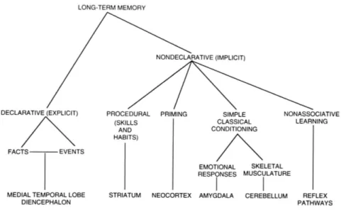
Memory has been classified as explicit (facts and events) and implicit (e.g. classical and operant conditioning and procedural memory; Fig 1.1.). Cerebral areas associated with these mnemonic processes are extensively studied by psychologists, neurophysiologists and psychiatrists. In a situation of learning, many of these brain areas may be working simultaneously, each one processing different streams of information about the perceived event (visual, location, sound, emotional content, etc.). The associative learning is believed to emerge from the coordinated activity of different brain areas. This is how organisms, including humans, encode causality in the perceived world.
Memory can also be classified as short- and long-term. Short-term memory has a limited temporal window (minutes) and needs to be continuously renewed to be maintained; long-term memory lasts longer (from hours to years).
Different functions are associated to specific brain regions, and the same applies for implicit and explicit memory processes (reviewed by Squire, 2004). In the 1940’s, Penfield’s electrophysiological experiments on epileptic patients and studies of hippocampal lesions in primates and humans were some of the first experimental evidences supporting the idea of structure - function relations for mnemonic processes (reviewed by Feindel, 1982). Before surgical ablation of the temporal lobe, Penfield’s patients, who were conscious throughout the procedure
(local anaesthesia), were electrically stimulated in different areas of the temporal cortex. During the stimulation, Penfield’s patients verbally referred back to past experiences.
Another famous example comes from the case of patient H.M., who suffered from an aggressive form of epilepsy. Bilateral partial ablation of the temporal lobes, including both hippocampi, was performed as a treatment. After the surgery, H.M. suffered from a severe form of anterograde amnesia: he could recollect everything that happened until the day of the surgery, but from that moment onwards his long-term memory was severely impaired although procedural memory processing remained intact.
In the coming years, lesion studies on primates and rodents demonstrated that the hippocampus and associated structures (perirhinal, entorhinal and parahippocampal cortices) are selectively involved in acquisition and consolidation of declarative but not implicit memory processes (reviewed by Squire, 2004).
Fig 1.1. Schematic representation and functional classification of long-term memory and associated brain structures (modified from Squire, 2004).
1.2. Cellular correlates of learning and memory: synaptic plasticity
What is memory from a physical point of view? In pure physical terms, memory is the ability of a system to acquire, store and recollect information. In a biological system, a complex phenomenon like memory is characterized by cellular and molecular mechanisms that have been the subject of extensive investigation in the last 40 years. Considering that most of the stored information is acquired from sensorial experience, the brain has to undergo many long-term functional and structural modifications corresponding to the mnemonic tracks left by perceived experiences. In his Croonian Lecture to the Royal Society in 1894, Ramòn y Cayal proposed that memory formation relies on reinforcement of the signalling between neurons in the involved areas:
[…] it can be accepted that the mental exercise brings to a major development of the dendritic apparatus and of the axonic collaterals system in the mostly used brain areas […].
In this concept, the notion of synaptic plasticity is already maturing: the ability of chemical synapses to increase or decrease the efficiency of transmission between neurons according to the frequency of the stimulation and to the previous history.
The idea that memory and learning result from the alteration of activity of specific synapses was further highlighted by the Canadian psychologist Donald Hebb in 1949 in his book titled “Organization of the behaviour”:
“When an axon of cell A is near enough to excite B and repeatedly or persistently takes part in firing it, some growth process or metabolic change takes place in one or both cells such that A's efficiency, as one of the cells firing B, is increased.”
This Hebbian rule has been formulated on pure theoretical basis, hypothesizing that such a mechanism could stabilize specific patterns of neuronal activity: if a
neuronal activity pattern corresponds to a specific behaviour, stabilizing that pattern means memorizing that behaviour (Hebb, 1949).
Most part of both excitatory and inhibitory synapses show a rich repertoire of plastic modalities that work on time scales comprised between milliseconds and weeks (e.g. paired-pulse facilitation, paired-pulse depression, long-term potentiation, long term depression, suppression of inhibition etc.). With regards to the cellular and molecular basis of memory and learning, long-term potentiation (LTP) and long-term depression (LTD) have represented the main experimental model in the last 30 years. Both LTP and LTD have been observed in many brain structures such as hippocampus, neocortex and subcortical structures. In 1973, Bliss and Lømo were the first to observe that high frequency stimulation (HFS) of the perforant path (PP) of the hippocampus of anesthetized rabbits in vivo determined a significant and prolonged potentiation of synaptic transmission in the dentate gyrus and termed this phenomenon LTP. It has been mainly explored in hippocampus, but it can also be induced in other brain structures, such as the perirhinal cortex (Prh). LTP is characterized by a long-term (more than 3 h) increment of synaptic strength following a short period of coordinated neuronal activity, such high frequency stimulation of afferent fibres. Even if LTP is persistent, it’s not irreversible: the synaptic strength can be returned to basal levels through low frequency stimulation (LFS) of the afferent fibres (depotentiation, DP; Barrionuevo et al., 1980). In addition, when LFS is applied to a non-potentiated pathway it leads to LTD (Bear and Dudek, 1992). LTD can be returned to basal levels after HFS through the process termed de-depression. Thus, the strength of synaptic transmission can be altered in a bidirectional and reversible way: the dynamic storage of large amounts of information at neuronal level may be constantly redefined. Furthermore, these forms of Hebbian plasticity
act through positive feedback processes, that if left without control measures may destabilize the neuronal networks by driving neurons into maximal and/or minimal firing frequency averages: by degrading the signals that propagate through the network, this ultimately disrupts the ability of the neurons to encode further plastic changes. Homeostatic forms of synaptic plasticity should integrate negative feedback systems in order to keep the synaptic transmission and plasticity within a dynamic functional range, either increasing or decreasing the strength of all the synaptic inputs: this can be achieved by either keeping at the same time their relative weight (synaptic scaling) or by modifying the ability of the synapses to undergo further plastic changes (metaplasticity; reviewed by Pérez-Otaño and Ehlers, 2005)
1.2.1. Long term potentiation (LTP): induction and expression mechanisms
LTP is defined as the long-term increase of synaptic strength subsequent to the application of a HFS (usually 100 Hz) on the presynaptic fibres. A lot of what we know about LTP arises from experiments conducted at Schaffer Collaterals (SC) / CA1 glutamatergic synapses. The induction of LTP at these synapses involves the activation of N-methyl-D-aspartate receptors (NMDAR), a class of ionotropic receptors for glutamate, permeable to calcium (Ca2+) (reviewed by Collingridge and Bliss, 1995). HFS or presynaptic stimulation coupled to post-synaptic depolarization removes the voltage-dependent block of the NMDAR by the displacement of the magnesium (Mg2+) ion placed on the extracellular side of the channel pore (Mayer et al., 1984). NMDAR are referred to as coincidence detectors, as the contemporary presence of glutamate in the synaptic cleft in combination with postsynaptic depolarization, determines the opening of NMDAR resulting in the influx of Ca2+. NMDAR are formed by hetero-oligomeric assemblies
of NR1 subunits with NR2 (A-D) and NR3A (Monyer et al, 1994). According to Liu et al., 2004, HFS applied to a glutamatergic pathway activates NR2A subunit containing NMDAR leading to the activation of Ca2+/calmodulin dependent Kinase II (CaMKII). On the other hand, if in the subunit NMDAR is comprised the NR2B subunit, the Ca2+ influx will result in the activation of the Ca2+/calmodulin dependent phosphatase calcineurin that is responsible for LTD induction (Mulkey et al., 1993, 1994).
The LTP expression mechanisms are not yet fully understood. The activation of CaMKII triggers the insertion of further GluR1 containing α-amino-3-hydroxy-5-methyl-4-isoxazolepropionic acid receptor (AMPAR) on postsynaptic dendritic processes (Malinow and Malenka, 2002). Recent studies highlighted two other main signal transduction pathways responsible for the long-term modifications needed for the consolidation of LTP: the cAMP-dependent protein kinase (PKA) and the mitogen activated protein kinase (MAPK). Intracellular Ca2+ influx induces the activation of adenylate cyclase (AC) leading to the subsequent increase of intracellular cAMP which activates PKA. The expression in transgenic mice of the inhibitory form of the regulatory subunit of PKA R(AB) does not affect the early phase of LTP but disrupts its consolidation. MAPK is also termed extracellular signal-related kinase (ERK). In the dentate gyrus, the application of a LTP-inducing stimulation determined rapid phosphorylation and nuclear translocation of MAPK (English and Sweatt, 1996; Davis et al., 2000 a). Both PKA and MAPK can phosphorylate and activate the transcription factor cAMP-responsive element binding protein (CREB) (Yin and Tully, 1996; Silva et al., 1998). CREB triggers the expression of genes responsible for long-term modifications (functional and structural) underlying the late phase of LTP. Finally, many lines of evidence have
consistently shown that protein synthesis blockade prevents the long-term expression of LTP, without affecting induction (reviewed by Blitzer et al., 2005).
Many recent studies have highlighted the key role of brain-derived neurotrophic factor (BDNF) in both induction and expression of LTP (Lu and Gottschalk, 2000; Aicardi et al., 2004; Santi et al., 2006; Minichiello et al., 2009). In particular, it has been suggested that activity-dependent release of BDNF from the presynaptic site is necessary for the induction of LTP, while the activity-dependent sustained production and secretion from the post-synaptic site is necessary for the expression of LTP (Aicardi et al, 2004; Reviewed by Lu et al., 2008). Pro-BDNF is cleaved to mature BDNF (mBDNF) via proteolytic cleavage by the tissue plasminogen activator (tPA). mBDNF binds the tyrosine-kinase coupled receptor B (TrkB) which in turn phosphorylates various substrates, including MAPK/ERK resulting in the activation of MAPK/ERK kinase (MEK). TrkB activation also leads to phospholipase C (PLC) activation, which in turn activates both PKC and calcium calmodulin kinases kinase (CaMKK), subsequently resulting in CaMKIV and CREB activation (Reviewed by Minichiello, 2009; Fig 1.3).
Fig 1.2. Schematic representation of LTP induction and expression mechanisms. The three most important downstream signalling pathways are CaMKII, MAPK and PKA. Ca2+ influx occurs through NMDAR and voltage-gated Ca2+ channels (VGCC). AC can also be activated by stimulation of Gs coupled receptors such as β-adrenergic receptors (modified from Blitzer et al., 2005).
Fig 1.3. Activation of TrkB triggers three main intracellular signalling pathways. i) Ras–MAPK, which promotes neuronal differentiation and growth through MAPK/ERK kinase (MEK); ii) phosphatidylinositol 3-kinase (PI3K) cascade, which promotes survival and growth of neurons and other cells through Ras or GRB-associated binder 1 (GAB1); iii) phospholipase Cγ1, which mediates synaptic plasticity through CaMKII and CaMKK/CaMKIV signalling cascade (modified from Minichiello et al., 2009).
1.2.2. Long term depression (LTD): induction and expression mechanisms
The first evidence for LTD came from the observation that LTP inducing protocols in hippocampal SC/CA1 synapses generated a reversible depression in the non tetanized pathway (Lynch et al., 1977); this phenomenon is called heterosynaptic plasticity. Homosynaptic depression was later shown in hippocampus: the application of LFS reversed LTP in a pathway previously tetanised; this phenomenon is now known as depotentiation (Barrionuevo et al., 1980). Later, it was demonstrated that LFS can induce LTD in CA1 even without previous LTP induction (Dudek and Bear, 1992; Mulkey and Malenka, 1992).
LTD can be induced in many brain areas other than the hippocampus including the visual cortex (Artola et al., 1990), striatum (Calabresi et al., 1994), perirhinal cortex (Ziakopoulos et al., 1999; Cho et al., 2000), amygdala (Wang and Gean, 1999), posterior cingulus (Hedberg and Stanton, 1995) and prefrontal cortex (Hirsch and Crepel, 1991).
The first studies on homosynaptic LTD showed that application of LFS consisting of 900 pulses delivered at 1 Hz, induces LTD that relies on the activation of NMDAR (Dudek and Bear, 1992; Mulkey and Malenka, 1992). NMDAR-dependency of LTD is often age-related (Kemp et al., 2000, Jo et al., 2006), consistently with the developmental change in subunit composition of NMDAR (Monyer et al., 1994).
It is of interest to note that LTD induction can be mediated by other receptors like kainate receptors (KAR), metabotropic glutamate receptors (mGluRs) and type I muscarinic acetylcholine receptors (M1) (reviewed by Kemp and Bashir, 2001). Group I and II mGluRs are involved in depotentiation in CA1 (Bashir and Collingridge, 1994); mGluRs agonists have been shown to induce LTD in CA1
(Fitzjohn et al., 2001), in the dentate gyrus (O’Mara et al.,1995a; Huang et al., 1999) and in Prh (McCaffery et al., 1999).
By far, the majority of AMPAR are impermeable to Ca2+, due to the editing of the mRNA of the glutamate receptor subunit 2 (GluR2). However, LTD in CA3 has been shown to require co-activation of mGluRs and Ca2+ permeable AMPA (Laezza et al., 1999). Increasing evidence suggests the pivotal role of AMPA trafficking in LTD. Activity-dependent internalization of AMPA receptors is central for recognition memory and LTD induction in Prh (Griffiths et al., 2008).
KAR involvement in LTD is not yet entirely demonstrated, but it’s suggested by the observation that the co-application of the mGluR antagonist MCPG and the AMPA/KA receptor antagonist CNQX inhibits LTD induction in the hippocampus (reviewed by Kemp and Bashir, 2001).
The application of LFS consisting of 3000 pulses delivered at a frequency of 5 Hz in Prh slices of juvenile rats resulted in M1-dependent LTD (Jo et al., 2006); in addition, the bath application of the acetylcholine analogue carbachol (Cch) was shown to induce chemical M1-dependent LTD in Prh slices of adult rats (Massey et al., 2001).
The central intracellular event for LTD induction is the cytosolic Ca2+ concentration increase, as observed for LTP (Lynch et al., 1983; Bliss and Collingridge, 1993): LTP-inducing stimuli determine rapid and high increases in intracellular Ca2+, while for LTD the increase is low and slow (Lisman, 1989). Presynaptic Ca2+ increase is also necessary for LTD induction (Kobayashi et al., 1996, 1999). The source for the increase of post-synaptic Ca2+ can be both extracellular and intracellular. In the first case, it relies on the activation of NMDAR, Ca2+ permeable AMPAR or voltage-gated Ca2+ channels (VGCC; Cummings et al., 1996; Christie et al., 1997; Wang et al., 1997 a; Norris et al.,
1998; Otani and Condor, 1998). In the second case, Ca2+ comes from intracellular stores as a consequence of the activation of Gq-coupled receptors like group I mGluRs or M1.
The increase in intracellular Ca2+ determines the formation of the Ca2+/calmodulin (CaM) complex activating Ca2+/CaM-dependent phosphatase calcineurin. This happens if the Ca2+ increase is low and slow. LFS-dependent calcineurin activation inactivates inhibitor 1, by dephosphorylation. This results in the activation of phosphatase 1 and 2 (PP1/2) which results in LTD via dephosphorylation of various targets such as AMPAR and CaMKII (reviewed by Kemp and Bashir, 2001; Fig 1.4.).
In the last three decades, LTD expression mechanisms have been deeply investigated in various brain regions. It was concluded that different induction mechanisms determine different expression mechanisms. Hippocampal NMDAR-LTD in CA1 relies on the increased internalization of AMPAR via
Fig 1.4. LTD induction requires increased intracellular calcium concentration from the extracellular space via NMDAR or VGCC activation or IP3-mediated opening of intracellular stores. The Ca2+-calmodulin complex activates the protein phosphatase calcineurin that dephosphorylates the phosphatase inhibitor 1 inactivating it. This enables the activation of protein phosphatase 1 (PP1) that dephosphorylates targets like CaMKII and AMPA receptors (Modified from Kemp and Bashir, 2001).
dephosphorylation of Ser-845 (target of PKA) and Ser-831 (target of CaMKII) of the GluR1 subunit, altering the conductance and the probability of opening of AMPARs. In addition, LFS determines increased internalization of postsynaptic AMPARs through a dynamin-dependent, clathrin-mediated process (Lüscher et al., 1999; Lüthi et al., 1999; Carroll et al., 1999; Man et al., 2000; Beattie et al., 2000; Wang and Linden, 2000; Henley, 2003; Collingridge et al., 2004; Griffiths et al., 2008). In specific, it has been shown that AP2, a clathrin adaptor protein, is important for the internalization of surface AMPARs and for the expression of NMDAR-dependent LTD (Lee et al., 2002).
The last event that temporally characterizes LTD is a change in protein synthesis, which is essential for mGluR-dependent LTD in CA1 (Huber et al., 2000) and for Cch-induced LTD in Prh (Massey et al., 2001): LTD induction has been shown to correspond to long-term changes in spine morphology, specifically in a reduction in spine density (Halpain et al., 1998). Fig 1.5. illustrates the main LTD induction pathways and related post-synaptic changes.
It is of interest to note that the immature form of BDNF, proBDNF, has been demonstrated to be involved in LTD induction via activation of the pan-neurotrophin receptor 75 (p75NTR) (Woo et al., 2005; Rösch et al., 2005).
1.2.3. Biological relevance of LTP and LTD
Many studies showed that both LTP and LTD can be induced in the same pathway depending on the frequency of firing of the presynaptic fibres (Bliss and Collingridge, 1993; Bear and Malenka, 1994).
Certain properties of LTP and LTD provide support to the hypothesis that they underlie complex mnemonic processes. Firstly, synapses can be independently modified: only co-active synapses participate in the plastic change, i.e. stimulation does not affect surrounding synapses, thus is input-specific. This evidence
suggests that every single synapse can be individually used to store information. Since every neuron is surrounded by about 10,000 synapses and brain areas involved in learning and memory contain billions of neurons, the mnemonic capacity of the brain is extraordinary. Secondly, LTP and LTD are associative phenomena: this property allows for small changes in certain synapses in order to produce a distributed storage in a complex and organic memory within a neuronal network. This implies that LTP and LTD cannot be triggered by the activity of a single neuron, but only as a consequence of associate activation of many inputs. This functional need comes from the fact that both LTP and LTD induction require a sufficient degree of depolarization of the postsynaptic membrane (Bilkey, 1996; reviewed by Diamond and Rose, 1994). LTP and LTD have a reciprocal connection in the generation of stored memories. LTP is associated with the storage of new information, but, since the brain represents a system with limited capacity, soon the newly stored information would reach a saturation point, destabilizing the system and making the recollection more challenging. LTD has the role to improve the signal/noise ratio associated to the acquisition and recollection of newly stored information (reviewed by Rosenzweig and Barnes, 2003; Fig 1.6).
Fig 1.6. Schematic representation of an information storage system with limited capacity. If we assume that a neural network coincides with a sheet of white paper, we can draw on it different shapes, coincident to new encoded patterns of neural activity. If the network contains too much information it becomes useless. Acquisition of new shapes/patterns is mediated by LTP. If the system keeps accumulating patterns one on another, soon every single shape becomes indistinguishable from others: recollection is therefore impossible and most of the information will be lost. The role of the LTD is to organize the patterns and select the stronger ones to increase memory capacity and recollection (Rosenzweig and Barnes, 2003).
Fig 1.5. Schematic representation of the main LTD induction pathways. Abbreviations: AC, adenylate cyclase; AA, arachidonic acid; CaMKII, calcium/calmodulin-dependent kinase II; CREB, cAMP responsive element binding protein; IP3, inositol trisphosphate; Ka, kainate receptor; mGluR, metabotropic glutamate receptor; MAPK, mitogen activated protein kinase; PI, phosphatidil inositol; PLC, phospholipase C; PKA, protein kinase A; PKC, protein kinase C; PP1/2 A, protein phosphatase 1/2 A; TKR, tyrosine kinase receptor (Modified from Blitzer et al., 2004).
1.3. Perirhinal cortex (Prh): anatomy and functions
At the beginning of the 1980’s, the systematic exploration of amnesia in animal models lead to the identification of cortical structures involved in declarative memory formation; these were primarily located in the medial temporal lobe, comprising the hippocampus, dentate gyrus, subiculum, entorhinal cortex (EC), parahippocampal cortex (Prp) and Prh (Zola-Morgan et al., 1986; reviewed by Squire and Zola-Morgan, 1991). Prh is a periallocortex, located on the ventral surface of the temporal lobe. In both primates and rodents, Prh comprises two cytoarchitectionally different regions: 35 and 36 Brodman areas (Fig. 1.8.). Area 35 is thin and agranular (IV layer is missing) and it is located ventrally to the rhinal sulcus; area 36 contains a thin IV layer, in which granular cells are mixed with pyramidal cells from III and V layer (Burwell, 2001).
Prh has strong reciprocal connections with the hippocampus and the subiculum. These connections are both direct through the lateral perforant path, and indirect through EC (Deacon et al., 1983; Burwell et al., 1995; Liu and Bilkey, 1996; Naber et al., 1999). Furthermore, Prh receives both unimodal inputs from associative unimodal cortices (i.e. visual, somatosensory, auditory and olfactive) and multimodal inputs from associative multimodal cortices (i.e. medial and ventro-lateral prefrontal cortex, cingulate anterior area, retrosplenial cortex; Fig 1.7). Visual information, transmitted from unimodal associative visual cortex, is then transmitted to the Prh (Meunier et al., 1993; Wiig and Bilkey, 1995; Ennaceur and Aggleton, 1997). Since a few years ago, Prh was merely considered a structure where sensory information was transmitted from sensory related cortices to the hippocampal formation, without ascribing any role in memory formation. Since the early 1990’s many studies highlighted that Prh interacts, directly or via integration with other brain areas, for various mnemonic functions, with a crucial role in visual
recognition memory (Buffalo et al., 1998; reviewed by Brown and Aggleton, 2001; Massey et al., 2001; Warburton et al., 2003; Massey et al., 2008) and fear conditioning (Corodimas and LeDoux, 1995). In the last decade many studies clarified the cellular and molecular mechanisms underlying Prh-dependent visual recognition memory.
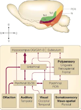
Fig 1.7. Location of the Prh in the rat brain and schematic representation of connections to and from other brain areas. Rat Prh network is characterized by 3 main connections. The first one is a strong reciprocal connection with the hippocampal formation through lateral EC (Suzuki and Amaral, 1994). The second one is characterized by afferent projections from unimodal and multimodal associative cortices, including visual, somatosensory, auditory and olfactory associative areas, medial and ventro-lateral prefrontal cortex, anterior cingulus and retrosplenial and Prp cortices (Burwell et al., 1995). The third one is characterized by the reciprocal connection with the amigdaloid complex. The thickness of the arrows indicates the connection density (Modified from Brown and Aggleton, 2001).
1.3.1.Perirhinal cortex and visual recognition memory
Recognition memory is a primary aspect of our ability to remember. It is based on the capability of both identifying and judging the prior occurrence of a visually perceived experience (Mandler et al., 1980).
Recognition memory is a main component of the sort of memory lost in anterograde amnesia. However, it has been proposed that within the two main components of recognition memory, identification and judgement of prior occurence, only the latter is directly compromised in anterograde amnesia (Mandler et al., 1980; Jacoby et al., 1981; Gardiner et al., 1990; Aggleton and Brown, 1999).
The visual component of recognition memory corresponds to the sensation of familiarity of a visual stimulus without active recollection of the attributes of that vision. This discrimination of familiarity is defined as ‘knowing’ (I know I’ve seen this thing before) whereas the active recollection of the attributes is defined as ‘remembering’ (I actively remember the name of the object, what the material is made of, where I’ve seen it before etc.). There are two main models describing recognition memory. The first model is the single process model. In this model ‘knowing’ and ‘remembering’ are considered a single process that differ only quantitatively: knowing corresponds to a weaker mnemonic trace whereas remembering is related to recollection and hence is related to a stronger mnemonic pattern. The second model is the dual process model, according to which ‘knowing’ and ‘remembering’ are two qualitatively distinct processes mediated by two distinct structures. In the dual model the hippocampus is usually related to ‘remembering’ because of its structural complexity and the many lines of evidence showing its role in recollection; on the other hand Prh has been shown to
be selectively involved in recognition memory. So far, the dual model is the most accepted (reviewed by Brown and Aggleton, 2001).
Prh is critically involved in recognition memory as shown by several lesion studies in both primates and rodents (Zola-Morgan et al., 1989; Meunier et al., 1993; Mumby et al., 1994; Meunier et al., 1996; Winters et al., 2004). Other evidence highlighting the pivotal role of Prh in visual recognition memory comes from electrophysiological recordings from the medial temporal lobe of monkeys performing recognition memory tasks (Brown and Wilson, 1987; Fahi et al., 1993; Li et al., 1993; Miller et al., 1993; Xiang et al., 1998). These studies consistently showed a decrease in neuronal responsiveness subsequent to the presentation of a previously encountered visual stimulus (Brown, 1996; Desimone, 1996; Eichenbaum et al., 1996; Ringo, 1996; Brown and Xiang, 1998; Suzuki and Eichenbaum, 2000; Eichenbaum, 2000). The decrease in the overall neuronal responses brings into account information about prior occurrence of a stimulus mediating the discrimination between familiarity or recency of that stimulus (Miller et al., 1993; Sobotka and Ringo, 1993; Xiang and Brown, 1998). These response reductions are observed in Prh (in ~25% of the recorded neurons) and rarely in the hippocampus (<~1%) (Brown and Wilson, 1987; Miller et al., 1993; Sobotka and Ringo, 1993; Xiang and Brown, 1998). In the last ten years many studies have aided in the clarification of the molecular mechanisms underlying the acquisition, consolidation and recollection of visual recognition memory. Three main approaches have been followed, primarily conducted in rats: 1) behavioural measures in animals with bilateral cannula into Prh: the local infusion of drugs or transfecting viruses in the Prh and the consequent deficit or enhanced performance in an object recognition task has teased about the roles of various membrane receptors and associated cellular signalling pathways involved in
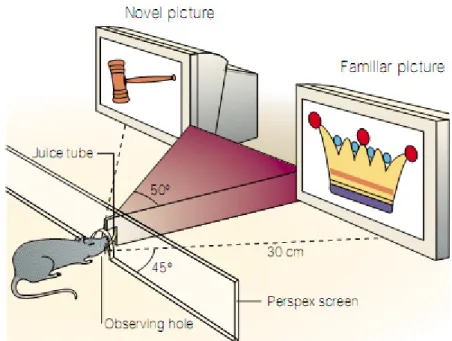
recognition memory processes. The most applied behavioural protocol used to investigate visual recognition memory in rats is the spontaneous novel object exploration task, described in section 1.3.1.1; 2) Electrophysiological recordings on acute brain slices: the measure of basal synaptic transmission and synaptic plasticity in Prh slices has helped to define which are the cellular correlates of recognition memory. This in vitro approach can be carried out on: i) naive slices treated with various drugs ii) slices from transgenic animals iii) slices from animals locally transfected in Prh with viruses inducing a genetical modification, and finally iv) slices from animals that underwent the paired-viewing protocol (described in Brown and Aggleton 2001). This protocol consists in keeping the animal’s head in a window facing a screen showing two different images. The two images are projected in a manner as to occupy the monocular visual field of each eye independently. Sets of images are then repetitively projected. At the end of a period of training, a set of novel images are projected towards only one eye, in order to encode novelty in one hemisphere and familiarity in the other. Note that the visual information perceived in one eye is processed in the contralateral hemisphere of the brain. This treatment results in one of the hemispheres being conditioned with ‘familiar’ and the other hemisphere with ‘novel’ images respectively (Fig 1.8.). Upon surgical removal of these hemispheres, electrophysiological experiments (Warburton et al., 2003; Massey et al., 2008) and immunohistochemical studies can be performed in order to evaluate how the encoding of familiar or novel information influences neuronal activity. 3) Immunohistochemical imaging has been used for the evaluation of molecular changes in the Prh of rats that underwent the paired-viewing protocol; it can be examined in a quantitative manner by analysing the differential expression of
genes, e.g. the immediate early gene (IEG) c-fos levels in the ‘novel’ versus the ‘familiar’ hemisphere.
1.3.1.1. Perirhinal cortex and visual recognition memory: behavioural studies
In order to better understand the cellular correlates underlying visual recognition memory formation, many studies evaluated the visual recognition performance with the spontaneous novel object exploration task on intra-Prh cannulated rats. This test consists of a training phase where the animal is placed in an arena with two identical objects where it is then left to explore. In a second phase of the test, a delay is introduced that varies between minutes to hours (2 min to 24 h). During the test phase, the animal is placed back into the same arena with one familiar and one novel object: if the animal remembers the familiar object, it would preferentially explore the novel one due to innate behavioural patterns; if not, it would explore the novel and familiar objects equally (Warburton et al.,
Fig 1.8. In the paired-viewing protocol, a rat is exposed to two pictures simultaneously, one novel and one familiar. Each figure is projected on a screen in order to occupy the visual field of only one eye and it is therefore selectively processed in the contralateral hemisphere (Modified from Brown and Aggleton, 2001).
2003). The infusion technique can be used to analyse different stages of memory formation according to the different stage of the experiment when the drug is applied: i) acquisition: if the drug is infused before the training phase ii) consolidation: after the training phase iii) retrieval before the test phase (Ennaceur and Delacour, 1988; reviewed by Brown et al., 2010; Warburton et al., 2003).
The reversible inactivation of Prh with the glutamatergic AMPAR antagonist CNQX has been shown to impair both acquisition and retrieval of visual recognition memory in the spontaneous novel object preference task (Winters and Bussey, 2005). In addition, the selective blockade of NMDAR or group I and II mGluRs produced an impairment at longer (24 h) but not at shorter (20 min) delays (Barker et al., 2006a,b), when the drug is applied before the training phase. This treatment however did not affect retrieval. The blockade of glutamatergic KAR produced deficits after a 20 min delay but not after 24 hours (Barker et al., 2006b). This unusual temporal pattern (amnesia and then remembering) can be perhaps explained because some neurons (called ‘familiarity’ or ‘slow change neurons’) in the Prh of monkeys responded less strongly if a novel stimulus was repeated after a certain amount of times (min to h); if the delay was shorter they kept responding strongly (Xiang and Brown, 1998). Other neurons have reduced responses in monkey even when stimuli are repeated after very short delays (even <1 s; Miller et al., 1993). Thus, two different populations of neurons within Prh provide the information of prior occurence of the stimulus at different time delays (20 min and 24 h). In both cases AMPAR-dependent transmission seems to be essential for recognition memory, while NMDAR blockade induces a deficit in acquisition at longer delays and KAR at shorter delays.
A similar pharmacological dissociation was also observed as a consequence of the antagonism of muscarinic or nicotinic receptors. The muscarinic antagonist
scopolamine was shown to impair visual recognition memory at 20 minutes but not at 24 hours; the opposite pattern was observed with the α 7 nicotinic antagonist MLA (Tinsley et al., submitted). Therefore, the muscarinic receptor and the KAR appear to be involved in a form of acquisition that takes place in the first 20 min, without however affecting the performance at longer delays.
The role of cholinergic transmission in visual recognition has been deeply investigated and many studies highlight its pivotal role in this form of memory. It has been demonstrated that scopolamine partially impairs object recognition memory at 24 hours (Winters and Bussey, 2005). The selective targeting of basal forebrain cholinergic fibres with the immunotoxin 192 IgG-saporin caused a permanent cholinergic denervation of fibres targeting the Prh, impairing spontaneous novel object exploration in rats (Winters and Bussey, 2005). Also, it has been shown that scopolamine affects both visual recognition memory and synaptic plasticity in Prh: systemic injections or intra-Prh infusions of scopolamine before the test phase in the spontaneous object recognition memory task significantly impaired object recognition memory at 15-20 min delays, and disruptions in the decremental responses to familiar versus novel pictures in Prh neurons as measured with Fos expression were observed. Furthermore, bath application of scopolamine prevented in vitro LTD but not LTP in Prh slices (Warburton et al., 2003; Massey et al., 2001). Another study showed that intra-Prh infusion of scopolamine disrupted recognition memory at 24 h whereas infusion before the retrieval phase improved the performance, an effect probably due to the elimination of interferences coming from cholinergic fibres in the retrieval phase (Winters et al., 2006; Winters et al., 2007). To sum up, if AMPARs mediate several aspects of acquisition, consolidation and retrieval, NMDARs are considered crucial for consolidation and acquisition, whereas muscarinic cholinergic transmission in
Prh seems to be selectively involved in acquisiton (reviewed by Winters et al., 2008).
Acquisition is also influenced by the intra-Prh infusion of lorazepam (Brown and Brown, 1990; Wan et al., 2004) and L-type voltage-dependent calcium channel blockers (verapamil, diltiazem, nifedipine; Seoane et al., 2009).
Interfering with intracellular signalling pathways that play an important role in Prh synaptic plasticity produces recognition memory impairments: recognition memory is impaired at 24 hours by blocking CaMKII (Tinsley et al., 2009) while blocking CaMKK has been shown to impair consolidation (Tinsley et al., 2011). Of interest, blocking BDNF expression via intra-Prh infusion of antisense oligodeoxynucleotides blocks visual recognition memory acquisition tested at 24 h (Seoane et al., 2010).
Viral transduction of Prh neurons prevented the phosphorylated CREB (pCREB)-mediated signalling, which impaired visual recognition at 24 hours but not at 20 min (Warburton et al., 2005). Another recent study showed that viral transduction of Prh neurons with a lentivirus expressing a peptide considered able to block AMPAR internalization, impaired visual recognition at both 24 hours and 5 min (Griffiths et al., 2008). When the Prh is conditioned by direct infusion of a transfecting virus, it is not possible to distinguish if the deficit seen is related to impairments in acquisition, consolidation or retrieval, since the transfection is a stable modification and requires days after the infusion to be expressed. Nonetheless, the advantage of evaluating memory deficits in an animal model locally transfected with a virus remains in the ability to evaluate how the expression of a gene, or the integrity of a pathway in a specific brain region, affects memory in a wild-type animal. The only alternative would consist in a transgenic animal model, which carries a mutation affecting every tissue of the
organism and therefore all brain areas. The non specificity of the transgene expression could possibly affect the experiment in a way not directly related to the functions of the brain area under examination.
1.3.1.2. Perirhinal cortex and visual recognition memory:
electrophysiological recordings
As previously described (section 1.2), synaptic plasticity is thought to represent the cellular correlate of memory and learning. In order to better clarify the cellular and molecular mechanisms underlying visual recognition memory, in vitro electrophysiological recordings on Prh acute slices were carried out by several research groups, providing a collection of data complementing and enriching behavioural observations already described in section 1.3.1.1.
Both LTD and LTP can be observed in Prh after the application of the appropriate stimulation protocol (Bilkey, 1996; Ziakopoulos et al., 1999; Cho et al., 2000; Massey et al., 2001, 2004; Aicardi et al., 2004). Input specific LTP in Prh is NMDAR dependent (Bilkey, 1996). Also, LTP induction is strictly layer dependent: HFS (100 Hz) of the superficial layer does not cause LTP induction, whereas stimulation of layer II/III induces a robust LTP in the Prh and is strictly NMDAR-dependent (Ziakopoulos et al., 1999). Further studies have shown that the NR2A subunit containing NMDAR is necessary for LTP induction in Prh, whilst NR2B is deemed necessary in LTD induction alone (Massey et al., 2004). In addition, LTP induction in Prh requires BDNF-mediated activation of TrkB and the application of HFS (100 Hz) determines an activity-dependent increase of the basal secretion of BDNF in horizontal Prh slices (Aicardi et al., 2004). Finally, inhibition of pCREB-mediated signalling blocks LTP induction in Prh (Warburton et al., 2005).
LTP may be involved in refining stabilized patterns in Prh network: such synaptic changes may be necessary for long-term maintenance of visual
information, essential for familiarity discrimination processing: note that NMDAR activity (Barker et al., 2006a) and pCREB signalling (Warburton et al., 2005) are necessary for acquisition of visual recognition memory with a delay of 24 hours but not of 20 min, suggesting that LTP may indeed have a role as the cellular correlate of this acquisition process. Assuming that LTP plays a role in visual recognition memory, we have to consider that electrophysiological recordings in the Prh of animals performing visual recognition tasks showed a decrease in the neuronal responsiveness (Brown and Wilson, 1987; Fahi et al., 1993; Li et al., 1993; Miller et al., 1993; Xiang et al., 1998). Thus, LTD may well overrule the importance of LTP as cellular correlate of visual recognition memory. Some elegant computational studies show how LTD could represent, in an in vitro model, the decreased long-term neuronal responsiveness necessary to encode the information concerning familiarity (Brown and Bashir, 2002). Supporting this hypothesis, a recent study showed that both LTD and depotentiation are prevented in Prh slices obtained from the ‘familiar’ brain hemisphere of an animal that underwent the paired-viewing protocol. Interestingly, the possibility to induce both LTD and depotentiation was restored in this hemisphere by bath application of the muscarinic antagonist scopolamine, indicating a pivotal role for cholinergic neurotransmission in both visual recognition memory and synaptic plasticity in Prh (Massey et al., 2008). Several other studies have focused on the cellular mechanisms involved in LTD induction and expression in the Prh, leading to interesting results. LTD in Prh can be induced with both pharmacological (Massey et al., 2001) and electrical stimulation (Cho et al., 2000; Ziakopoulos et al., 2000); its induction relies on the activation of NMDAR, mGluRs, KAR, muscarinic receptor 1 (M1), depending on age, excitation level of the network and stimulation protocol. As observed for LTP, one form of LTD requires glutamate receptors: at
variance with other brain areas, LTD in Prh requires both mGluRs and NMDAR activation (Cho and Bashir, 2002; Cho et al., 2000; McCaffery et al., 1999). An elegant study by Cho et al. (2000) showed that the activation of both group I and II mGluRs in combination with the co-activaction of NMDAR may be necessary for LTD induction in Prh slices. This mechanism is voltage dependent: in resting conditions (-70 mV), the application of LFS (1 Hz) induced LTD relying on the co-activation of group I and II mGluRs and NMDAR, whereas depolarization of the postsynaptic membrane (-40 mV) generated the condition where LTD induction required only group I mGluRs and NMDAR co-activation. From these results, it was suggested that in resting conditions the Ca2+ influx via NMDAR activation and intracellular Ca2+ mobilization after group I mGluR activation is insufficient for LTD induction, requiring the contemporary activation of group II mGluRs. In depolarized conditions, NMDAR are sufficiently activated in order to allow through enough Ca2+ influx to trigger the molecular machinery necessary for LTD induction (Cho et al., 2000). It has also been shown that LTD of KAR-dependent synaptic transmission (that is different from AMPAR-dependent synaptic transmission) requires mGluR5 activation, increased intracellular Ca2+ from internal stores, PKC activation, protein interacting with kinase C 1 (PICK1) and PDZ domain interactions (Park et al., 2006). Another study showed that application of LFS consisting of 3000 pulses delivered at 5 Hz induces a robust LTD in Prh relying on the activation of mGluR5 in neonatal rats (p7-12). This induction mechanism undergoes a developmental switch in juvenile rats (p28-35) where the same stimulation protocol induces LTD relying on the activation of M1. This molecular switch is mediated by sensory information resulting from the opening of the eyes and relies on the subsequent increase in M1 expression; in fact, M1 is poorly expressed in neonatal rats Prh. Both M1 expression in Prh and the switch from mGluR5 to M1 dependency of the
LFS 5Hz-induced LTD are blocked if neonatal rats are kept in the dark during development (Jo et al., 2006). This introduces another important factor for the induction of LTD: the cholinergic pathway. As previously discussed, cholinergic neurotransmission appears to play a pivotal role in visual recognition memory (see section 1.3.1.1). It has been shown that bath application of the cholinergic agonist charbachol (Cch) at 50 µM for 10 minutes in Prh slices induces robust LTD relying on M1 activation, Ca2+ release from intracellular stores and protein synthesis, and is independent from PKC and protein phosphatase activation (Massey et al., 2001). Cholinergic and glutamatergic dependent synaptic plasticity within Prh are probably both synergistic and independent in influencing different facets of object recognition memory processes (Massey et al., 2008). Furthermore, it has been shown that L-type VGCC are involved in both LTD and depotentiation but not in LTP (Seoane et al., 2009).
Recent studies tried to clarify the cellular processes involved in LTD induction downstream to the activation of different receptors. It was found that LTD in neonatal rats (p7-13) can rely on the activation of NMDAR or mGluR5 depending on the stimulation protocol applied. NMDAR-LTD is induced by application of LFS (1 Hz) in depolarized neurons (-40 mV) in Prh slices, while mGlu-LTD is induced by 5Hz-LFS. In both cases an increase in cytosolic Ca2+ concentration is required: Ca2+ acts as a second messenger, activating different Ca2+ sensors: NMDAR-LTD requires calmodulin, mGluR-LTD requires neuronal cell sensor protein 1 (NCS-1) that binds to PICK-1 through its bar domain. NCS-1/PICK-1 and PKC activation has been shown to be strictly involved in mGluR LTD (Jo et al., 2008). Furthermore, it has been demonstrated that the internalization of AMPARs is necessary for 5Hz LTD induction in Prh of adult animals (7-12 weeks). In this study adult rats were intra-Prh transfected with a recombinant lentivirus expressing
a peptide which blocks the interaction between the AMPAR subunit GluR2 and the clathrin adaptor protein 2 (AP2), necessary for endocytosis. These animals were both impaired in visual recognition memory and in LTD induction, but LTP induction was normal (Griffiths et al., 2008). Notice that in these animals 5Hz LTD was NMDAR dependent and not M1 dependent as observed in juvenile (p28-35) rats by Jo et al. (2006), suggesting a further developmental switch between adolescence and adulthood. Finally, it has been shown that both 1 Hz LFS and 5 Hz LFS determine an activity-dependent decrease in the basal secretion of BDNF in juvenile rat Prh slices (Aicardi et al., 2004).
These evidences strongly confirm the role of LTD as a plausible in vitro model for visual recognition memory acquisition, although LTP may play a role as well, possibly in encoding long-term modifications necessary for familiarity discrimination.
1.3.1.3. Perirhinal cortex and visual recognition memory:
immunohistochemical studies
The study of visual recognition memory and its proposed cellular correlate, LTD in Prh, has been extended to the investigation of the molecular mechanisms underlying these processes. The basic question was: which changes in gene expression are induced by acquisition and consolidation of a visual recognition memory or by protocols inducing synaptic plasticity? The expression of C-fos, an IEG mainly expressed in activated neurons, was used as an indicator of neuronal activity (Dragunow et al., 1996; Herdegen et al., 1998): interestingly, neurons were found to be activated in the Prh by the novel rather than by the familiar stimulus (reviewed by Brown and Aggleton, 2001), and intra-Prh infusion of scopolamine blocked the decrease in c-fos expression observed in the ‘familiar’ hemisphere (Warburton et al., 2003); a similar effect was observed with the L-type VGCC
antagonist verapamil (Seoane et al., 2009), or by blocking the pCREB signalling pathway (Warburton et al., 2005).
The results coming from this technique, together with behavioural measures and electrophysiological recordings, have so far confirmed the association between visual recognition memory and neuronal activity in the Prh, expressed as a decrease in neuronal responsiveness in the Prh network. LTD, the best in vitro model underlying this cognitive function, is induced by the activation of different neurotransmitter receptors depending on the state of excitation of the network, the developmental stage and the induction protocol. These phenomena also involve changes in IEG expression; in particular, visual familiarity is encoded by the decreased levels of c-fos expression within Prh neurons.
1.4. Nitric oxide (NO)
NO is a ubiquitous amphiphilic highly diffusible molecule, synthesized intracellularly. It is a free radical, and because of its instability and ability to freely diffuse through the plasma membrane it mainly acts as a paracrine modulator. NO is involved in many physiological and pathological processes. It emerged as a neuronal messenger about 20 years ago, when searches focused on endogenous modulators showed that NMDAR activation caused increases in cGMP concentration in surrounding neurons (reviewed by Garthwaite, 2008). NO is involved in the regulation of peripheral organs (digestive, urogenital, respiratory) through nitrergic nerves, and acts as a modulator mediating relaxation of smooth muscle tissue (reviewed by Rand and Li, 1995; Toda and Okamura, 2003; Toda and Herman, 2005). It plays a major role in the regulation of blood flow: it is produced from endocytes and it primarily acts within smooth muscle tissue of blood vessels as a vasodilator. In addition, it is produced by many blood cells involved in immunity such as neutrophiles, macrophages and circulating
monocytes, eosinophiles and even platelets: it is involved in both the physiological regulation of the metabolism of these cells and as an inflammatory mediator. In the vertebrate CNS, NO-dependent transmission is involved in several neuronal functions and complex behaviours such as learning and memory formation, sensory and motor function, sleeping, feeding, and reproductive behaviours. Indeed, NO-dependent transmission is highly conserved in evolution as a modulator of behaviour. For instance, NO synthesis in the jellyfish induces cGMP production via activation of a receptor very similar to the mammalian soluble guanilate cyclase (sGC), and cGMP plays a role in swimming patterns associated with feeding (Moroz et al., 2004). In molluscs and insects, NO mediates olfactive, feeding and learning related behaviours (reviewed by Davies, 2000). In the last few years many studies clarified the role of NO in vertebrate CNS functions like neurogenesis, neural development and differentiation (Mize and Lo, 2000; Contestabile and Ciani, 2004; Estrada and Murillo-Carretero, 2005), memory and learning (Susswein et al., 2004), and neuropathology (Contestabile et al, 2003). In the sections to come, I have reviewed NO-dependent transmission and its importance in memory and learning.
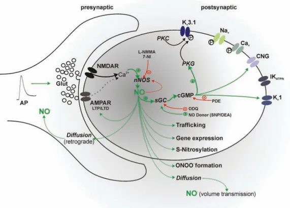
1.4.1. Nitric oxide synthesis, receptors and downstream signalling
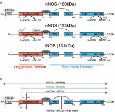
NO is intracellularly synthesized by a group of enzymes known as NO-synthases (NOS). NOS are complex enzymes expressed in 3 different isoforms: endothelial (eNOS), neuronal (nNOS) and inducible (iNOS). The first two isoforms (nNOS and eNOS) are constutively expressed in the CNS and they are mainly involved in physiological processes, whilst the third one (iNOS) is prototypically expressed in macrophages and glia in the CNS as a consequence of immunological activation. All three isoforms synthesise NO and citrulline from