A
A
l
l
m
m
a
a
M
M
a
a
t
t
e
e
r
r
S
S
t
t
u
u
d
d
i
i
o
o
r
r
u
u
m
m
–
–
U
U
n
n
i
i
v
v
e
e
r
r
s
s
i
i
t
t
à
à
d
d
i
i
B
B
o
o
l
l
o
o
g
g
n
n
a
a
DOTTORATO
DI
RICERCA
Biodiversità ed Evoluzione
Ciclo XXSettore/i scientifico disciplinari di afferenza: BIO/18
TITOLO TESI
Wing Shape Evolution:
A Role for Cell Competition in Shaping the Proximal Distal Axis of Drosophila Wing.
Presentata da: Dott. Marcello Ziosi
Coordinatore
Dottorato
Relatore
Prof. Giovanni Cristofolini Prof.
Sandro
Cavicchi
T
ABLE OFC
ONTENTSGeneral Itroduction...1
Oogenesis... 2
Embryogenesis... 2
Larval and Pupal Development
...3Wing imaginal disc structure, specification and development ...4
The wing ...6
Developmental Imaginal Disc Program: Axes and Boundaries ...
7Anterior-Posterior determination ...9
Dorsal-Ventral determination ...9
Proximal-Distal determination ...10
Cellular dynamics involved in growth and shape control in the imaginal disc
...14Cell proliferation ...14
Spatial control of proliferation. Oriented cell division and cellular reallocation. ...15
Cell competition ...18
fat(ft), dachsous (ds) and four jointed (fj) ...21
Hippo pathway ...25
Hippo pathway core members ...25
Salvador (Sav) ...25
Warts and Hippo ...25
Yorkie ...26
Hippo pathway accessorial members ...27
Expanded (Ex) and Merlin (Mer) ...27
Disc overgrown/Double Time (Dco) ...28
Dachs (D) ...28
Hippo pathway downstream target genes ...28
myc (diminutive in Drosophila)
...30How development cant contribute to evolutive tematics:
Evolution and Development (Evo-Devo)...
34Pleiotropy and Evolution ...37
Selector Genes ...38
Regulatory Architecture ...39
Thesis Outline
...41General Material and Methods
...42Nomenclature, Breeding Conditions and Used Stocks ...42
Genetic Methods ...44
UAS-Gal4 system (Brand and Perrimon, 1993) ...44
FLP-FRT (Xu and Rubin, 1993) ...45
Flp - out (Neufeld, 1998) ...46
MARCM system (Tee and Luo, 1999) ...48
Immunostaining ...49
X Gal Staining ...50
in situ hybridization ...51
Results ...
52Chapter 1: Myc and Yki
...52dmyc is a target of dachsous and fat tumor suppressors genes ...53
Yki is a dmyc activator ...55
dmyc repression affect yki activity ...56
Yki ectopical expression transforms cells into supercompetitors ...58
yki and compartmentalization ...60
dmyc competition assay ...62
dmyc fails to rescue yki LOF viability ...64
vg is implicated in dMyc regulation along the proximal distal axis ...68
Chapter 2: morphological analysis...69
Effect on the whole wing of fat, ds and four jointed UAS lines ...72
Phenotipes Obtained with the MS1096-GAL4 driver (whole wing) ...74
Wing Area ...75
Cell Area and Cell Number ...77
Continuos analysis along the proximal distal axis ...79
Trend in Cell Area Variation along the proximal distal axis ...81
Trends of Wing Area, Cell Number and Cell Area of the UAS lines overexpressed with MS1096-GAL4 ...82
Ectopic Patterned Expression along the Proximal Distal Axis ...83
Ds expression pattern constrain the shape of the wing ...90
TABLES ...91
Discussion and Perspectives
...92About Cell Competition ...92
On Genes and Form ...94
On the Evolution of Animal Form ...95
GENERAL INTRODUCTION
Drosophila melanogaster (Fruit fly) has from nearly a century a fundamental
role in the understanding of the basis of genetic mechanisms, due to the relative semplicity of its genome, to the short life cycle, to the abundance of the progeny and to the great quantity of genetic markers of its body wall.
From the pioneering studies of Thomas Hunt Morgan and the members of his laboratory, the fruit fly has early become the most characterized model organism for genetic studies.
The development of mutagenesis techniques by nobel prizes Christianne Nüsslein-Volhard and Eric Wieschaus has permitted the isolation and characterization of a large number of genes involved in each step of the early development.
Antonio Garcia-Bellido, John Merriam and Peter Bryant, by clonal analisys techniques have characterized the basis of the genetic programs involved in the development of the adult appendages.
With the development of molecular biology techniques and by the genome sequentations, this animal represents today an excellent model to understand the genetic mechanisms at the basis of almost all metazoan development.
The fruit fly is an holometabolous organism, characterized by a life cycle that starts from an embryonal stage, made inside the egg, followed by three larval instars (L1, L2, L3), during which the larva increases its dimensions. At the end of the third larval instar, at the pupal stage, a catastrophic metamorphosys occurs characterized by the histolysis of the larval tissues and by imaginal tissues differentiation, responsible for the adult appendages definition.
At the end of this stage the adult insect (also called imago) ecloses. The life cycle of a wild type organism is about ten days at 25°C.
OOGENESIS
The oogenesis is the process that drives the fomation of the oocyte in the ovary.
Each ovary is composed by 10-17 ovarioles, in wich the most apical portion is called germary, that is responsible for the creation of pro-oocyte cells. They migrate basally in a region called vitellary, where they reach the maturation before the release into the oviduct.
The oogenesis begins in the germary, in which a germinal stem cell will divide, generating another stem cell and a cytoblast that in turn originates the pro-oocyte and the nurse cells.
The oocyte and the nurse cells are surrounded by somatic cells called follicular cells; these three kinds of cells together build the egg chamber (Carpenter, 1975).
The Drosophila oocyte development is subdivided in 14 stages (King,1970). At the beginning of stage 8 the vitellum accumulation occurs in the oocyte, that rapidly increases at the expenses of the nurse cells; as a result at the end of oogenesis the egg chamber is entirely occupied by the oocyte.
EMBRYOGENESIS
Drosophila embryo develops immediatly after the egg laying (AEL), and it is
characterized by a superficial meroblastic segmentation.
The mature oocyte is quiescent at the metaphase of the first mitotic division of the meiosis until fertilization. After that, male and female pro-nuclei perform a sincronous division followed by fusion, that generate the zygote.
The first 12 rounds of cell division are syncytial and lead to the syncytial blastoderm formation; at the end of this stage a large number of nuclei migrate to the embryo surface (Foe and Alberts, 1983), with the formation of the polar cells, successively there is the cellularization of the nuclei and the beginning of the blastodermal stage with the creation of the somatic and germinal cell lines (the second derived by the polar cells), 6.3h AEL (Glover 1991).
This is a key step because while the early stages are governed by the maternal genome, from this moment the development is driven by the zygotic genome.
During the successive phases of gastrulation, major migratorial events (medioventral furrow and cefalic furrow) occur, generating mesodermal structures and the head region. During this stage, mesoderm is subdivided in splancnopleura, responsible for visceral muscles, and somatopleura, from wich all the other mesodermal structures originate (muscles, circulatorial system, fat bodies and somatic components of the gonads).
Nervous system is constituted by a series of elements distributed longitudinally to the embryo body.
Successively to the formation of the germinative “stria”, there is the invagination of the polar cells and other structures, and the formation of the tracheas.
The last morphological event is head involution (Fullilove and Jacobson, 1978). The mascellar, mandibular and labial segments of the larval head and the cells of the imaginal discs that are external invaginate inside the embryo, and this process is followed by an external cuticular deposition and by the differentiation of internal organs (Wieschaus and Nüsslein-Volhard, 1986).
LARVAL AND PUPAL DEVELOPMENT
After 23h AEL the L1 larva emerges; the three larval instars are spaced out by moults and followed by metamorphosis.
Moults and metamorphosis are governed by ecdyson peaks, a steroid hormon produced by the ring gland.
The larva is characterized by two cellular types: larval cells, that are polyploid, and imaginal cells that are diploid.
Imaginal cells segregate precociously from the surrounding larval cells, forming small cell groups at 9-10h AEL and are organized in two fundamental groups, imaginal discs and abdominal histoblasts.
Imaginal discs begin an intense proliferative activity from the second larval instar until pupariation, while abdominal histoblasts proliferate later, during the pupal stage. At this moment the majority of larval cells are eliminated and substituted by imaginal cells that originate the integument and the adult appendages.
Imaginal discs originate the structures of the head, thorax external appendages, genitalia and adult muscles. The histoblats originate the abdomen structures at the exception of the 8th segment that derivates from the genital imaginal disc.
During the metamorphosis three important steps occur: 1.elongation
2.eversion of the imaginal structures
3.enlargement and fusion of the imaginal structures, with the formation of a continuum epithelium (Fristrom and Fristrom, 1993).
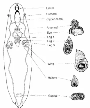
Figure 1 – Localization of the imaginal discs in the larva at the L3 instar (Bate e
Martinez-Arias, 1991).
Wing imaginal disc structure, specification and development
Each imaginal disc at the end of larval development is constituted by a pseudostratified columnar epithelial tissue, that represents the actual imaginal disc, and by a squamous epithelium that forms the peripodial membrane.
The first one originates the integument and the appendices, the second one originates the epithelial veil that welds the structures derived from different imaginal discs.
One interesting feature of the imaginal disc is the possibility to transplant it in adult abdomens, in which they can survive, metamorphose and differentiate (Schubinger et al, 1969; Simcox et al., 1989).
Fragments of imaginal discs can regenerate the entire disc structure and experiments in this direction have allowed the characterization of “fate maps” of regeneration (Bryant, 1978).
The wing imaginal disc is an excellent model for the elucidation of organogenesis and proliferation mechanisms; at the end of L3 it is subdivided in a series of folds. The centrifugal regions of the imaginal disc originate the thorax structures, the notum and the pleura. The middle region originates the hinge region while the central region is the presumptive territory that makes the wing lamina.
Imaginal tissues are virtually two-dimensional structures, but they originate adult appendages with three axes (AP, DV, PD); this is due to a mechanism of eversion during methamorposis, in which the most central regions of the disc originate the distal structures (wing from distal to proximal), while the external regions originate the most proximal structures of the appendage.
During the embryogenesis the action of the segment-polarity genes generate parasegmental restriction boundaries and positional information for the specification of the imaginal discs primordia (Choen, 1990).
The patterned expression of the selector gene engrailed is present in the anterior region of the parasegmental compartment and in the posterior region of the wing imaginal disc, suggesting a derivation from two different parasegments (Kornberg, 1981a, 1981b; Kornberg et al., 1985; DiNardo et al., 1985; Ingham, 1985)
Imaginal discs primordia are constituted by 20 founder cells, identified by the expression of specific markers.
The direct source for the genesis of positional signals for the founder cells is Wingless. Without this signal, no imaginal structures arise.
Escargot (Esg) is a marker of all the primordial imaginal cells and it is indispensable for the diploid maintainment.
Several indications exist that wing imaginal disc originate ventrally and represent a branch of the second leg imaginal discs (Cohen,1997). The activation of differential selector genes separates the two structures early making them
become independent, and the imaginal disc grows dorsally, while legs are ventral appendages.
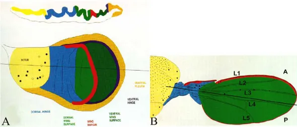
Figure 2 – Schematic representation of the wing imaginal disc at L3 instar (A) and of the
adult wing (B). Different colors in the figure A indicate the presumptive territories of the structures visualized in B (from Bate and Martinez-Arias, 1991).
The wing
The wing is composed of a plate of epithelial bi-stratified cells in which a ventral and a dorsal surface are present.
x
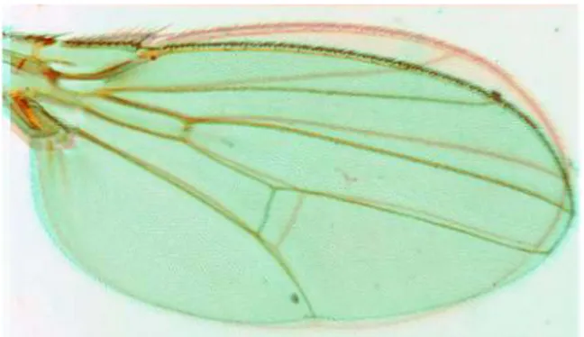
Figure 3- Vein and Intervein Territories of Drosophila wing,
Longitudinal veins are five and are called L1-5; there are also two cross-veins, the anterior (acv) and posterior (pcv) cross-veins. Intervein compartments are also five and named A-E.
The proximal region is called the hinge, and it connects the wing to the thorax, dorsally (notum) and ventrally (pleura) respectively.
The wing lamina is called wing blade, and in the anterior border it is provided with three series of sensorial bristles. The wing blade is constituted by two types of cells: vein and intervein.
Veins are constituted by alive epithelial cells that form tubular structures that confere stiffness to the wing and accomodate tracheas and neuronal sheafs.
Intervein cells are dead and chitinized, and are characterized by the differentiation of a single trichome for each cell.
DEVELOPMENTAL IMAGINAL DISC PROGRAM:AXES AND BOUNDARIES
Transplantation assays demonstrated that developmental patterning is not determined by external influences but by a disc-specific intrinsic developmental program. Originally defined in the wing disc by Antonio Garcia-Bellido in 1973, the compartments are the basic components of the Drosophila body plan. In genetic terms they are parts of the body that originate from the same cell lineage. From the beginning of the embryogenesis, as a consequence of the pair rule gene expression and segmental differentiation of embryo, the primordia of the imaginal cells of the wing contain two separate cell lineages, which form the anterior and posterior compartments.
This is a consequence of the patterned activation of the homeobox gene
engrailed (en), which segregates anterior and posterior compartment cells.
Antonio Garcia-Bellido in 1973 showed by clonal analysis the existence of a restriction boundary between the anterior and posterior regions of the wing imaginal disc that clones of cells can’t cross during proliferative stages.
The discovery of compartments surely plays a pivotal role in the developmental genetics of the past 35 years and the use of mitotic analysis is yet today the war horse of developmental geneticists.
The existence of restriction boundaries in the Drosophila wing imaginal disc allowed to elaborate a series of developmental models about the genetic program
involved in morphogenesis. The Entelechia model by Antonio Garcia-Bellido proposes that the restriction boundaries are the organizers of developmental programs responsible for the achievement of correct size and shape of the organ, by the activation of specific hypotesized genes called “martial genes”. The expression level of those genes is gradual and decreases from the boundary. Cells respond to martial genes information and stop dividing when reach the limiting quantity of signal. (Garcia-Bellido and De Celis, 1992; Garcia-Bellido and Garcia-Bellido, 1998).
The other current model is the morphogen one, in which diffusible factors generated along the restriction boundary (Dpp or Wingless for example), generate positional cues that cells detect as the quantity of factor received. A different distance from the boundary is responsible for diverse abundances in factor caption and this gradient drives cell proliferation, allocation and survival (Wolpert, 1969; 1971).
After the characterization of the AP restriction boundary, other boundaries where discovered. The DV boundary, formed by the activity of Notch and Wingless and the less canonical proximal-distal boundary that separates the wing blade from the hinge and the hinge from the thorax.
Anterior-Poserior Determination
One of the first events in the imaginal disc development is the formation of the Anterior Posterior polarity, as a consequence of the activation of the gene
engrailed. engrailed is expressed in all the posterior compartment and its protein
induces the expression of hedgehog (hh) in the whole posterior compartment (Tabata and Kornberg TB, 1994; Zecca, Basler, Struhl G, 1995).
Hh protein is a morphogen that spreads in a line of cells along the anterior-posterior border where it induces the expression of another morphogen, Dpp (Tabata and Kornberg TB, 1994; Zecca, Basler, Struhl G, 1995).
This process is the basis of a complex regulative pathway, that leads to the activation and the repression of a large number of genes complexes. The final result is the definition of the anterior-posterior axis and the definition of positional information that leads to the creation of the anterior-posterior boundary and the specification of the vein and intervein regions.
Dorsal-Ventral Determination
Dorsal-Ventral polarity is the second axis of polarity to form in a temporal succession. The definition of this axis occurs at the beginning of the second larval instar (Garcia-Bellido, 1973).
The dorsal-ventral restriction border originates through the action of the selector gene apterous, that is expressed only in the dorsal compartment (Diaz-Benjumea and Cohen, 1993; Blair, 1993).
Ap protein activates fringe in the dorsal compartment (Irvine and Wieschaus, 1994), a secretion factor that induces the activation of Serrate (Kim et al.,1995), a Notch ligand (Rebay et al.,1991), and inhibits the expression of Delta, another Notch ligand (de Celis et al., 1996; Milan and Cohen, 2000).
Fringe is a glycosil transferase that modifies Notch receptor, inhibiting its affinity for Serrate but increasing its affinity for Delta, that in turn increases the affinity for Notch.
The final picture is that dorsally Serrate is inhibited by the activity of Fringe, while ventrally Delta can’t activate Notch.
The only region where Serrate and Delta can both activate Notch is at the boundary, where Apterous is expressed in two cell lines (de Celis, 1996).
Notch activates the expression of wingless, cut and vestigial and, as a consequence, the DV restriction boundary is formed and the wing blade presumptive territory begins to be defined.
Proximal-Distal Determination
While much is known about the development of the patterning mechanisms that specify the A/P and D/V axes, very little is known about the determinationof the P/D. The formation of the three structures of this axis (notum, hinge, wing
Figure 5 - Genes involved in Proximal Distal Axis formation show a circular
blade) is due to the sequential activation-repression of a moltitude of selector genes, in response to the formation of the AP and DV axes (engrailed-AP axis and apterous-D/V axis) and to the generation of morphogen activity at the boundary of the two axes (Dpp at the AP border and Wg at the DV border).
The correct wing patterning requires the activity of Wg morphogen; in factthe reduction of Wg function causes a complete loss of wing structures and notum duplication (Morata and Lawrence 1977).
Wg expression begins in a ventral-anterior wedge of the early second instar wing disc and is extremely dynamic throughout the larval stages (Couso 1993; Ng 1996). At this stage, after the formation of AP and DV axes, another subdivision occurs in the formation of the proximal-distal axis that subdivides the wing disc in the Notum, Hinge and Blade regions (Klein, 2001). The activity of Wingless promotes the repression of teashirt homeobox gene that produces the imaginal body wall formation, and vg expression (Wu and Cohen, 2002).
At the second instar Wg activity is repressed in the Dorsal and Posterior compartments by the activity of EGFR (Baonza, 2000), the EGFR activity promotes the specification of the notum identity in the proximal region by the activation of the iroquois complex (Iro-C) genes (Zecca and Struhl, 2002), ectopic expression of EGFR at this stage causes a notal duplication similar to the Wg loss (Wang SH, Simcox A, Campbell G: 2000). During the second instar, the combined activities of Wg and Notch signalling pathways induce the expression of the nuclear protein Vestigial (Vg), wich is essential for the wing blade development. Vg is firstly just along the D/V boundary by the activation of theso called boundary enhancer (vgBE; Couso 1995; Kim 1996: Klein and Martinez Arias 1998, 1999; Neumann and Cohen, 1997; Williams et al, 1991, 1994).
In the early third instar, N, Wg and Vg act together to activate a second enhancer, the vg quadrant enhancer (vgQE), that induces the vg expressionacross the wing pouch (Klein and Martinez Arias, 1998). Later the vgQE is regulated in a dosage-dependent manner by the activity of Wg and Dpp signaling pathways (Kim 1996; Klein and Mrtinez Arias, 1998).
Vestigial is a transcription factor (see also cap1) that acts in a complex with the product of the scalloped gene (sd). vg loss of function prevents the development of the wing blade (Delanoue 2004, van de bor 1999) while its
overexpression may promote cell proliferation (Delanoue et al. , 2004; Halder et al., 1998; Kim et al., 1996; Simmonds et al., 1998; Baena-Lopez et al., 2003).
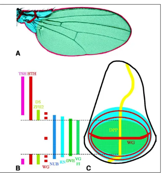
The integration of the signal derivating by Dpp and Wg-N produces a graded activity of Vg along the proximal-distal axis, and this distribution can trigger local cell interactions between neighboring cells, globally modulating the growth and shape of the wing blade (Baena-Lopez, 2006), so generating positional information.
Wg is also required for the correct development of the proximal region of the wing (Neumann and Cohen, 1996; Witworth and Russell, 2003), where Vg is not expressed. This leads to the activation of the homeobox gene homothorax (hth) by Wg activity. Here Hth plays a dual role, to limit the size of developing distal wing, by repression of target genes, and to upregulate Wg expression (Azpiazu and Morata, 2000; Casares and Mann, 2000).
A key step in the distal identity formation is the extreme dynamics of pattern expression of Wg, that change during the third instar; in particular Vg acts in the activation of Wingless expression in a ring of surrounding cells expressing the Sd-Vg complex (Liu et al., 2000; del Álamo Rodriguez et al., 2004) .
At the end of the third larval instar Vg is expressed in the distal part of the wing while Wg is expressed in the proximal region of the wing in two concentric rings, one mediated by the Vg-Sd activity and another due to the activation of an enhancer called spad-flag (Neumann and Cohen, 1996).
In particular, Wg activity in the proximal region promotes the initial expression of zfh2 homeodomain containing gene, a proximal determinant, and the homothorax expression (Witworth and Russell, 2002; Azpiazu and Morata, 2000); the function of Wg is fundamental for the identity, proliferation and survival of the proximal region of the wing (Neumann and Cohen, 1996; Johnston, 2002).
Recapitulating, at the first larval instar there is the subdivision of the wing region in the notal versus wing structures due to the repression of teashirt in the distal region by Wingless; successivley, during the second instar, the hinge region is defined by the repression of teashirt and by the activation of homothorax by Wingless, and wing identity results from the activation of the selector gene
The events that drive the proximal-distal axis formation in the wing blade region are poorly understood. There are a series of mutant conditions that lead to a reduction of specific regions along this axis: rotund (rn) (Kerridge and Thomas-Cavallin, 1998) mutation leads to the reduction, mutations in fat (ft), dachsous (ds), approximated (app) and four jointed (fj) interfere with the development of medial and proximal regions of the wing (Lindsley and Zimm, 1992; Garoia et al., 2002-2004). nubbin is responsible for a reduction of the distal regions of the wing (Cifuentes and Garcia-Bellido, 1997).
Clonal analysis experiments have failed to detect restriction boundaries between proximal and distal regions of the wing blade, nevertheless the selector gene nubbin appears to have an important role in the development along this axis. At the end of L3 nub is expressed in the region of the wing blade (Cifuentes and Garcia-Bellido, 1997).
Clonal analysis has revealed that nubbin mutant clones, even if small, may affect the entire P/D axis pattern when arising in the proximal region of the wing; on the contrary distal clones do not (Ng and Garcia-Bellido, 1998), suggesting the existence of a proximal-distal organizing center in the proximal region.
In the determination of proximal and distal elements of the wing, Wingless and Vestigial act in the regulation of homothorax and teashirt. The interaction between Wg and Vg expression is important for the differentiation of wing blade structures, while the action of Wingless alone is important in the hinge definition (Klein and Martinez-Arias, 1998).
Wg appears to have a dual role. While in the distal region of the wing it contributes in the wing blade formation, in the proximal region Wg and Teashirt act together in repressing Vg and therefore the expansion of wing blade territory (Casares and Mann, 2000).
In the hinge and proximal wing structures fomationm, dachsous has been seen to be determinant, and appears to act as a Wg modulator. ds LOF mutants often show notum duplication instead of the wing blade, similarly to the phenotype commonly observed in Wg LOF mutants (Rodriguez, 2004).
CELLULAR DYNAMICS INVOLVED IN GROWTH AND SHAPE CONTROL IN THE IMAGINAL WING DISC.
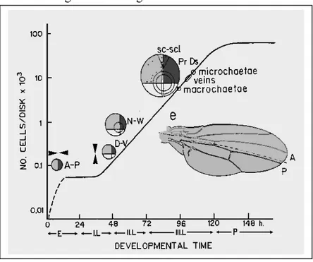
Cell proliferation
The wing imaginal disc represents an outstanding model in the understanding of proliferative mechanisms involved in organognesis. This structure is indeed characterized by a rapid growth that, from few initial cells (20-30), in almost 120 hours allows to reach a final number of 50.000 cells, with a proliferative rate of about 8.5 hours/cell division.
The proliferation is intercalar and exponential in almost all the wing pouch (Garcia-Bellido, 1965; Resino et al., 2002). Imaginal disc cells show a sinchrony of divisions and growth preferentially in the proximal to distal direction (Milan et al.,1996).
Final shape and dimension are genetically programmed, but the mechanisms are poorly know. The postulated models that try to explain the spatial control of the proliferation inside the discs are the morphogen model, proposed by Peter Lawrence, and the Entelechia model proposed by Antonio Garcia-Bellido.
According to the first model, cell proliferation follows the signals dictated by diffusible morphogens originated along the restriction boundaries; in the second model, direct interactions between cells mediated by adhesion molecules are involved in which Fat is a possible candidate (Le Cuit and Le Goff, 2007). Still today proofs in favour or in discussion of these two model are provided, and none can be discarted in favour of the other.
Mutations in cell cycle components have an effect on cell growth without affecting final size and shape of the disc. Weigman and collaborators (1997) stopped mitotic divisions specifically in the anterior compartment of the wing disc inactivating the cdc2 kinase, a mitosis promoter. The result was that imaginal disc with normal dimensions and shape grew in which the anterior compartment was composed of fewer but larger cells.
Neufeld and collaborators (1998) induced mophogenetic clones overexpressing cell cycle regulators: cycE and CDC25 or the gene E2F. In this case mitotic rate is increased, producing more cells of reduced size, preserving the compartment size and shape.
These results show that genes that affect cell cycle can affect cell number or cell size without affecting the final size and shape of the disc.
Experiments on the Minute mutation allowed to discover the phenomenon of
cell competition (discussed more in detail bellow) an important mechanism of
compensation involved in the homeostasis of organs.
The insulin pathway (Inr) is a cell autonomous process involved in cell growth control. Manipulations at different levels of this pathway can affect both cell proliferation and cell growth. Loss of function mutations of the Inr gene lead to a delayed development with a smaller final dimension of the fly, less and smaller cells (Böhni et al., 1999).
Mutations of the gene S6K, involved downstream in the Insulin pathway, lead to smaller individuals in which the reduction is only due to a smaller cells.
Other mutations are of interest in cell cycle control, for example ras1,
cycD/cdk4 and myc that are discussed in detail in this thesis.
Finally, transmembrane cadherins and cytoplasmic associated catenins hare the fulcrum of adherens junctions, and are responsible for cell shape, because they are a link between cell membrane and cytoskeleton.
Classical cadherin mutations are often associated with cancer onset, and Fat and Dachsous, two protocadherins, are involved in proliferation control.
Spatial control of proliferation. Oriented cell division and cellular reallocation.
Clonal analysis has highlighted that cell divisions in imaginal discs are intercalar (Garcia-Bellido, 1994), this means that proliferation is not generated in specific regions to follow migratory events, but it is homogeneous inside almost all the disc.
This process is cell autonomous and small fragments of imaginal disc can regenerate an entire compartment (Briant, 1975). Despite that, a series of non cell autonomous interactions between mutant and wild type cells in mosaics show effects of accomodation, demonstrating that cell proliferation is the result of collaboration between neighboring cells (Resino, 2004).
Imaginal disc cells appear organized in small groups, not clonally related, that during the disc growth proliferate sincronically; these groups are not fixed and often change during development (Milan et al., 1996).
Cell-cell contacts are fundamental in those processes, and coordinate the growth of this monostratified epithelium. Cell junctions are indeed docking structures for a multitude of tumor suppressor genes (TSG).
Clonal analysis has permitted to understand the lineage of single marked cells during development, finding that clone shape is related to organ shape (Garcia-Bellido, 1994; Resino et al., 2002; Dolan et al. 1998).
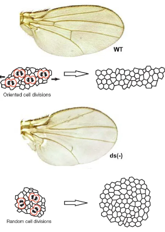
In the wing, clones of cells show an elongated shape, and grow prevalently in the proximal-distal direction (Resino, 2002). In 2005 Luis Alberto Baena-Lopez showed that this situation was largely determinated by the orientation of mitotic divisions (OCD), and by the fact that after dividing, cells maintain their position (Baena-Lopez et al.,2005). Cells of the wing blade proliferate in PD direction with the exception of the wing margin in which cells proliferate in DV direction.
Mutants that affect OCD also affect clone shape that appears rounded, and the entire organ shape, that appears affected in the proximal-distal direction.
Genes involved in this process are often involved in planar cell polarity (PCP), but the two processes appear indipendent (matakatsu, Strutt, lawrence).
Also in this process fat and dachsous are implicated, underlining the great pleiotropy of these genes and the importance of cell adhesion in communication among cells.
ft and ds mitotic clones show rounded shape instead of the wild type elongated
shape, and grow prevalently in the proximal wing regions while the wild type twin grow preferentially in the distal direction (Garoia et al., 2000-2004).
C
Cell competition
C
Cell competition was discovered in imaginal discs over 30 years ago (Morata and Ripoll, 1975), in which cells with a lower dividing rate, although viable, were eliminated by flanking faster dividing cells.
The Minute mutation is subjected to cell competition. Homozygous cells (M/M) die but heterozygous (M/M+) are viable, but with a lower dividing rate. When M/M+ cells are flanked by wild type cells (by mitotic recombination),
Minute heterozygous cells are eliminated through apoptosis and the process is
called “Cell competition” (Cytagon).
There are more than 60 Minute loci in the Drosophila genome, each showing, if mutated, growth delay due to some defects in ribosomal proteins or activity. Mutations at the diminutive locus (dmyc) of the homologous human oncogene
c-myc, has also been shown to trigger cell competition (for review see Moreno,
2008).
Clones of epithelial cells carring a dmyc hypomorfic condition are outcompeted and die if flanked by a wt twin. By contrast, the same cells are viable when allocated within cells with the same hypomorphic condition.
Many other genes have been shown to regulate cell competition, in some cases genes regulating dmyc activity. Those genes include the homologues of the retinoblastoma family (Rbf), of the E2F family of transcription factors, of the Ras family of proto-oncogenes. Members of the decapentaplegic (Dpp) signaling pathway such as the transcription factor Brinker (Brk) or the Dpp receptor Thickveins (Tkv) might also trigger cell competition, and it is not clear if
components of the insulin pathway are also involved (De la Cova, 2004; Moreno and Basler, 2004).
It is possible that cell competition may act through different mechanisms and does not use a universal pathway, but this is not currently known.
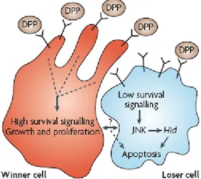
Interestingly, outcompeted cells seem to have a decreased activation of survival signaling pathways, in particular the Dpp pathway (Moreno 2004).
Moreno, Morata and Basler have formulated the ligand-capture hypothesis, which proposes that cells compete for the active uptake of extracellular survival and growth factors, and winner cells may inhibit or limit the loser cells in the uptake of survival factors, eventually causing their death.
However this mechanism is not clear, and evidence suggests that ligands cells compete for are not in a limiting supply, for example Dpp (moreno 2007).
Cell competition is not a passive mechanism in which winner cells induce loser to suicide, and cell culture experiments suggest that soluble factors are involved secreted by both winner and loser populations of cells (Johnston 2007).
Cell competition apoptosis is mediated by Jun N-terminal kinase (JNK) and the proapoptotic genes hid (also called Wrinkled) and rpr (Johnston 2004), and winner cells appear to amplify this death signals through the activation of a mechanism called “engulfment” (Li and Backer, 2007). Cell competition in wing
imaginal disc cannot occur without engulfment and it has been proposed that with this mechanism winner cells can take the position of losing cells by phagocytosis.
Genes involved in engulfment are known (draper, WASp, PSR) but mechanisms through which winner cells activate its machinery remain to be determined.
Notably, cell competition appears to act within a small range of 5-8 cells from the dmyc overexpressing cells, but is incapable to cross compartment borders.
Laura Johnston (2004-2007) and Eduardo Moreno (2002, 2004, 2007) showed that cell competition is relevant both to evolution and cancer onset.
The evolutive hypothesis of Laura Johnston is that cell competition may act like a mechanism intrinsic to the imaginal disc or possibly to another tissue involved in the maintainment of developmental homeostasis, conferring a flexibility important to the elimination of misspecified or growth-impaired cells without affecting the final developmental architecture. This is supported by the fact that dmyc overexpression leads to cell competition through the substitution of the surrounding wild type tissues, without affecting the final dimension and shape of the organ (2004).
Recently Nicholas Baker has demonstrated, by genetic experiments and imunofluorescence, that fat and other genes of the Hippo pathway protect cells from competition (Baker 2006).
Proliferation, oriented cell division and cell competition appear to converge on
fat and dachsous genes, that may play a fundamental role in orchestrating two
fat (ft), dachsous (ds) and four jointed (fj)
Recessive mutations of fat tumor suppressor gene leads to hyperplastic extragrowth and morphogenetic defects.
fat gene encodes for a very large transmembrane protein, member of the
protocadherins and is constituted by a lot of protein domains: an extracellular portion with 34 cadherin homology domains, 4 EGF-like and 2 LamininG domains, a transmembrane domain and a cytoplasmic domain (Mahoney et al.,1991).
Cadherins are transmembrane glycoproteins Ca2+ dependent that mediate cell adhesion prevalently by homophylic interactions (Tomschy et al., 1996).
This mechanism is essential for the organization of almost all methazoan tissue development and integrity (Hyafil et al., 1981; Gumbiner, 1996).
Cadherins and protocadherins can inhibit cell proliferation acting on cycline dependent inhibitor kinases, interacting with different signaling pathways as the EGFr (Levenberg et al., 1999; Hermiston et al., 1996; Garoia et al., 2004), by cytoskeleton stabilization (Chausovsky et al., 2000) and by planar cell polarity (PCP) control (Muller, 2000; Woods et al., 1997).
Fat also interacts with the Wingless pathway in more than one manner: mutants for fat can release more Armadillo (β-catenin) into the cytoplasm (Greaves et al., 1999), and fat appears to regulate glycoproteins levels that mediate Wingless spreading (Baena-Lopez, personal communication).
The interaction between Fat and Wingless plays an important role in the proximal to distal axis organization (Cho et al., 2004-2006), Fat can inhibit Wg expression in certain locations in developing wings, and the increased Wg expression in fat mutants partially accounts for the hyperplastic growth that occurs in the wing, that can be suppressed by loss of Wg, but only in the proximal region of the wing with a former role in the proximal-distal signaling (Cho et al., 2006).
Viable hypomorphic alleles like ft1 show anterior to posterior reduction of abdominal structures, larger thorax and a proximal to distal reduction of all the appendages with the presence of planar cell polarity defects (Lindsley and Zimm, 1992).
Alleles like fat8 or fat Gull revertant (ftG-rv) (Bryant et al., 1998b) are lethal recessive with a hyperplastic extragrowth of larval imaginal discs, in particular in the proximal region of the wing imaginal disc that shows extra folds but maintain a monostratified structure.
Cell dimensions are generally smaller than the control and during the differentiation show strongest cuticolar posing, probably due to cytoskeleton defects (Garoia et al., 2000).
In fat8 mutant pupariation is delayed of 3.2 days at 25°C, and the final cell number is about 122.000 against the 50.000 of the wild type (Briant et al.,1988b).
ftGull is an anthimorphic allele, that in heterozygosis can resemble the viable phenotypes of the other recessive alleles (Mahoney et al.,1991) and can be rescued by the lethal recessive allele ftG-rv.
ft is involved into the proximal-distal axis shapening of the appendage
development and this role appears to be related to that of another protocadherin, Dachsous, and to a Golgi transmemembrane protein, Four jointed (Villano and Katz, 1995).
ds encodes for a protocadherin similar to Fat, constituted by 27 cadherin
domains, a transmembrane domain and a cytoplasmatic domain (Clark et al, 1995). ds viable mutants show strong similarities to ft viable mutants and the suppression of ftGull phenotype by ds1 viable allele has suggested a possible interaction (Mohr, 1929).
Heterophyilic interactions between those proteins have been demonstrated (Matakatsu and Blair, 2004), and are singular in cadherins that prevalently interact in a homophylic manner. Ft-Ds interaction is important for cell adesion, for the activation of different signaling pathways, probably involving also the Hippo pathway (Choo et al., 2006, my data) and for the regulation of the planar cell polarity (PCP) in the eye, in the wing (Matakatsu and Blair, 2004-2006) and in the abdomen (Laurence 2007), by the frizzled (fz) regulation and by direct binding with the co-repressor Atrophin (Fanto et al.,2003).
Remarkably, Dachsous is the only cadherin expressed in a particular pattern in the developing wing, with a preference for the proximal cells representative of the hinge (Clark et al.,1995); low level of dachsous are also present in more distal regions with a decrescent intensity from proximal to distal (Strutt and Strutt, 2002; Ma et al., 2003).
Garoia et colleagues (2000) have demonstrated that fat clones are allocated prevalently in the proximal region of the wing. fat cells grow prevalently in proximal direction, differently from the wt cells that grow prevalently in distal direction.
Also the majority of growth defects are localized prevalently in the proximal regions.
ft clones are rounded and planar cell polarity inside the clones is strongly
perturbed. Cells tend to be located at the clonal boundary, losing the normal proximal-distal grow direction; this is prevalently due to a loss in oriented cell division direction (Baena-Lopez et al. 2005) responsible for strong defects in the entire organ.
Fat is an EGFR antagonist (Garoia et al. 2004); double clones for fat18 and EGFR pathway mutants are smaller and tend to be distributed all along the proximal-distal wing region (Garoia 2000, 2004), and this is an indication that fat participates in the differentiation vs proliferation program of the imaginal discs.
fat clones fail to grow in a diminutive (dm) background (Garoia et al., 2004),
suggesting a role in cell competition (demonstrated in this thesis).
Fat is the first transmembrane protein recently connected to the Hippo pathway (Bennett and Harvey, 2006; Cho, E. et al., 2006; Silva, E. et al., 2006; Willecke et al., 2006; Tyler et al., 2007) and a dramatic suppression of fat mutant overgrowth phenotype can be obtained by removal of just a single copy of yki. Thus Fat signaling is highly sensitive to Yki dosage.
fat mutants increase the levels of Cyc-E and dIAP1, like all the genes of the
hippo pathway.
Probably the link between Ft and the Hippo pathway is represented by Expanded, but some data indicate that they act in a independent manner (Feng J. and Irvine K.D., 2007).
Fat and Expanded strongly co-localize at apical junctions, but attempts to detect a direct interaction between these two proteins have not yet been successful.
Expanded is absent from the membrane in fat mutants. It seems reasonable to suggest that the ability of fat to recruit Ex to the membrane enables Ex to signal
efficently to Hippo and Wts, potentiating the hippo signalling (Yin and Pan, 2007).
How Fat receive signals and how it exerts its receptor property is an unknow matter; the only known protein that is suspected to interact extracellularly with Fat is Ds (Matakatsu and Blair, 2006). Cells that express Fat preferentially adhere to cells that express Ds (Matakatsu and Blair, 2004), and the expression of both Ft and Ds is required for the intracellular positioning of proteins that are involved in planar cell polarity as well as proximal-distal patterning.
Dachsous is involved in the specification of the proximal-distal axis in the early wing imaginal disc. At the present it is the only known cadherin in
Drosophila that shows a spatially restricted pattern of expression in wing imaginal
discs from early stages onwards, and it is considered one of the earliest specific markers for the hinge territory (Rodriguez, 2004).
The pattern of expression of Ds is relevant for the control in the proximal-distal axis shape of the wing (see results).
Four jointed (Fj) is a type 2 transmembrane protein localized in the Golgi membrane. It has a specific distal pattern of expression in the wing disc, and fj mutants do not to affect the proximal region of wing disc (Villano and Katz, 1995; Brodsky and Steller, 1996). The pattern of expression of ds is complementar to that of four jointed, and this has been shown to be important for the polarization of the eye in PCP (Zeidler, Perrimon and Strutt, 1999) defects. The transcription of four jointed is repressed by Fat activity via the Hippo pathway (Choo et al., 2006), suggesting that Fat is activated by a proximal source, possibly Dachsous.
In a model proposed by Cho and Irvine (2004), fat, dachsous and four jointed act together in regulating the proximal-distal axis development, acting on Wingless and Rotund activity, mediated by Dachs and the Hippo pathway (Cho et al., 2006).
Hippo pathway
The size of developing organs is controlled by cell growth, cell proliferation and apoptosis, and the Hippo pathway has recently been described to be involved in those processes.
An important function of this pathway is to limit imaginal disc size, and loss of function of those genes leads to hyperplastic overgrowth (due to cell growth and proliferation), defects in apoptosis or cell competition and in the oriented cell divisions and Planar Cell Polarity (PCP).
This genetic pathway has been proposed to act like a “size checkpoint” operating in a tissue autonomous manner, sensing the total mass of the organ, rather than the size.
Genes owing to the Hippo pathway can be divided in “core genes” and “accessory genes”, the signal from the core converges on the co-transcriptional factor Yorkie.
Hippo pathway core members Salvador (Sav)
It is a scaffold protein containing a WW domain that can interact with WTS, and a C-terminal SARAH domain that mediates the bond to Hippo (in competition with RASSF). The primary function of Sav is to facilitate the close association of Wts and Hpo kinases, and/or to recruit Hpo to its site of activation (Tapon et al., 2002).
Warts and Hippo
They are both serine/threonine kinases that belong to the nuclear NDR and sterile-20 kinase families, respectively.
Hippo phosporylates Warts, Drosophila inhibitor of apoptosis 1 (dIAP1) and Salvador, whereas Warts phosporylates the co-transcriptional factor Yorkie (Harvey and Tapon, 2007). The founding member of this pathway is Warts (wts; also known like large tumour suppressor or lats), a gene coding for a kinase
similar to the human Dystrophin. Mutations in this gene result in dramatic outgrowths in the epithelial tissues, however cells do not lose their identity and the tumoral behaviour is hyperplastic (Justice et al., 1995; Xu et al., 1995).
The phosporylation of Salvador by Hpo activity leads to a physical contact between Hpo and Wts mediated by Sav scaffold activity.
The successive phosphorylation of Wts mediated by Hpo activates the pathway by the inhibition of the co-activator Yki.
Yorkie
Yki, wich was identified in a yeast two-hybrid screen for interactors of Wts (Huang et al., 2005), is an oncogene that regulates the transcription of genes involved in cell proliferation and apoptosis.
It is the homologue of the human YAP that has been reported to bind and regulate the activity of various transcriptional regulators, including p73, p53BP2 and several TEAD/TEF-type transcription factors. A point of interest in my thesis, successively discussed, is the very recent finding that Yki can bind the TEAD/TEF protein Scalopped, the transcriptional factor also complexed with Vestigial, the selector gene of the wing (Wu S. et al., 2008; Goulev Y. et al, 2008).
Dephosphorylated forms of Yki go into the nucleus where they activate a series of target genes; the sequential activation of the Hpo pathway drives to the phosphorylation of Yki by Wts. This process leads to the functional repression of Yki through its sequestration in the cytoplasm (Huang et al., 2005).
Yki phosphorylated form binds to 14-3-3 protein, via a specific motif. 14-3-3 is a protein that leads to the shuttling to the cytoplasm of phosphorylated proteins (Dong et al., 2007).
Activated forms of Yki control the transcription of several genes implicated in cell cycle, like Cyc-E, or in apoptosis repression, like dIAP1, or both, like the miRNA bantam. In this thesis I expose how Yki leads also to the transcriptional activation of the oncogene dmyc, involved in cell cycle regulation, apoptosis and cell competition.
YAP activity is influenced both in in vivo and in vitro assays, where the cytoplasm sequestration is dependent by the cell density status. This finding leads
to the hypothesis that also in vitro cell-cell contact interactions stimulate the activation of the Hippo pathway, mediating the growth inhibition by confluency, in this model, Fat and Dachsous are two possible surface receptors (Bin Zao et al, 2007).
Hippo pathway accessorial members
More than ten proteins have been implicated so far as components of the Hippo signaling pathway in Drosophila, from transmembrane proteins to nuclear transcription factors.
Expanded (Ex) and Merlin (Mer).
Ex and Mer are part of the 4.1 family of proteins. Mammalian members of this protein family such as Ezrin, Radixin and Moesin are thought to relay signals from cell surface receptors to the cytoskeleton. The mammalian orthologue of
Drosophila MER is the tumour-suppressor protein NF2 (Mc Clatchey et al.,
2003).
Phosphorylated forms of Ex and Mer activate the Hippo pathway .
Discs overgrown/ Double Time (Dco)
It is a highly conserved protein kinase orthologue of mammalian Casein Kinase 1ε (CK1ε). Dco is a pleiotropic protein involved in a wide series of processes, and it is important for the control of Hedgehog signaling, the Wnt-Wingless (Wg) signaling, circadian rhythms, planar cell polarity and organ size (Kloss et al., 1998; Zilian et al., 1999).
Dachs (D)
It is an unconventional myosin that is predicted to function as a motor protein, or a scaffold molecule. Genetic epistasis experiments place dachs upstream of
warts and downstresam of fat and dco (Choo 2004-2006). Mutations in dachs can
suppress excessive growth and proliferation in a tissue that lacks either ft and dco. Dachs can complex with Wts in vitro, but the mechanism by which it suppresses the Hippo pathway is unclear (Choo 2006).
dRASSF
It shares homologies with many human proteins and antagonizes the Hippo pathway by competing with Salvador for Hippo (Polesello 2006). RASSF proteins have several functional domains: a Ras-association domain, which mediates binding to Ras and other small GTPases; a SARAH domain, which homotypically or heterotypically binds to other SARAH domain proteins. In addition they have a C1 or LIM domain that mediate binding to membrane (Schell and Hofman, 2003).
Hippo pathway downstream target genes
Yki activation has been shown to phenocopy hpo, wts and sav loss of function mutations, whereas yki loss of function clones have opposite effects and do not survive (Huang et al., 2005).
Strongly candidate target genes for yki exist, but no direct targets have been formally defined.
DIAP1 and cycE were the first discovered targets of this pathway, where mRNA for these genes is increased in mutant clones for sav, hpo, fat and other pathway members (Tapon et al., 2002; Udan et al., 2003; Wu et al., 2003; Huang et al. 2005).
DIAP1 (for Drosophila Inhibitor of Apoptosis1) has the function to inactivate caspases activity and to prevent apoptotic processes (for a review see Montell, 2006). In yki loss of function clones, DIAP1 overexpression permits cells to survive (Thompson and Cohen, 2006).
Cyc E is a cell cycle effector whose mutations limits the G1-S progression in epithelial tissues and it is often overexpressed in neoplastic human cells (Hwang et al., 2005).
bantam (ban) miRNA was identified as a component of the Hpo pathway
owing to phenotypic similarities (Thompson and Cohen, 2006). Bantam was identified as a growth promoter and apoptosis inhibitor (Brennecke et al., 2003). The only described target of ban is the IAP inhibitor hid (Brennecke et al., 2003). Genetic experiments of epistasis place ban downstream of yki, and yki activity is sufficient to trigger ban sensor in imaginal tissues, and bantam overexpression in
yki loss of function clones makes them survive and accounts for the 70% of yki
phenotype (Thompson and Cohen, 2006).
Although the transcriptional inhibition of bantam, cyclin E and DIAP1 by Hippo signaling in flies provide an appealing explaination for the overgrowths resulting from a loss of function in Hpo pathway genes, the contribution of these factors to the overgrowth has not been rigorously tested and they appear not to be sufficient to induce the hyperproliferative phenotype.
In particular there is a lack of information regarding how Hpo pathway regulates cell growth, or better how Hpo pathway can supply the strong resource accumulation needed to support the strong proliferation rate.
Several components of the Hpo pathway are regulated by a feedback loop, in particular four jointed appears to be positively regulated by yki and is also increased in cells in which the Hpo pathway is inhibited as in fat loss of function clones (Cho et al., 2006).
myc (diminutive in Drosophila)
Myc is a member of a family of transcription factors of the basic-helix-loop-helix-leucine zipper (BHLH-LZ) class, involved in several fundamental processes of cellular biology: cell growth, cell division and survival.
Myc acts together with a binding partner called Max and with a repressor called Mad/Mxi/Mnt, this network is evolutionarily conserved in almost all metazoan phyla (Gallant P, 2006). Interestingly, although C. elegans has functional Max and Mnt orthologs, Myc is apparently absent from its genome (Yuan J et al., 1998), also if it has recently been identified a protein named MML1 for “Myc and Mondo-like 1” that forms a complex with Max and functions prevalently in cell migration (Pickett et al., 2007). In humans three Myc proteins are known (c-, N-, L-) with different patterns of development; and
Drosophila has one homologue for each member of the network: dMyc, Max and
Mnt. This absence of redundance, added to the advanced genetic tools available, makes Drosophila the best choice for studying the Myc family members.
The first myc mutant was identified as a spontaneous mutation in Drosophila in the 30s by Eleanor Nichols-Skoog and Calvin Bridges. They called it
diminutive (dm) for its small body size.
dm was then characterized like a mutation in the Drosophila myc gene (dmyc)
in 1996, by Peter Gallant in the laboratory of Prof. Eisemann (Gallant et al., 1996). Afterwards, all the the findings related to this gene are relatively recent and a lot of aspects related to the network of regulation of this gene are still unclear.
dMyc heterodimerizes with the partner dMax and binds as a complex a DNA sequence called E-box (CACGTG) (Gallant et al.,1996). The protein dMnt (Mad) is a functional antagonist of dMyc for the binding to dMax.
Within the N-terminus of c-Myc there are two short motifs that are highly conserved among vertebrates, known as Myc-box I (MBI) and Myc-box II (MBII) (Grandori et al., 2000). In Drosophila the MBI is not conserved but anyway dMyc can rescue c-Myc defective cells in a transactivation assay in human cell culture (Gallant et al., 1996); can transform rat embryo fibroblasts when expressed along with human RasV12 (Schreiber-Agus et al., 1997) and can rescue growth defects in mouse fibroblast derived from c-myc conditional knock-outs (Trumpp et al. 2001).
In Drosophila, some variations in dmyc transcript length have been reported, but only one form of dMyc protein has been isolated (Benassayag et al. 2005).
dMnt was identified in a two-hybrid screen as a dMax interacting protein (Loo et al., 2005). In transactivation assays dMnt/dMax heterodimerize and repress transcription from canonical E-box sequences (Loo et al., 2005).
Three variants exist of dMnt as a result of differential splicings, that appear necessary in cell-size regulation. dmnt mutants are viable with defects opposed to
dmyc, they are characterized by a large body size for an effect in cell volume, but
they have a shortened life span (Loo et al., 2005).
dmax is poorly known and the characterization of mutant forms is actually on
course (Peter Gallant, personal comunication). It was found that dMax bound to a large number (365) of genes not bound by dMnt or dMyc, and it is possible that the mechanism of action of dMax does not only involve the presence of the E-boxes (Orian et al., 2003).
As expected, flies lacking dmax share some phenotypes with dmyc mutants. Surprisingly, however, these defects are much less severe than those of dmyc mutants, demostrating a dMax function independent from dMyc.
All dmyc mutations deeply affect growth processes, null mutant individuals fail to grow and die early during the second larval instar (Pierce et al., 2004).
Hypomorphic alleles are lethal at progressively later stages of development, depending on severity (Pierce et al., 2004, Maines et al., 2004).
In animals bearing weak alleles like dm1 and dmycP0, development is delayed and yelds to smaller flies due to smaller cells (Johnston et al., 1999).
Animals carrying the stronger dmycP1 allele also show a significant reduction in cell number. The reduction of dimension is interesting because the allometric proportions are not affected (Johnston et al.,1999). Proliferating mutant dmyc imaginal cells are markedly reduced in size and spend a lot of time of the cell cycle in the G1 phase; this cells are also smaller in G2 and M phases (Johnston et al., 1999). Conversely, overexpression of dmyc increases cell growth and when it is overexpressed throughout the animal the size of the fly is increased by nearly 30% (Johnston et al., 1999).
Both flies and mice carrying myc mutations are small in size, but the basis for this effect appears to be different. Like dmyc mutant flies, c-myc null mice
generated with conventional “knock-out” techniques die at early stages (Davis et al. 1993).
Conditional floxed c-myc tissues avoid lethality and are smaller (Trumpp et al., 2001). In mouse the smaller size of tissues appears to be due solely to cell death, while in Drosophila the reduction of body size appears to be mainly due to cell size.
dMyc is required for an efficent transition from G1 onto S phases, dmyc mutations stall cells in G1 and its overexpression accelerates this phase (Pierce et al., 2004; Maines et al., 2004; Johnston et al., 1999).
In endoreplicating cells the role of dmyc is not clear but several indications shows a competition between dmyc and the PI3K adaptor p60 of the insuline pathway (Johnston et al., 2006).
In Drosophila imaginal cells the regulators of the G1/S and G2/M transitions are Cyclin E and Cdc25 phosphatase, String, respectively (Neufeld et al.,1998). Overexpression of dMyc increases Cyc-E levels and accelerates the G1/S transition (Johnston et al.,1999; Prober and Edgar, 2000) prevalently by post-transcriptional regulation. dMyc also regulates the levels of E2F/RB complex increasing both the mRNA and protein levels.
G2 regulation appears independent from dmyc activity (de la Cova and Johnston, 2006).
Interestingly, for developmental and evolutive aspects dmyc expression is regulated by at least two of the major developmental signaling pathways that regulate patterning in Drosophila, Wingless and Dpp. This suggests that dmyc is involved not merely in size control but it contributes to the definition of body size and proportions, and possibly in the maintainment of a correct homeostasis; evidence of that emerged in the works made by Claire de la Cova and Laura Johnston in 2002 and 2004, confirmed by Eduardo Moreno and Conrad Basler in 2004.
Myc is important for almost two developmental processes: patterned cell cycle arrest and cell competition. Developing wing cells exit cell cycle at the end of development, but specific cells at the Dorsal-Ventral (DV) boundary arrest more than a day earlier than the rest as part of the neural differentiation program and form the zone of non-proliferating cells (ZNC) (Johnston and Edgar, 1998).
Those cells are arrested in G1 phase by the activity of Rbf. Overexpression of CycE is sufficient to prevent this arrest by repressing Rb. Since dMyc is a strong regulator of CycE expression, this activity must be inhibited in the ZNC. During the later phases of development dmyc is repressed in the ZNC by the activity of Wg, by the expression of a dmyc repressor called Halfpint (Hfp).
The activity of dmyc in cell competition is a very recent finding (2004) and is also discussed in chapt... . The mechanism by which myc triggers cell competition is not completely clear, there are two models, one proposed by Basler and Moreno that involves growth factors deprivation (such as Dpp, Wg and EGF). This model is supported by a report in which some Minute cells are deficient in responding to Dpp (Moreno and Basler, 2004). However Claire de la Cova and Laura Johnston have shown that dmyc expression does not alter the levels of Dpp and Wg response in neighboring cells and have proposed a model of ligand-capture (de la Cova and Johnston, 2006-2007), in which physical contacts between cells is not necessary, but cells can sense dMyc levels of neighboring cells and this activates an apoptotic program that triggers the elimination of cells in which dMyc levels are lower.
Interestingly, Minute mutants and
dmyc mutants show defects in ribosomal
biogenesis.
Experiments in mouse blastocyst have demonstrated that cell competition involving riboproteins also occurs in mice (Oliver et al., 2004), this opens the perspective that this mecanism is evolutionarily conserved, and can be an important process in the definition of size and shape of organs in response to patterning signals.
HOW DEVELOPMENT CANT CONTRIBUTE TO EVOLUTIVE TEMATICS: EVOLUTION AND DEVELOPMENT (EVO-DEVO)
Evolutionary Developmental Biology (Evo-Devo or Devo-Evo) is a relatively new discipline, or more correctly a new multidisciplinar approach, that investigates the developmental mechanisms involved in the phenotypic evolutive changes of the organisms.
The main questions investigated by this discipline are: How did development originate?
How did the evolution repertoire evolve?
How are developmental processes modified by evolution?
To answer that, Evo-Devo exploits developmental genetics tools to address evolutive thematics, in particular developmental genetics can be useful to understand how development can influence phenotypic variation, how development can contribute in phenotypic novelty and how development affects the final organization of phenotypes.
The relevance of developmental mechanisms in the understanding of evolutive thematics arose immediately after the publication of “On the Origin of Species” by Charles Darwin (1859), in wich Darwin underlines the importance of Embriology in providing convincing evidence of the evolutionary processes, sensing the importance of “growth correlations” between organisms of different species that during the early steps of development showed strong similarities; this perception was lately recapitulated and formalized by Ernst Haeckel in 1916, who showed how developmental processes proceed in a homologous manner in different organisms, demonstrating a common lineage for all animal species.
Thomas Huxley himself, a great supporter of Darwin theories, in the text “Man’s Place in the Nature” (1863) largely referred to the embryological data of the age to bring humans into the animal kingdom and to irrefutably demonstrate the theory of evolution.
Although Darwin and Huxley considered with greath insight the development as the key of the evolution, during the first half of the XX century evolution and development advanced as separate disciplines, and when the first one focalized on categorization and relationships between different species (phylogenesis), the study of development focalized on the understanding of the origin of the single
individual and of its components (onthogenesis) to establish relationships among different body districts.
The famous cartesian coordinates of D’Arcy Wentworth Thompson published in the trattato “On growth and Form” in 1917, put the bases of the study of allometries and underlined how animal forms follow mathematical and physical laws to be generated.
The visual approach of Thompson has influenced many generations of biologists to come.
In 1932 Julian Huxley published “Problems of Relative Growth” and rised the question of how different organs and body districts relate with the final dimensions of the organism.
Despite those fundamental contributions, also after the “darwinian neosynthesis”, in wich several current opinions in biology were refuted, from paleonthology to classic morphology, from embryology to genetic mendelism, rised in the first half of the XX century, development was not sufficiently considered in evolutive terms.
With the findings of mechanisms involved in genetic regulation, the DNA sequentiation and gene cloning, in other words with the impact of the molecular biology in the study of biological questions, emerged the importance of developmental mechanisms in the understanding of evolutive processes.
The Evo-Devo concept was attributed to Stephen J. Gould, who in 1977 published “Onthogenesis and Phylogenesis”, in which he enlighted the importance of heterocrony as a mechanism of evolutive variation but the most convincing evidence of the importance of development in evolutive thematics came from the discovery of homeotic genes by Lewis in 1978 and Ghering in 1985 who highlighted the existence of specific genetic programs in the creation of the various body structures.
Gould analyzed the Evo-Devo perspective from paleonthological data, Lewis and Ghering from developmental genetics, thus one of the most relevant features of Evo-Devo is the multidisciplinar approach. Evo-Devo can be considered a small synthesis inside the big darwinian neo-synthesis.
The mechanisms of development are characterized to be hierarchical, and are involved in a huge series of genetical and epigenetical processes, such as the epigenetic maternal control of the zygote, cell-cell interactions that drive most of