UNIVERSITA’ DELLA CALABRIA
Dipartimento di Elettronica,
Informatica e Sistemistica
Dottorato di Ricerca in
Ingegneria dei Sistemi e Informatica
XXIII ciclo
Tesi di Dottorato
BIOINFORMATIC METHODS FOR GENE DISCOVERY AND
PROTEIN PREDICTION
Ofelia Leone
Coordinatore
Supervisore
Prof. Luigi Palopoli
Prof. Luigi Palopoli
CONTENTS
1. INTRODUCTION ... 3
1.1 CONTRIBUTIONS ... 4
1.2 PLAN OF THE THESIS ... 5
2. STATE OF THE ART ... 7
2.1 BIOLOGICAL CONTEXT ... 7
2.1.1 Generals about gene finding ... 8
2.2 TECHNOLOGICAL CONTEXT ... 12
3. GENE FINDING BY TAG SEARCH ... 16
3.1 INTRODUCTION ... 16
3.2 IP6K IN INOSITOL POLYPHOSPHATES METABOLISM ... 16
3.2.1 Is there IP6K in plants? ... 21
3.2.2 RNA editing ... 23
3.3 CONTRIBUTIONS ... 25
3.4 METHODOS ... 26
3.4.1. Gene finding by tag search: a multi-tool methodology ... 26
3.4.2. L-SME ... 30
3.4.3 BLAST ... 31
3.5 EXPERIMENTAL RESULTS ... 32
3.5.1 IP6K gene search ... 32
3.5.1.1. Validation of the method ... 34
3.5.1.2. IP6K search results ... 36
4. PROTEIN PREDICTION ... 45
4.1 INTRODUCTION ... 45
4.2 THE PLANT MITOCHONDRIAL DNA... 45
4.3 CONTRIBUTIONS ... 51
4.4 METHODOS ... 53
4.4.1 RNA Editing Simulator ... 53
4.4.2 Protein prediction: a multi-step methodology ... 56
4.5 EXPERIMENTAL RESULTS ... 57
4.5.1. Known proteins search ... 57
4.5.2 Mitochondrial proteins prediction ... 57
4.5.3 Some further analysis ... 60
5. CONCLUSIVE REMARKS ... 64
ACKNOWLEDGMENTS ... 67
LIST OF FIGURES AND TABLES ... 68
LIST OF PUBLICATIONS ... 70
1. INTRODUCTION
Nucleic acids and proteins, the most important biomolecules, are linear sequences of nucleotide and aminoacid units respectively, that can be represented by letters. There are two different nucleic acids in cells, the desossiribonucleic acid (DNA), that constitutes the genomes, and the ribonucleic acid, or RNA. Nucleotidic sequences have an alphabet of four characters (A, C, G, T or U), in which every letter represents a nucleotide; protein sequences have an alphabet of 20 characters, each one representing an aminoacid. Thus, from an information point of view, both nucleic acids and proteins are letter strings, on which ad hoc softwares can work. Of course, the character string is only a simplified representation of corresponding nucleic acid or protein, but it represents faithfully the primary structure and it permits to carry out interesting analysis not possible otherwise. For instance, to determine how much two sequences are similar, it is necessary to find the best way to align them.
The development of molecular biotechnologies and, in particular, the improvement of sequencing techniques led to production of enormous amount of biological data. Today we know whole genomic sequences of many species, belonging to all five kingdoms. Computational technology supported the development of modern biology by providing databases and algorithms to get and analyze them. Analysis of biological sequences is the issue of sequence-oriented Bioinformatics, a field of Bioinformatic that focusing on analyzing complete genomes, studying properties of biomolecule strings, rebuilding philogenetic relationships and so on.
Two major goals in sequence analysis are to identify sequences that encode proteins, and to discover sequences that regulate the expression of genes or other cellular processes. In the last years many genomes of different organisms have been completely sequenced, but the simple knowledge of nucleotidic sequences do not imply detailed knowledges on how DNA brings its informations. Thus, in spite of the availability of sequence data, most of informational content of genomes remain still undiscovered. The number of genes contained in sequenced genomes is not completely clear, and many genes are not yet been identified. It is known that DNA
is made not only of genes, but also of sequences involved in regulation of gene expression, and long sequences whose meaning is still unknown. Thus, a relevant challenge is to assign a meaning to millions of nucleotides towards and to understanding the functions of all different parts of genomes.
Several computer programs have been developed to scan genomic sequences in order to find genes, particularly those encoding proteins. These programs usually are based on sequence similarity, thus they are not suitable to be applied to contents where the expected homology between the gene searched for and the known sequences is low. Furthermore, computational methods are very useful in searching for genes with standard structure, but they fail in identification of encrypted genes, that is, the genes hidden in genomes because of the many complications in genic organization.
1.1 Contributions
This thesis has his focus located in the context just mentioned above. From the biological point of view, two are the main problems this thesis deals with: the first, a specific one, is the identification of specific genes; the other, more general, is the discover in plant mitochondrial genomes, of genes encoding proteins not yet identified and difficult to search for using standard search tools and methodologies. In particular the contributions of the thesis can be summarized as follow:
First, as far as methods and tools are concerned, two new approaches to gene search have been proposed to deal with the two problems, that are:
Gene finding by tag search
A multi-tool methodology have been developed to discover specific genes;
One of the supporting tools is an ad hoc one, specifically designed to implement a particular kind of tag search.
Protein prediction
A multi-tool methodology has been defined to predict undiscovered mitochondrial proteins;
A system for Open Reading Frame (ORF) extraction from genomes, based on automatic RNA-editing simulation, was designed to support the methodology.
Second, concerning the experimental analysis, we have developed several campaigns,specifically:
Gene finding by tag search
a. The methodology based on tag search was applied to find a specific gene, the IP6K, in plants. All sequenced mitochondrial DNA (mtDNA) of plants and nuclear genome of two model plants were analyzed.
b. To show the goodness of the new approach the search of a known gene in plant genome was performed.
Protein prediction
c. In order to predict possible undiscovered protein, the new method for ORF extraction was applied to mtDNA of a model plant.
d. In order to validate our approach the search of known proteins generated by RNA editing was performed.
1.2 Plan of the thesis
This thesis is organized as follow. After this brief introduction, in chapter 2 the state of the art is illustrated, with regard to both biological and technological contexts.
In chapter 3 the problem of specific gene finding is addressed. In particular in section 3.2 the question of IP6K identification in plant is illustrated; then the function of the gene we looked for in cellular metabolism is described. In section 3.3 the contribute given by this thesis, to solve this problem, is stated. In particular, in the subsequent section 3.4, the multi-tool methodology based on tag search that we designed is described, as well as other tools supporting the methodology. In section
3.5 the main results of our analysis are illustrated and their biological relevance is discussed.
Chapter 4 concerns the prediction of genes encoding proteins not yet identified with classical searching tools. First, in section 4.2 the peculiar structure of plant mitochondrial DNA is described and the editing phenomenon is these organelles is illustrated. Then, in section 4.3, the contribution given by this thesis in this context is stated. In particular, in section 4.4 a multi-tool methodology for protein prediction is described as well as the system for automatic RNA-editing simulation, that we designed in supporting to the methodology. In section 4.5 the experimental results are illustrated and some further analysis are outlined.
2. STATE OF THE ART
2.1 Biological context
The importance of data collected from genome sequencings, concerns their biological content. The great interest on nucleotide sequences is because the four letters A, T, C, G, assembled in a suitable way, assume particular meanings. The nucleotide sequences that have a biological meaning, are called genes. A gene can be better defined as a nucleotide sequence coding for a functional biological product.
Some genes code for other nucleic acids, the ribosomal RNA (rRNA) and the transfer RNA (tRNA), both implicated in cellular pathway of protein synthesis. However, most of genes codes for proteins, the main macromolecules involved in biochemical and metabolic cellular processes. Actually, only a small percentage of the genomic sequences is known to encode proteins because of the presence of introns within coding regions and other non coding regions in the genome. In superior eukaryotes it is estimated that this percentage is between 1 and 5%, more than 98% being non coding DNA. The role of this large part of genome is not yet completely clarified, and it was termed “junk DNA” because believed without any function. Recently it was found that some of the apparently not meaningful sequences actually contribute to protein synthesis (Labrador M et al, 2001). This discovery opened a new research field and encouraged enormously studies in sequence DNA analyses.
With the publication of the human genome sequence in 2003 (Venter J. Craig et al., 2001), achieved with the international efforts of researchers joined of the Human Genome Project, the “genomic era” was born. Sequencing projects of both prokaryotic and eukaryotic organisms were rapidly completed, and the number of genome sequences available is today in continuous increase. It can be said that now we are entering in the “post-genomic” era, in which the efforts are concentrated on harvesting the fruits hidden in the genomic text.
2.1.1 Generals about gene finding
Once the genome of a species has been sequenced, the first and most important step to understand its meaning, is gene finding. First of all, it is important to know how many genes it contains and their locations, then the nucleotide sequence with the gene structure, and finally the function of the encoded protein. Via the genetic code, the gene determines the exact amino acid sequence of the protein chain. The transcription machinery of the cell reads genes and translates them into the appropriate protein chain. But a gene is much more than a simple coding sequence along the genome; it has a complex internal structure and there are also associated sequence regions that help regulate the transcription.
The first attempts of gene finding were made with biological methods. Starting from known proteins (the gene products), it was possible to create genetic maps, establishing the rough location of genes relative to each other on a certain chromosome. There are two different way of mapping: genetic mapping, using classical genetic methods, like pedigree analysis or breeding experiments, and physical mapping, using molecular biology techniques. When the location of a gene is known, it is possible to clone and characterize a gene with recombinant DNA techniques. Another biological method to gene finding is to compare RNA transcripts to genomic DNA sequences by experimental analysis. The genes that can be identified by this method are restricted to ones expressed in the cell at the time of experimental RNA isolation. Indeed, while DNA is static, with the same sequence present in all cells, RNAs are synthesized only when the cell needs them. Generally speaking, genes are expressed according to cell requirement, that depends on the kind of cell or tissue, the distinct steps of cellular differentiation, and on external signals.
Today, with powerful computational resources at the disposal of the research community, gene finding has been largely redefined as a computational problem. Computational programs are available for identifying elements on genomes, a process called gene prediction. Then, genome annotation is the following step of attaching biological information to the predicted gene sequence.
2.1.2 Troubles in gene finding
Because of the rapid advancements in genetics and molecular biology knowledge, the gene concept, long regarded as a unit of inheritance, undergoes continuous transformations to accommodate novel structures and modes of action. The advent of DNA sequencing led to the concept of the gene as an open reading frame, and the post-genomic era has challenged the very idea of the gene.
According to the classical concept, each gene corresponds to only one protein, so that the transfer of genetic information results, in a sense, linear. This concept constitutes the central dogma of biology, based on univocal correspondence between a gene and the encoded protein, through an intermediate step involving another kind of ribonucleic acid, the messenger RNA or mRNA.
The transfer of information from DNA to mRNA is called transcription, while the subsequent step from mRNA to protein is called translation. Transcription is the first step leading to gene expression. In this process, a complementary RNA copy of a sequence of DNA is created; the stretch of DNA transcribed into an RNA molecule and translated in a protein is a gene. In eukaryotic cell, a second step is required to get RNAs ready for protein synthesis, that includes some modifications of RNA transcript. The mature mRNA reaches the cellular cytoplasm and are decoded by the ribosome to produce a specific amino acid chain, or polypeptide, that will later fold into an active protein. The correspondence between the nucleic acid alphabet of 4 letters and which of proteins, made of 20 letters, is the genetic code. The code is read in triplets, so that each sequence of three nucleotides, called codons, specifies a single amino acid. There are 4³ = 64 different codon combinations possible with a triplet codon of three nucleotides; all 64 codons are assigned for either amino acids or stop signals during translation. In particular, the nucleotide triplets tag, tga and taa, are stop codons, not corresponding to any aminoacid in genetic code. The codon atg, corresponding to methionine, is considered the start codon, because it is the beginning signal during translation.
However, the effective implementation of these logical steps implies some problems. Confronted with the very small percentage of DNA coding for proteins, the amount of RNA carrying the genetic information, is much larger. Thus, the univocal correspondence “one gene, one protein”, stated by the central dogma of
biology and considered unquestionable until to the year 2000, collapsed after the completion of genome sequencing. Surprisingly, in the human genome, only 25000 genes were found, while in human cells there are at least 90000 different types of proteins.
Some exceptions to the standard gene structure have been discovered that account for part of this disagreement. For instance, it is known that two genes can overlap and that a gene can be codified in part from a strand and in part from the other strand of DNA. However, these mechanisms are not enough to explain the great difference between protein number and DNA sequences that generate them. Therefore, it was considered that the transfer of genetic information is not always linear, but there are some processes that break this linearity and increase the diversity of proteins produced by a certain DNA sequence.
In this respect, it is today clear and accepted that the information content of a single gene can be modified so that the protein diversity of an organism can be increased. These modification processes are prevalently post-transcriptional events, and the most common of them are trans splicing and RNA-editing.
Trans splicing is a process in which two RNA molecules, produced by different DNA regions (even very distant from one another), are joined into a single RNA molecule able to produce a protein.
Differently, RNA editing is a process in which some bases of an RNA molecule are enzymatically modified, so that its information content is altered. RNA editing occurs in the cell nucleus and cytosol, as well as in mitochondria and plastids. Many molecular editing mechanisms are known, including nucleoside modifications such as cytidine (C) to uridine (U) and adenosine (A) to inosine (I) deaminations, as well as non-templated nucleotide additions and insertions.
The result of editing process is that the amino acid sequence of the encoded protein is effectively altered, so that it differs from that predicted by analyzing the genomic DNA sequence. Unfortunately, but interestingly, the differences between RNAs and their coding sequences can be so large as to hinder both experimental and computational research of genes.
2.1.3 Biological problems of interest
Although there has been a huge progress in developing computational methods for analyzing genomic sequences and finding protein-encoding regions, these methods do not offer complete support to all discovery tasks. Therefore, even if many gene have been identified by computational analysis of genomes, much remains still to be discovered, especially in vegetal organisms.
Very intriguing is the case of genes whose existence is supposed on the basis of specific biological considerations, but they are not yet been discovered. One of these genes is IP6 Kinase (IP6K) in plants, the gene that encodes the enzyme converting
inositol hexakisphosphate (IP6 or phitic acid) in diphosphoinositol
pentakisphosphate (IP7 or PP-IP5). Although IP6K has not yet been identified in plant chromosomes, there are many clues suggesting its presence in plant cells.
Notably, the classical methods to gene search failed to find IP6K in plant. Thus new approaches are required to identify this interesting and elusive gene.
A more general question is the identification of new proteins in plant mitochondrial genomes. Mitochondrial DNA is very different from nuclear DNA. It is typically made of only one type of circular molecule, occurring in several copies per cell. This molecule contains additional information with respect to nuclear DNA, concerning a certain number of mitochondrial components, such as some tRNAs, rRNAs and proteins. Genes coding for proteins are present on both strands of mtDNA.
There are important structural differences between mitochondrial genomes of animals and plants. While the nucleotide sequences of the former are almost entirely codifing, plant mtDNAs contain a large amount of apparently non-coding sequences. Furthermore, plant mitochondrial genomes have several characteristics that hinders the search of genes. It is considered that the number of proteins present in plant mitochondria is certain higher than Open Reading Frame known in mtDNA to date; identifying all the proteins resident in this organelle represents a major challenge in cell biology.
To discover mitochondrial genes that elude classical gene finding techniques, an approach seems necessary that takes into account the mechanisms of mtDNA gene expression.
2.2 Technological context
The rapid development of biotechnologies introduced the need of computational techniques backing modern biology and there are several biological fields in which computer support is now indispensable. One of the most important function of bioinformatics is to provide systems to collect the enormous amount of biological data and to provide algorithms to analyze them. Many biological databases are available, designed as containers built to store data, so that all users can easily get them. There are primary database (for nucleotide and aminoacid sequences), and specialized databases (including protein motifs and domains, protein structures, genes, transcriptoma, expression profiles, metabolic pathways ecc). The most important primary databases are GenBank (http://www.ncbi.nlm.nih.gov), managed by the National Center for Biotechnology Information (NCBI); the DNA Databank of Japan (DDBJ;http://www.ddbj.nig.ac.jp); and the European Molecular Biology Laboratory (EMBL)/EBI Nucleotide Sequence Database (http://www.embl-heidelberg.de). Collection of genome data is a process in continuous evolution. To have an idea of the growth rate, Genebank had 606 sequences in 1982, and 70 millions in 2007 (Greene E. A, et al., 2000). New sequences are submitted daily to the GenBank, EMBL, and DDBJ databases. The NCBI reviews new entries and updates existing ones. Several biological databases are available in flat-file format, that is sequential files in which every class of information is reproduced on one or more consecutive lines, identified by a code. Most databases provide special tools to process the data, like tools for database screening (BLAST, FASTA), tools for multiple sequence alignments (BLAST, ClustalW, AntiClustAl (Di Pietro et al., 2003), T-Coffee, ProbCons); tools for identification of exons and regulation elements, that is gene and promoter prediction (GeneScan, Promoser). An important development of herewith is the implementation of databases resulting from a generic container with the aim to provide more specific and accurate informations. For instance the EST (Expressed Sequence Tags) database is a collection of partial sequences of expressed genes, useful in finding new genes.
Frames (or ORFs). An ORF is DNA sequences that does not contain any stop codon in a given reading frame; thus, it is made of a stretch of DNA that contains a contiguous set of codons, each of which specifies an amino acid. There are six possible reading frames in every DNA sequence, three starting at positions 1, 2 and 3 of a given strand, and three starting at positions 1,2 and 3 of the complementary sequence, all going in the 5‟ to 3‟ direction of the strand1
.
In prokaryotic genomes, DNA sequences that encode proteins are transcribed into mRNA, and the mRNA is usually translated directly into proteins without significant modification. In these organisms an ORF running from the first available start codon on the mRNA to the next stop codon in the same reading frame generally provide a good, even if not assured, prediction of a protein-encoding region. The presence of many in-frame stop codons in a reading frame gives rise to short ORFs that do not encode any protein. In eukaryotic organisms, protein synthesis is a more complex process. Gene transcription begins at specific promoter sequences and it is followed by removal of noncoding sequence (introns) from primary RNA transcript by a splicing mechanism. The mature RNA, arising from post-transcriptional processing, can be translated in the 5‟ to 3‟ direction, usually from the first start codon to the first stop codon. As a result of the presence of introns in the genomic DNA sequences of eukaryotes, the ORF corresponding to an encoded gene will be interrupted because of the presence of stop codons in introns.
A considerable percentage of genes identified within genomic sequencing projects encode proteins previously unknown. Hence the need of computational methods to predict new gene structures, that can make it easier to annotates gene and to provide a guide for experimental validation. There are two kind of methods to gene finding: the predictive and the comparative ones. Predictive methods can be content-based, analyzing the global properties of sequences under study, or
1
Each nucleotide in a DNA molecule is made of three components: a nitrogenous base, a phosphate group and a sugar (2-deoxyribose). The backbone of the DNA strand is made from alternating phosphate and sugar residues. The sugars are joined together by phosphate groups that form phosphodiester bonds between the third and fifth carbon atoms of adjacent sugar rings. These asymmetric bonds gives a direction to a strand of DNA. In a double helix the direction of the nucleotides in one strand is opposite to their direction in the other strand: the strands are antiparallel. The asymmetric ends of DNA strands are called the 5′ (five prime) and 3′ (three prime) ends, with the 5' end having a terminal phosphate group and the 3' end a terminal hydroxyl group.
based, that focus on the presence or absence of specific signals (pattern or consensus sequences). Because the single parameter have a very low predictive value, prediction softwares use a combination of content- and site-based approaches. Thus, these methods identify the most likely protein-encoding regions in a DNA sequence on the basis of models of gene structure which incorporate descriptions of the basic transcriptional, translational and splicing signals, as well as length distributions and compositional features of exons, introns and intergenic regions. Several gene prediction softwares are now available, like GeneScan (Burge C et al., 1997), Fgenesh (Salamov A. et al.,2000), HMMgene (Krogh A, 1997), GeneParser (Snyder EE, 1993; Snyder EE et all., 1995). They all have in common the ability to differentiate between characteristic sequences of expressed genes from other non-gene sequences that lack these patterns. Because these non-gene sequences as well as gene structure (the number and sizes of exons and introns) vary from one organism to another, a program trained on one organism is not generally useful for another organism. Reliability tests of gene prediction programs have shown that the available methods for predicting known gene structure are, in general, anyway, error-prone.
The alternative to this “ab initio” gene discovery is the comparative gene finding, based on sequence similarity.
It consists in performing a database search by translating nucleotidic sequences in all possible reading frames and comparing them to a protein sequence database using the BLASTX or FASTX programs. Homology criteria can allow for the identification of new proteins in the organism under analysis. Alternatively, if a genomic sequence is to be scanned for a gene encoding a particular protein, the protein can be compared to a nucleic acid sequence database that includes genomic sequences and is translated in all six possible reading frames by the TBLASTN or TFASTX/TFASTY systems. For proteins that are highly conserved, these methods can give a very good, albeit approximate, indication of the gene structure. If the proteins are not highly conserved, or if the exon structure of a gene is unusual, these methods may not work.
Available software tools are trained especially for nuclear genes prediction, but they do not take into account phenomena like editing, occurring mostly in
content is still in hiding. This is because of a major structural complexity of plant mitochondrial genomes, that show an unusual organization and unique gene-processing mechanisms.
Thus, computational methods for gene finding are not exhaustive, because they can work very well for searching some genes, but they fail in solving particular problems in gene finding.
3. GENE FINDING BY TAG SEARCH
3.1 Introduction
Even if several genes have been discovered by computational genome analysis, many challenges still remain open. Indeed, although such computational methods are very helpful in finding canonic genes, there are situations in which they fail in discovering genes encrypted in the genome due to several complications that may possibly arise.
This is the case of IP6K gene. This gene belongs to an inositol polyphosphate kinase superfamily, the IPKs (Pfam PF03770), that evolved from a common ancestor. IP6K has been found in all eukaryotes analyzed but not in plant, where it has been searched by common software of gene finding.
Although functionally conserved, IPK genes present very low sequence homology in different organisms, with less than 24% identity showed in some pairwise combinations (Ives EB et al., 2000). The sequence identity is limited to a few small regions with high homology. The considerable sequence heterogeneity among the several known IPKs, is the cause of failure of common homology search programs in searching this gene in plants, suggesting the need to define new approaches not based on gene sequence homology.
We addressed the problem of identification of IP6K gene by proposing a new approach to gene finding based on tag search. In this part of the thesis I will describe the problem and our contribute towards its solution. In particular, this chapter is organized as follow. In the section 3.2 I will describe the metabolic role of the IP6 Kinase and the problem of the lack of this enzyme in plants. In the section 3.3 I shall illustrate our contributions in this context, specifying the methodological approaches (section 3.4) and the experimental results (section 3.5).
3.2 IP6K in Inositol Polyphosphates metabolism
Inositol polyphosphates (IP) are an important class of signaling molecules controlling disparate cellular functions. The first inositol polyphosphate (inositol hexakisphosphate or IP6) was described about 90 years ago in plant seeds (Posternak S., 1919). Inositol polyphosphates derivates from myo-inositol, the most abundant inositol isomer in nature, with the six-carbon ring harboring one axial hydroxyl at the
diversity of inositol derivatives is achieved with multiple combinations of mono- and pyro-phosphate groups attached to each of the six hydroxyls moieties.
Interest in inositol polyphosphates dramatically increased about thirty years ago when the role of inositol 1,4,5-trisphosphate (Ins(1,4,5) P3) in mobilization of Ca2+ from intracellular stores was discovered (Irvine R., 2003). Today it is known that the cytoplasmic functions of IP include essential structural and signaling roles in vesicular trafficking, actin cytoskeleton rearrangements, and Akt signaling (Strahl and Thorner 2007). Recently some roles in nuclear processes have been discovered for soluble inositol polyphosphates (IPs), like gene expression, (DNA) repair, telomere homeostasis, and kinase-free phosphorylation of proteins within the nucleus (Alcázar-Román AR et al., 2008).
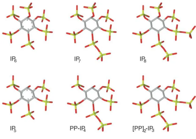
Inositol hexakisphosphate (also known as phytic acid) is the most abundant inositol polyphosphate in eukaryotic cells. It is a major component of plant seeds representing 0,1 – 1% of its dry weight and 60 – 80% of total phosphate content (Shears S.B., 2003). Significantly, IP6 is the precursor of a novel class of more anionic inositol polyphosphates, the inositol pyrophosphates, in which the fully phosphorylated IP6 ring is further phosphorylated to create high-energy pyrophosphate groups (Fig.3.1).
Figure 3.1. Inositol pyrophosphate chemical structure. The figure shows the
structure of phytic acid with its pyrophosphate derivate IP7 and IP8, as well as IP5, with the derivate pyrophosphates PP-IP4 and [PP]2-IP3. In the figures, carbon, oxygen and phosphate atoms are coloured grey, red and green, respectively (Bennet et al., 2006).
The best charateryzed inositol pyrophosphates are the diphosphoinositol pentakisphosphate (IP7 or PP-IP5) and the bis-diphosphoinositol tetrakisphosphate (IP8 or [PP]2-IP4), with one and two pyrophosphate group, respectively (Bennett M et al., 2006). Since their discovery in the early 1990s, inositol pyrophosphates have been found in all eukaryotic cells analyzed, from yeast to mammalian neuron.
Inositol pyrophosphates are important cellular messengers that control a wide range of cellular function, including endocytosis (Saiardi A et al., 2002), apoptosis (Gong B et al., 2002), telomere length (Saiardi A., et al., 2005), DNA recombination (Luo H., et al., 2002). The high energy pyrophosphate bond of IP7 can directly
transductional protein modification (serine pyro-phosphorylation) (Bhandari R., et al., 2007).
The first enzyme able to synthesise inositol pyrophosphates was purified to homogeneity from rat brain (Voglmaier S. M., et al., 1996). The new enzyme was called inositol hexakisphosphate kinase (IP6K) and it was show to be able to convert IP6 plus ATP to IP7 and ADP; ATP cannot be substituted by other nucleotides such as CTP or GTP (Fig.3.2).
Fig 3.2. Convertion of IP6 plus ATP to IP7; the reaction is catalyzed by IP6K.
IP7 plus ATP can be converted in IP8 in a following reaction.
Later the same laboratory characterized one yeast and two mammalian proteins with inositol hexakisphosphate kinase activity, called KCS1 (Kinase C Suppressor 1, also known as yIP6K), IP6K1 and IP6K2, respectively (Saiardi A., et al., 1999). Subsequently, a third mammalian gene, IP6K3, was cloned (Saiardi A. et al., 2001). All the mammalian IP6Ks phosphorylate in vitro IP6 to IP7, IP5 to PP-IP4 and [PP]2-IP3 (Saiardi et al., 2001, Saiardi et al., 2000). Later the enzyme was cloned in other mammalians, and its high evolutionary conservation was regularly observed, which facilitated the identification and cloning of IP6K enzymes from distant organisms, including yeast and the amoeba Dictyostelium (Luo H. et al., 2003).
The cloning of IP6Ks also helped to identify an evolutionarily conserved family of inositol polyphosphate kinases known as inositol polyphosphate multi-kinases (IPMKs) (Saiardi A. et al., 1999), that phosphorylate a broad range of substrates to yield a variety of inositol polyposphate reaction products (Stevenson-Paulik J., et al., 2002, , Eskin E. et al., 2002, Larkin M. et al., 2007, Altschul S. et al., 1997),
including some inositol pyrophosphate species (Shears SB, 2004, Luo H. et al., 2003, Loomis W. et al., 1995).
Altogether, the IP6Ks, IMPKs and IP3-3Ks (the enzymes that convert I(1,4,5)P3 to I(1,3,4,5)P4 -Schell M. J. et all., 1999-) belong to an inositol polyphosphate kinase superfamily, the IPKs (PFAM accession number PF03770), that evolved from a common ancestor. Phylogenetic analysis of their sequences predicts that IP6Ks arose first, followed by IPMKs, and lastly IP3-3Ks (Fig. 3.3).
Fig3.3. Phylogenetic tree of IPK proteins family members. The following abbreviations are used: At, Arabidopsis thaliana; Ce, Caenorhabditis elegans; Dd,
Dictyostelium discoideum; Dm, Drosophila melanogaster; Hs, Homo sapiens; Lu, Linus utilissimum; Os, Oryza sativa; Sc, Saccharomyces cerevisiae; Sp, Schizosaccharomyces pombe.
3.2.1 Is there IP6K in plants?
Plants have played a special role in inositol polyphosphate research since in plant seeds was discovered the first IP, the fully phosphorylated inositol ring of phytic acid (IP6). Although plants were instrumental to the early discovery of inositol polyphosphates, and remain experimental systems with large agri-business commercial interests, the majority of studies are now performed in mammalian cells, amoeba or yeast. Nevertheless, studies in plants have demonstrated that IP6 is an endomembrane-acting regulator of calcium-release and controller of K+ channels in guard cell (Lemtiri-Chlieh F., et al., 2000). IP6 is a major component of plant seeds representing 0,1-1% of its dry weight and 60-80% of total phosphate content (Raboy V., 2003, Shears S.B., 2001). In the late nineties, significant steps were made in the elucidation of the biochemical pathway responsible for IP6 synthesis in crop and model species, enabling subsequent genetic analysis of plant mutants with impaired phytic acid synthesis (Stevenson-Paulik J. et al., 2005, Sweetman Det al., 2006, Xu J. et al. 2005) .
The presence of IP7, the more anionic inositol polyphosphates derived from IP6, has been demonstrated in vegetal organisms, both in monocotyledonous and in dicotyledonous plants (Flores S. 2000, Brearley C. 1996). Furthermore, the conversion of IP6 to IP7 has been detected in Arabidopsis cells and leaf tissue in the presence of ATP, demonstrating IP6- kinase activity in plant extracts (Saiardi A., Azavedo C., unpublished manuscript).
These findings, together with the observed high conservation through the evolution of IP6K, strongly suggest the presence of this enzyme in vegetal cells. Therefore, IP6K enzyme was searched in plant genomes by homology based methods, but all studies have failed to reveal its presence.
Two IPMK proteins (called AtIPK2a and AtIPK2b in Arabidopsis thaliana) have been identified so far (Stevenson-Paulik J. 2002, Xia H, 2003). The substrate ambiguity is a general property of IPKMs, that phosphorylate a broad range of substrates and some IPMK proteins are able to synthesise inositol pyrophosphate species (Nalaskowski M. M., 2002, Saiardi A., 2001, Zhang T.,2001). In rice and barley an IPMK able to phosphorylate all intermediates from inositol bisphosphate to IP6 has been characterized (Josefsen L., 2007). To date IPMKs have been identified in dicotyledonous and in monocotyledonous plants, as well as in algae.
However, in Arabidopsis it has been shown that the two IPMK proteins contribute to inositol 1,3,4,5,6-pentakisphosphate(IP5) production, but do not show any inositol pyrophosphate enzymatic activity (Stevenson-Paulik J. 2002, Xia H, 2003).
Thus, an enzyme must exist in plants converting IP6 in IP7. The observed evolutionary conservation strongly suggests that this enzyme is indeed an IP6 Kinase.
There are many clues connecting IP6K to cell mitochondria. It was shown that human IP6K2 moves from nuclei to mitochondria and provides physiologic regulation of apoptotic process by generating IP7 (Nagata E. et al., 2005). Furthermore, yeast deficient in KCS1 (yeast IP6-Kinase), kcslΔ, do not survive if they are grown in conditions in which survival is dependent from mitochondrial function, thus demonstrating the importance of IP6K for this organelles (Saiardi A., unpublished manuscript).
Some further observations could suggest that the corresponding gene might be found in plant mtDNA, probably encripted and hidden by virtue of editing and/or trans splicing processes. It is known that most of mtDNA information concerns genic products acting inside the mitochondrion itself. Plant mitochondrial genomes have several peculiar characteristics such as the large size (from 200Kb to 2400Kb), the presence of introns and genetic material of chloroplast or nuclear origin (Palmer J et al., 2000). Furthermore, mitochondrial genome is characterized by occurrence of RNA editing and trans splicing mechanism enlarging protein variability (Takenaka M. et al., 2008).
On the basis of the above considerations, we have indeed hypothesized that IP6K gene is present in plants, nested in mitochondrial DNA, probably encrypted and hidden by virtue of editing and/or trans-splicing processes. But before going into more detail on the technique we have developed in order to verify such an hypothesis, let us take a closer look into the RNA editing process, which is of central interest to the methods we propose, which is accounted for next.
3.2.2 RNA editing
RNA editing is a molecular process in which some bases of a RNA molecule are altered by specific enzymes. It may be broadly defined as any process (co- or post-transcriptional) that changes the primary nucleotide sequence of an RNA molecule from that encoded by the corresponding gene. Since the initial discovery in trypanosome mitochondria (Benne R. et al., 1986) various types of RNA editing have been described. The diversity of RNA editing mechanisms includes site-specific insertion or deletion of one or a few nucleotides (insertional/deletional type of editing) as well as specific identity changes of individual nucleotides (conversional type of editing) such as cytidine (C) to uridine (U) and adenosine (A) to inosine (I) deaminations. Most of this systems are localized in mitochondria and act on transcripts of protein-coding genes; however, editing of the transcripts of certain mammalian nuclear genes does occur as well (Hodges P., et al., 1992). The extent of sequence changes introduced by editing can range from massive (e.g. certain trypanosome transcripts, where upwards of 50% of the sequence is contribute by editing process- Stuart K., 1991-) to minimal (e.g. the single nucleotide editing in the mRNA encoding a mammalian glutamate receptor protein –Sommer B. et al., 1991-). The biological consequences of editing differ with the system. Editing in trypanosome mitochondria is required to generate translatable mRNAs from transcripts that otherwise could not produce functional proteins. In other cases, both unedited and edited forms of a mRNA may give rise to biologically active proteins having different physical and/or functional properties. Messenger RNA editing usually involves changes to internal codon positions, but it may also create new initiation and termination codons and alter 5‟- and 3‟- noncoding regions and intron sequences.
The mechanism and informational basis for the selection of individual nucleotides as editing sites is unknown. No consensus sequence at an editing site has been identified in either mitochondrial or chloroplast RNAs. Nucleotide sequence comparison of mitochondrial editing sites indicated that the edited cytosine usually does not have a purine, especially a guanine, as the 50 nucleotide (Covello P.S., et al., 1990). In addition, some editing sites bear similarities to each other, and this could reflect different „classes‟ of editing sites (Gualberto J.M., et al. 1991). Apart from these simple trends, no specific sequence or secondary structural motifs have
been identified from the hundreds of mitochondrial editing sites examined (Gray M.W., 1992; Wissinger B., et al., 1992).
RNA editing is particularly diffuse in plant mitochondria and chloroplasts. Often the genomic information encoding an open reading frame is often incomplete in these organelles, and RNA editing is necessary to yield a functional product.
3.3 Contributions
The hypothesis at the basis of this part of the work is that IP6K gene lies in DNA of plants, in an encrypted form. Therefore, we decided to first search IP6K gene in mtDNA of plants, where the occurrence of mechanisms interrupting the linearity of genetic information is high.
Because of the considerable sequence heterogeneity among the several known IPKs, common homology search programs are not useful to our aim, Furthermore, these softwares cannot detect possible changes in nucleotide sequences due to RNA editing mechanisms.
The main intuition behind here is that some specific gene families, such as all IPK genes, are characterized by the presence of specific tags, short sequences of few amino acids, often corresponding to functional regions. Thus, we decided to use this new approach of looking not for the gene sequence as a whole, but for a specific tag sequence, characterizing IPK gene family. This is possible only when a gene, or a gene family, contains a region (usually a short sequence) that is indispensable and always present in the gene sequence. In fact, alignment studies between IPKs from different organisms allowed to identify several conserved motifs in the amino acids sequence. These motifs comprise the ATP binding site, first characterized in IP3-3K (Communi D. et al., 1993), the C-terminal motif (last 19 amino acids), important for the catalytic activity (Togashi S., 1997), the “SSLL” motif, required for enzymatic activity of IP6K (Saiardi A. et al., 2001) and the P-XXX-D-X-K-X-G tag, a sequence of nine amino acids with four of them very conserved among IPKs (Saiardi A. et al., 1999). Despite the considerable sequence heterogeneity of IPKs, this last motif represents a unique consensus sequence and it can be considered a specific tag of IPK gene family. The consensus sequence P-XXX-D-X-K-X-G (Fig.3.4) is a very important functional region, identifying the inositol binding site of the enzyme (Saiardi A. et al., 2000). Here, the functional role explains its strong conservation through evolution.
InsP3 kinase A 243 YLQLQDLLDGFDGPCVLDCKMGVRTY 268 InsP3 kinase B 453 YNQMDDLLADFDSPCVMDCKMGIRTY 478 InsP6 kinase 1 (KIAA0263) 205 YKFLLLENVVHHFKYPCVLDLKMGTRQHGDDA 236 InsP6 kinase 2 (PIUS) 200 YKFILLENLTSRYEVPCVLDLKMGTRQHGDDA 231 IPMK (ArgRIII) 112 KQYLVLENLLYGFSKPNILDIKLGKTLYDSKA 143 yInsP6 kinase (KCS1) 758 KFILLEDLTRNMNKPCALDLKMGTRQYGVDA 788
Consensus: PXXXDXKXG
Fig. 3.4 Alignment of the inositol-phosphate-binding motif of the different inositol
polyphosphate kinases. Identical amino acids are shown in bold. The GenBank accession numbers of the different sequences are: rat InsP3 kinase A, GI:124808; rat InsP3 kinase B, GI:1170577; Saccharomyces cerevisae yInsP6 kinase, GI:1078508; and IPMK (inositol polyphosphate multikinase), GI:114134. Numbers to the right and left of the sequences indicate their positions in the respective complete amino-acid sequences. The consensus sequence is written in Prosite format, where X represents any amino acid (Saiardi et al., 1999).
Thus, we chose the P-XXX-D-X-K-X-G tag to perform IP6K gene search. This tag search, however, had to be supported by a suitable methodology and associated software tools. The following sections indeed account for the multi-step methodology that we developed to discover specific genes and one of the supporting tools specifically designed to implement this particular kind of tag search. Then, the so developed method was indeed applied to searching for IP6K gene. The thus obtained experimental results are also presented below.
3.4 Methodos
3.4.1. Gene finding by tag search: a multi-tool methodology
When a gene is characterized by a tag, the finding of the tag sequence in a genome indicates that possibly this genome contains the gene under study. In fact,
candidate gene sequence is found, further analyses are required to confirm the discovery. To find possible specific, still undiscovered, genes in cell genomes we developed a multi-tool methodology. The new approach is based on tag search, and includes a series of analyses aimed to analyze the candidate gene sequences eventually found.
The tag search is a motif discovery task, since a tag can be viewed as a subsequence whose structure is not completely specified a priori, that is, a special kind of motif. In order to solve the tag search problem, we designed the methodology summarized by the pseudocode illustrated in Figure 3.5. In particular, we consider a tag sequence and a mtDNA sequence in input, and we aim at generating all the mtDNA subsequences containing the tag. Then the search can be performed by following the procedure illustrated in Figure 3.5.
Pseudocode Input:
a string t defined on the alphabet = (A, C, T, G, X) representing the “tag” a string tag of shape A1-D1-…-Dn-1-An defined on = (Aminoacid, X),
representing the “tag”
a string s defined on the alphabet = (A, C, T, G) representing the mtDNA of a given organism
Output:
a set S of strings that are subsequences of s containing tag
Begin
Let S be an empty set S= 0
while there is a triplet t in s
if t matches the aminoacid A1
set j to 1
while j is smaller than n
skip Dj triplets in s
get a triplet a in s
if a does not match the aminoacid Aj+1
break else increment j by one end end; if j is equal to n
build Si as the portion of s starting from i and matching the tag
add to Si 200 triplets after Si
add to Si 200 triplets before Si
add Si to S end end increment i by one end Return S end
Fig.3.5. Tag search pseudocode.
Once a tag is identified, we extracted a nucleotide sequence surrounding the consensus sequence. This sequence was then examined as a candidate gene and submitted to further analyses. To this aim, we implemented the procedure described below by exploiting a set of existing software tools, suitably fixed, as showed in
Fassetti F. et al., 2008. The system is able to take into account both the genetic code degeneration and possible RNA editing events.
The extracted nucleotidic sequences were translated into amino acid sequences by using the Transeq (Rice P. et al., 2000) software.
Then, in order to detect possible homologies, we performed sequence alignments using ClustalW (Larkin M.A., 2007) and BLAST (Altschul S.F., 1997). Finally, using the TBLASTX and TBALSTN algorithms, we screened expressed sequence tag (EST) databases for proteins containing the sequences identified by our tag search. These databases include short fragments of DNA derived from a longer cDNA sequence and representing part of the expressed genome.
The methodology can be, therefore, shortly summarized as follows:
1. (Tag Definition) set a (partially undefined) sequence representing the specific tag to searched for;
2. (Genome Scanning) scan a genome sequence (or a set of genome sequences) to individuate possible instances of the tag;
3. (Post-processing Analysis) analyze the candidate subsequences extracted by the previous step in order to verify the presence of the gene in the considered genomes.
A (somewhat more detailed) summary of the methodology is illustrated in the figure 3.6.
Fig.3.6. Summary of the gene finding methodology based on tag search.
3.4.2. L-SME
L-SME (Fassetti F. et al., 2008) is a system designed to mine general kinds of motifs where several “exceptions” may be tolerated; that is, it is able to handle different complex kinds of pattern variabilities. Roughly speaking, a motif is a pattern composed by two or more substrings (called boxes) separated by a number of symbols (called gaps).
In the following, the main characteristics of L-SME are briefly presented. The basic configuration of L-SME allows the user to specify the minimum and the maximum length of each box composing the pattern and the minimum and the maximum size of each gaps between boxes. But, motivated by biological observation, L-SME supports also approximated matching of the pattern, namely, the occurrences of the pattern may differ in some characters. Also this configuration is
Futhermore, L-SME is able to take into account box skips (deletions) and box swaps (box invertions). The former ones consist in mining occurrences where at most a user-defined number of boxes are missing, while the latter ones consist in mining occurrences where at most a user-defined number of swaps between adjacent boxes occur. Finally, L-SME allows the user to specify some anchors for each box, namely, the user can constrain a box to be equal to one of the substrings specified as anchor for that box. Despite the complexity of the addressed pattern variabilities, the system is able to exhibit very good performances.
The flexibility of the method in specifying variable distances between two boxes easily allows possible introns to be taken into account. In order to limit the great variability introduced by considering introns, we adopted an incremental approach consisting in iteratively increasing the number of introns. In particular, we did not take care of any intron at the beginning and, then, we considered the presence of n introns by incrementing the distance between n pair of boxes of the typically maximum length of an intron (e.g., 100 bases for Arabidopsis thaliana).
3.4.3 BLAST
BLAST (Basic Local Alignment Search Tool) (Altschul S.F., 1997) is an algorithm for comparing primary biological sequence information, such as the amino-acid sequences of different proteins or the nucleotides of DNA sequences. It is one of the most widely used bioinformatics programs, because it addresses a fundamental biological problem. A BLAST search enables comparison of a query sequence with other sequences or a database of sequences, and identify sequences that resemble the query above a certain threshold. BLAST directly approximates alignments that optimize a measure of local similarity, the maximal segment pair (MSP) score. The basic algorithm is simple and robust; it can be implemented in a number of ways and applied in a variety of contexts including straight-forward DNA and protein sequence database searches, motif searches, gene identification searches, and in the analysis of multiple regions of similarity in long DNA sequences. Input sequences are in FASTA format or Genbank format. BLAST output can be delivered in a variety of formats. These formats include HTML, plain text, and XML formatting.
The following different types of BLASTs are available according to the query sequences. Nucleotide Blast: search a nucleotide database using a nucleotide query; Protein Blast: search protein database using a protein query; Blastx: search protein database using a translated nucleotide query; tblastn: search translated nucleotide database using a protein query; tblastx: Search translated nucleotide database using a translated nucleotide query
3.4.4.Clustal
Clustal (Larkin M.A., 2007) is a general purpose multiple sequence alignment tool for DNA or proteins. There are two main variations: ClustalW, command line interface and ClustalX, with a graphical user interface. It produces biologically meaningful multiple sequence alignments of divergent sequences. It calculates the best match for the selected sequences, and lines them up so that the identities, similarities and differences can be seen.
The program accepts a wide range on input format, including NBRF/PIR, FASTA, EMBL/Swissprot, Clustal, GCC/MSF, GCG9 RSF, and GDE. The output format can be one or many of the following: Clustal, NBRF/PIR, GCG/MSF, PHYLIP, GDE, or NEXUS. The program includes three main steps: performs a pairwise alignment, creates a phylogenetic tree (or use a user-defined tree), uses the phylogenetic tree to carry out a multiple alignment.
3.5 Experimental results
3.5.1 IP6K gene search
As stated above, several considerations of functional and evolutionary order, led us to suppose that IP6K gene is present in plant genomes, most likely in mitochondrial DNA. Thus, we decided to first apply the new method of gene search on mitochondrial DNA of plants, where such a gene could have been nested. Then we looked for the gene in nuclear genome of two model plants, Arabidopsis thaliana and Oryza sativa.
Tag Definition
As stated above, the most important tag for IPK gene is the P-XXX-DX-K-X-G motif, corresponding to the inositol binding site of the enzyme. Thus, for the identification of IP6K gene in plant DNA, we focused on the nucleotide sequence corresponding to this specific IPK tag.
In particular, the sequence associated to this the tag, is made of both symbols in the alphabet Σ = {A,C,G,T}, representing nucleic acids, and a generic symbol X that can be associated to a subset of Σ. This way, step 2 can be carried out by performing an approximate search of the motif represented by the tag sequence.
Genome Scanning
We analyzed all the published mtDNA sequences (available at
http://www.ncbi.nlm.nih.gov/sites/entrez) and the whole nuclear genome of two plants and performed motif extraction from them.
For the purposes of this research, we looked for the pattern:
[CC{T,C,A,G}] ---[GA{T,C}] --- [AA{A,G}] --- [GG{T,C,A,G}] ,
where the square brackets delimitate the boxes and the hyphens denote the distances between boxes. For this motif extraction we used L-SME, setting the configuration parameters as reported in Figure 3.7. Note that, because of the genetic code degeneration, the fixed aminoacids are specified by more than one nucleotidic triplet. All the possible C->T editing events giving rise to the fixed tag aminoacid are included in the search.
Fig. 3.7. L-SME configuration parameters.
Post-processing Analysis
For each identified tag, we extracted a sequence of about 1200 nucleotides surrounding the consensus sequence and examined it as a candidate IP6K gene. We first translated the extracted nucleotide sequences and then examined the identified amino acid sequences, looking for other IP6Ks conserved domains. Then we performed sequence alignments using both ClustalW and BLAST. Finally we screened expressed sequence tag (EST) databases in order to verify if the candidate gene sequences are actually transcripted.
3.5.1.1. Validation of the method
In order to confirm the goodness of our method, we applied it on nuclear DNA of Arabidopsis thaliana, searching a known gene, already identified by biological techniques. Such a gene is the one coding for inositol 1,3,4,5,6-pentakisphosphate2-kinase (InsP5 2-1,3,4,5,6-pentakisphosphate2-kinase or Ipk1), the enzyme responsible for the production of inositol hexakisphosphate (IP6).
Ipk1s are unique among inositol phosphate kinases in that they phosphorylate the axial 2-position of the inositide ring, whereas other enzymes act on equatorial
identified in yeast (York J.D. et al., 1999) and in other different fungal species. Although functionally conserved, IPK genes present very low sequence homology in different organisms, with less than 24% identity in pairwise combinations across the fungal proteins. The sequence identity is limited to a few small regions with high homology. This lack of significant homology initially disallowed the discovery of non-fungal Ipk1. After characterization of human Ipk1 (Verbsky J.W. et al., 2002), the gene was cloned in Arabidopsis thaliana using molecular strategy based on the presence of specific tags in the protein (Sweetman D., 2006). As a consequence consequence, we searched Ipk1 in Arabidopsis thaliana genome with the modified L-SME software, exploiting the presence of specific tag in Ipk1 gene family.
As for the validation carried on the Ipk1 gene, we looked for the pattern composed by the two regions EIKPK and R-XX-MHQ-X-LK characterizing the gene because both present in all Ipk1 genes discovered up to now; they are separated each other from a variable number of amino acids (19 amino acids in human and rat Ipk1, 9 in yeast Ipk1). Then, the corresponding pattern in the genome sequence is:
GA{A,C}AT{T,C,A}AA{A,G}CC{T,C,A,G}AA{A,G}--. . . --
{AGA,AGG,CGT,CGC,CGA,CGG}---{ATG}CA{T,C}CA{A,G}--- {TTA,TTG,CTT,CTC,CTA,CTG}AA{A,G}
where the number of boxes is 11 and the length of each box is 3. The distance between the fifth and the sixth box, corresponding to the distance between the region EIKPK and the region R-XX-MHQ-X-LK, is set to the interval [27 − 57]. As for the distances between the other boxes, they are set according to the above described method.
We found the conserved sequence on chromosome 5 of Arabidopsis thaliana genome. In particular we had only one positive match when we considered the possibility of occurrence of an intron between the third and fourth amino acid of EIKPK motif. The intron length resulted to be 82 nucleotides and distance between two EIKPK and R-XX-MHQ-X-LK motifs 63bp. We extracted a sequence of about 2000 nucleotides around the tags. BLAST allignements showed that the sequence was Arabidopsis thaliana Ipk1 gene.
This approach led us to easily find the gene, thereby allowing to validate the method, that appears general and very useful when homology search strategies cannot be used.
3.5.1.2. IP6K search results
Due to the numerous suggestions relating IP6K to cell mitochondria, we decided to first perform the IP6K gene search on mitochondrial DNA of plants. To date the full mitochondrial genome sequence is known for 42 different vegetal organisms, belonging to various Phyla, even very distant from one another from the evolutionary point of view. The specific IP6Ks tag (P-XXX-D-XK-X-G) was searched over the overall sequenced mitochondrial genomes available to date and both DNA strands were analyzed. Twentythree genomes out of 42 gave at least one positive match. Interestingly, we noted that some tag sequences (9 amino acids) were identical among different organisms. For each identified tag we extracted a sequence of about 1200 nucleotides surrounding it. To find out possible relevant homologies, we performed alignments among the sequences found in different vegetal organism. All the sequences sharing the same tag showed high similarity in the region surrounding the consensus sequence, while alignment with IP6K known genes (Saccharomyces cerevisiae KCS1 or human IP6K1) showed only a weak similarity.
Furthermore, in order to confirm the identity of our putative hit, we looked for other IP6Ks conserved motifs in the identified putative amino acids sequence like the ATP binding site, the C-terminal motif (last 19 amino acids), and the “SSLL” motif. These analyzes led us to focus on the sequence PVGTDRKGG, that was found in mitochondrial genome of Tripsacum dactyloides, Sorghum bicolour, and three different species of Zea genus (Zea mays, Zea perennis and Zea luxurians). Alignment between the 410 aminoacid around the PVGTDRKGG sequence of Tripsacum dactyloides and the human IP6K gene showed an interesting correspondence of the consensus region (see Figure 3.8).
Fig.3.8 Alignment between the 410 amino acids around the PVGTDRKGG sequence
of Tripsacum dactyloides and the human IP6K gene (ClustalW2). “*” = residues identical in the two sequences in the alignment; “:” = conserved substitutions; “.” = semi-conserved substitutions.
To verify if the Tripsacum dactyloides sequence was an actively transcribed gene, we analyzed the Expressed Sequence Tags (ESTs) databases using the region surrounding the PVGTDRKGG tag of Tripsacum dactyloides. This search failed to find any EST matching indicating that our putative hit is unlikely to be transcribed in mRNA. Finally, we used a region of 50 amino acids of Tripsacum dactyloides mtDNA surrounding the consensus sequence to perform a multiple alignment with corresponding regions of inositol phosphate kinases (IPMK, IP6K, IP3-3K) from different organism using ClustalW2. As shown in Figure 3.9, our sequence resulted
to be an outsider. This result indicated that the identified mitochondrial tag does not belong to any subgroup of kinases composing the IPK gene family.
Fig.3.9. Phylogenetic tree from multiple alignment of a 50 amino acid region of
Tripsacum dactyloides mtDNA surrounding the tag with corresponding regions of inositol phosphate kinase (IPMK, IP6K, IP3-3K) from different organisms (Clustal W2). Branch lengths are proportional to the amount of inferred evolutionary change.
Once excluded the presence of IP6K gene in mtDNA, we decided to look in nuclear genome of plants where, up to now, the search has been performed only by methods based on sequence similarity. We analyzed all chromosomes of Arabidopsis thaliana and Oryza sativa, a dicotyledonous and a monocotyledon plant respectively. Arabidopsis thaliana is a small flowering plant, belonging to eudicot, the largest group of flowering plants on the planet. Because of its short generation time and compact size, it is used as a model organism in plant biology and genetics. Its nuclear genome comprises five chromosomes, with a total size of approximately 125 Mb (megabases). It is one of the smallest genome among plants, and it was the first plant genome to be sequenced in 2000 (Initiative T., 2000). Oryza sativa (rice) was the second plant genome to be published (Lan S., 2002) the first among monocot. It has the smallest cereal genome consisting of just 430 Mb across 12 chromosomes and it is routinely used as a model organism in cereal genomics.
In each chromosome of both plants, we found dozens of tags, but only few tag sequences per chromosome resulted in good candidates to be specific IP6K tags. In fact, too polar or too big amino acids between the four fixed positions of the tag are not consistent with the tag sequence functionality. In particular we considered as good candidate a tag sequence including amino acids L, V,T,M,I,A,S,G,C between the four fixed positions, and we rejected others. For each identified tag, we extracted a sequence of about 400 amino acids surrounding the tag. Each sequence was examined as a candidate IP6K gene as described above for mtDNA. We did not find any strong homology with known IP6Ks. This result was not surprising, because only a weak similarity is anyhow expected between organisms very distant from an evolutionary point of view. Thus, the selected sequence to be actually interesting was established on the basis of other parameters, like alignment of tag sequences, presence of other conserved amino acids and of sequence in EST database.
A very promising sequence was found on chromosome 5 of Oryza sativa, around the tag PLLVDSKLG. The sequence comprises 198 amino acids without any stop codon. As shown in Figure 3.10, the ClustalW alignment of this sequence and Saccharomyces cerevisiae KCS1 gene gave positive score with an alignment in correspondence of the inositol-binding region.
Alignment with human IP6K (hIP6K) gave a lower score, but still maintained the correspondence between tags (see Figure 3.11).
Fig.3.11. Alignment between the 198 amino acid sequence around the PLLVDSKLG
tag of Oryza sativa and the human IP6K gene (Clustal W2). ”*” = residues identical in the two sequences in the alignment; ”:” = conserved substitutions; ”.” = semi-conserved substitutions. The P-XXX-D-X-K-X-G tag is underlined.
We performed a multiple alignment (ClustalW2) between the region of 50 amino acids of Oryza sativa surrounding the tag and the corresponding regions of inositol
phosphate kinases (IPMK, IP6K, IP3-3K) from different organisms. This analysis revealed (Figure 3.12) that the Oryza sativa sequence, although appears dissociated from other IPK family members, shows a certain degree of similarity with Giardia lamblia IP6K, that itself appears to be a distant member of the IPK genes family.