SAPIENZA UNIVERSITÀ DI ROMA
DEPARTMENT OF MOLECULAR MEDICINE
IMMUNOLOGICAL HEMATOLOGICAL AND REUMATOLOGICAL SCIENCE
CURRICULUM: IMMUNOLOGY AND IMMUNOPATHOLOGY
Adoptive immunotherapy using PRAME-specific
T cells in medulloblastoma
Supervisor:
Dr. Biagio De Angelis
Co-supervisors:
Prof. Concetta Quintarelli
PhD: Domenico Orlando
Index
1.INTRODUCTION ... 1
1.1. Medulloblastoma ... 1
1.2. Molecular characterization of MBsubgroups ... 2
1.3. MB Therapeutic approaches ... 5
1.4.Immunotherapy for MB ... 7
1.5.A new target for MB immunotherapy ... 13
2.AIM OF WORK ... 15
3.MATERIALS AND METHODS ... 16
3.1. Cell lines ... 16
3.2. Healthy Donor’s PBMCs, MB patients and controls ... 16
3.3. Retroviral constructs ... 17
3.4. Generation and expansion of polyclonal iC9-SLL-TCR T cells ... 18
3.5. Activation of the suicide gene ... 18
3.6. Immunophenotyping ... 19
3.7. ELISpot assay ... 19
3.8.Co-Culture assays ... 19
3.9. IFNγ ELISA ... 20
3.10. Chromium-release assay ... 20
3.11. RNA isolation and quantitative real time PCR ... 20
3.12. Molecular detection of retroviral transduced T cells ... 21
3.13. Xenograft mouse model for in vivo studies ... 21
3.14. Histological and immunohistochemistry data ... 22
3.15. Mouse Behavioural studies ... 23
3.16. Statistical analysis ... 23
4.RESULTS ... 25
4.1. PRAME expression in MB at diagnosis and relapse ... 25
4.2. Retroviral vector carrying iC9 and PRAME-SLL specific αβTCR allows stable and functional expression of the transgene ... 35
4.3. PRAME-specific TCR-redirected T cells exert antitumor activity toward HLA-A-*02-matched MB cell line ... 41 4.4. iC9-SLL TCR T cells exert antitumor activity in vivo in xenogenic mouse model of MB ... 46
5.DISCUSSION ... 51
1
1. Introduction
1.1. Medulloblastoma
Medulloblastoma (MB) is the most common malignant pediatric brain tumor. It represents about 15–20% of the central nervous system (CNS) malignancies, 40% of which are located in the posterior fossa.(1) Despite the evidence that survival rates have increased by 30% in the past 20 years, today many patients still have a poor prognosis. (2) MB are embryonic CNS tumors constituted by primitive-appearing cells that can differentiate along multiple lineages. It arise from cerebellum and it is not completely understood which specific cells subset gives rise to these malignancies. One common theory is that MB starts from mutations in proliferating granule neuron precursors (GNPs) in the external germinal/granular layer (EGL). (3) The histological disease features reveal a heterogeneous disorder, in fact four variants of MB have been identified: classic, desmoplastic/nodular (D/N), extensive nodularity (MBEN), anaplastic and large cell, the latter often coexisting in the large cell/anaplastic (LC/A) variant. (4,5) MB is not a single disease, but a heterogeneous mixture of various subgroups with distinct features. The genomic profiles show four distinct molecular subgroups that have been identified: Wingless (WNT), Sonic Hedgehog (SHH), Group 3 and Group 4. (6,7)(Figure 1) Each subgroup corresponds to specific survival rate, age demographics, and genetic aberrations.(8) WNT and SHH subgroups are named after the discovery of the signaling pathway that plays a prominent role in the pathogenesis. (9) Molecular subgroups are correlated to clinical outcome. For this reason, they were integrated in the most recent version of WHO classification of CNS malignancies (5) and applied into the clinical practice. These MB subgroups remain stable at recurrence and are likely remindful of original cells. Recent research on 763 MB samples by Cavalli and colleagues (10) has shown the presence of inter-tumoral heterogeneity within the four MB subgroups delineating the presence of 12 subtypes based on gene expression and DNA methylation features, but the degree of heterogeneity and the extent of overlap are still unknown. This newfound heterogeneity within subgroups could explain previously unknown variation.(10) Thus, much emphasis has been recently placed on further identifying new alterations that may occur in each subtype pathway. Moreover, Northcott et al. described the
2
whole genome landscape of MB subtypes analysing 491 patient tumours.(11) They showed that driver mutations could be assigned to the two most malignant forms, group 3 and group 4 MB. In particular, in-frame insertions hotspot that target KBTBD4 (Kelch Repeat And BTB Domain Containing 4) and activation of PR/SET Domain 6 (PRDM6) were identified.(11) These and other novel findings identified in the MB subgroups could lead to the discovery of attractive targets for MB, which have not previously been explored in detail.
1.2. Molecular characterization of MB subgroups
WNT is the less common molecular MB subgroup and accounts for about 10– 11% of all MB. (12) It shows the main incidence at 10–12 years of age, being very rare in infants, with no differences in gender predilection. The typical histology is most frequently encountered, with rare cases of the LC/A variant and metastatic dissemination. (13) The main characteristic in WNT tumors is the somatic activating mutations in exon 3 of β-catenin (CTNNB1), shown in 85% of WNT MB with a predictive good outcome for patients. (14) However, the link between a better prognosis due to biological effect of Wnt/β-catenin signaling activation have not been fully clarified yet. (15) Monosomy 6 is the primary recurrent structural chromosomic alteration, present in the 80–85% of cases. Other common mutations are in DDX3X gene and the missense mutations in TP53 (15% of cases), that is a marker of high risk in the SHH subgroup but with no evidence in differences in the WNT patients survival rate. (16)
The SHH subgroup that represents 28–30% of all MB, has a bimodal age distribution, being more frequent in infants and adults, with a weak male predominance among infants. (17) The prognosis depends on the associated mutation meaning that these patients have an intermediate prognosis, with 5-years survival ranging between 60% and 80%, but the mutations in MYCN or TP53 oncogenes worsen the prognosis. (18) D/N and MBEN histology are the main features associated with the SHH subtype, however, it is also possible to observe classic or LC/A histology. (13) There are typical genetic alterations that lead to the SHH signaling over-activation: the most common ones are the somatic or germ-line inactivating alterations or the loss of
3
PTCH1 and SUFU, or the somatic missense mutations activating SMO. (19,20) Few SHH patients showed high-risk disease due to co-amplification of MYCN and GLI2, accompanied by inactivation of TP53. (16) Patients with Gorlin syndrome, carriers of a germ-line mutation on PTCH1, have a predisposition to develop basal-cell carcinoma and MB, especially MBEN. (21) Unusual SHH subgroup MBs has also been reported in patients with rare diseases such as Fanconi anemia. (22)
The Group 3 has around 25-28% incidence of all MB and these patients exhibit the worst survival rate and the highest rate of metastatic dissemination. (23) Group 3 MBs are very rare in adults but are common in infants and younger children, with a male to female ratio of 2:1. (13) LC/A is the principal histology and the tumor genome is considerably unbalanced. Several numbers of broad alterations were find such as gain of chromosome 7 and iso-chromosome 17q. There are also associations with neurocutaneous syndromes such as tuberous sclerosis complex that have been described. (24)
Group 4 MBs share many of these alterations.(25) The most common event is the MYC amplification (10– 20%), followed by OTX2 transcription factor mutations, which affect the 10% of patients and is mutually exclusive of MYC amplification. The incidence of Group 4 is about 34–35% of all MBs, and it is the most common subgroup. (12)This subgroup is rare in infants, it exhibit a peak incidence in 10-years-old children, with a prevalence in males (sex ratio 3:1). They can have classic or LC/A histology. Group 4 MBs have an intermediate prognosis and show a high rate of metastasis and relapse. MYCN amplification is not associated with the worst outcome such as the SHH subgroup. (26)
4
Figure 1
Figure 1. Molecular subgroups of medulloblastoma. WNT: wingless, SHH: sonic
5
1.3. MB therapeutic approaches
Nowadays the treatment for MB is based on surgery followed by craniospinal chemotherapy and irradiation. However, CNS radiation therapy can be harmful to the developing brain and is usually spared in children under the age of three, but this can affect the disease control and survival. Moreover, the post-surgery treatment with chemotherapy and irradiation are fundamental to eliminate any residual tumor cells leading to the reduction of metastases risk occurrence. However, it is relevant to consider that the same treatments leading to increased survival rates are also causing potentially debilitating physical deficits, such as endocrine dysfunction, neuropsychological deficits or subsequent malignancies. Indeed, the outcomes in 51 long-term survivors of paediatric MB were comprehensively assessed in terms of tumour control, neurological, endocrine, and neurocognitive complications and their impact on behavioural and psychological adjustment, and health-related quality of life (QoL). (28) Endocrine deficits occurred in 61%, neurological complications in 72%, and significant school problems in 72%. All patients had significant deficits in neurocognitive functioning: attention and processing speed was impaired in 79%, learning and memory in 88%, language in 56%, visual perception in 50%, and executive functions in 64%. Because of their treatment, including craniospinal radiotherapy, MB long-time survivors are not only at great risk for neurological, endocrine, and neurocognitive complications, but also of social isolation thereby decreasing self-rated QoL substantially.
Thus, there is a need for the development of new strategies that may significantly ameliorate outcome and QoL especially in the younger population and decrease potential side effects. MB tumors possess peculiar features different from peripheral tumors, and many factors such as the blood brain barrier (BBB) and the hostile tumor microenvironment should be considered. The disease understanding is critical, in fact one of the major obstacles is the drug delivery due to the MB brain localization. The tumor cells usually grow in the cerebellum midline. The patients present increased intracranial pressure (ICP) and typical signs of obstructive hydrocephalus. The symptoms usually are headaches and nausea/vomiting for weeks to months prior to disease discovery. Irritability and poor feeding could also affect young children, with severe cases that show altered mental status. Other physical signs found during patients’ examination are bradycardia, hypertension and widened pulse pressure.
6
With a severe hydrocephalous degree we can have ocular findings such as papilledema and 6th nerve palsies.(29) The perfect scenery for MB patients is the maximal tumor resection and the first step is the surgery. Approximately 25% of MB patients develop the posterior fossa syndrome after surgery, leading to some degree of cerebellar mutism where patients try to produce words but are unable and start to be extremely irritable, and provoke hypotonia and ataxia. The syndrome is usually exhibited 24 hours after resection of a posterior fossa tumor and the duration is variable from weeks to months, but the language difficulties may be lifelong.(30) Other complications of surgery include disruption of the blood supply leading to infarction of surrounding normal brain, and intra-operative bleeding leading to subdural hematomas. 30 days after definitive surgery the patients is recruited for the radiation therapy (XRT).(31,32) Daily fractions of 1.8 Gy to a final dose of 54 Gy - 59.4 Gy are used. There is high risk to develop complications due to XRT for pediatric patients with MB. Radiation therapy could lead to anorexia and nausea due to the proximity of the spinal fields to the gastro intestinal tract. It is fundamental to control the patient’s weight and nutrition during the treatment. The bone marrow of the vertebral bodies is active and important for the maintenance of normal blood counts, so cytopenias often appear during this period. Most MB patients require XRT for long term survival, and treatment costs are very expensive.
Moreover, another approach is the pharmacological treatment. It is based on chemotherapy drugs administration and the aim is to enhance the local control of tumor and the management of micrometastatic disease. The drugs’ mechanism affects rapidly dividing cells and also damages the gastro intestinal tract, hair follicles, and bone marrow. This leads to risk of nausea and vomiting, diarrhea and/or constipation, hair loss and myelosuppression. The chemotherapy is used specifically depending on the MB patient characteristic and there are protocols to well establish the correct treatment. Some examples of used drugs are: Cisplatin, mechanism of action: induces cellular apoptosis by cross-linking DNA;(33) Cyclophosphamide, mechanism of action: alkylating agent; Lomustine, mechanism of action: alkylating agent; Vincristine, mechanism of action: microtubule inhibitor that prevents cell division by binding to the tubulin component of microtubules and leading to metaphase arrest.(34)
7
1.4. Immunotherapy for MB
Cancer immunotherapy is a new advanced medicine approach that allows triggering the cytotoxic potential of the immune system against cancer cells. In particular, T cells adoptive immunotherapy is one of the best-characterized and used strategies nowadays, with several clinical trials opened worldwide. T cells are the cells responsible to fight foreign pathogens that pretend to be ‘self’. T cells are able to recognize mutated or infected cells through antigens (small peptides) presented by the major histocompatibility complex (MHC) class. (35)Tumor cells evolve through mutations from the phenotype of their origin cells and de-differentiate and up-regulate the expression of proteins normally not exhibited by the origin cells, the tumor associated antigens (TAA). Cancer immunotherapy is based on the possibility of T cells (or other immune system component) to recognize TAA. Unlike traditional pharmacologic agents that require increased titration for full effect (often with dose limiting toxicities that preclude rampant dose escalation), immunotherapy is a biologic agent that may not necessitate the same dose-dependent increases for maximal impact. (36) For instance, the law of the impassable BBB is now changing opening new scenery for the in “vivo biology” respect the difficulties of the bio-distribution of pharmacological treatment. Recently, lymphatic drainage networks have been described within the CNS that communicate with deep cervical draining lymph nodes. (37-41) Activated T cells able to recognize tumor-specific antigens are able to traverse the BBB through adhesion markers (i.e., VLA-4), allowing them to penetrate the tumor microenvironment and induce their effector functions against pediatric cancer cells. (37-41)
For children with malignant brain tumors, immunotherapy may be used in conjunction with standard therapy but we should consider the impact of traditional protocols on the immunotherapy treatment. XRT may confound Th1 type responses by decreasing CD8+ dendritic cells (42,43) but can damage the tumor environment with the release of damage associated molecular patterns (DAMPs) and tumor antigens that can be processed by endogenous antigen presenting cells (APCs) for presentation to T cells in local draining lymph nodes. (44)Instead, chemotherapy can be directly toxic to activated T cells, but may eliminate regulatory populations (i.e., regulatory T cells, myeloid derived suppressor cells) that compete for essential cytokines.(45-47) The adoptive cellular therapy is best utilized after radiation and chemotherapy; moreover,
8
all these treatment modalities may synergize also with a vaccine immunotherapy. (48,49) Preclinical and clinical investigations have shown that vaccination through repeated cycles of chemotherapy enhances antigen specific T cell immunity.(49–51) We can image to use an adoptive cellular therapy followed after by chemotherapy with continued vaccination cycles to maintain immunologic memory.
Focusing our attention on pediatric brain tumors, appropriate tumor antigens have to be selected and targeted, avoiding immunologic targeting of epitopes expressed on normal tissue that may elicit autoimmunity. (52) Most pediatric brain tumors have low mutational rate at the genomic level but are prompted by epigenetic deregulatory mechanisms that lead to the re-expression of developmental fetal antigens in malignancies, such as MB and primitive neural ectodermal tumors (PNETs). (53-55)
The immune system actively survey against cancer and is able to recognize and remove cells that are undergoing oncologic transformation.(56,57)The immune system triggers a response when a critical mass of cancer cells develops, but consequently, tumor cells evolve to escape the immune surveillance and re-populate the mass.(56,57) (Figure2) This new tumor microenvironment becomes comprised of immunoregulatory populations, including FoxP3+ regulatory T cells, tumor associated macrophages, and myeloid derived suppressor cells, which actively suppress the cytotoxic effect modulating effector T cells.(58–60) Immunotherapy can be employed to help the cytotoxic components to escape the hostile tumor environment in order to develop a novel effective tumor response.
Immune checkpoint blockade antagonizes inhibitory receptor/ligand interactions (i.e., programmed death-ligand 1 (PD-L1) on APCs and tumor cells/programmed death-1 (PD-1) receptor on T cells) designed to inhibit activated T cells.(61)Today with monoclonal antibodies (mAbs) we are able to block these inhibitory axes in order to re-activate T cells and to elicit their anti-tumor effector functions.(61) Checkpoint inhibitors have been shown remarkable results against adult cancers (i.e., skin and lung), but pediatric malignancies are distinct.(62) Moreover, immune checkpoint mAbs are not expected to cross the BBB; however, in the presence of tumor, chemotherapy and radiation, this barrier may be perturbed, allowing these molecules to translocate. (63,64) Clinical trials are currently underway in children with high grade gliomas (HGGs) and DIPGs (phase I, clinicaltrials.gov: NCT02359565) to
9
determine the utility of these agents against pediatric CNS malignancies.
Since childhood brain cancer is a developmental topic, the generation of novel immunotherapy strategies is necessary. While checkpoint inhibitors (i.e., PD-1 and PD-L1 mAbs) are promising candidates that can be leveraged for all patients, they appear to require tumors with high mutational burdens and PD-1+ intratumoral T cells (or high expressing PD-L1+ tumors).(61,62) CD47 is an immune evasion marker on the surface of many solid cancers that could be used as a tumor immunosuppressive target. (65) CD47 is overexpressed by malignant cells to evade the innate immune response and may be particularly enriched in pediatric brain tumors including MB, acute teratoid rhabdoid tumor, pediatric glioblastoma, and DIPG.(65) Pre-clinical studies with a humanized mAbs antagonizing CD47 have demonstrated that macrophages can be unlocked to phagocytize tumor cells in several pediatric brain tumor models. (65) In addition to CD47 targeting, tumor immunosuppression can be overcome through targeting indoleamine 2,3-dioxygenase (IDO).(66,67) IDO is an enzyme used by effector T cells to deplete tryptophan (critical for cells survival and longevity), an essential amino acid, and this protein is overexpressed in malignant brain tumors. (66,67) Tumors that utilize IDO deplete tryptophan from T cells forming kynurenines, which predispose an exhausted and regulatory T cell phenotype.(66,67) IDO blockers have been developed and are currently evaluated in children in early phase studies (phase I, clinical trials.gov: NCT02502708). (68)
Alternatively, other strategies discussed above have been employed to activate immunologic responses de novo against pediatric brain tumors, like cancer vaccines. The treatment consist to deliver tumor antigens to APCs allowing them to process and present these epitopes on the surface of their MHC class I and II molecules for presentation to T cells. (69) Glioma associated peptide vaccines with poly-I/C have been investigated in HLA-A*002 positive patients with DIPG and HGGs. (70) These peptides (commonly expressed in childhood gliomas) included EphA2, interleukin-13 receptor alpha, and survivin. (70) Thirteen of twenty-one children treated with these peptide vaccines had an immune response to the antigens. (70)These results are promising but HLA-A*002 restricted constraining eligibility to patients that express this haplotype. Alternatively, DNA or RNA based vaccines bypass HLA haplotype restriction, allowing an individual’s cell machinery to transcribe/translate nucleic acids into proteins for personalized processing. (71-74)
10
Like vaccines, oncolytic viral therapy intends to induce endogenous immune responses. (75-79) Oncolytic viruses are attenuated or genetically modified in order to preferentially propagate in tumor cells to allow the recognition of oncologic cells by the immune system for rejection. (75-79) Oncolytic viral therapy has been employed in several cancers with promising results and is currently being evaluated in phase I studies in children with recurrent supratentorial tumors (using herpes simplex virus; clinicaltrials.gov: NCT02457845). (80)
Nowadays, the best developing approach is the engineered chimeric antigen receptor (CAR) modified T cells, which can be utilized to target surface antigens. (81-86) Recently, in the field of CNS tumors, CAR T cells have been shown to mediate significant anti-tumor activity against adult glioblastoma by targeting HER2 or IL13Ra2, which can be employed against pediatric high-grade gliomas (phase I, clinicaltrials.gov: NCT02208362). (87,88) CAR T cells have the binding antibody capacity with the cytotoxicity of a T cell, with the possibility off-target side effects. (81-86)The targeting of normal antigens by CAR T cells in the brain may lead to CNS inflammation, neurotoxicity, and risks for an increased intracranial pressure and herniation. Future development of CAR T cell therapies that activate only after binding to multiple tumor surface antigens may limit these off-target effects. (89)Instead, we can genetically modified T cells in order to express an exogenous TCR with the same CAR based immunosuppressive cancer effect. Exogenous TCR are able to recognize antigens presented by MHC class I or II, thus implicating haplotypes restriction.
Other types of adoptive cell therapies for pediatric brain tumors include the transplantation of autologous/allogeneic tumor-specific cytotoxic T lymphocytes and natural killer (NK) cells. In children with posterior fossa tumors, autologous NK cells are being be expanded ex vivo before forth ventricular infusion (via ommaya reservoir) (phase I, clinicaltrials.gov: NCT02271711). Allogeneic T and NK cells are also being administered after non-myeloablative haploidentical transplant (reduced intensity conditioning regimens) to enable graft versus tumor killing against high risk solid tumors (i.e., pediatric brain tumors) (phase II, clinicaltrials.gov: NCT01804634 and NCT02100891).
11
speed up the fight against pediatric brain tumors. These early phase studies are important to better understand the optimal dose, safety, and efficacy of these investigational agents. While aggressive surgical resection, radiation, and chemotherapy have improved the cure rates for many pediatric patients with malignant brain tumors, the important side effect lead to a poor patients QoL, and these cancers remain the most common cause of solid cancer death in children. (90) New targeted therapies are vital to improve treatment outcomes, but must be developed to enable trafficking across the BBB.
12
Figure 2
Figure 2. Cancer immunoediting paradigm. It defines the dynamic relationship between
13
1.5.A New Target for MB Immunotherapy: PRAME
Cancer cells must be recognized from their surrounding environment to allow adoptive T cell immune mediated tumor toxicity. Nowadays, a well-established characterization of TAAs is critical for the correct development of new strategies in order to obtain a potential tumor-specific antigenic repertoire. Due to brain complexities, it is fundamental to discard immunological off-targets. In MB contest, among the potential tumor antigens, some of the best typified are the so-called cancer testis antigens (CTAs).(93,94) CTAs (the most common are MAGE and GAGE proteins) were initially described in melanoma, where they were able to trigger an autologous T-cell-mediated anti-tumor response. CTAs are proteins normally expressed in normal male germ cells but over-expressed in several types of cancer cells and not in usual somatic cells, (95) CTAs are considered efficient biomarkers to target cancer cells due to their expression pattern and to develop immunotherapy strategies. (96,97) CTAs constituted an enormous genes’ family of closely associated members and are classified into two groups: the CT-X antigens that are encoded by the X chromosome and the non-X CTAs that are encoded by the autosomes. To date, 228 CTAs have been discovered: 120 CTAs (52%) map to the X chromosome, while all the others are present on the autosomes and Y chromosome. (98,99) Starting from melanoma, these antigens were also found being over-expressed in different cancer types like breast cancer, lung cancer, esophageal cancer, hepatocellular carcinoma, Acute Lymphoblastic Leukemia, and MB and their expression correlates with aggressive tumor phenotype and resistance to chemotherapy. (93) MB treated with current therapies exhibits compliances due to the severity of the treatment itself and is an ideal target for immunotherapy development. In the previous paragraph, we showed the feasibility of this strategies in the context of brain tumors, where immunotherapies are already been applied but improvement are necessary. These “biological technologies” has the potentiality to improve the current outcome in MB high risk patients. It is more effective in killing tumor cells and less toxic for the brain development than the current treatments.
Recently, Amir Al et al, discovered a new HLA-A*002 restricted TCR able to recognize the preferentially expressed antigen in melanoma (PRAME) with high
14
affinity. (100) They showed the great capacity of this selected TCR to recognize and trigger an immune response against cells expressing PRAME. PRAME belongs to the cancer-testis antigens (CTA) family, and its expression has been demonstrated in several tumors (including melanoma, non-small cell lung carcinoma, breast carcinoma, renal cell carcinoma, head and neck cancer, Wilms' tumor and Hodgkin's lymphoma) and in germ-line tissues, whereas it has limited expression in healthy adult tissues. (101)
PRAME may be explored for its feasibility of adoptive T cells immunotherapy for treatment of patients with MB, studying its up-regulation in this malignancy.
PRAME was originally identified as a gene coding for HLA-A*024-presented antigens, able to stimulate tumor-specific cytotoxic lymphocytes (CTL) derived from melanoma patients. (102) PRAME is a protein of 509 amino acids with uncertain function that is encoded by the PRAME gene located on chromosome 22 (22q11.22).(102) Few quantities of PRAME transcripts were displayed by trophoblasts and testis, normal adrenal, ovarian and endometrial cells. It is demonstrated that a clinically relevant characteristic of PRAME protein is an immunogenic nonapeptide able to induce a cytotoxic activity by T cells when presented by HLA-A24, thus, inspiring a potential role of PRAME as a target for immunotherapy.(102) Recently, a study of the PRAME physiologic role demonstrates that overexpression of this protein confers growth or survival advantages by antagonizing Retinoic Acid Receptor (RAR) signaling (103), and is causally involved in tumorigenic process. These features make PRAME a suitable target antigen for tumor immunotherapy. (104,105) Despite the possibility of in vitro reactivation of T cells progenitors specifically directed against PRAME peptides able to target leukemia HLA- matched cells (106-108), this approach remains difficult to translate into clinical application, for several reasons, including the scarce availability of “clinical grade” reagents, (produced with good laboratory procedures in compliance with “Good Manufacturing Practices”(GMP));the need to generate autologous antigen-presenting cells, as well as, the prolonged in vitro culture needed to generate clinically meaningful numbers of specific T cells.
15
2. AIMS OF WORK
We chose to focus our project on PRAME as a target for an immunotherapy approach in patients with MB.
To this end, the following aims were pursued:
• Evaluation of PRAME expression in MB subgroups and its correlation with survival rate and patients outcome.
• Cloning of a HLA-A*002+-restricted αβTCR specific for SLLQHLIGL PRAME-derived peptide (SLL-TCR) to target HLA-A*002+ MB tumor cell lines.
• Evaluation of in vitro functionality of SLL-TCR T cells against HLA-A*002+ MB tumor cell lines.
• Development of a xenograft orthotopic mice model to assess the in vivo anti-tumor activity of SLL-TCR T cells towards MB anti-tumor.
16
3. Materials and Methods
3.1. Cell lines
The following cell lines: DAOY (desmoplastic cerebellar medulloblastoma, HLA-A*02+), D283 Med (medulloblastoma, down-regulated HLA-A*02), CEMT2 (hybrids of human T and B lymphoblastoid cell lines, HLA-A*02+), RS4:11 (acute lymphoblastic leukemia, HLA-A*02-), U266 (myeloma, HLA-A*02+), HDML2 (Hodgkin lymphoma, HLA-A*02-) and HEK 293T/17 (Embryonic Human Kidney used to produce retroviral supernatant), were obtained from the American Type Culture Collection Company (ATCC). RS4:11, U266, HDML2 and CEM-T2 were maintained in culture with RPMI 1640 medium (EuroClone, Italy). DAOY and D283 MB cells were cultured in IMDM medium (EuroClone, Italy), while HEK 293T/17 in DMEM medium (EuroClone, Italy). Mediums contain 10% heat-inactivated, fetal bovine serum (EuroClone, Italy), 2 mM L-glutamine (GIBCO, USA), 25 IU/mL of penicillin, and 25 mg/mL of streptomycin (EuroClone, Italy). Cells were maintained in humidified atmosphere containing 5% CO2 at 37°C. Identity of all cell lines was confirmed by an external lab (BMR Genomics srl) through PCR-single-locus-technology (Promega, PowerPlex 21 PCR); mycoplasma test was performed every three months.
3.2. Healthy Donor’s PBMCs, MB patients and controls
Peripheral blood mononuclear cells (PBMC) were isolated from either peripheral blood (PB) or buffy coat obtained from 8 healthy donors after obtaining written informed consent, in accordance with rules set by the Institutional Review Board (IRB) of Bambino Gesù Children’s Hospital of Rome (OPBG) (Approval of Ethical Committee N°969/2015 prot. N° 669LB), using Lymphocyte Separation Medium (Eurobio; France). MB specimens were obtained from a cohort of 60 patients with histologically-confirmed diagnosis who had undergone surgical resection at the OPBG between August 2011 and April 2015. All specimens were formalin-fixed, sectioned, stained with hematoxylin and eosin (H&E) and examined through
17
microscopy. In order to minimize inter-observer variability, histology was reviewed by an experienced neuropathologist, F.G., according to the international staging system for pediatric brain tumors. (109,110) The investigation was approved by the Institutional Review Board (Prot. N. 21LB; Study N 730/2013 OPBG). Informed consent was obtained from patient’s parents or legal guardians (as required per institutional review board policy). For all samples, around 0.5 cm3 of tumor’s tissue was also snap-frozen in liquid nitrogen and stored at -80°C until ready for RNA extraction. Clinical (age, gender and outcome), molecular and histopathological details of all 60 patients and of the 51 of them followed in our Institution are reported in Table 1. RNA of normal human cerebellum (10 adult samples from 25- to 70-year-old subjects and 8 samples from 22 to 36-week-70-year-old foetuses) was purchased from Biocat (Heidelberg, Germany), Ambion (USA) and BD Biosciences (USA). MB primary cell lines were obtained from fresh patient’s tissue samples. In detail, tissues were collected in HBSS media (Thermo Fisher Scientific, USA) supplemented with 0.5% glucose and 2% Penicillin/Streptomycin, grossly triturated with serological pipette and treated with DNAse-I (Sigma-Aldrich, United Kingdom) to a final concentration of 0.04% for 20 min. Finally, cells aggregates were mechanically dissociated using pipettes of decreasing bore size to obtain a single-cell suspension that was grown in DMEM/F12 medium + 10% FCS and 2% Penicillin/Streptomycin at 5% CO2. After 1 week, the supernatant was removed from cultures and replaced with fresh medium. Two weeks after the start of the culture, cells were harvested and characterized for the expression of PRAME and neural markers (B3TUBB, S100A, GFAP) and re-plated to perform the experiments. In selected experiments, cell lines or primary MB patient-derived cells were pre-treated with 1,000 IU/mL of IFNγ (R&D System, USA) for 48 hours before their use as target cells.
3.3. Retroviral constructs
The complete PRAME specific αβTCR recognizing SLL-peptide in the context of HLA-A*02 (100) was cloned in a retroviral vector containing in frame the iC9 suicide gene sequence (iC9-SLL TCR). An additional retroviral vector encoding eGFP-Firefly-Luciferase (eGFP- FFLuc) (111) was used in selected experiments to label
18
tumor cells (DAOY-FF-Luc.GFP and RS4:11- FF-Luc.GFP) for in vitro and in vivo studies, as previously described.(112)
3.4. Generation and expansion of polyclonal iC9-SLL TCR T
cells
T lymphocytes were activated with immobilized OKT3 (1 µg/ml, e-Bioscience Inc., USA) and anti-CD28 (1 µg/ml, BD Biosciences, USA) antibodies in the presence of recombinant human interleukin-2 (IL-2, 100 U/ml; R&D, USA). Activated T cells were transduced on day 3 in 24-well plates pre-coated with recombinant human RetroNectin (Takara-Bio, USA), using a SFG retroviral supernatant specific for iC9-SLL TCR. On day 5 from transduction, T cells were expanded in “complete medium” containing 45% RPMI1640 and 45% Click’s medium (Sigma-Aldrich, United Kingdom) supplemented with 10% FBS and 2 mMGlutamax, and fed twice a week with IL-2 (50U/ml). In selected experiments, iC9-SLL TCR T cells were generated from CD8+ or CD4+ T cells prepared by positive immunomagnetic sorting (MiltenyiBiotec, Germany), following the manufacturer's instructions. iC9-SLL TCR T cells were also sorted by anti- Allophycocyanin (APC) magnetic microbeads (MiltenyiBiotec, Germany), to select T cells previously stained with SLL-dextramers conjugated with APC (JPT, Germany).
3.5. Activation of the suicide gene
The chemical inducer of dimerization (CID) (AP1903; ARIAD Pharmaceuticals, USA) was kindly provided by Bellicum Pharmaceuticals (Houston, Texas, USA) and added at the indicated concentrations to either control T cells or iC9-SLL TCR T cells. The elimination of transgenic cells co-expressing the iC9 suicide gene was evaluated 24 to 48 hours later by FACS analysis, enumerating the percentage of AnnexinV-/7-AAD- Vβ1+ CD3+ T cells in the culture.
19
3.6. Immunophenotyping
Expression of cell surface molecules was evaluated by flow-cytometry using standard methodology. The following monoclonal antibodies (mAbs) were used: SLL dextramer conjugated with APC (JPT, Germany); CD3 peridinin chlorophyll protein (PerCP)-conjugated monoclonal antibody; CD8 fluorescein isothiocyanate (FITC)-conjugated monoclonal antibody; T- cell receptor-Vβ1 phycoerythrin (PE)-(FITC)-conjugated monoclonal antibody (all Ab were purchased from Becton Dickinson, USA). Control samples, labeled with an appropriate isotype-matched antibody, were included in each experiment. Samples were analyzed with a BD LSRFortessa X-20. Flow cytometry profiles were analyzed using the FACSDiva software (BD Biosciences). For each sample, we analyzed a minimum of 100,000 events.
3.7. ELISpot assay
We used an IFNγ ELISpot assay, as previously described. (66) Briefly, iC9-SLL TCR T cells were plated in triplicate, serially diluted from 1 × 10⁵ to 1 × 10⁴ cells/well, and then CEM-T2 (at the ratio 1:1) loaded with either the specific SLL peptides or ALY-PRAME-derived irrelevant peptide were added at the indicated concentration (all peptides from JPT were dissolved in DMSO as indicated by the manufacturer). As a positive control, T cells were stimulated with 25 ng/mL of phorbolmyristate acetate (PMA) and 1 µg/mL of ionomycin (Sigma-Aldrich, United Kingdom). The IFNγ+ spot-forming cells (SFCs) were enumerated (ZellNet, USA).
3.8. Co-Culture assays
For co-culture experiments, un-transduced control (CNT) and iC9-SLL TCR T cells were plated at 0.5x10⁶ cells/well in 24-well plates at the indicated Effector:Target (E:T) ratios (1:1 and 5:1). Following 7 days of incubation at 37°C, adherent tumor cells and T cells were collected, and both residual tumor cells and T cells assessed by FACS analysis based on CD3 expression (Effector T cells) and GFP [(DAOY-FF-Luc.GFP cell line (PRAME+HLA-A*02+) and RS4:11- FF-[(DAOY-FF-Luc.GFP cell line (PRAME+ HLA-A*02neg.ve)].
20
3.9. IFNγ, IL2, TNFα ELISA
Supernatant from Effector/Target co-culture was collected at 24 hours to measure IFNy, IL2 and TNFα released by iC9-SLL TCR T or NT T cells. The supernatant was analyzed by ELISA assay (R&D System, USA), following the manufacturer’s instructions.
3.10. Chromium-release assay
The cytotoxic specificity of T cells was evaluated using a standard 4- hour 51Cr-release assay. Target cells were incubated in medium alone or in 1% Triton X-100 (Sigma-Aldrich, UK) to determine spontaneous and maximum 51Cr release, respectively. iC9-SLL TCR T cells or NT T cells were plated in triplicate on PRAME+ HLA-A*02+ target cells [DAOY, CEM- T2 or CEM-T2 SLL (loaded with SLL-PRAME peptide) cell line] or on PRAME+ HLA-A*02- target cells (RS4:11 and D283). In selected experiments, we used sorted CD8+ T cells transduced with iC9-SLL TCR as effector cells. After 6 hours of co-culture of effector and target cells, as described previously (106), the supernatant was collected and radioactivity measured with a gamma counter. The mean percentage of specific lysis of triplicate wells was calculated as follows: [(Experimental release-spontaneous release)/(maximal release-spontaneous release)]x100.
3.11. RNA isolation and quantitative Real-Time PCR
Total RNA was purified and reverse transcribed (Thermo Scientific, USA) as previously described.(19,20) Quantitative RT-PCR (qRT-PCR) was performed employing Viia7 Sequence Detection System (Thermo Scientific, USA), using best coverage TaqMan gene expression assays, specific for each mRNA analysed (PRAME, ßIII-tubulin S100A, GFAP, GAPDH, ß-ACTIN, and ß2-MICROGLOBULIN). Expression signature for MB sub-grouping was performed by qRT-PCR using TaqMan probes. TaqMan Low Density Array was custom-designed with TaqMan assays for genes of interest. Each amplification was performed in triplicate, and the average of the three threshold cycles was used to calculate the amount of transcripts (Thermo Scientific, USA). Transcript quantification was expressed in arbitrary units
21
(AU) as the ratio of the sample quantity to the mean values of control samples (PBMCs of 8 healthy donors). Relative gene expression was calculated using the 2(2DCt) method, where DCt indicates the differences in the mean cycle threshold (Ct) (113) between selected genes and three endogenous gene controls (GAPDH, ß-ACTIN, and ß2-MICROGLOBULIN; data were shown only with respect to ß-ACTIN normalization).
3.12. Molecular detection of retroviral transduced T cells
Total DNA was purified according to manufacturer indications (Qiagen, USA). Retroviral vector was amplified by using TaqMan probe/primers directed towards an invariant region of the plasmid located between LTR and the transgene iC9-SLL TCR. Vector copy numbers for cells were normalized with respect to the copy numbers of the housekeeping gene ß-Actin.
3.13. Xenograft mouse model for in vivo studies
All immunocompromised NSG (NOD scid gamma) mice (strain NOD.Cg-Prkdcscid Il2rgtm1Wjl/SzJ) were purchased from Charles River and maintained in the animal facility at Sapienza University (where orthotopic models using stereotaxic MB implantation in mouse brains were performed) or in Plaisant Castel Romano (where Intraperitoneal (i.p.) model for the bioluminescence monitoring of the tumour using IVIS Image System was performed) in Rome. All procedures were performed in accordance with the Guidelines for Animal Care and Use of the National Institutes of Health (Ethical committee for animal experimentation Prot. N 03/2013 for University of Rome Sapienza, and Prot. N 088/2016-PR for Plaisant Castel Romano, respectively). For the orthotopic in vivo model, adult female NSG mice were anesthetized by intraperitoneal injection of ketamine (10 mg/kg) and xylazine (100 mg/kg). The posterior cranial region was shaved and placed in a stereotaxic head frame. DAOY cells were prepared from fresh culture to ensure optimal viability. MB cells (2 x10⁵ per 5 µl) were stereotaxically implanted into the cerebellum at an infusion rate of 1 µl/min by using the following coordinates, according to the atlas of Franklin and Paxinos: 6.6 mm posterior to the bregma; 1 mm lateral to the midline; and 2 mm ventral from the surface of the skull. After injection, the cannula was kept in place for about 5 min for equilibration of pressures within the cranial vault. The skin
22
was closed over the cranioplastic assembly using metallic clips. Ten days following tumor implantation, the animals were randomly divided into three groups: Group 1 = intracranial injection of CTRL -T lymphocytes; Group 2: intracranial injection of PRAME-T lymphocytes; Group 3: no lymphocyte injection. In selected experiments, T cells were inoculated intravenous (i.v.) into the tail vein. On the same day of T-cell infusion, IL2 i.p. treatment was started (1.000 U/animal in PBS; administered twice a week). After 4 weeks, animals were sacrificed and brains were fixed in 4% formaldehyde in 0.1 M phosphate buffer (pH 7.2) and paraffin embedded. For brain tumor volume calculation, serial thick coronal sections (2 µm) starting from the mesencephalon to the end (HALF) of cerebellum were performed. In order to in vivo estimate tumor control within a setting of bulky tumor, we also carried out an i.p. model of MB. In particular, in NSG male mice of 5 weeks age, we engrafted i.p. 2x10⁶ PRAME+ tumor cells (DAOY-FF-Luc.GFP) re-suspended in Matrigel (Becton Dickinson, USA). Ten days later, when the light emission of the tumor was consistently measurable, mice received i.p. injection of 107 iC9-SLL TCR T cells or control, un-transduced T cells. Tumor growth was evaluated using IVIS imaging system (PerkinElmer, USA). Briefly, a constant region of interest was drawn over the tumor regions and the intensity of the signal measured as total photon/sec/cm2/sr (p/s/cm2/sr), as previously described.(24) For In vivo CID study, we genetically modified T cells (CNT and iC9-SLL TCR T cells)to express the Fire Fly luciferase and these cells monitored through IVIS technologies (DAOY cell line was wt and stereotaxically implanted). After 4 days, the drug AP1903 (100mg/mouse) was infused and mice analyzed for the iC9-SLL TCR T cells presence.
3.14. Histological and immunohistochemistry data
The histopathologic H&E analysis for the orthotopical models was performed on 40 sections (2 µm each), sampled every 40 µm on the horizontal plan of the cerebellum, in which the cerebellum was identified and outlined at 2.5x magnification. Tumor area of every slice was evaluated with a microscope (Axio Imager M1 microscope; Leica Microsystems GmbH, Germany) equipped with motorized stage and Image Pro Plus 6.2 software. The following formula was used to calculate the mouse brain tumor volume: tumor volume = sum of measured area for each slice x slice thickness x
23
sampling frequency. For immunohistochemistry (IHC) analysis, the sections were deparaffinized with xylene, sequentially rehydrated in ethanol, and incubated in 0.3% hydrogen peroxide for 10 minutes to quench endogenous peroxidase activity. Immunostaining was performed using the Vectastain Elite ABC kit (Vector Laboratories, Burlingame, CA). Nonspecific binding was blocked by incubation with normal rat serum for 30 minutes. The sections were then incubated with anti-CD3 antibody (1:100, DAKO) or anti-PRAME (1:100, ABCAM) at 4°C for 60 minutes. Sections were incubated with a biotinylated secondary antibody (mouse or anti-rabbit IgG) for 30 minutes, washed, and incubated for another 30 minutes with ABC (avidin and biotinylated enzyme complex) reagent. Colourwas developed by adding peroxidase substrate diaminobenzidine. Sections were counterstained with Mayer's hematoxylin (Sigma-Aldrich) and, finally, mounting solution and coverslips were added.
3.15. Mouse behavioural studies
Mice were stereotaxically implanted with DAOY (0.2x10⁶/mouse). Ten days after, CNT or iC9-SLL TCR T cells (1x10⁷ cells/mouse) were inoculated i.v. into the tail vein. To evaluate the neurological effect of potential side effects due to T cell expansion, beginning on day before T cell infusion, throughout the period of tumor eradication (day 14), all the animals (n=8; 4 in ech cohort of treatment) were assessed using a modified Smith-Kline Beecham, Harwell, Imperial College, Royal London Hospital, phenotype assessment (SHIRPA) protocol (114). This comprehensive behavioural assessment involves a battery of 33 semi-quantitative tests for general health and sensory function, baseline behaviours and neurological reflexes. The procedures were carried out in an open testing environment away from the home cage, and took 15–20 min per animal daily.
3.16. Statistical analysis
All data are presented as means ± SD. Student t test was used to determine the statistical differences between samples, and p value <0.05 was considered to be statistically significant. Maximum likelihood method (115) using R2: Genomics Analysis and Visualization Platform (http://r2.amc.nl) was applied to calculate the expression level of 19.2x10³ AU as cut-off to stratify patients based on PRAME
24
expression. The Kaplan-Meier method was used to estimate overall survival (OS) probabilities; differences between groups were compared with the log-rank test. Hazard ratio for death was calculated with 95% confidence interval (CI). No evaluable samples were excluded from the analyses. Animals were excluded only in the event of death after tumor implant, but before T-cell infusion. Neither randomization nor blinding was done during the in vivo study. However, mice were matched based on the tumor signal for control and treatment groups before infusion of control or gene-modified T cells in the i.p. bulky MB tumor model. In this last model, to evaluate tumor growth over time, bioluminescence signal intensity was collected in a blind fashion. Bioluminescence signal intensity was log transformed and then compared using a two-sample t-test. The analysis of the neuropathologist (F.G.), aimed at quantifying tumor volume, was performed in a blind fashion. We estimated the sample size considering the variation and average of the samples. We tried to reach a conclusion using a sample size as small as possible. We estimated the sample size to detect a difference in averages of 2 standard deviation at the 0.05 level of significance with an 80% power. Graph generation and statistical analyses were performed using Prism version 6.0d software (GraphPad, USA).
25
4. Results
4.1. PRAME expression in MB at diagnosis and relapse
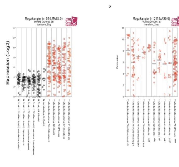
We evaluated CTAs gene expression data-sets through bioinformatics analysis. The data evidenced that the majority of known CTAs are either downregulated or not modulated (figure 3A and B) in MB samples respect to normal cerebellum. Only CTAs PRAME (Figure 3C) and CT22 (a CT-antigen, also known as Sperm Autoantigenic Protein 17 - SPA17, with a wide expression in somatic tissues) have a different expression pattern. (116) Starting from these observations, we decided to focalize our attention on PRAME, analyzing its mRNA expression levels in 10 normal adult (NAHC) and 8 foetal cerebella (NFHC), underlining a less expression than mononuclear cells derived from 8 healthy donors (PBMC, Figure 4A). We then studied PRAME mRNA levels in tumor samples collected at diagnosis from 60 MB patients diagnosed/treated at OPBG. The clinical-pathological data of the considered MB patients are summarized in Table 1. The 82% (49 out of 60) of all tumor samples showed a significative higher PRAME expression (average 92,2x10³±248x103; range, 0.9x10³-1500x103 AU) respect to NAHC tissues (average, 0.8x10³ AU; p<0.0001), with no relevant differences among the four molecular subgroups (Figure 4A). Noteworthy, Kaplan–Meier analysis evidenced that high PRAME mRNA expression correlates significantly with a worse Overall Survival (OS) probability in the 51 patients for which follow-up data were available in our Institution. This correlation remains statistically significant considering the PRAME mRNA expression cut-of as the maximum likelihood estimation threshold (117) of 19.2 X 10³ AU (p=0.0004; Figure 4B), the median value of 7.705 X 10³ AU (p=0.0003; Figure 4E), the first quartile value of 0.381 X 10³ AU (p=0.002; Figure 4F) and third quartile value of 36.221 X 10³ AU (p=0.0006; Figure 4G) quartile. Indeed, the median overall survival was 29.1 months (95% CI, 6 to 62) in high-PRAME-expressing patients group versus 59.4 months (95% CI, 6 to 158) in low-PRAME-expressing patients group (p=0.0004; Figure 2B. Hazard ratio for death, 4.258; 95% CI, 2.288 to 15.39; p=0.0031). Considering patients according to the four molecular subgroup, we verified a significant correlation between high PRAME expression and worse OS in SHH-MB (n=9; Figure 4C, p=0.0038) and G3-MB (n=19; Figure 4D, p=0.0075) subgroups; this
26
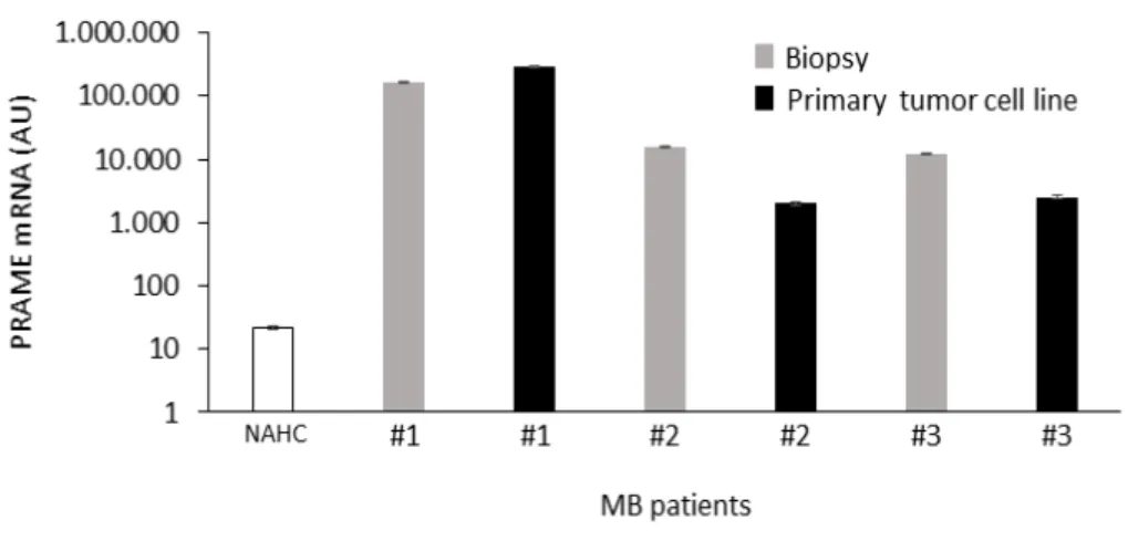
correlation was not statistically significant in WNT-MB (n=9) and G4-MB (n=14) due probably to the low number of patients studied. PRAME expression also correlated in our patient’s cohort with male gender (p=0.0279). We also analyzed PRAME expression in two MB patients (one case of SHH-MB and one of Group 3-MB) for which tumor biopsies were available both at diagnosis and at time of relapse, demonstrating high level of the antigen in recurrent MBs (Figure 5A). After all, IHC analysis shows rilevant PRAME protein expression in MB tumour samples, ranging from 20% to more than 90% of positivity in tumour cells, whereas the expression of PRAME protein were negligible in normal brain (Figure 5B).
27
Table1
28 Figure 3 A B
29
C
Figure 3.A, CTA down modulated in MB respect normal cerebellum. CTA mRNA gene
expression profiles across Gilbertson (GEO ID: GSE37418), Kool (GEO ID: GSE10327),Pfister (P=GEO ID:GSE49243; P2=ps_avgpres_mb500affym223_u133p2), Fattet/Delattre (GEO ID: GSE74195) datasets exploring CTA expression in either normal brain (N) or patients with MB. Numbers in the legend indicate the number of samples evaluated in each dataset. We have included in the analysis the following CTAs: MAGEA1 (melanoma antigen family A1), NA88 – VENTX (VENT homeobox), GAGE1 (G Antigen 1), SSX1 (synovial sarcoma, X breakpoint 1), MAGEA4 (melanoma antigen family A4), CAGE1 (cancer antigen 1), LUZP4 (leucine zipper protein 4), TPTE (Transmembrane phosphatase with tensin homology), MAGEC1 (melanoma antigen family C1) and MAGEA3-6 (melanoma antigen family A3/6). B, CTA not modulated in MB respect normal cerebellum. CTA mRNA gene expression profiles across Gilbertson (GEO ID: GSE37418), Kool (GEO ID: GSE10327), Pfister (P=GEO ID: GSE49243; P2=ps_avgpres_mb500affym223_u133p2), Fattet/Delattre (GEO ID: GSE74195) datasets exploring CTA expression in either normal brain (N) or patients with MB. Numbers in the legend indicate the number of samples evaluated in each dataset. We have included in the analysis the following CTAs: SLCO6A1 (Solute carrier organic anion transporter family, member 6A1), FMR1NB (fragile X mental
30
retardation 1 neighbor), HCA661 (transcription factor Dp family, member 3), OY-TES-1 (ACRBP, acrosin binding protein), SYCP1 (synaptonemal complex protein 1), NY-ESO-1, FATE (fetal and adult testis expressed 1), SPANXA1/B1 (Sperm protein associated with the nucleus, X-linked, family member A1), MAGEB1 (melanoma antigen family B1GAGE1), BAGE (B melanoma antigen), SPANXB1 (family member B1). C, PRAME mRNA
expression data from public datasets of expression profiles in MB patients. (1) PRAME
mRNA gene expression profiles across Gilbertson (GEO ID: GSE37418), Kool (GEO ID: GSE10327), Kool/Pfister (GEO ID: GSE49243), Fattet/Delattre and de Bont (GEO ID: GSE74195) datasets exploring PRAME expression in either normal brain (black plot circles) or patients with MB (red plot circles). Numbers indicate the number of samples evaluated in each dataset. (2) PRAME mRNA gene expression profiles across Gilbertson, Kool and Pfister datasets, exploring PRAME expression in patients with different MB-subtypes. Numbers indicate the number of samples evaluated in each MB subtype.
31 Figure 4 E F G
32
Figure 4. PRAME mRNA expression and its correlation with clinical feature of MB.A,
Relative expression of PRAME mRNA in PBMCs isolated from 8 healthy donors, 10 normal adult human cerebella (NAHC), 8 normal fetal human cerebella (NFHC), biopsies from 10 patients with WNT-MB pathway subtype, 14 patients with SHH-MB pathway subtype, 20 patients with Group 3-MB, and 16 patients with Group 4-MB. Transcripts quantification was expressed in AU versus the average expression of PRAME mRNA observed in PBMCs isolated from 8 healthy donors. *≤ P 0.05; **≤ P 0.001; ***≤ P 0.0001; ****≤ P 0.00001. B, Kaplan–Meier analysis for OS in all 51 patients with MB with a known follow-up (n=51), stratified by PRAME mRNA expression >19.2x10³ AU or 19.2x10³ AU, respectively. Differences between groups were compared with the log-rank test. C and D, Kaplan–Meier analysis for OS in patients with SHH-MB (C) and G3-MB (D) with more than 5 years of follow-up (n=51), stratified by PRAME mRNA expression >19.2x10³ AU or 19.2=103 AU, respectively. E, F, G, Differential cut-off for PRAME mRNA expression and their
correlation with clinical feature of MB. Kaplan-Meier analysis for overall survival (OS) in
all 51 MB patients with a known FU (n=51), stratified by PRAME mRNA expression considering the cut-off of median value of 7.705 x 10³ AU (E), 1° quartile value of 0.381 x 10³
AU (F) or 3° quartile value 36.221 x 10³ AU (G), respectively. Differences between groups
33 Figure 5 A B
34
Figure 5. A, MB tissues from two patients in relapse show high level of PRAME expression. Relative expression of PRAME mRNA from matched primary-metastases MB
tissues of patient with Group3-MB or with SHH-MB at diagnosis (dark gray bar) or at the time of relapse (light gray bars). PRAME mRNA expression in PBMC from 8 healthy donors has been added for comparison (black bars). B,Immunohistochemistry results of histological
sections to evaluate PRAME expression. IHC images of PRAME staining of testis (positive
control), normal cerebellum and three MB patients. Original magnification 10X and 20X .
35
4.2. Retroviral vector carrying iC9 and PRAME-SLL specific
αβTCR allows stable and functional expression of the
transgenes.
TCR (α and β chains) sequences specific for PRAME-SLL in frame with iC9 sequences (100), were cloned, codon-optimized, and encoded into a retroviral vector (iC9-SLL TCR, Figure 6A). Then, primary T cells or CD8+ sorted T cells (derived from PBMCs of healthy donors) were transduced with the generated retrovirus and expanded in the presence of cytokine IL2. About Six days after transduction, 53%±8,6% of CD3+ T cells stained for TCRVβ1 (to which PRAME-SLL TCR β chain belongs) and 32%±7,8% of CD8+ T cells were detected positive for SLL dextramer (Figure 6B shows an explicative example, whereas the median level of transduction reached in 8 independent experiments is shown in Figure 6C). In particular, CD4+ T cells were also significantly transduced with iC9-SLL TCR, as shown by the expression of TCRVβ1; however, using SLL-dextramer staining, CD4+ cells resulted negative for the SLL-TCR pairing, since SLL-dextramer staining is specific only for CD8+ cells (Figure 6B). Moreover, no significant differences in the transduction level (considering TCRVβ1 or dextramer staining analysis) were detectable between total T cells and CD8+ selected T cells.
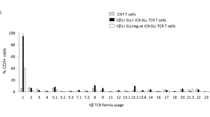
Starting from this observation, we decided to select through a SLL-dextramer microbeads, from the genetically modified T cells, the subset that express the correct SLL-TCR α and β chains pairing. Our in vitro data demonstrated that the selected SLL-TCR+ T cells stably expressed the transgene (Figure 6B) and were able to expand, generating a great number of iC9-SLL TCR T cells. Analyzing these genetically modified T cells, they showed a polyclonal phenotype after transduction, since no preferential TCRβ family usage has been observed in SLL-dextramer+ T cells, beside SLL-specific Vβ1 repertoire (Figure 6E). Furthermore, no proliferative differences were found between transduced T cells or non-transduced T cells (CNT T cells) when co-cultured with the pleiotropic cytokine IL2 (10,7±7,6 and 17.5±16,5 fold expansion at day 15, respectively, Figure 6D). The exogenous expressed SLL TCR was functional, since iC9-SLL TCR T cells produce IFN-γ in response to the CEM-T2 cell line loaded with the SLL peptide (until 10-5 M concentration), but not with the irrelevant PRAME-peptide ALY (Figure 7A). In potency experiments, through Cr51
36
release assay, we demonstrated that SLL peptide-pulsed CEM-T2 cells were lysed by iC9-SLL TCR T cells at higher extent than un-loaded CEM-T2 (i.e. a HLA-A*02+ cell line characterized by low PRAME expression, as shown in Figure 5E; 69,8%±6.5% vs 8.0%±1.8% specific lysis, respectively at the Effector:Target (E:T) ratio of 20:1,p≤0.005 Figure 7B). Noteworthy, iC9-SLL TCR T cells, when co-cultured with the tumor cell line U266 (HLA-A02+ PRAME+) in a ratio 1:1 (effector:target), were able to eliminate tumor cells without SLL-peptide pre-loading (Figure 7G). As previously mentioned, to improve in a clinical perspective the safety of our transduced T cells, we included in the retroviral vector the iC9-suicide gene. In order to verify the capability of iC9 to induce apoptosis in transduced cells we perform functional experiments. Results demonstrated that also the second transgene is active, since iC9-SLL TCR T cells were promptly eliminated upon 24-hour exposure to 20 nM AP1903 (Figure 7C). The residual CD3+ Vβ1+ cells (average, 6.3%±3.9%) still alive after 72 hours of AP1903 exposure were further expanded in culture with IL2, and tested for the presence of vector DNA, showing the absence of genetically modified T cells (Figure 7D).
37
38
Figure 6.Generation of iC9-SLL TCR T cells.A, α and β chains of SLL-PRAME TCR were
cloned in frame with the suicide gene iC9 in a retroviral vector, with the separation of the transgenes through 2A sequences. B, Flow cytometry analyses in an esemplificative donor of untransduced (CNT T cells, top) or transduced with the retroviral vector iC9-SLL TCR (bottom) polyclonal T cells in vitro, activated through OKT3/CD28. TCR Vβ1 staining is shown in total CD3+, CD3+/CD4+, CD3+/CD8+ cells, whereas SLL-dextramer+ staining is shown in total CD3+ and in SLL-dextramer–sorted T cells (postsorting). C, The average of the positive TCR Vβ1 cells was shown in total CD3+, CD3+ CD8+, and CD3+ CD4+ T cells, whereas the average of positive SLL-dextramer cells was shown in CD3+ CD8+ and CD3+ CD4+ T cells. Data are expressed as average ± SD from 8 healthy donors at day 15 of in vitro expansion.D, Fold expansion of untransduced T cell (CNT, gray dashed line) and iC9-SLL TCR T cell (black line), evaluated by Trypan blue count assay. Data represent results from 8 healthy donors. E, TCR-Vβ repertoire analysis in iC9-SLL TCR T cells. TCR-Vβ repertoire analysis showed no significant difference in TCR Vβ usage between control T cells (CNT) and the transduced iC9-SLL TCR T cells (positive for Vβ1) subdivided in the T cell subset positive or negative for SLL tetramer staining.
39 Figure 7 E J I H G F
40
Figure 7. A-D, In vitro functional analysis of iC9-SLL TCR T cells. A, iC9-SLL TCR
avidity assessed by IFNg ELISpot assays of CEM-T2 cell line loaded with an irrelevant peptide (gray bars) and the SLL-specific peptide (black bars). Ionomycin/phorbolmyristate acetate (I/PMA) was used as positive control. SFCs per 105 cells. Data represent the mean ± SD of triplicate experiments. B, In vitro51Cr release assay evaluating cytolytic activity of iC9-SLL TCR T cells on CEM-T2 tumor cell line loaded with an irrelevant (irr; gray line) or iC9- SLL-specific peptide (black line). C, Evaluation of percentage of alive (Annexin-V_/ 7AAD_) T cells
grown in IL2 and exposed to 20 nmol/L AP1903 for 24, 48, or 72 hours. CD3+ TCR Vβ1+ T cells negative for Annexin-V/7AAD staining were considered to be alive after the activation of the iC9 suicide gene. Data from four healthy donors are expressed as average± SD. D, Analysis of the presence of retroviral vector sequence in iC9-SLL TCR T cells residual after AP1903 exposition. Quantitative PCR targeting specific retroviral sequence was carried out to establish whether Vβ1+ T cells residual after AP1903 exposition were genetically modified. Data from three independent experiments show that Vb1þ residual cells did not carry iC9-SLL TCR vector. E-J, PRAME expression in tumor cell lines and in vitro functional
analysis of iC9-SLL TCR T cells towards neoplastic cell lines. E, Average of relative
PRAME mRNA expression (AU) in 8 PBMC samples of adult healthy donors, MB cell lines DAOY (HLA-A*02+), D283 (HLA-A*02+, down-regulating HLA Class I molecule), Multiple Myeloma cell line U266 (HLA-A*02+), Lymphoma cell line HDML2 (HLA-A*02-), leukemia cell line RS4;11 (A*02-), and lymphoblastoid cell line CEM-T2 (A*02-). F, HLA-A*02 expression in D283 cell line untreated (light gray histogram) and after treatment with IFNγ (1000U/mL) for 48hrs (dark gray histogram). G, 7-days co-culture assay between effector cells [control CNT T cells (gray bars) or iC9-SLL TCR T cells (black bars)] and PRAME+ HLA-A*02+ multiple myeloma U266 or PRAME+ HLA-A*02- leukemia RS4;11 cell lines (1:1 Effector:target ratio). Data are expressed as average ± SD from 3 donors. H, in vitro 51Cr release assay evaluating cytolytic activity of un-transduced (CNT) T cells (dotted line) and total CD3+ iC9-SLL TCR T cell (continuous line) vs DAOY cell line. I, in vitro 51Cr release assay evaluating cytolytic activity of CD8+ selected T cells un-transduced (CNT) (dotted line) or transduced with iC9-SLL TCR T cell (continuous line) vs HLA-A*02+ DAOY cell line. J, in vitro 51Cr release assay evaluating cytolytic activity of un-transduced (CNT) T cells (dotted line) and total CD3+ iC9-SLL TCR T cell (continuous line) vs HLA-A02- RS4;11 cell line. n=3 replicates per point; representative of four donors. * value=<0.05, ** p-value=<0.01, *** p-value=<0.001.