Facoltà di Scienze Matematiche, Fisiche e Naturali Dipartimento di Biologia
Scuola Dottorale in Biologia
Sezione di Biologia Applicata alla Salute dell’Uomo (BASU)
– XXIV ciclo –
“STUDY OF THE ROLE OF PRODUCTS AND
ENZYMES OF CHOLESTEROL BIOSYNTHETIC
PATHWAY IN MUSCLE TISSUES”
“STUDIO DEL RUOLO DEI PRODOTTI E DEGLI
ENZIMI DELLA VIA BIOSINTETICA DEL
COLESTEROLO NEI TESSUTI MUSCOLARI”
Ph.D. Student: Dr. Laura Trapani
Supervisor: Prof. Valentina Pallottini
Coordinator of Biology Applied to Human Health section: Prof. Paolo Visca
To my own grandmother
who meant the world
to me
INDEX
ABSTRACT………..1
RIASSUNTO………...….2
INTRODUCTION………..………...12
1. Mevalonate pathway……….……….….…....12
2. 3β-hydroxy 3β-methylglutharyl coenzyme A reductase ……….14
2.1 HMGR regulation………...16
3. Mevalonate pathway’s main end-products……….……..18
3.1.1. Cholesterol….………….………...………...18
3.1.2. Cholesterol functions in vertebrates……….…...20
3.2. Ubiquinone ……...……….21
3.3. Dolichol………...……….23
3.4. Prenylated proteins……….24
4. Muscle tissue……....………...……….26
4.1. Skeletal muscle development……….28
4.2. Skeletal muscle regeneration………..29
5. HMGR inhibition: the statins..………..31
AIM………...………...40 PAPERS CONCERNING PhD PROJECT
3-Hydroxy 3-Methylglutaryl Coenzyme A Reductase Increase Is Essential for Rat Muscle Differentiation (Paper 1)
Mechanism Underlying Long-Term Regulation of 3-Hydroxy-3-Methylglutaryl Coenzyme A Reductase During L6 Myoblast Differentiation (Paper 2)
3-Hydroxy 3-Methylglutaryl Coenzyme A Reductase inhibition impairs muscle regeneration (Paper 3)
Effects of myosin heavy chain (MHC) plasticity induced by HMG CoA-reductase inhibition on skeletal muscle functions (Paper 4)
HMG CoA reductase inhibition gets rat β-Myosin Heavy Chain disappeared: a statin paradox (Paper 5)
ABSTRACT
The mevalonate pathway is an important metabolic pathway providing cells with vital bioactive compounds such as cholesterol, prenyls, ubiquinone and dolichol. All of them play pivotal roles in muscle structure and functions being involved in ATP production, contractile regulation and myoblast fusion into multinucleated syncitia. The key and rate limiting step of the pathway is the reduction of 3-hydroxy-3-methylglutaryl Coenzyme A in mevalonate, reaction catalized by the enzyme 3-hydroxy-3-methylglutaryl Coenzyme A reductase (HMGR) that, as a central modulator of cholesterol homeostasis, is highly regulated. Statins, drugs widely used in ipocholesterolemic therapies, competitively inhibit HMGR: besides the well known lipid lowering properties they can also exert muscle side effects with symptoms ranging from weakness and pain to symptoms associated with rabdhomyolysis, a life threatening condition. Statin-induced muscle adverse effects suggest that HMGR could play a central role in muscle physiological processes. The majority of the studies so far are limited to the definition of statin-induced myotoxicity without investigating whether and how HMGR inhibition can affect muscle physiology. Thus the PhD project was aimed at providing a comprehensive analysis of the role played by HMGR and its main end-products in skeletal muscle development, repair and functionality and in cardiac muscle structure and metabolism.
The thesis provides evidence that HMGR inhibition negatively affects myoblast differentiation and fusion, delays muscle regeneration, impairs the mechanical and functional features of glycolytic fibers, leads to changes in cardiac fiber phenotype, highlighting the crucial role exerted by this enzyme and its end-products in muscle tissues.
Thus, statin users might not only suffer from myopathy but also might not be able to repair any muscle damage. Furthermore, even though statins are supposed to reduce the risk of cardiovascular disease, they can also affect energy-dependent myocardial functions.
In conclusion the results suggest that more efforts should be spent to find alternative pharmacological approaches to statin treatment, able to block cholesterol biosynthetic pathway downstream to HMGR, the inhibition of which can affect body health at multiple levels.
RIASSUNTO
Introduzione
Il pathway biosintetico del mevalonato è deputato alla produzione di isoprenoidi sterolici e non sterolici di primaria importanza nella fisiologia cellulare. Tra i prodotti finali del pathway si annoverano il colesterolo, precursore degli ormoni steroidei e degli acidi biliari nonché entità strutturale delle membrane plasmatiche e del sistema dei tubuli T nelle cellule muscolari, l’ubichinone, trasportatore di elettroni nella catena respiratoria mitocondriale, il dolicolo, intermedio della glicosilazione in N di proteine, alcune delle quali responsabili della fusione dei mioblasti in sincizi multinucleati durante lo sviluppo embrionale del muscolo scheletrico, i prenili fondamentali per la modificazione post-traduzionale di proteine, quali RhoA, coinvolta nel differenziamento dei mioblasti e nella regolazione della contrazione del muscolo liscio.
La tappa velocità limitante la sintesi del mevalonato è la deacilazione del 3-idrossi-3-metilglutaril coenzima A (HMG CoA) in mevalonato, reazione catalizzata dall’enzima 3-idrossi-3-metilglutaril coenzima A reduttasi (HMGR) che, come regolatore centrale dell’omeostasi del colesterolo, è finemente modulato da meccanismi quali:
- una regolazione a breve a termine che agisce sull’attività enzimatica della proteina mediante reazioni di fosforilazione e defosforilazione;
- una regolazione a lungo termine che ne modula i livelli intervenendo sulla trascrizione e sulla degradazione dell’enzima in dipendenza del contenuto di steroli presenti nella cellula.
- una regolazione ormonale che influenza ambedue le modulazioni descritte. Il tessuto muscolare, parenchima costitutivo di tutti i tipi di muscolo (liscio, scheletrico e striato cardiaco) e responsabile, insieme allo scheletro, della locomozione e del movimento del corpo, svolge le sue funzioni estrinsecando capacità contrattile: la trasformazione di energia chimica in energia meccanica -per scissione enzimatica di molecole di ATP- consente lo scorrimento di filamenti di dimensioni ultrastrutturali in cui si organizzano miosina, actina e proteine regolatorie
.
La miosina, in particolare, è una proteina esamerica costitutita da quattro catene leggere (MLC) e due catene pesanti (MHC) ad attività ATPasica le cui diverse isoforme sono responsabili della velocità di contrazione delle fibre muscolari e della forza sviluppata durante il processo contrattile.Le prevalenti isoforme di MHC e il metabolismo cellulare delle fibre muscolari scheletriche ne consentono la classificazione in:
-fibre lente ossidative (esprimenti MHCI) -fibre rapide ossidative (esperimenti MHCIIa/x) -fibre rapide glicolitiche (esprimenti MHCIIb)
Nel muscolo cardiaco MHC α e β generano, nei ventricoli, tre differenti isoforme della miosina definite:
-V1, omodimero αα -V2, eterodimero αβ -V3, omodimero ββ.
Gli atri contengono una quarta isoforma definita Va, un omodimero αα con un differente set di catene leggere rispetto all’isoforma ventricolare V1.
L’embriogenesi del muscolo scheletrico è un processo attraverso cui le fibre muscolari sono generate da mioblasti mononucleati che, in seguito all’arresto dell’attività proliferativa differenziano in miociti capaci di fondere in sincizi multinucleati.
La riparazione del muscolo segue processi che ricapitolano solo in parte l’quelli dell’embriogenesi. Infatti, poiché i nuclei presenti nelle fibre muscolari sono incapaci di replicare essendo irreversibilmente fuoriusciti dal ciclo cellulare e trovandosi pertanto in uno stato postmitotico permanente, la fibra di per sé non è in grado di riparare eventuali perdite di tessuto dovute a traumi o miopatie degenerative, non potendo ripristinare l’attività mitotica dei suoi nuclei. La riparazione avviene mediante la capacità di peculiari cellule staminali definite “satelliti” di ricostituire il tessuto: queste sono elementi mononucleati, normalmente quiescenti situati tra il plasmalemma e la lamina basale. In seguito ad eventuali lesioni, le cellule satelliti sono stimolate a proliferare, generando una popolazione cellulare che mantiene uno stato quiescente ed elementi che dopo un certo numero di mitosi, si fondono tra loro formando strutture sinciziali capaci di esprimere i prodotti differenziati propri delle fibre e di ricostituire quindi l’integrità del tessuto.
L’HMGR è un target terapeutico appetibile per la cura farmacologica dell’ipercolesterolemia: l’inibizione intracellulare della sintesi del colesterolo induce una risposta omeostatica che determina l’incremento dell’esposizione di recettori superficiali di membrana in grado di mediare l’assunzione cellulare delle lipoproteine aterogeniche quali le LDL (Low Density Lipoprotein) e le VLDL (Very Low Density Lipoprotein).
Evidenze sperimentali e cliniche tuttavia dimostrano che il trattamento con le statine, potenti inibitori dell’HMGR largamente impiegati nella terapie ipocolesterolemizzanti, provocano effetti collaterali quali mialgia, miosite e
rabdomiolisi e l’insorgenza di malattie clinicamente silenti quali la distrofia miotonica o la malattia di Kennedy e di McArdle. Ne consegue che l’interesse principale della comunità scientifica nel campo sia stato quello di definire i meccanismi alla base della miotossicità provocata dalle statine piuttosto che di comprendere la funzione dell’HMGR nella fisiologia del muscolo scheletrico e cardiaco.
Pertanto, obiettivo del progetto di dottorato, è stato l’analisi del ruolo dell’HMGR e dei suoi prodotti finali nello sviluppo, nella rigenerazione e funzionalità del muscolo scheletrico, così come nella struttura e nel metabolismo del muscolo cardiaco.
Risultati
HMGR e suoi prodotti finali nello sviluppo del muscolo scheletrico
(3-Hydroxy 3-Methylglutaryl Coenzyme A Reductase Increase Is Essential for Rat Muscle Differentiation. J Cell Physiol. 2009 Aug;220(2):524-30) Paper 1
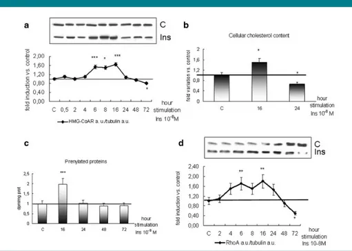
Per comprendere il ruolo dell’HMGR e dei suoi prodotti finali nello sviluppo del muscolo scheletrico, come modello sperimentale sono stati impiegati mioblasti fetali di ratto L6, il cui differenziamento è stato indotto mediante insulina. Per valutare se l’HMGR fosse soggetto a modulazione durante il processo miogenico, i livelli proteici dell’enzima e dei suoi prodotti finali quali colesterolo, proteine prenilate e tra queste RhoA, sono stati monitorati in un time corse di 72h. L’iniziale incremento dell’enzima è seguito da un decremento nelle fasi terminali del processo differenziativo: l’andamento di colesterolo, proteine prenilate e RhoA è simile a quello dell’HMGR (CFR fig. 2 paper 1).
Il ruolo dell’enzima nello sviluppo del muscolo scheletrico è stato indagato valutando i livelli proteici dei marker precoci e tardivi del differenziamento muscolare (miogenina e MHC embrionale) a 16 e a 24h dalla stimolazione con insulina, gli indici di fusione e differenziamento dei mioblasti a 72h dall’induzione della miogenesi, in presenza di 25OH colesterolo o mevinolina in grado, rispettivamente, di stimolare la degradazione e inibire l’attività dell’enzima. In ambedue le condizioni sperimentali i livelli dei marker del differenziamento e gli indici di fusione e differenziamento delle cellule manifestano una significativa riduzione rispetto ai controlli suggerendo che l’inibizione dell’HMGR possa rallentare il processo miogenico (CFR fig. 3, 4 paper 1).
Per comprendere quali proteine potessero essere coinvolte nella modulazione dell’HMGR durante il differenziamento dei mioblasti indotto da insulina, è stato inizialmente valutato lo stato di attivazione di una proteina di segnale, p38, in grado di indurre p21waf che, favorendo la fuoriuscita dal ciclo cellulare dei mioblasti in fase G1 ne consente il differenziamento. Accertato l’incremento della fosforilazione di p38 in seguito a stimolazione con insulina (CFR fig. 5 paper 1), è stato indagato il cross-talk tra p38, HMGR, p21waf e RhoA inibendo alternativamente p38 e HMGR e analizzando i livelli delle proteine precedentemente menzionate. L’inibizione di p38 determina la riduzione dei livelli proteici dell’HMGR, p21waf e RhoA; l’inibizione e la down-regolazione dell’HMGR riduce i livelli di p21waf e RhoA (CFR fig. 7, 8 paper 1).
I risultati ottenuti suggeriscono pertanto che l’insulina incrementa la fosforilazione e attivazione di p38 coinvolta nell’up-regolazione dell’HMGR a sua volta responsabile dell’aumento dei livelli di RhoA e di p21waf, eventi che, nelle fasi iniziali, favoriscono l’evolversi del processo miogenico.
(Mechanism Underlying Long-Term Regulation of
3-Hydroxy-3-Methylglutaryl Coenzyme A Reductase During L6 Myoblast Differentiation. J Cell Biochem. 2010 May 15;110(2):392-8) Paper 2
Per comprendere pienamente i meccanismi alla base della modulazione dell’HMGR nel differenziamento dei mioblasti fetali di ratto sono stati analizzati il pattern d’espressione dell’enzima e delle proteine coinvolte nella sua regolazione a lungo termine nonché il tasso di degradazione enzimatica. L’analisi condotta dimostra come l’enzima sia regolato da meccanismi differenti durante le varie fasi del processo miogenico. A tempi precoci del differenziamento, l’incremento dei livelli dell’mRNA e proteici dell’HMGR è imputabile all’induzione trascrizionale e al rallentamento della velocità di degradazione dell’enzima: il ridotto contenuto cellulare di Insig-1 (CFR fig. 2 paper 2) infatti consente la migrazione del complesso SREBP-Scap dal reticolo endoplasmatico all’Apparato di Golgi e il conseguente rilascio di nSREBP, fattore trascrizionale del gene codificante l’HMGR; i ridotti livelli di Insig-1 inoltre provocano il decremento del tasso di ubiquitinazione e degradazione proteosomale dell’enzima. La successiva riduzione dei livelli proteici dell’HMGR sembrerebbe essere dovuta all’aumentata velocità di degradazione dell’enzima, come giustificato dall’incremento del contenuto cellulare di Insig-1 e dal decremento dei livelli di nSREBP (CFR fig. 2, 3, 5
.paper 2). Negli stadi tardivi del differenziamento la riduzione dei livelli di mRNA e proteici dell’HMGR è imputabile ad un diminuito tasso trascrizionale: infatti, Insig-1, il cui contenuto cellulare si mantiene elevato nelle fasi terminali del processo miogenico,, trattenendo il complesso SREBP-Scap nel reticolo endoplasmatico impedisce il rilascio di nSREBP e la conseguente trascrizione del gene codificante l’HMGR, senza influenzare la velocità di degradazione dell’enzima (CFR fig. 2, 3, 5 paper 2 )
HMGR e suoi prodotti finali nella rigenerazione del muscolo scheletrico
(3-Hydroxy 3-Methylglutaryl Coenzyme A Reductase inhibition impairs muscle regeneration. Pending editorial decision: Journal of Cellular Physiology) Paper 3
Poiché la rigenerazione muscolare è un processo che ricapitola gli eventi principali che si succedono durante la miogenesi, è stato ipotizzato, sulla base dei risultati ottenuti sui mioblasti fetali di ratto, che l’HMGR potesse svolgere un ruolo chiave nel differenziamento delle cellule responsabili della riparazione del tessuto muscolare.
Pertanto nei mioblasti derivati da cellule satelliti murine (SCDM) (gentile concessione del Dottor Marco Crescenzi, Istituto Superiore di Sanità) è stato indotto il differenziamento mediante cambiamento di terreno e riduzione di siero, e sono stati monitorati i livelli proteici dell’ HMGR e delle differenti isoforme di MHC in un time course di 72h.
I risultati ottenuti indicano che i livelli proteici dell’enzima e di MHC embrionale raggiungono il mssimo incremento 6 ore dopo l’induzione del differenziamento e si riducono più tardivamente; l’aumento delle isoforme adulte di MHC (rapide e lente) si osserva 24 ore dopo il cambiamento del terreno e la riduzione del siero (CFR fig. 1 paper 3). La simile modulazione dei livelli proteici dell’HMGR durante le prime fasi del differenziamentp osservata nei mioblasti fetali di ratto (paper 1) e nelle SCDM murine (CFR fig. 1 paper 3) suggerisce che l’enzima possa svolgere un ruolo determinante anche nel differenziamento delle cellule responsabili della rigenerazione muscolare.
Le SCDM pertanto sono state trattate per 72h con simvastatina ad una dose paragonabile a quella dosata nel plasma di pazienti sottoposti a trattamento cronico con il farmaco; sono stati successivamente valutati l’indice di fusione delle cellule in differenziamento e i livelli proteici delle isoforme embrionali e adulte di MHC. Le SCDM trattate con il veicolo
generano miotubi ramificati e con un elevato numero di nuclei, le SCDM trattate con simvastatina danno origine a miotubi piccoli, sottili e con un ridotto numero di nuclei. Il trattamento con la statina inoltre previene la riduzione dei livelli di MHC embrionale e l’incremento delle isoforme adulte rapide e lente dell’MHC, eventi necessari per l’evolversi del processo riparativo del tessuto. Il co-trattamento con il mevalonato, prodotto della reazione catalizzata dall’HMGR, ripristina la morfologia dei miotubi e il pattern proteico delle isoforme dell’MHC (CFR fig. 2 paper 3).
Con il supporto dei risultati ottenuti in vitro, è stata studiata in vivo la risposta al danno muscolare indotto mediante iniezione di cardiotossina nel muscolo tibialis anterior di topi trattati quotidianamente, per 21 giorni, con iniezioni intraperitoneali di simvastatina (1,5 mg/kg comparabile alla dose massima impiegata nella terapia ipocolesterolemizzante) o di simvastatina e mevalonato. I dati ottenuti indicano che 3 settimane dopo l’insulto miotossico, la citoarchitettura dei muscoli tibiali anteriori dei topi sottoposti a trattamento con il veicolo e a co-trattamento con simvastatina e mevalonato è completamente ristabilita, contrariamente a quella dei muscoli prelevati dai topi trattati con la sola simvastatina, ove la rigenerazione delle fibre non è ultimata ma ancora in corso, come dimostrato dalla presenza di nuclei centrali all’interno delle fibre (CFR fig. 4 paper 3). Analisi condotte mediante Western-blot indicano inoltre che il livello delle isoforme adulte, rapide e lente, di MHC è ridotto nei muscoli di animali trattati con il farmaco rispetto ai veicoli e ai topi sottoposti a co-trattamento (CFR fig. 5
paper 3).
I dati ottenuti sia in vitro che in vivo pertanto indicano che l’inibizione dell’HMGR riduce il differenziamento delle SCDM e rallenta la riparazione del tessuto muscolare: gli esperimenti condotti mediante l’impiego di mevalonato dimostrano che gli effetti osservati sono riconducibili all’inibizione dell’enzima e non ad un effetto tossico provocato dal farmaco indipendentemente dal suo meccanismo d’azione.
HMGR e suoi prodotti finali nel muscolo adulto
(Effects of myosin heavy chain (MHC) plasticity induced by HMG CoA-reductase inhibition on skeletal muscle functions. FASEB J. 2011 Nov;25(11):4037-47. Epub 2011 Jul 28) Paper 4
Per comprendere il ruolo svolto dall’HMGR e dei suoi prodotti finali nel tessuto muscolare adulto, ratti Wistar di 3 mesi sono trattati con
iniezioni intraperitoneali di simvastatina (1,5 mg/kg) per 21 giorni, a conclusione dei quali gli animali sono stati sottoposti a test comportamentali e quindi sacrificati; il prelievo di sangue e muscoli ha consentito l’effettuazione di analisi biochimiche e meccaniche.
Gli esperimenti sono stati condotti sul muscolo estensor digitorum
longus (EDL) poichè costitutito prevalentemente da fibre glicolitiche, le
più suscettibili agli effetti collaterali delle statine.
Il trattamento cronico con il farmaco non ha determinato cambiamenti significativi nel peso corporeo dei ratti, ma, come aspettato, una riduzione dei livelli plasmatici di colesterolo e trigliceridi a dimostrazione dell'efficacia del trattamento. La simvastatina ha inibito del 20% l’attività dell’HMGR nel muscolo scheletrico e conseguentemente incrementato i livelli del recettore LDL sulla membrana delle fibre (CFR fig. 1 e tabella 1 paper 4). Il trattamento non ha provocato danni muscolari, in termini di necrosi o apoptosi, come dimostrato dalla mancata variazione dei livelli di creatin chinasi plasmatiche e di PARP-1 nell’EDL (CFR fig. 2
paper 4). L’inibizione cronica dell’HMGR non ha causato variazioni nello
stato d’attivazione della proteina prenilata RhoA, né nel contenuto muscolare di colesterolo e ubichinone (CFR fig. 2 e tabella 2 paper 4).
Il trattamento con simvastatina ha tuttavia provocato una riduzione dei livelli proteici dell’isoforma rapida glicolitica MHCIIb ed un incremento delle isoforme rapide-ossidative MHCIIa/x (CFR fig. 4 paper 4). Per verificare se lo shift delle isoforme potesse determinare variazioni nella performance muscolare dell’EDL, il muscolo è stato sottoposto ad esperimenti meccanici che hanno consentito la valutazione di diversi parametri inerenti la contrazione. I risultati ottenuti dimostrano che l’inibizione cronica dell’HMGR riduce la potenza contrattile e la velocità di accorciamento delle fibre senza determinare alcun cambiamento nella forza isometrica, nell’affaticamento e nel recupero muscolare dell’EDL (CFR fig. 6, 7 e tabella 3 paper 4). Per valutare se la riduzione dei parametri meccanici causata dal trattamento con simvastatina potesse provocare modificazioni funzionali, sono state valutate la coordinazione motoria e l’attività locomotoria basale degli animali sottoponendo i ratti al test del rotarod e dell’open field. I dati ottenuti indicano che l’inibizione cronica dell’HMGR pur non modificando l’attività locomotoria basale, come dimostrato dall’assenza di variazioni nella frequenza di attraversamento dei quadrati in cui l’arena dell’open field test è stata suddivisa, peggiora tuttavia la capacità degli animali di sostenere condizioni che richiedano sforzi muscolari più intensi (CFR fig. 8, 9 paper 4).
Complessivamente i dati ottenuti in questa fase del progetto dimostrano che il trattamento cronico con simvastatina determina nel muscolo EDL uno shift delle isoforme rapide dell’MHC verso un fenotipo più lento, una conseguente riduzione della potenza e della velocità di accorciamento delle fibre, un peggioramento funzionale: questo potrebbe spiegare i fastidi muscolari di pazienti sottoposti a trattamento con statine privi di un incremento delle creatin chinasi plasmatiche.
HMGR e suoi prodotti finali nel muscolo cardiaco
(HMG CoA reductase inhibition gets rat β-Myosin Heavy Chain disappeared: a statin paradox. Subitted to Journal of Cellular Biochemistry) Paper 5
Effetti avversi delle statine sono stati descritti anche a carico del muscolo cardiaco. Pisarenko e colleghi hanno dimostrato che il trattamento di un mese con lovastatina determina una riduzione dell’eiezione cardiaca e dell’indice di contrattilità: gli autori hanno attribuito gli effetti descritti alla riduzione dei livelli intratissutali di ubichinone.
Poiché le statine, come precedentemente dimostrato, sono in grado di modificare il pattern d’espressione delle diverse isoforme di MHC, evento potenzialmente responsabile della modulazione delle proprietà contrattili del cuore, e poiché le conoscenze circa la capacità delle statine di ridurre i livelli intratissutali di ubichinone sono limitate, sono stati valutati gli effetti dell’inibizione dell’HMGR sul fenotipo strutturale e metabolico delle fibre cardiache. Ratti Wistar di 3 mesi sono stati trattati con iniezioni intraperitoneali di simvastatina (1,5 mg/kg) per 21 giorni, a conclusione dei quali gli animali sono stati sacrificati; è seguito quindi il prelievo del sangue e del cuore per l’effettuazione di indagini biochimiche.
Dopo aver accertato l’efficacia del trattamento, dimostrato dalla riduzione dei livelli plasmatici di colesterolo e trigliceridi (CFR fig. 1 paper 5), è stato misurato, mediante HPLC, il contenuto di colesterolo e ubichinone in omogenati di ventricoli cardiaci: l’inibizione cronica dell’HMGR determina un incremento dei livelli di colesterolo ed una riduzione di ubichinone (CFR tabella 1 paper 5). La valutazione del pattern delle isoforme MHC ha dimostrato che nonostante non siano state individuate variazioni significative nel contenuto di MHCα, l’isforma β subisce un gravoso decremento che tuttavia non si può escludere venga compensato da in aumento di α (CFR fig. 2 paper 5). La mancata
rilevazione di variazioni significative dell’MHCα potrebbe essere riconducibile all’elevato contenuto di questa isoforma nel ventricolo e alla sensibilità del Western-blot come metodo di indagine; infatti piccole variazioni compensatorie potrebbero non essere apprezzate dalla metodica utilizzata. Inoltre, poiché MHC α e β sono regionalmente distribuite nel tessuto muscolare cardiaco per rispondere alle esigenze strutturali e funzionali del cuore, variazioni anche minime dei livelli delle isoforme potrebbero determinare conseguenze funzionali: ne deriva che sebbene l’isoforma β sia poco rappresentata rispetto ad α (5:95), la sua riduzione potrebbe tradursi in una insufficiente funzionalità tissutale.
I risultati ottenuti sembrano pertanto mettere in luce un paradosso: le statine, farmaci impiegati per ridurre la colesterolemia e per prevenire il rischio di malattie cardiovascolari, possono modificare le proprietà contrattili dell’organo che si suppone debbano proteggere: il cuore.
Conclusioni
Nel complesso i dati ottenuti durante lo svolgimento del progetto di dottorato mettono in luce come l'HMGR e i suoi prodotti finali siano fondamentali per il corretto svolgimento di molti processi fisiologici che coinvolgono il muscolo scheletrico come lo sviluppo, la rigenerazione e la contrazione muscolare nonchè nel mantenimento del fenotipo strutturale e metabolico delle fibre cardiache.
A causa della crescente incidenza delle malattie cardiovascolari il cui principale fattore di rischio è l’elevato livello di lipidi plasmatici, il controllo della colesterolemia, effettuato con farmaci come le statine, che inibiscono l'HMGR, enzima chiave della sintesi del colesterolo, è divenuto un’esigenza primaria per il mantenimento dello stato di salute.
Resta tuttavia da stabilire fino a che punto il rapporto rischio/beneficio suggerisca l’impiego di tali farmaci.
Pazienti sottoposti a trattamento con statine, infatti, potrebbero non soltanto lamentare fastidi alla muscolatura scheletrica, ma non essere in grado di riparare eventuali danni. Inoltre, seppur impiegate per ridurre il rischio di sviluppare malattie cardiovascolari, le statine possono alterare le funzioni cardiache.
Pertanto, considerando il ruolo cruciale dell’HMGR nella crescita, nella proliferazione, nel differenziamento e nella sopravvivenza cellulare e la necessità di mantenere a livelli ridotti il contenuto plasmatico di colesterolo per prevenire l’insorgenza di patologie cardiovascolari, dovrebbero essere impiegate nuove strategie terapeutiche nel trattamento
dell’ipercolesterolemia mirate all’inibizione esclusiva della sintesi di colesterolo a valle dell’HMGR la cui downregolazione e/o ridotta attività possono provocare effetti dannosi alla salute dell’uomo.
INTRODUCTION
1. Mevalonate pathway
Mevalonate (Mva) pathway (Fig. 1) is an important metabolic pathway that provides cells with essential bioactive molecules, vital in multiple cellular processes. This pathway converts Mva into sterol isoprenoids, such as cholesterol, essential precursor of bile acids, lipoproteins, and steroid hormones, and into a number of hydrophobic molecules, nonsterol isoprenoids, among others dolichol, ubiquinone (CoQ) and isoprenes. These intermediates play important roles in the post-translational modification of a multitude of proteins involved in intracellular signaling and are essential in cell growth, differentiation, gene expression, protein glycosylation and cytoskeletal assembly (Buhaescu and Izzedine, 2007).
Figure 1. Scheme of mevalonate pathway.
Acetyl-CoA HMG-CoA Mevalonate Isopentenyl-PP Geranyl-PP Farnesyl-PP Squalene CHOLESTEROL Geranylgeranyl-PP 2,3-Oxydosqualene ISOPRENYLATED PROTEINS DOLICHOL UBIQUINONE HMGR
Initially, the pathway involves repeated condensation of acetyl-CoA units, resulting in 3β-hydroxy 3β-methylglutharyl coenzyme A (HMG-CoA). The next reaction, which is a reduction of the substrate to mevalonate consuming two molecules of NADPH in a two-step reaction, is mediated by
3β-hydroxy 3β-methylglutharyl coenzyme A reductase (HMGR) (Fig. 2). This protein is considered to be the major regulatory enzyme in cholesterol biosynthesis and is one of the most intensively investigated proteins in biochemistry.
Mva is phosphorylated in two consecutive steps, which requires two molecules of ATP (Tanaka et al., 1990). Dehydration-decarboxylation of Mva-PP gives isopentenyl pyrophosphate (IPP), the basic building block for all products of the pathway (Rozman and Monostory, 2010). IPP can isomerize to dimethylallyl pyrophosphate via isopentenyl pyrophosphate isomerase (ILI1). IPP and dimethylallyl pyrophosphate condense to form the C-10 geranyl pyrophosphate which in turn condenses with a second molecule of isopentenyl pyrophosphate to produce the C-15 farnesyl pyrophosphate by farnesyl pyrophosphate synthase (FDPS) (Rozman and Monostory, 2010). The terminal steps of Mva pathway involve the branch-point enzymes that utilize FPP, i.e., squalene synthase, trans-prenyltransferase, cis-prenyltransferase and farnesyl- or geranylgeraniol- protein transferases for cholesterol, CoQ, dolichol and isoprenylated proteins, which are considered to be rate-limiting for the terminal portion of the biosynthetic sequences (Bentinger et al., 2011).
Figure 2. The reductive deacylation of HMG-CoA to mevalonate is thought to proceed in three steps, with mevaldyl-CoA and mevaldehyde as reaction intermediates (Istvan and Deisenhofer, 2000).
2. 3β-hydroxy 3β-methylglutharyl coenzyme A reductase
HMGR is the key and rate-limiting enzyme of Mva pathway, catalyzing a NADPH-dependent reduction of HMG-CoA to mevalonate, the first committed step in cholesterol biosynthesis (Rozman and Monostory, 2010). The enzyme is a transmembrane glycoprotein anchored to the endoplasmic reticulum (ER). The human HMGR protein consists of three domains: the N-terminal 339 residues form the membrane-associated domain; the C-terminal catalytic region (residues 460–888) located in the cytosol and the residues 340–459 of the linker domain that connect the two portions of the protein (Friesen and Rodwell, 2004).
Figure 3. Ribbon diagrams of human HMGR. (A and B) Structure of the HMGR homotetramer. (C) Stereo diagram of the HMGR dimer structure (Istvan and Deisenhofer, 2000).
The functional enzyme is a tetramer with the dimeric active sites at the interface of the two monomers (Istvan and Deisenhofer, 2000)(Istvan et al., 2000). The HMG moiety of the substrate, which is bound to a single HMGR monomer, comes into proximity of the nicotinamide ring of a NADPH molecule, whose binding pocket is located in the neighbouring monomer. The substrate-binding pocket is characterized by a loop of residues 682–694. The cis-peptide (Cys688– Thr689) within this region is essential in the formation of the HMG-CoA binding site, and is critical in positioning the residues participating directly in HMGR mediated reduction (Istvan and Deisenhofer, 2000) (Istvan et al., 2000) (Fig. 3). The substrate-binding pocket also accommodates statin molecules upon competitive inhibition of the enzyme (Istvan and Deisenhofer, 2001) (Fig. 4).
Figure 4. Statins exploit the conformationl flexibility of HMGR to create a hydrophobic binding pocket near the active site. (A) Active site of human HMGR in complex with HMG-CoA, and NADP. (B) Binding of rosuvastatin to HMGR (Istvan and Deisenhofer, 2001).
As the central enzyme of the cholesterol biosynthetic pathway, HMGR is hightly regulated (Goldstein and Brown, 1990). In particular, the enzyme undergoes short-term, long-term and hormonal regulations.
2.1. HMGR regulation
HMGR short term regulation is achieved by phosphorylation and dephosphorylation reactions both able to affect the enzyme activity (Fig. 5 A). The phosphorylation of enzyme’s residue S872 decreases HMGR catalytic activity whereas the removal of the phosphate reactivates the enzyme (Beg et al., 1985). AMP activated kinase (AMPK) appears to be the major HMGR kinase in the liver, where the cholesterologenesis takes place. AMPK is known to be involved in the regulation of energy homeostasis responding to changes in cellular AMP to ATP ratio (Towler and Hardie, 2007). HMGR dephosphorylation (activation) is operated principally by Protein Phosphatase 2A (PP2A), an abundant cellular serine/threonine phosphatase that regulates a significant network of cellular events (Janssens and Goris, 2001). Aside from short-term regulation, HMGR is subjected to transcriptional, translational, and post-translational control (Xu et al., 2005). To monitor levels of membrane sterols, cells employ in addition to HMGR another membrane-embedded protein of the ER, Scap (SREBP cleavage activating protein), both containing a polytopic intramembrane sequence called sterol-sensing domain (SSD).
Scap is an escort protein for Sterol Regulatory Element Binding Proteins (SREBPs), membrane bound transcription factors able to induce the expression of genes required for cholesterol synthesis and uptake, such as HMGR and Low Density Lipoprotein receptor (LDLr) (Brown and Goldstein, 1997). In sterol-deprived cells, Scap binds SREBPs and escorts them from the ER to the Golgi apparatus where SREBPs are proteolytically processed to yield active fragments that enter the nucleus and induce the expression of their target genes (Brown and Goldstein, 1999). When cholesterol builds up in ER membranes, the Scap/SREBP complex fails to exit the ER, the proteolytic processing of SREBPs is abolished and the transcription of the target genes declines. ER retention of Scap/SREBP is mediated by sterol-dependent binding of Scap/SREBP to Insig (INSulin Induced Gene), an ER resident protein (Fig. 5 B) (Yang et al., 2002). Intracellular accumulation of sterols induces HMGR to bind Insig: the interaction promotes the ubiquitination and proteasomal degradation of the enzyme (Sever et al., 2003) (Fig. 5 C) .
Several hormones, including insulin, glucagon, glucocorticoids, thyroid hormone, and estrogen, regulate the expression of hepatic HMGR in animals. Insulin likely stimulates HMGR expression by increasing its rate of transcription, while glucagon opposes this effect. Hepatic HMGR activity undergoes significant diurnal variations due to changes in the levels of immunoreactive proteins, which are primarily mediated by changes in
insulin and glucagon levels. Thyroid hormone increases hepatic HMGR levels by acting to increase both transcription and mRNA stability, while glucocorticoids decrease hepatic HMGR expression by destabilizing HMGR mRNA. The effects of estrogen on HMGR expression are still debated. Some studies suggest that estrogens act to increase hepatic HMGR activity primarily by stabilizing HMGR mRNA and that deficiencies in those hormones that act to increase hepatic HMGR gene expression lead to elevated serum cholesterol levels (Ness and Chambers, 2000). On the other hand, studies using the DLD1 cell line suggest that estrogens induce an early increase in LDLr at both mRNA and protein level and later cause decreases in HMGR activity and protein expression (Messa et al., 2005).
Figure 5. Schematic illustration of HMGR short (A) and long (B, C) term regulations. In particular, panel B and C show the regulation of HMGR transcription (B) and degradation (C) as a function of intracellular sterol amount and of cholesterol uptake (Burg and Espenshade, 2011).
3. Mevalonate pathway’s main end-products 3.1.1. Cholesterol
Cholesterol (Cholest-5-en-3-ol) is the most abundant sterol in vertebrates. First isolated from bile stones by Poulletier de La Salle in 1770, cholesterol was later isolated from unsaponifiable fraction of animal fats by Chevreul (1815), who named it Cholesterine (Greek χόλη, bile and στέρεος, solid). The exact formula of cholesterol (C27H46O) was proposed by
Reinitzer in 1880, and the right structure and exact steric representation were elucidated between 1900 and 1932, mainly by works from Wieland and Windaus. Meanwhile, Schmidt (1914) measured for the first time high serum cholesterol levels in patients with xanthomatosis, thus recognised it as essential hypercholesterolemia. By the early 1950s, Lynen demonstrated that the acetylation of Coenzyme A is the key first step in a chain of reactions that result in the formation of cholesterol and fatty acids. Together with Bloch, Lynen successively received the Nobel price for medicine (1954) and Nobel price for physiology and medicine (1964) for their discoveries concerning the mechanism and regulation of cholesterol and fatty acids metabolism (pathway from “activated acetic acid” to terpenes and fatty acids). Meanwhile, Tavormina et al. (1956) discovered that mevalonic acid is quantitatively incorporated in cholesterol in cell-free systems with loss of carbon dioxide. The biological function of cholesterol gained insights from work by Fisher (1976) who, by freeze-fracture and biochemical analysis in human erythrocytes, demonstrated that cholesterol is asymmetrically distributed across the plane of the membrane, being more present on the exterior side than on the interior side. Works to elucidate the very nature and metabolism of cholesterol were just starting that it was already pointed for its involvement in atherosclerogenesis. The first hint came from work by Windaus who reported that atherosclerotic plaques from aortas of human subjects contained 20- to 26- fold higher concentrations of cholesterol than did normal aortas. This observation opened nearly a century of research, which leaded to the recognition of the cholesterol carrying low density lipoprotein (LDL) particles as the primary cause of atherosclerosis (Rader et al., 2003) With the discovery of the mevalonic aciduria as first inborn error of cholesterol biosynthesis (Hoffmann et al., 1986), a new era of cholesterol history started, era in which cholesterol was no longer considered just as heart breaking molecule, but also as a key player in developmental processes.
One of the sources for the acquisition of cholesterol by animals and human beings is the absorption of sterol ingested in the diet. During
digestion, cholesterol esters are broken into unesterified cholesterol and long chain fatty acids; such monomers of unesterified cholesterol can diffuse directly up to the microvillus border of the intestinal epithelial cell and be absorbed passively (Dietschy, 1984). After entering the enterocytes, approximately half of the cholesterol molecules move to the endoplasmic reticulum (ER) where cholesterol is esterified by acyl-CoA:cholesterol acyltransferase (ACAT) before incorporation into nascent chylomicron (CM), particles produced by the small intestine soon after a meal. CMs carry a lot of triglycerides (TGs) as well. These particles are released from the base of the intestinal epithelial cell and enter the intestinal lymphatic vessels, where they interact with high-density lipoprotein (HDL) to acquire apoproteins C and E (Havel, 1982). The liver is the most important site for body synthesis, but all organs in the body are capable of significant rates of cholesterol synthesis (Dietschy, 1984). The rate of cholesterol synthesis in the liver varies inversely with the amount of dietary cholesterol reaching it in the CMs (Nervi et al., 1975). Hence, sterol synthesis within the intestinal hepatic axis tends to accommodate to changing rates of dietary cholesterol entrance into the body. Cholesterol moves out of the liver largely carried in VLDL (Very Low Density Lipoproteins) that are gradually converted into IDL (intermediate-density lipoproteins) and LDL. Cells that have a demand for cholesterol bind LDL through their LDLr and then take up the complete particle through receptor-mediated endocytosis. This type of transport is mediated by depressions in the membrane (“coated pits”), the interior of which is lined with the protein clathrin. After LDL binding, clathrin promotes invagination of the pits and pinching off of vesicles (“coated vesicles”). The clathrin then dissociates off and is reused (Brown and Goldstein, 1985). After fusion of the vesicle with lysosomes, the LDL particles are broken down, and cholesterol and other lipids are used by the cells. The HDLs also originate in the liver. They return the excess cholesterol formed in the tissues to the liver. While it is being transported, cholesterol is acylated by lecithin cholesterol acyltransferase (LCAT). Formed cholesterol esters are no longer amphipathic and can be transported in the core of the lipoproteins. In addition, HDLs promote CM and VLDL turnover by exchanging lipids and apoproteins with them (Nilsson and Duan, 2006).
3.1.2. Cholesterol function in vertebrates
The cholesterol molecule consists of three main parts: a rigid lipophilic steroid core, which can trigger anchoring to lipid bilayer, a non-polar hydrocarbon tail that can undergo oxidation, and a non-polar hydroxyl head, which can take part in several esterification reactions (Fig. 6). As can be predicted from its molecular structure, the biochemistry of cholesterol covers a wide spectrum of biological processes, a lot of which is still to be understood.
More than 80% of the cellular pool of cholesterol is located in the plasma membrane (Hoekstra and van ISC, 2000), in which it accounts for 20 to 25% of the lipid molecules (Dietschy and Turley, 2001). The plasma membrane (PM) cholesterol functions not only as a structural entity, but influences many properties of the PM. In addition to generally affecting membrane fluidity and permeability (Khan et al., 2003), as well as integral protein function, cholesterol-induced membrane packing in lateral microdomains (rafts) can provide a scaffold for variety of membrane associated signalling proteins (Tabas, 2002). Rafts are small platforms, composed of sphingolipids and cholesterol in the outer exoplasmic leaflet, and connected to phospholipids and cholesterol in the inner cytoplasmic leaflet of the lipid bilayer. These assemblies are fluid but more ordered and tightly packed than the surrounding bilayer. The presence of this liquid-ordered microdomains in cells transforms the classical membrane fluid mosaic model into a more complex system, where proteins and lipid rafts diffuse laterally within a two-dimensional liquid.
In particular, in muscle cells, the sarcolemma and the T-tubular system emanating from it, are enriched in cholesterol and differ in this respect from the internal membranes of the sarcoplasmic reticulum (Carozzi et al., 2000). Is therefore possible that fluctuations in cellular cholesterol content predominantly affect the T-tubular membranes (Draeger et al., 2006).
Apart from its function as component of the PM, free cholesterol is the precursor in the biosynthesis of physiologically important substances like bile acids and steroid hormones, linking this fascinating molecule to vital functions like nutrition and reproduction (Tabas, 2002).
3.2. Ubiquinone (CoQ)
CoQ consists of a benzoquinone ring which is highly substituted and at carbon 6 an all-trans polyisoprenoid side-chain is attached (Fig. 7). The number of isoprenoid units in the prenyl side chain often varies among species. Some organisms have more than one type of CoQ, but usually there is one most abundant homologue. Most mammals, including humans, have mainly CoQ10 and small amounts of CoQ9, whereas CoQ9 prevails in rodents. In invertebrates, CoQ8 to CoQ10 were identified (Nowicka and Kruk, 2010).
CoQ biosynthesis involves a series of steps. The first is the synthesis of the ring from aromatic amino-acids. The other is the synthesis of the isoprenoid side chain, followed by prenylation of the ring with the aid of 4-hydroxybenzoate prenyltransferase. After the ring has been prenylated, a sequence of reactions involving the modifications of the ring, such as decarboxylation, hydroxylation and methylations, occurs.
Following its isolation and characterization in 1955, CoQ was originally shown to be a necessary component of the mitochondrial respiratory chain two years later. It functions as an electron carrier from complex I and II to complex III and, according to Mitchell’s protonmotive Q cycle, production of ubisemiquinone accounts for the energy conservation occurring at coupling site 2 of the respiratory chain (Mitchell, 1975). It is hypothesized that disruption of ubiquinone production may lead to dysfunction of the electron transport chain, which could reduce muscle cell ATP levels, thus affecting muscle contraction, increase radical production and lead to apoptosis.
Today, several other important functions are also associated with this lipid.
1. The PM of most cells contains a CoQ-dependent NADH-oxidase which regulates the cytosolic NAD+/NADH ratio and ascorbate reduction and is involved in regulation of cell growth and differentiation (Gomez-Diaz et al., 1997).
2. CoQ is our only lipid-soluble antioxidant synthesized endogenously, and efficiently prevents oxidation of proteins, lipids and DNA. Effective enzymatic systems strive continuously to maintain this compound in its active reduced form (Bentinger et al.2011).
3. Opening of the mitochondrial membrane transition pore allows the translocation of molecules as large as 1500 Da in size, which leads to a collapse of mitochondrial functions. CoQ10 is one of the compounds that prevent such pore opening, thereby it is counteracting apoptotic events such as ATP depletion, release of cytochrome c into the cytosol, caspase-9 activation, depolarization of the mitochondrial membrane potential and DNA fragmentation (Papucci et al., 2003).
4. Uncoupling proteins present in the inner mitochondrial membrane can translocate protons from the outside to the inside of this membrane as a result of which the proton gradient formed by the respiratory chain is uncoupled from oxidative phosphorylation and heat is produced instead. These protons are delivered from fatty acids to the uncoupling proteins with the assistance of oxidized CoQ, which is thus an obligatory cofactor in this process (Echtay et al., 2001).
5. CoQ exerts multiple anti-inflammatory effects by influencing the expression of NFκB1-dependent genes (Schmelzer et al., 2007). Apparently, uptake of CoQ into lymphocytes and monocytes initiates the release of mediators and signal substances into the blood that subsequently modify such expression in a variety of tissues (Bentinger et al., 2011).
6. By protecting LDL from oxidation, this lipid has also anti-atherosclerotic properties. Moreover, it reduces the levels of lipid peroxides associated with lipoproteins in atherosclerotic lesions, as well as the size of such lesions in the aorta. Furthermore, CoQ decreases the levels of β2-integrin CD11b in monocytes, which counteracts monocyte–endothelial cell interactions (Thomas et al., 1996).
7. CoQ helps counteract endothelial dysfunction by stimulating endothelial release of nitric oxide (Hamilton et al., 2007).
8. CoQ mediates both oxidation of sulfide in yeast and the introduction of disulfide bonds into bacterial proteins (Bentinger et al., 2011).
3.3. Dolichol
Dolichol is widely distributed in all tissues and membranes and besides carrying a terminal free hydroxyl group it exists in a phosphorylated, dephosphorylated or in an esterfied form (Holstein and Hohl, 2004) (Fig 8). Although knowledge on dolichol function is limited, it has been proposed as a biomarker for ageing (Parentini et al., 2005).
Briefly, the unesterfied dolichol was shown to modify the organization and packing of phospholipids in model membranes (Vigo et al., 1984), the free and the phosphorylated forms were found to be mediators of protein N-linked glycosylation or as sugar carriers (Houten et al., 2003), the lipophilic molecule dolichol intercalates into the bilayer of cell membranes and interacts with the phospholipids (Parentini et al., 2005).
Figure 8. Structure of dolichol molecule.
In particular, it was found that alteration of cell-surface glycoproteins, using oligosaccharide-processing inhibitors that interfered with the synthesis of the high-mannose type of N-linked oligosaccharide, resulted in the inhibition of both the fusion reaction and biochemical differentiation of rat
skeletal muscle precursors, L6 myoblasts. Ketoconazole, compactin, and lovastatin, which affect dolichol and cholesterol biosynthesis, were also potent fusion inhibitors. These observations, coupled with earlier studies on the characterization of fusion-defective myoblast cell lines defective in glycoprotein biosynthesis, point to the importance of surface glycoproteins in cellular recognition in L6 myoblasts (Jamieson et al., 1992) (Belo et al., 1993).
In comparison to dolichol phosphate, the role of the free alcohol and its carboxylic ester is less established. An important question is the effect of these lipids on the physical properties of biological membranes. Biophysical studies have shown that dolichol and dolichol phosphates destabilize the bilayer structure of model membranes, and also increase their fluidity (Valtersson et al., 1985).
Furthermore dolichol enhances vesicle fusion whereas dolichol phosphate stimulates the fusion of rat liver microsomes. In this way, dolichol might play a more general role in membrane trafficking.
Some studies (Sato et al., 1999) (Belgareh-Touze et al., 2003) suggest that free dolichol is involved in vesicle trafficking, a function in agreement with an enrichment of the Golgi apparatus for this lipid. A specific role for the dolichol-ester has not been clearly established. Besides an effect on membrane properties, the esterified form was suggested to be necessary for intracellular dolichol transport (Tollbom and Dallner, 1986). An oxidized derivative of dolichol, dolichoic acid has also been identified in human brain (Ward et al., 2007): it is speculated to result from dolichol catabolism, but its exact function is unknown (Cantagrel and Lefeber, 2011).
3.4. Prenylated proteins
Protein prenylation is a post-translational modification that consists of the addition of 15 (farnesyl) or 20 (geranylgeranyl) carbon isoprenoid units to specific cysteine residues positioned near the C-terminus of a protein (Fig. 9) (Shepherd et al., 1995). Based on labelling experiments with tritiated Mva, it has been estimated that 2% of the mammalian proteome is prenylated (Anderson et al., 1995). Thus, essentially all signal transduction pathways involve prenylated proteins. The frequent occurrence of this modification, coupled with its crucial role in regulating cellular processes has generated intense interest in the enzymology and function of protein prenylation. The “CAAX” box is the recognition sequence for prenylation. The mature isoprenylated protein arises from a three-step process that consists of prenylation, followed by proteolysis of the “AAX” sequence,
and methyl esterification of the new C-terminal cysteine. Progress has also been made in understanding the function of protein prenylation. In some cases, isoprenylation serves to direct membrane association, (Ridker et al., 2001) whereas in other situations, the prenyl group is involved in mediating protein-protein interactions (Yamazaki et al., 1995).
Figure 9. Structure of a prenyl group.
Thus, protein isoprenylation allows the covalent attachment, subcellular localization, and intracellular trafficking of membrane-associated proteins. Importantly, members of Ras and Rho GTPase family are major substrates for posttranslational modification by prenylation. Ras translocation from the cytoplasm to the plasma membrane is dependent on farnesylation, whereas Rho translocation is dependent on geranylgeranylation (Nakagami et al., 2003).
Concerning the role played by prenylated proteins in muscle function, GTP RhoA interacts and activates the phosphotransferase activity of Rho-kinase. Rho-kinase is able to regulate the phosphorylation of myosin light chian (MLC), a component of the muscle contractile protein myosin, by both the direct phosphorylation of MLC, and by the inactivation of myosin phosphatase through the phosphorylation of MBS (Myosin Binding Subunit of myosin phosphatase). Rho-kinase and myosin phosphatase thus coordinately regulate the phosphorylation-state of MLC, which is thought to induce smooth muscle contraction and stress fiber formation in non-muscle cells (Amano et al., 1996).
Moreover, Fortier and co-workers (2008) showed that RhoE, a member of Rho family, accumulates in elongated, aligned myoblasts prior to fusion and that its expression is also increased during injury-induced skeletal muscle regeneration. Furthermore, although RhoE is not required for myogenesis induction, it is essential for myoblast elongation and alignment before fusion and for M-cadherin expression and accumulation at the cell–cell contact sites. RhoE physiological upregulation is also
responsible for the decrease of RhoA and Rho-kinase activities, which are required for myoblast fusion process (Fortier et al., 2008).
4. Muscle tissue
Muscle is a very specialized tissue that has both the ability to conduct electrical impulses and to contract. Muscles are classified both structurally as either striated or smooth and functionally as either voluntary or involuntary. From this, three types of muscles emerge: skeletal muscle associated with the body's voluntary movements, cardiac muscle whose cells are joined to one another by intercalated discs which allow the synchronization of the heart beat, and smooth muscle, the intrinsic component of the wall of internal organs and blood vessels.
The prime mechanical functions of skeletal muscle are to produce force, to generate power and to act as a brake. Skeletal muscle maintains the integrity of the skeleton, allows walking, running, jumping, talking, eating, breathing and undertaking activities essential for life and living. These highly diverse requirements of muscle are met through a number of factors, such as the intricate neural control mechanisms, the different architectural arrangements, the heterogeneity of fiber types, and the ability to use different sources of fuel to keep the muscle machine working. However, within the basic functional unit of contraction, the sarcomere, there are a multitude of different structural, regulatory and contractile proteins, many of which exist as different isoforms, giving skeletal muscle a multiplicity of isoform expression (Schiaffino and Reggiani, 1996). The ability to increase the number of sarcomeres (i.e. muscle size), together with an ability to alter protein isoform expression, gives muscle the ability to adapt to the different challenges that may be placed upon it (Harridge, 2007).
Several classification techniques differentiate fibers based on their physiologic capabilities and their content of different isoforms of myosin, the sarcomere protein forming the thick filaments (Barany, 1967).
Myosin is a hexameric protein (molecular mass 480 kDa) comprising two myosin heavy chains (MHC) and four myosin light chains (MLC). At the COOH terminus, the myosin molecule is rod-shaped, as a result of the dimerization of the two heavy chains (molecular mass 200 kDa) into a 200-nm α-helical tail (the S-2 fragment). Bundled together, this portion of the MHC forms the thick filament backbone. At the NH2 terminus, the heavy
chains separate and branch out to form two distinct heads (the S1 fragment), which contain both actin and nucleotide binding domains.
Chemomechanical transduction occurs at the S1-actin interface, where the intramolecular conformational change in the S1 structure induced by cleavage of the γ-phosphate of ATP is reversed by strong binding of the S1 fragment to the actin molecule (strongly bound cross-bridge formation) and subsequent release of ATP hydrolysis products (Pi and ADP) (Gordon et al.,
2000). The detailed mechanisms of the actomyosin ATPase reaction have been extensively studied in solution in permeabilized skeletal muscle fibers (Cooke, 1997).
Studies on chemically skinned single human fibers showed that, as with rodent muscle, it is the MHC isoform the prime determinant of the velocity at which the pick power of contraction occurs (Bottinelli et al., 1996) as well as the rate of force development (Harridge, 2007) in a fully active fiber.
According to the majority of MHC isoforms found in adult mammalian skeletal muscles, the following pure fiber types exist: slow type I with MHCI, and three fast types, namely type IIA with MHCIIa, type IIX with MHCIIx, and type IIB with MHCIIb. The coexpression of specific pairs of these major MHC isoforms results in the formation of hybrid fibers, which can be subdivided based on the predominant MHC isoform. Fiber type-specific programs of gene expression are not restricted to the MHC isoforms, but exist for many other muscle proteins (Pette and Staron, 1997). For example, fiber type-specific isoforms exist for the essential and regulatory myosin light chains (MLC), the three troponin subunits, tropomyosin, α-actinin, and various Ca2+ regulatory proteins (e.g., sarcoplasmic reticulum Ca2+ ATPase, calsequestrin, and the α -subunit of the dihydropyridine receptor) (Pette and Staron, 1997). In addition to these qualitative differences, the expression levels of some proteins vary in a fiber type-specific manner.
Muscle fibers can be classified as a function of their metabolic pathways that can be either aerobic/oxidative, anaerobic/glycolitic. This kind of classification leads to 3 fiber types: twitch glycolytic (FG), fast-twitch oxidative (FOG), and slow-fast-twitch oxidative (SO) (Pette and Staron, 1997). Although a good correlation exists between type I and SO fibers, the correlations between type IIA and FOG and type IIB and FG fibers are more varied.(Hamalainen and Pette, 1995).
In cardiac myocytes, the two MHCs are encoded by two separate genes, designated as α and β genes (Jostarndt-Fogen et al., 1998). The two MHCs, α and β, generate three myosin isoforms in the ventricles, named V1, V2, and V3, based upon their electrophoretic mobility in the native state. The three myosin isoforms differ in the composition of α and β
MHCs, but have the same two pairs of MLCs, refereed as essential (LC-1) and regulatory (LC-2) light chains. The V1 myosin isoform is an αα homodimer and V3 is a ββ homodimer, whereas V2 is an α/β heterodimer. The atria contain a separate myosin isoform (Va) that is made up of two
αMHCs and a separate set of light chains (LC-1a and LC-2a) (Matsuoka et
al., 1991) (Saez et al., 1987).
4.1. Skeletal muscle development
The development of skeletal muscle is a multi-step process by which new muscle fibers are formed from precursor muscle cells.
Figure 10. Control of skeletal myogenesis by bHLH and MEF2 transcription factors (modified from Lluis et al., 2006).
Mononucleated undifferentiated myoblasts grow in proliferating conditions, characterized by a high mitogen content (proliferation); upon mitogen
Id Id Id Id Myf5 E Protein MyoD E Protein MyoD E Protein Myogenin MEF2 MEF2 E Protein MRF4 Id
Proliferation Early differentiation Late differentiation Terminal differentiation
Induction of muscle differentiation specific genes α-Actin MHC MCK Others
withdrawal, myoblasts differentiate into mononucleated myocytes (early differentiation) that subsequently start to fuse into multinucleated myotubes expressing muscle-specific proteins (late differentiation), to form the mature muscle. Progression through the different myogenic stages is controlled by the sequential activation of four myogenic regulatory factors (MRFs) belonging to the basic helix–loop–helix (bHLH) family of transcription factors (Myf5, MyoD, myogenin and MRF4), which cooperate with the ubiquitously expressed E proteins (the E2A gene products E12 and E47, and HEB) and myocyte enhancer factor 2 (MEF2), transcriptional regulators to activate the transcription of muscle-specific genes, coding for structural and enzymatic muscle proteins such as α-actin, MHC or muscle creatine kinase (MCK) (Fig. 10).
Studies using both primary cultures of skeletal muscle and established muscle cell lines (which partially recapitulate myogenesis, thus being extensively used as myogenic model systems) have confirmed the expression of MyoD and Myf5 in undifferentiated myoblasts, whereas myogenin and MRF4 are activated at early and late differentiation stages, respectively. In proliferating myoblasts, MRFs and E proteins associate with the HLH protein Id (inhibitor of differentiation). Id expression is downregulated at the onset of differentiation, allowing the formation of the functional MRF–E-protein heterodimers (Lluis et al., 2006).
4.2. Skeletal muscle regeneration
As previously reported, mammalian skeletal muscles consist of multinucleated myofibers which are formed during development by fusion of mononucleated muscle progenitors, some of which remain associated to adult myofibers as satellite cells, a specific type of stem cells located under the basal lamina of muscle fibers. The satellite cells are responsible for both muscle growth and regeneration, a process that in many but not all respects recapitulates the sequence of events observed during embryonic myogenesis.
Following muscle damage, myofibers are sheared or torn exposing the intracellular contents to the extracellular environment. Activation of calcium-dependent proteases leads to the rapid disintegration of the myofibrils whereas the activation of the complement cascade induces chemotactic recruitment of neutrophils and later macrophages who begin the process of digestion of the necrotic myofibers and cellular debris by phagocytosis. The neutrophils and macrophages release cytokines that amplify the inflammatory response and recruit the satellite cells.
Satellite cell proliferation leads to the formation of both new stem cells, which maintain an undifferentiated state, and of committed myogenic precursors which express the MyoD family regulators of muscle determination and differentiation (MRFs), including MyoD, Myf5, myogenin and MRF4. The transition from the quiescent to the activated state is rapidly followed by muscle differentiation, with expression of MHCs and fusion of myoblasts to each other forming myotubes.
Figure 11. Major events in muscle regeneration. Schematic representation of the temporal sequence of inflammatory and regenerative events following muscle injury (Ciciliot and Schiaffino, 2010).
The growth of regenerated muscle may vary according to various factors, including the type of muscle injury, the involvement of blood vessels and the re-establishment of neuromuscular and myotendinous connections. A crucial factor for successful muscle regeneration is the maintenance of the basal lamina of muscle fibers. Within the intact basal lamina satellite cells can proliferate and generate myoblasts which fuse to form almost normal muscle fibers in a short time period. Regenerated fibers can be recognized by the presence of centrally located nuclei which in rodents, but not in human muscle, are a hallmark of previous muscle regeneration (Ciciliot and Schiaffino, 2010).
5. HMGR inhibitors: the statins
Statins competitively inhibit HMGR occupying the catalytic portion of the enzyme, specifically the binding site of HMG-CoA, thus blocking access of this substrate to the active site (Istvan, 2002)(Istvan, 2003). The structures of the catalytic domains of the enzyme in complex with statin molecules have been identified (Istvan and Deisenhofer, 2000).
By interrupting cholesterol synthesis in the liver, statins activate the production of microsomal HMGR and cell surface LDLr (Lennernas and Fager, 1997). This results in a predictable increased clearance of LDL from the bloodstream and a decrease in blood LDL cholesterol levels that may range from 20% to 55% (Jones et al., 1998) (Jones et al., 2003). Statins have shown strong evidence-based capacity of decreasing the cardiovascular morbidity and mortality both in primary (Shepherd et al., 1995)(Downs et al., 1998),and secondary prevention settings (Sacks et al., 1996)(Tonkin et al., 2000). Because of these properties, statins are amongst the most widely used pharmaceutical agents in the world (Golomb and Evans, 2008).
Although statins are generally well-tolerated, they have potentially serious adverse effects on skeletal muscle. While fatal rhabdomyolysis is a rare event, with 0,15 deaths per million prescriptions, rhabdomyalgia and myositis are much more common, with an estimated occurrence in 1-5% of patients (Draeger et al., 2006).
The development of myopathy can impact daily living and quality of life by affecting the patient’s ability to accomplish even simple tasks or may prevent partecipation in once enjoyable physical activities. Currently, the only treatment for statin-induced myopathy is the discontinuation of statin use in affected patients, in which symptoms are most often reversible (Dirks and Jones, 2006).
Moreover a range of cases have now been reported in which statin use has “uncovered” previously clinically silent or clinically tolerated conditions, including McArdle disease, myotonic dystrophy (Tsivgoulis et al., 2006) acid maltase deficiency (Voermans et al., 2005) and possible Kennedy disease (Tsivgoulis et al., 2006). Statin has also exacerbated known muscle conditions, such as myasthenia gravis. In case of mitochondrial myopathies, the relative degree to which statins have unmasked versus induced disease may not always be clear (Golomb and Evans, 2008).
The pathogenesis of muscle damage is not known, nor it is clear why skeletal muscle should be thus affected (Draeger et al., 2006). However, given that cholesterol-lowering drugs in general can elicit myalgia, several