1
Alma Mater Studiorum – Università di Bologna
DOTTORATO DI RICERCA IN
SCIENZE BIOMEDICHE
Ciclo XXVIII
Settore Concorsuale di afferenza: 06/F4 Settore Scientifico disciplinare: MED/33
TITOLO TESI
EVALUATION OF THE EFFECTIVENESS OF
FEMORAL NECK PROPHYLACTIC SURGERY IN
ELDERLY OSTEOPOROTIC PATIENTS TO
PREVENT HIP FRACTURES
Presentata da: Dott. Eugenio Chiarello
Coordinatore Dottorato Relatore
Prof. Lucio Ildebrando Prof. Roberto Emanuele Cocco Buda
2
CONTENTS
1. INTRODUCTION
3
2. MATERIALS AND METHODS
10
3. RESULTS
14
4. DISCUSSION
18
5. CONCLUSIONS
20
6. FIGURES AND TABLES
21
3
1. INTRODUCTION
Osteoporosis is a skeletal disorder characterized by an increased risk of fractures due to a compromised bone strength [1, 2]. The strength reflects both density and quality of bone, therefore the decrease of bone mass and the micro-architectural deterioration that occur in this disease cause bone frailty leading to low energy fracture [3-5].
Fragility fractures are one of the major causes of morbidity and mortality worldwide. In Italy, there are 80.000 new femoral neck fractures due to osteoporosis every year, with a high prevalence in women (72%) [6]. Moreover, was estimated that in 2012 the cost of femoral fractures was 1.1 billion euro [7].
Osteoporosis prevalence is likely to rise due to an aging population: people older than 60 will increase by 50% over the next 40 years. Although numbers are uncertain, the latest pessimistic estimates lead us to expect a doubling of fragility fractures by 2050 [8, 9].
Moreover, hip fractures are associated with an increased mortality up to 25-30% within the first year [10] and an increase of 2.5 times risk of a new fracture [11]. One year after a hip fracture 40% of patients are still unable to walk independently, 60% have difficulty in at least one of the normal daily living activities and 80% experience limitations in other activities such as driving and shopping. In addition, 27% of patients were hospitalized in a long-term care facility following a hip fracture [12].
This scenario shows how osteoporosis and femoral neck fractures represent a tremendous concern in economic and social terms, therefore new strategies must be sought for the prevention and treatment of this pathology.
4
1.1 PREVENTION OF THE SECOND CONTRALATERAL FEMORAL NECK FRACTURE
In literature the incidence of second contralateral hip fractures in elderly osteoporotic patients ranges from 7 to 12% within two years after the first femoral neck fracture, with a high percentage of symmetry between the two fractures which varies from 70% to 83% [13, 14].
Therefore, it is mandatory to adopt appropriate strategies to prevent the second fracture in these patients. Currently secondary prevention focuses on pharmacological and non-pharmacological therapy.
The pharmacological secondary prevention is based on the prescription of anti-osteoporotic drugs.
In the market, there are several classes of drugs with different mechanism of action: anti-absorbable, anabolic, hormone replacement and selective estrogen receptor modulators and monoclonal antibodies.
Bisphosphonates are a class of anti-absorbable drugs with high tropism for the mineralized tissues. They are able to concentrate electively on remodeling bone surfaces, blocking osteoclast activity [15-18].
Teriparatide (rh-PTH) is an anabolic drug that stimulates bone formation and increases bone mineral density [19, 20].
Hormone replacement therapy, as the name suggests, is based on the substitution of estrogens whose production decreases in menopausal women; however, this class of drugs is associated with a high risk of uterine and breast cancer and increased cardiovascular risks [21-23]. In order to overcome these complications, selective estrogen receptor modulators (SERMs) such as Raloxifene and Bazedoxifene were introduced. These drugs explicate their action on
5
the estrogen receptors on the bone cells without having the negative effects on breast and uterus [24-26].
The last class of drug introduced in the marked are the monoclonal antibodies. Currently only Denosumab is available; this is an IgG2 human monoclonal antibody directed against RANK-L, which binds with high affinity and specificity. These bindings prevent the activation of its receptor RANK present on osteoclasts and their precursors’ surface thereby inhibiting their formation, functionality and survival and thus reducing both cortical and trabecular bone resorption. [27, 28].
Although there are so many drugs for the treatment and prevention of osteoporosis, it has been showed that none of these attain significant efficacy for the prevention of hip fractures below three years of continuous treatment [29, 30]. This combined with poor patient compliance results in a lack of efficacy of drugs for the secondary prevention of femoral neck fractures [31]. Recently some authors argue that evidence for drug therapy to prevent hip fracture is insufficient to warrant the current approach. They believe that pharmacotherapy can achieve at best a marginal reduction in hip fractures at the cost of unnecessary psychological harms, serious medical adverse events, and forgone opportunities to have greater impacts on the health of older people. Therefore, they propose to regret the current approach to hip fracture prevention because it is neither viable as a public health strategy nor cost effective [32].
The non-pharmacological prevention is based on modification of environmental risk factors, on a healthy diet with daily supplements of calcium and vitamin D and on the use of hip protectors. A Cochrane review on the use of hip protectors has demonstrated that their effectiveness in reducing fractures in nursing home patients but
6
equally it has shown that they are less effective in patients living in community. These results are probably related to the adherence of patients in wearing hip protectors due to their discomfort [33].
Currently, in addition to pharmacological and non-pharmacological prevention we need to add a new type of prevention: the surgical one.
1.2 SURGICAL PREVENTION OF FEMORAL NECK FRACTURE
The cortical thinning and the trabecular bone loss are both important in the frail osteoporotic bone. The cortical thinning of long bones is the consequence of endosteal resorption and normally is compensated by periosteal bone apposition, leading to an increase in the diameter of the bone. The femoral neck is not covered by periosteum because it is intracapsular, and therefore there isn’t bone apposition [3]; this may partially explain why, in osteoporotic femur, the neck is the "locus minoris resistentiae".
Moreover Holzer et al. show in an in vitro study that in the femoral neck the cortical bone and its geometry are primarily responsible for the bone strength, whereas the trabecular bone gives a marginal contribution (less than 10%) due to morphological changes [34].
The rationale of surgical reinforcement is the need to increase the resistance of the neck to the compression and distraction forces acting on it [34, 35].
During gait, the major stresses occur in the subcapital and middle-cervical regions: high compressive stress occurs inferiorly and mild distraction stress occurs superiorly [36]. During a fall to the side with impact on the greater trochanter, the stresses are reversed: on the superior side of the femoral neck, a huge compressive stress occurs while on the inferior side there is a distraction stress [36, 37].
7
The concept of surgical reinforcement of the femoral neck was proposed for the first time in 1960 by Crockett [38] who described a reinforcement technique of the femoral neck characterized by percutaneous insertion of stainless-steel nails under local anesthesia. In the conclusion of his paper the author affirmed that in case of a fracture in the reinforced neck, the patient would have a non-displaced fracture and therefore the treatment required was only rest and walking with 2 crutches.
More recently, Heini et al. [39] in 2004 described another experimental technique called “femoroplasty” consisting of injection of poly-methyl-methacrylate (PMMA) inside osteoporotic femoral neck. The author used 20 pairs of osteoporotic femurs, each pair as a case-control, to assess the surgical reinforcement. The author inserted a low viscosity cement in a 4.5 mm hole on the lateral cortex at the base of the greater trochanter. Subsequently the femurs were tested by simulating a fall. Fracture type observed in control group matched those commonly seen in vivo; in the study group, different fracture patterns were observed: trochanteric and medial fractures of the femoral neck and in three cases subtrochanteric fractures. Moreover, all the fractures occurred at the bone-cement interface. In this study group femurs had an increased breaking load greater than 82% compared to controls, and an increase in absorbed energy of 188%. However the author concluded that there is concerns in the application of this technique in vivo due to the high volume of PMMA necessary which generates enormous heat during polymerization (up to 60° in vivo) leading to necrosis of the femoral head. Moreover, revision surgery in event of fracture would be technically very difficult. Other authors [40] tested ten pairs of osteoporotic human femurs, each pair as a case-control, augmented with about 40 ml of another
8
low viscosity cement. They simulated a fall on the greater trochanter and confirmed the increase of breaking load and absorbed energy in the augmented femurs; however it was unknown if this increase would be enough to prevent fracture in vivo. Moreover, they found that the stiffness was not significantly different between the two groups. They hypothesized that these results were due to the composite nature of the augmented femur: the bone governs the pre-yield behavior and once fracture occurs, it is likely that the composite formed by trabecular bone and cement determines the mechanical response.
To overcome the high temperature of polymerization, Beckmann et al. [41] tested a not-resorbable composite consisting of crosslinking resins and reinforcing glass ceramic particles already used for vertebral augmentation instead of PMMA. The author used nine pairs of femur as case-control; they recorded the temperature of polymerization and simulated a fall on the great trochanter. Subsequently the fractured femurs were stabilized using cannulated screws, a dynamic hip screw or a proximal femoral nail and they were then biomechanically tested again. As expected, breaking load and absorbed energy were significantly increased. The maximum temperature elevation (about 11°) was lower if compared with PMMA but still high if compared to the near iso-thermic polymerization cement based on calcium phosphate. However, the authors expressed concern regarding the revision surgery of the reinforced femur especially in the drilling: the composite was even harder to drill than the PMMA. Moreover, femoroplasty may directly influence the subsequent fracture of the augmented region: a distal shift of the fracture location could be assumed for in vivo condition.
9
Moreover De Bakker et al [42] in a finite elements study have shown how, reinforcing the femur with a Gamma nail, there was a 100% increase in the resistance to fracture.
Currently, to our knowledge, there is only a device on the market, for the prevention of the femoral neck. Fractures. Recently it has been published a finite element analysis showing that this device has led to a decrease in the risk of femoral neck fracture (−28%) and trochanteric fracture (−52%) [43].
1.3 AIM
The aim of our study is to evaluate the efficacy and safety of a new device: the Prevention Nail System (PNS), made by Medacta (Medacta International Castel San Pietro Switzerland) for the surgical prevention of femoral neck in elderly patients with severe osteoporosis.
Secondary objectives of the study are to evaluate the bone-screw integration, the range of motion of the reinforced hip, the incidence of intra and post-operative complications such as infection and femoral fractures, the neck-screw angle (neutral, varus, valgus).
In addition, the number of falls and the ambulatory patient autonomy will be evaluated.
10
2. MATERIALS AND METHODS
2.1 DEVISE DESCRIPTION
The PNS is a device that consists of a self-tapping cephalic screw [Figure 1]; it is made in a titanium alloy (Ti6Al7Nb; ISO 5832-11), with a Young's modulus of 14.1 10¹¹ N / m². The thread is hydroxyapatite coated with a diameter of 13 mm and a pitch of 3 mm; it is available in several sizes from 70 mm up to 110 mm. The screw head is tapered (taper 10-12 mm) that allows, in case of medial neck femoral fracture, avascular necrosis or arthritis of being coupled with a prosthetic metal head.
The PNS is introduced with a minimally invasive percutaneous technique in the femoral neck through the lateral cortex below the greater trochanter after performing the treatment of contralateral fractured femoral neck.
In case of throcanteric or below the trochanter fractures of the reinforced femur, a plate is available to fix the fracture using the PNS as a “lag screw”
2.2 PATIENTS
The local ethical committee approved the randomized, controlled trial (RCT) and all patients enrolled signed an informed consent.
The inclusion criteria were:
• Patients able to understand the purposes of the study and signing the informed consent.
• Age ≥ 65 years.
• Diagnosis of medial fractures of the femoral neck.
• Osteoporosis or osteomalacia confirmed by DXA in the not fractured femoral neck.
11
The exclusion criteria were:
• Patient unable to understand the purposes of the study and / or sign the informed consent.
• Previous femoral neck fractures. • Pathological fracture.
• Paget's disease.
• Primary hyperparathyroidism.
Patients were randomized using a specific computer program.
To evaluate the bone-screw integration a CT and X rays in AP and lateral view of the reinforced hip has been performed.
In the AP radiograph it was evaluated the CCD angle of the femur (the angle in the frontal plane between the axis of the femoral neck and diaphyseal axis) [44] and the angle between the neck and the screw. An angle of 0 ° is an angle positioned in the middle of the neck, in our analysis, positive values mean a valgus positioning of the screw while negative values imply a varus position.
2.3 SURGICAL THECNIQUE
The PNS system can be performed under spinal or general anesthesia after performing the surgery of fractured side.
The patient is supine, the hip is tractioned and the other limb is positioned with the thigh flexed to 90° and abducted to allow the passage of the C arm.
The incision is sub-trochanteric with a longitudinal extension of 2-4 cm. After the positioning of the guide wire and checked by the image intensifier in both planes: antero-posterior and axillary.
The measurement of the length of the femoral neck is performed with a special device in order to choose the right size of the PNS.
12
The drill bit is inserted to open the lateral cortex and then the PNS is inserted with a cannulated T shaped handle.
A "recall" screw is inserted inside the PNS to protect the internal thread.
Subcutaneous tissue and skin are sutured with 2-0 absorbable wire.
2.4 FOLLOW UP
Three postoperative follow up (FU) were performed at: three months, twelve months and 24 months.
During each FU X rays of the pelvis in AP and lateral view of the hip, a CT scans and DXA of the reinforced hip were carried out to evaluate bone-screw integration.
Radiographic check also aims to describe and quantify the presence of complications such as heterotopic ossification, areas of osteolysis and loosening of the PNS. For this reason, we have identified four regions of interest (ROIs). [Figure 2]
The limb function is evaluated through the range of motion (ROM) by measuring the degrees of flexion, extension, abduction, adduction, internal and external rotation, and by the presence or absence of pain measured with Visual Analogue Scale (VAS). All falls and their consequences will also be recorded.
2.5 STATISTICAL ANALYSIS
The Alpha level for statistical significance was 0.05. The descriptive analysis includes the arithmetic mean, standard deviation, the confidence interval, the median, the first and third quartile and the number of observations is not missing in the case of continuous variables. For categorical variables the absolute and relative frequencies were given. In addition to the primary variable will be
13
presented with a confidence interval of 95%. Changes from baseline status will be determined if required.
14
3. RESULTS
We enrolled 80 patients with a diagnosis of femoral neck fracture: 46 (57.5%) in the study group (A) and 34 (42.5%) in the control group (B).
The mean age at surgery was 82.94 ± 5.49 years; 83 ± 5.9 years in group A and 82.9 ± 4.9 in group B; (p = ns).
61 patients (76.2%) were females: 39 (63.9%) in group A and 22 (36.1%) in group B; 19 (23.8%) were males: 7 (36.9%) in group A and 12 (63.1%) in group B.
The preoperative DXA was -3.28 ± 0.64. Group A -3.3 ± 0.6 and -3.3 ± 0.7 group B. [Table 1]
All patients underwent surgery within 48 after hospital admission. We performed total hip arthroplasty in five patients (6.3%): (three in group A and two in group B); 60 patients (75%) underwent hemiarthroplasty (34 in group A and 26 in group B); fifteen patients (18.7%) underwent synthesis with cannulated screws (nine in group A and six in group B). [Table 2]
In group A, in the postoperative AP X ray, the neck-screw angle was measured: 24 patients (52.2%) had a valgus position of the screw in the femoral neck ranging from 12.6° to 1° (average 5.8°); six patients had a neutral position of the screw and sixteen patients had a varus position ranging from -1° to -12.2° (average -4.8°). [Table 3]
The average length of hospital stay was 11.9 ± 3.9 days in group A and 11.4 ± 4.3 days in group B (p = ns).
All patients the first day began physical therapy; according to the rehabilitative treatment, full weight bearing on the reinforced limb (group A) or on the non-operated limb (group B) was allowed.
15
FU were carried out at three months, one year and two years. [Figures 3 and 4]
In every FU, in the group A patients, an AP radiogram was performed to assess the presence of osteolysis around the screw, or if there was loosening of the device. None of the patients with surgical reinforcement reported areas of osteolysis or implant loosening in four Regions of Interest (ROIs).
On the three months CT scan, in the transverse plane, the integration of the screw in the bone was evaluated.
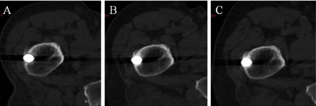
At the level of the lateral cortex, there was one mm gap between the edge of the cortical bone and the surface of the screw. This gap was no longer evident in the CT scan performed at twelve months FU and on the two years CT scan was even possible to observe an increasing integration. [Figure 5]
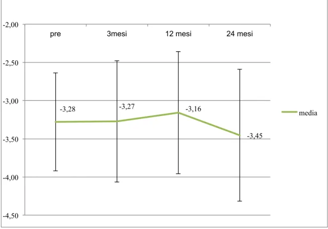
The mean DXA T-score of the reinforced hip at three months was -3.3 ± 0.8 at one year it was -3.2 ± 0.8, and at two years was -3.5 ± 0.9. In all FU the DXA T-scores were not statistically significant compared with the preoperative. [Graphic 1]
In every follow up the assessment of pain was performed with the VAS; at three months FU only one patient in group A had a VAS score of 8 but the pain was no more present at 12 months FU.
At the last FU, the assessment of the walk ability was performed with a score developed by our group: 10 points in case of walking without aids; 8 in case of walking with a stick; 7 with a crutch; 6 with two crutches; 4 with a walker; 2 if assisted by a care giver; and 0 if not able to walk. [Table 4]
All patients when admitted to hospital were not taking osteoporosis drugs.
16
17 patients (21.2%) died before the 24 months follow up: 9 in group A (52.9%) and 8 in group B (47.1%). Six patients died after the two years follow up: five in group A and one in Group B.
At the last follow up, 59 patients (73.7%) haven’t reported new falls; 21 patients (26.3%) reported one or more falls. [Table 5]
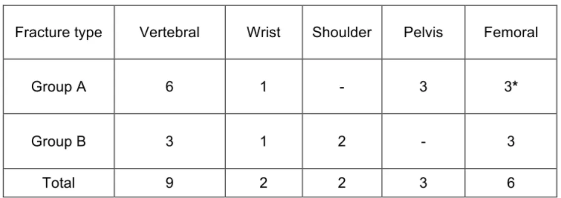
Of these patients, nine have reported one or more osteoporotic non-femoral fractures: six in group A (66.7%) and three in group B (33.3%).
In group A were recorded a wrist fracture, four vertebral collapse and three low energy pelvis fractures; in group B were recorded a wrist fracture, two humeral fractures and two vertebral collapse. [Table 6] Six patients: three for each group reported a second contralateral proximal femur fractures. In group A in all of the three patients was observed a below the trochanter fracture with the spiroid fracture rime to set off from the screw hole on the lateral cortex; all the patients experience the second fracture within a month after surgery. Only in one case the fracture was secondary to a fall; in the other 2 cases no falling were reported.
Two out of three patients were admitted in the hospital for a new fracture synthesis with PNS related plate. The third patient was operated in another hospital by removing the PNS.
All the three patients in group B reported a contralateral femoral fracture secondary to a new fall (two syntheses with intramedullary nail and an hemiarthroplasty were performed).
One patient in group A, in which was used the plate after the contralateral fracture, reported a non-union of the fracture with progressive varization of the proximal epiphysis of the reinforced femur; one year later a new surgery with explant of the proximal
17
femur was performed and was implanted an hemiarthroplasty with a revision stem.
On the epiphysis was performed a micro-tomographic analysis that showed the high rarefaction of trabecular bone of the femoral head and also highlights the osteointegration of the screw. [Figure 6]
Histological analysis confirmed the severe osteoporosis but however an important presence of newly formed bone on the thread of the screw that confirms osteointegration. [Figure 7]
18
4. DISCUSSION
Every year, there are over two million osteoporosis-related fractures in US, including hip, spine and wrist. Among these, the femoral neck fractures have the greatest significance in terms of morbidity and mortality; in addition, the direct and indirect costs of osteoporosis and related fractures are enormous. Due to the aging of the population by 2025, the direct annual cost of osteoporosis will reach over 25,3 billion dollars. Osteoporosis therefore has significant physical, financial and emotional consequences. [45]
Thus, strategies must be adopted to reduce osteoporosis-related fractures; currently the only strategies to decrease the incidence of fractures are pharmacological and non-pharmacological prevention. The device we developed for secondary prevention of femoral neck fractures may be a viable solution.
PNS resulted well tolerated: at one year FU no patients had pain in the reinforced hip and at 24 months FU, ROM of reinforced hip was wide and comparable to the not reinforced hip in group B.
Regards the walking ability, by summing the score according to the degree of independence during walking and dividing the score obtained for the number of patients per group, it resulted in a mean score of 5.6 in group A and 4,0 in group B. The PNS does not therefore affect walking ability of the patients.
The DXA examination carried out in the reinforced hip between the preoperative and the last FU performed at two years did not show a statistically significant difference.
The three months CT compared to those performed at twelve and 24 months showed good osteointegration and also no signs of sclerosis or osteolysis and that was also confirmed with the plan X rays of the
19
hip into the four regions of interest. The radiographic evaluation of neck-screw angle has not shown a correlation with the risk of fracture. The contralateral hip fractures in the reinforced side can be considered a technical error due to surgical instruments. In fact, during the surgery, in all three cases there was a difficult implant that likely resulted in the formation of micro-cracks that determined the fracture when the patients started to walk; another confirmation is the observation that the fracture line originated in all cases from the lower part of the screw hole on the lateral aspect of the femur.
If we exclude the two cases of spontaneous fracture of the femur, in the study group, there was just a fracture of the femur against the three occurred in the control group.
Limitations of this study were the lack of a system able to assess the strength of falls in order to understand when the energy of the fall was sufficient to cause a fracture in the reinforced and in the non-reinforced femur.
20
5. CONCLUSIONS
Surgical prevention could become a viable solution in the prevention of second femoral neck fracture in patients at risk. The right selection of patients is mandatory but also trials with a larger cohort should by designed in order to prove the efficacy of the treatment.
Regarding the device safety, it could be increased by performing some technical improvement on the instruments in order to avoid the risk of fracture in the reinforced femur.
21
6. FIGURES AND TABLES
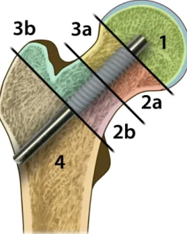
Figure 1: A. Two different PNS length; B. PNS positioned in the femoral neck; C. PNS with the related plate designed in case of sub-trochanteric fractures of the reinforced neck.
Figure 2: Draft of the four ROIs for the evaluation of osteolysis in the AP hip X ray.
C A
22
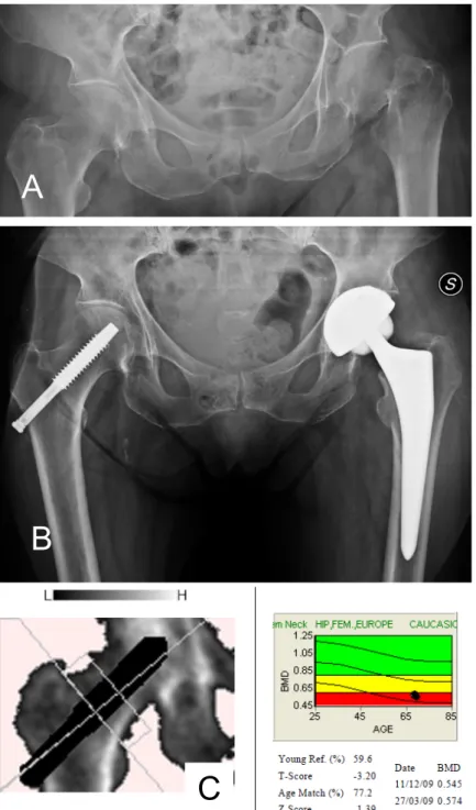
Figure 3: 67 year old female patient; A. AP pre-operative X ray shows a left femoral neck fracture; B. post op x ray: on the affected hip a total hip arthroplasty was performed and in the right size was performed the surgical reinforcement with the PNS; C. DXA shows a severe osteoporosis.
A
C
23
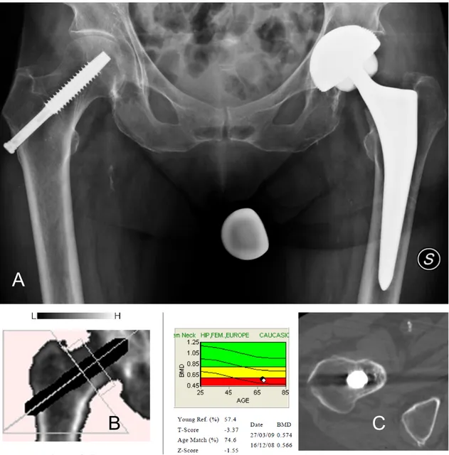
Figure 4: 12 months FU. A. No osteolisys are present in the reinforced neck. B. DXA shows a T-score slightly higher compared with the 3 months exam. C. CT assail slice shows good osteointegration.
A
24
Figure 5: CT slices of the same patient at different FU; A. Three months FU: there is a gap between the screw and the lateral cortex. B. 12 months FU: the gap although present is less evident. C: 24 months FU: the gap is no more present demonstrating the osteointegration capacity of the PNS.
Figure 6: Micro-tomographic analysis of the femoral head explanted during the revision surgery. The longitudinal cat on the major axis of the screw demonstrate the rarefaction of the cancellous bone.
25
Figure 7: Histological analysis of the femoral head explanted during the revision surgery. The pictures highlight osteointegration on the thread of the screw: A. 4x magnification; B. 10x magnification; C. 17x magnification. The presence of cartilage and osteoid shows also new bone formation.
26
Graphic 1: T score values (mean and SD) of the reinforced hip pre-operatively, three months, 12 months and 24 months.
N. Mean age Sex Pre-op. DXA Drugs taken at the H. admission Hospitalization days Patients 80 82,9±5,5 61 F; 19 M -3,28 ± 0,64 3,9 ± 2,5 12 ± 4 A (PNS) 46 (57.5%) 83 ±5,9 39 F; 7 M -3,3 ± 0,6 3,8 ± 2,5 11,9 ± 3,9 B (Control) 34 (42.5%) 82,9±4,9 22 F; 12 M -3,3 ± 0,7 3,9 ± 2,6 11,4 ± 4,3 Table 1: Patients enrolled in the study.
Surgery Arthroplasty Total Hip Hemiarthroplasty screw synthesis Cannulated Total
Group A 3 (6.5%) 34 (73.9%) 9 (19.6%) 46
Group B 2 (5.9%) 26 (76.4%) 6 (17.7%) 34
Amount 5 (6.3%) 60 (75%) 15 (18.7%) 80
Table 2: Surgery at the fractured hip.
-3,28 -3,27 -3,16 -3,45 -4,50 -4,00 -3,50 -3,00 -2,50 -2,00
pre 3mesi 12 mesi 24 mesi
27
Screw placement Valgus Neutral Varus
Patients 24 6 16
Neck-screw angle
(Mean SD) 5.8°± 3.5° - -4.8°± 2.8°
Table 3: Evaluation of neck-screw angle in patients underwent surgical reinforcement.
Walking ability Score Group A Group B
No aids 10 14 8 One cane 8 7 4 One crutch 7 2 6 Two crutches 6 3 1 Walker 4 7 2 Care giver 2 5 1
Not able to walk 0 8 12
Table 4: Walking ability of patients underwent surgical reinforcement at two years F.U.
Patients referred
one or more fall Total falls Total fractures
Non-femoral fractures
Group A 13 34 13 10
Group B 8 19 9 6
Total 21 53 22 16
Table 5: Patients that reported one or more falls after surgical reinforcement.
Fracture type Vertebral Wrist Shoulder Pelvis Femoral
Group A 6 1 - 3 3*
Group B 3 1 2 - 3
Total 9 2 2 3 6
Table 6: fractures divided by anatomical region.
28
7. REFERENCES
1. Seeman E. Pathogenesis of bone fragility in women and men. Lancet 2002; 359:1841-1850.
2. Turner CH. The biomechanics of hip fracture. Lancet 2005; 366:98–99. 3. Cullinane DM, Einhorn TA. Biomechanics of bone. In Bilezikian JP, Raisz
LG, Rodan GA (eds.) Principles of Bone Biology, 2nd ed., Academic Press, San Diego, CA, USA 2002; pp. 17–32.
4. Keaveny TM, Yeh OC. Architecture and trabecular bone-toward an improved understanding of the biomechanical effects of age, sex and osteoporosis. J Musculoskelet Neuronal Interact 2002; 2:205–208.
5. Seeman E, Delmas PD. Bone quality–the material and structural basis of bone strength and fragility. N Engl J Med 2006; 354:2250–2261.
6. Piscitelli P, Brandi ML, Tarantino U, Baggiani A, Distante A, Muratore M, Grattagliano V, Migliore, Granata M, Guglielmi G, Gimigliano R, Iolascon G. Incidenza e costi delle fratture di femore in Italia: Studio di estensione 2003-2005. Reumatismo, 2010; 62(2):113-118.
7. Italian Health Policy Brief June 2012
8. http://www.nof.org/professionals/clinical-guidelines.
9. Harvey N, Dennison E, Cooper C. Osteoporosis: impact on health and economics. Nat. Rev. Rheumatol. 2010; 6, 99-105.
10. Roberts SE, Goldacre MJ. Time trends and demography of mortality after fractured neck of femur in an English population, 1968-98: database study. BMJ. 2003 Oct 4;327(7418):771-5.
11. Colón-Emeric C, Kuchibhatla M, Pieper C, et al. The contribution of hip fracture to risk of subsequent fracture: Data from two longitudinal studies. Osteoporos Int. 2003;(14):879-883.
29
12. Cooper C. The crippling consequences of fractures and their impact on quality of life. Am J Med. 1997 Aug 18;103(2A):12S-17S; discussion 17S-19S.
13. Boston DA. Bilateral fractures of the femoral neck. Injury. 1982 Nov;14(3):207-10.
14. Lönnroos E, Kautiainen H, Karppi P, Hartikainen S, Kiviranta I, Sulkava R. Incidence of second hip fractures. A population-based study. Osteoporos Int. 2007 Sep; 18(9):1279-85.
15. Wells GA, Cranney A, Peterson J, Boucher M, Shea B, Robinson V, Coyle D, Tugwell P. Alendronate for the primary and secondary prevention of osteoporotic fractures in postmenopausal women. Cochrane Database Syst Rev 2008; (1):CD001155.
16. Wells G, Cranney A, Peterson J, Boucher M, Shea B, Robinson V, Coyle D, Tugwell P. Risedronate for the primary and secondary prevention of osteoporotic fractures in postmenopausal women. Cochrane Database Syst Rev 2008; (1):CD004523.
17. Black DM, Delmas PD, Eastell R, Reid IR, Boonen S, Cauley JA, Cosman F, Lakatos P, Leung PC, Man Z, Mautalen C, Mesenbrink P, Hu H, Caminis J, Tong K, Rosario-Jansen T, Krasnow J, Hue TF, Sellmeyer D, Eriksen EF, Cummings SR. Once-yearly zoledronic acid for treatment of postmenopausal osteoporosis. N Engl J Med 2007; 356:1809–1822.
18. MacLean C, Newberry S, Maglione M, McMahon M, Ranganath V, Suttorp M, Mojica W, Timmer M, Alexander A, McNamara M, Desai SB, Zhou A, Chen S, Carter J, Tringale C, Valentine D, Johnsen B, Grossman J. Systematic review: comparative effectiveness of treatments to prevent fractures in men and women with low bone density or osteoporosis. Ann Intern Med 2008; 148:197–213.
19. European Medicines Agency. European Public Assessment Report (EPAR) FORSTEO. EPAR summary for the public. (2009).
30
20. Stroup J, Kane MP, Abu-Baker AM. Teriparatide in the treatment of osteoporosis. Am J Health Syst Pharm 2008; 65:532–539.
21. Cauley JA, Robbins J, Chen Z, Cummings SR, Jackson RD, LaCroix AZ, LeBoff M, Lewis CE, McGowan J, Neuner J, Pettinger M, Stefanick ML, Wactawski-Wende J, Watts NB. Effects of estrogen plus progestin on risk of fracture and bone mineral density: the Women's Health Initiative randomized trial. JAMA 2003; 290:1729-1738.
22. Torgerson DJ, Bell-Syer SE. Hormone replacement therapy and prevention of nonvertebral fractures: a meta-analysis of randomized trials. JAMA 2001; 285:2891-2897.
23. Rossouw JE, Anderson GL, Prentice RL, LaCroix AZ, Kooperberg C, Stefanick ML, Jackson RD, Beresford SA, Howard BV, Johnson KC, Kotchen JM, Ockene J. Risks and benefits of estrogen plus progestin in healthy postmenopausal women: principal results from the Women's Health Initiative randomized controlled trial. JAMA 2002; 288:321-333. 24. Silverman SL, Chines AA, Kendler DL, Kung AWC, Teglbjaerg CS,
Felsenberg D, Mairon N, Constantine GD, Adachi JD, for the Bazedoxifene Study Group. Sustained efficacy and safety of bazedoxifene in preventing fractures in postmenopausal women with osteoporosis: results of a 5-year, randomized, placebo-controlled study. Osteoporos Int 2012 Jan;23(1):351-63.
25. Cummings SR, Ensrud K, Delmas PD, LaCroix AZ, Vukicevic S, Reid DM, Goldstein S, Sriram U, Lee A, Thompson J, Armstrong RA, Thompson DD, Powles T, Zanchetta J, Kendler, Neven P, Eastell R. Lasofoxifene in postmenopausal women with osteoporosis. N Engl J Med 2010; 362:686-696.
26. Barrett-Connor E, Mosca L, Collins P, Geiger MJ, Grady D, Kornitzer M, McNabb MA, Wenger NK. Effects of raloxifene on cardiovascular events and breast cancer in postmenopausal women. N Engl J Med 2006;
31
355:125-137.
27. Cummings SR, San Martin J, McClung MR, Siris ES, Eastell R, Reid IR, Delmas P, Zoog HB, Austin M, Wang A, Kutilek S, Adami S, Zanchetta J, Libanati C, Siddhanti S, Christiansen C. Denosumab for prevention of fractures in postmenopausal women with osteoporosis. N Engl J Med 2009; 361:756-765.
28. Papapoulos S, Bone HGI, Brandi ML, Brown JP, Chapurlat RD, Czerwinski E, Daizadeh N, Grauer A, Haller C, Krieg MA, Libanati C, Man Z, Mellstrom D, Radominski SC, Reginster JYL, Resch H, Roman Ivorra JA, Roux C, Cummings SR. Four years of denosumab exposure in women with postmenopausal osteoporosis: results from the first year extension of the FREEDOM trial. J Bone Miner Res. 2012 Mar;27(3):694-701.
29. McClung MR, Geusens P, Miller PD, Zippel H, Bensen WG, Roux C, Adami S, Fogelman I, Diamond T, Eastell R, Meunier PJ, Reginster JY. Hip Intervention Program Study Group. Effect of risedronate on the risk of hip fracture in elderly women. Hip Intervention Program Study Group. N Eng J Med 2001;344:333-340.
30. Järvinen TL, Michaëlsson K, Jokihaara J, Collins GS, Perry TL, Mintzes B, Musini V, Erviti J, Gorricho J, Wright JM, Sievänen H. Overdiagnosis of bone fragility in the quest to prevent hip fracture. BMJ. 2015 May 26;350:h2088.
31. Black DM, Delmas PD, Eastell R, Reid IR, Boonen S, Cauley JA, Cosman F, Lakatos P, Leung PC, Man Z, Mautalen C, Mesenbrink P, Hu H, Caminis J, Tong K, Rosario-Jansen T, Krasnow J, Hue TF, Sellmeyer D, Eriksen EF, Cummings SR. Once-yearly zoledronic acid for treatment of postmenopausal osteoporosis. HORIZON Pivotal Fracture Trial. N Eng J Med 2007;356(18):1809-1822.
32
following fractured neck of femur: a survey of orthopaedic surgeons practice. Ir Med J. 2000Jun;93(4):105-7.
33. Parker MJ, Gillespie WJ, Gillespie LD. Hip protectors for preventing hip fractures in older people. Cochrane Database SystRev 2005;20: CD001255.
34. Holzer G, Von Skrbensky G, Holzer LA, Pichl W. Hip fractures and the contribution of cortical versus trabecular bone to femoral neck strength. J Bone Miner Res. 2009 Mar;24(3):468-74.
35. de Bakker PM, Manske SL, Ebacher V, Oxland TR, Cripton PA, Guy P. During sideways falls proximal femur fractures initiate in the superolateral cortex: evidence from high-speed video of simulated fractures. J Biomech. 2009 Aug 25;42(12):1917-25.
36. Lotz JC, Cheal EJ, Hayes WC. Stress distributions within the proximal femur during gait and falls: implications for osteoporotic fracture. Osteoporos Int. 1995;5(4):252-61.
37. Verhulp E, van Rietbergen B, Huiskes R. Load distribution in the healthy and osteoporotic human proximal femur during a fall to the side. Bone. 2008 Jan;42(1):30-5.
38. Crockett G.S. Osteoporosis in the elderly. Br J Clin Pract. 1960 May; 14:385-90.
39. Heini PF, Franz T, Fankhauser C, Gasser B, Ganz R. Femoroplasty-augmentation of mechanical properties in the osteoporotic proximal femur: a biomechanical investigation of PMMA reinforcement in cadaver bones. Clin Biomech (Bristol, Avon). 2004 Jun;19(5):506-12.
40. Sutter EG, Mears SC, Belkoff SM. A biomechanical evaluation of femoroplasty under simulated fall conditions. J Orthop Trauma. 2010 Feb;24(2):95-9. doi: 10.1097/BOT.0b013e3181b5c0c6.
41. Beckmann J, Ferguson SJ, Gebauer M, Luering C, Gasser B, Heini P. Femoroplasty-augmentation of the proximal femur with a composite bone
33
cement-feasibility, biomechanical properties and osteosynthesis potential. Med Eng Phys. 2007 Sep;29(7):755-64.
42. De Bakker PM, Guy P, Fernlund G, Oxland TR. P452SU. Prophylactic augmentation of the contralateral femur: A finite element study. Osteoporos Int. 2006 17 (Suppl 2).
43. Szpalski M, Gunzburg R, Aebi M, Delimoge C, Graf N, Eberle S, Vienney C. A new approach to prevent contralateral hip fracture: Evaluation of the effectiveness of a fracture preventing implant. Clin Biomech (Bristol, Avon). 2015 Aug;30(7):713-9.
44. F. Laurenza, A. Lispi, P. Ruo CCD and femoral off-set: a primery of orthopaedic biomechanics after Total Hip Arthroplasty. G.I.O.T. 2001;27:168-72.
45. Crockett G.S. Osteoporosis in the elderly. Br J Clin Pract. 1960 May; 14:385-90.