UNIVERSITÀ DEGLI STUDI DI ROMA
"TOR VERGATA"
FACOLTA' DI MEDICINA E CHIRURGIA
DOTTORATO DI RICERCA IN
MICROBIOLOGIA MEDICA ED IMMUNOLOGIA
XXII CICLO
CHARACTERIZATION OF HIV-1 POL SEQUENCES IN NEWLY HIV-1 DIAGNOSED PATIENTS:
IDENTIFICATION OF DRUG RESISTANCE MARKERS AND THEIR INVOLVEMENT IN THE TRANSMISSION EVENTS
Dottorando: Claudia Alteri
A.A. 2009/2010
Docente Guida: Prof. Carlo Federico Perno
Tutor: Dott.ssa Valentina Svicher
Italian Abstract
Ad oggi sono disponibili 25 farmaci per il trattamento dell’infezione da HIV. L’uso combinato di tali farmaci, la cosiddetta Highly Active Antiretroviral Therapy (HAART), ha consentito di migliorare le prospettive e la qualità di vita dei pazienti, rendendo possibile la soppressione della replicazione virale nella maggior parte dei soggetti infetti.
Tuttavia, se la terapia non riesce a sopprimere adeguatamente il virus, si osserva l’insorgenza di ceppi virali farmaco-resistenti in grado di compromettere inesorabilmente l’efficacia dei farmaci stessi. Attualmente, la presenza di virus farmaco-resistenti è messa in evidenza attraverso il test di resistenza genotipica che prevede il sequenziamento dei geni bersaglio dei farmaci antiretrovirali. Recentemente, sono state inoltre messe a punto delle metodiche in grado di rilevare ceppi resistenti anche in forma minoritaria, presenti cioè con una prevalenza inferiore al 20% nell’intera popolazione virale, il cui utilizzo nella pratica clinica è però ancora compromesso dai costi elevati. La più nota tra tutte è sicuramente il pyrosequencing.
Un problema strettamente connesso all’insorgenza di ceppi resistenti è rappresentato dalla loro trasmissione (sia come specie predominante che in forma minoritaria) a soggetti HIV-infetti che non hanno mai assunto alcun farmaco antiretrovirale (definiti drug-naïve). Attualmente, nei paesi occidentali, la prevalenza di virus resistente nei pazienti drug-naïve è stimata intorno al 10%, e diversi studi hanno evidenziato come tali ceppi (anche quando presenti in specie minoritarie) possano compromettere il successo della prima linea terapeutica.
In base a quanto riportato, l’obiettivo di questa tesi è stato quindi quello di caratterizzare la distribuzione e la diffusione della resistenza trasmessa in Italia, con particolare attenzione alle dinamiche di trasmissione della stessa e al ruolo che i soggetti drug-naïve rivestono in questo fenomeno.
Un ulteriore obiettivo è stato quello di definire nuovi marcatori genetici di HIV rilevabili attraverso metodiche standard di sequenziamento in grado di predire la presenza di varianti minoritarie resistenti nella popolazione virale. L’individuazione di mutazioni “sentinella” in grado di predire la presenza di quasispecie minoritarie resistenti riveste un ruolo centrale nel miglioramento dell’interpretazione genotipica e nella selezione di pazienti potenzialmente portatori di farmaco-resistenza.
Analizzando 255 pazienti di nuova diagnosi e naïve alla terapia antiretrovirale, abbiamo riscontrato che il 5,9% di questi mostrava resistenza trasmessa; in particolare il 3,9% era resistente alla classe farmacologica degli analoghi nucleosidici della trascrittasi inversa (NRTI), il 3,5% alla classe dei non-NRTI e lo 0,4% alla classe degli inibitori della proteasi (PI). Il 3,5% dei pazienti era inoltre portatore di un virus resistente a più classi farmacologiche. Comparando pazienti con infezione recente a pazienti con infezione cronica, abbiamo riscontrato che la prevalenza di virus farmaco-resistente era leggermente più bassa nella prima categoria rispetto alla seconda (3,4% versus 6,6%,
benché la fonte principale di trasmissione di virus farmaco-resistente fosse rappresentata dai pazienti in fallimento terapeutico, ben il 38,5% dei virus resistenti figurava in clusters composti esclusivamente da individui drug-naïve con infezione sia recente che cronica, sostenendo il ruolo di tali pazienti quale ulteriore fonte di farmaco-resistenza. Nella terza parte di questa tesi abbiamo identificato alcune mutazioni nella trascrittasi inversa di HIV, la cui presenza nel test di resistenza genotipico correlava in modo significativo con specie minoritarie farmaco resistenti. In particolare, l’analisi di di 40 pazienti drug-naïve ha mostrato come il 70% di pazienti con la mutazioni L210M presentasse specie minoritarie farmaco-resistenti (P=0.03), supportando il ruolo di tale mutazione come “sentinella” di farmaco-resistenza nascosta.
In conclusione, questa tesi ha consentito di caratterizzare la resistenza trasmessa e le dinamiche di trasmissione di ceppi HIV-farmacoresistenti in Italia. Queste informazioni sono di fondamentale importanza per impostare programmi di sorveglianza e prevenzione dell’infezione da HIV. Tale tesi ha portato inoltre all’identificazione di mutazioni “sentinella” in grado di predire la presenza di specie minoritarie resistenti non rilevabili dai test genotipici utilizzati comunemente nella pratica clinica. Ciò è di fondamentale importanza per ottimizzare la gestione clinica del pazienti infetto da HIV.
English Abstract
To date 25 drugs are available for the HIV-1 treatment. The combined use of these drugs, known as Highly Active Antiretroviral Therapy (HAART) has successfully suppressed the HIV-1 replication and has dramatically improved the prognosis of HIV-1 infected patients.
However, if viral rebound occurs during therapy, viruses with mutations conferring drug resistance can be selected. To date, drug resistant viruses can be detected by the genotypic resistance test, based on the sequencing of the genes target of the antiretroviral therapy. Recently, new assays (as ultra deep pyrosequencing) have allowed to assess antiretroviral drug resistance even when present as minority species (with a prevalence <20% of the entire viral population). However, their use is still limited in clinical practice due to their high cost.
The frequently selection of drug resistant strains in treatment failing patients can in turn increase the risk of their transmission (both in predominant and both in minority species) to HIV-1 infected individuals, naïve to the antiretroviral drugs (drug-naïve). To date, in countries with a wide access to HAART, the prevalence of drug resistant strains in drug-naïve patients is around 10%, and many studies have demonstrated that these drug resistance strains (also in minority species) are associated with an increased probability of virological failure to the first-line antiretroviral therapy.
In this light, the aim of this thesis was to characterize the distribution and the spread of HIV-1 drug resistance in Italy, with particular attention to the population dynamics of transmitted resistance and the role of untreated patients in the spread of drug-resistance. The last objective of this thesis was to define new genetic markers of HIV that can predict the presence of transmitted drug resistant minority species. The identification of these “sentinel” mutations could improve the genotypic interpretation and could help the selection of patients potentially reservoir of drug-resistance.
Among 255 newly diagnosed and untreated individuals, the 5.9% of patients showed signs of transmitted resistance; in particular, 3.9% of patients was infected with nucleoside reverse-transcriptase inhibitors (NRTI)-resistant viruses, 3.5% with non-NRTI-resistant viruses and 0.4% with protease inhibitors-resistant viruses. In addition, the 3.5% of patients carried HIV-1 resistant strains with more than one major drug resistance mutation. Comparing chronic infections with recent infections, we also found a decreased rate of resistance in recent infections (P=0.09), reflecting the increasing use of potent drugs and highly active antiretroviral regimens in recent years. Homosexual individuals were also more likely to harbour a virus with at least one primary resistance mutation (OR 7.7; 95% CI: 1.7–35.0, P=0.008).
In the second part of this thesis, we have investigated the epidemiological networks characterizing the HIV-1 infection by using a phylogenetic approach in a cohort of 884 patients. Of them, 306 were drug-naïve and 578 were HAART-treated individuals. Even if patients failing HAART remained the principal source of transmitted drug resistance,
(P=0.03), supporting the role of this mutation as “sentinel” of hidden drug resistance mutations.
In conclusion, this thesis was able to characterize the HIV-1 transmitted drug resistance and the transmission dynamics of HIV-1 drug resistant strains in Italy. These data were essential to improve the surveillance and the preventive programmes on HIV infections. This thesis has also characterized “sentinel” mutations able to predict minority drug resistance variants, undetectable by standard genotypic tests. Taking into account these genetic markers right from diagnosis can help clinicians to improve the management of HIV-1 infected patients.
Keywords: HIV-1; drug naïve patients; transmission of drug resistance; drug resistance markers
Contents
1 Introduction
1.1 HIV and the retroviruses 1
1.1.1 Morphology 1
1.1.2 Genome 2
1.1.3 Replication 4
1.1.4 Pathogenesis 11
1.1.5 Epidemiology 14
1.2 Drug resistance development
1.2.1 Antiretroviral treatment 16
1.2.2 Drug resistance 18
1.2.3 Mechanism of drug resistance 20
1.2.4 Combination therapy 27
1.2.5 Detection of drug resistance 28
1.3 Transmission of HIV-1 drug resistance
1.3.1 Evolution of drug resistance in treatment naïve patients 31 1.3.2 Mechanism of transmission of drug resistant virus 32 1.3.3 Estimation rate of transmission of drug resistant virus 36 1.3.4 Clinical implications of transmitted drug resistance 39
1.4 Rationale of the work 41
2 Methods
2.1 Characterization of the patterns of drug resistance mutations in
newly diagnosed HIV-1 infected patients naïve to the antiretroviral drugs 44
2.1.1 Study population 44
2.1.2 The IgG avidity assay 44
2.1.3 HIV sequencing 45
2.1.4 Phylogenetic analysis 46
2.1.5 Determination of drug resistance mutations 47
2.1.6 Statistical analysis 47
2.2 Epidemiological network analysis in HIV-1 B infected patients
diagnosed in Italy between 2000-2008 49
2.2.1 Study population 49
2.3.3 Determination of drug resistance minority variants 56
2.3.4 Statistical analysis 57
3 Results
3.1 Characterization of the patterns of drug resistance mutations in
newly diagnosed HIV-1 infected patients naïve to the antiretroviral drugs 58
3.1.1 Study population 58
3.1.2 Circulation of HIV-1 subtypes in newly diagnosed patients in Italy 59 3.1.3 Prevalence of major mutations associated with drug resistance 64 3.1.4 Association among drug resistance mutations in newly diagnosed
HIV-1 infected patients 65
3.2 Epidemiological network analysis in HIV-1 B infected patients
diagnosed in Italy between 2000-2008 68
3.2.1 Study population 68
3.2.2 Phylogenetic analysis 69
3.2.3 Identification of antiretroviral drug resistant HIV-1 lineages
in drug naïve patients 72
3.2.4 Description of the major lineages 75
3.2.5 Timing of origin of the major lineages 79 3.3 “Sentinel” mutations in standard population sequencing can predict
the presence of RT major mutations detected only by 454-pyrosequencing 80
3.3.1 Study population 80
3.3.2 Coverage profile and UDPS results 82
3.3.3 Role of atypical RT mutations in predicting minority resistance variants 88
3.3.4 Response to HAART 91
4 Discussion
4.1 Characterization of the patterns of drug resistance mutations in
newly diagnosed HIV-1 infected patients naïve to the antiretroviral drugs 92 4.2 Epidemiological network analysis in HIV-1 B infected patients
diagnosed in Italy between 2000-2008 96
4.3 “Sentinel” mutations in standard population sequencing can predict
the presence of RT major mutations detected only by 454-pyrosequencing 100
5 Conclusion 104
6 References 106
1 Introduction
1.1 HIV and the retroviruses
The human immunodeficiency virus (HIV), identified in 1983, is a member of the Lentiviruses genus which are exogenous, non-oncogenic retroviruses causing persistent infections leading to chronic diseases with long incubation periods (lenti for slow). Like the human T-cell leukemia virus (HTLV) family of primate onco-retroviruses, lentiviruses are complex retroviruses (Cullen, 1991). The significant characteristic of the complex retroviruses is the ability to regulate their own expression via virally encoded protein factors not found in other retroviruses. This property has been proposed to be essential for the long-term association of the complex retroviruses with the host and the generation of chronic active infections. The lentiviral complexity is reflected in their replication cycle, which reveals intricate regulatory pathways, unique mechanisms for viral persistence (Tang et al., 1999) and the ability to infect non-dividing cells.
1.1.1 Morphology
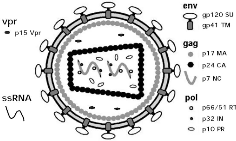
The HIV virion is a spherical virus particle of about 100 nm in diameter (Fig. 1.1). The viral envelope consists of a lipid bilayer derived from the host cell membrane during release of the newly produced particles from an infected cell. Embedded in the viral envelope are proteins from the host cell as well as viral protein complexes composed of the transmenbrane glycoprotein gp41 (TM) and the surface glycoprotein gp120 (SU).
shaped capsid. The capsid is made of ca. 2000 copies of the viral capsid protein p24 (CA). It encloses two single strands of the HIV RNA genome stabilized as a ribonucleoprotein complex with ca. 2000 copies of the nucleocapsid protein p7 (NC). Additionally, the capsid contains the three virally encoded enzymes, reverse transcriptase, protease, and integrase as well as accessory proteins such as nef, vif, vpr. There are three additional accessory proteins rev, tat, vpu, that are not packaged into the virion.
1.1.2 Genome
The genome of HIV has a length of approximately 9.2 kbp. Like all retroviruses it contains the characteristics: 5’- gag – pol – env - 3’ motif consisting of the three structural genes gag, pol, and env (Fig. 1.2). The Gag (group antigen) gene encodes the large precursor polyprotein p55 that is cleaved in four proteins: the matrix p17, the "core" capsid p24, the nucleocapsid p7 and the p6 (Freed, 1998). The pol (polymerase) gene encodes the synthesis of three viral enzymes: protease p10, reverse transcriptase/ribonuclease H complex p51 and p66, integrase p32. The env (envelope) gene directs the production of an envelope precursor protein gp160, which undergoes cellular proteolytic cleavage into the outer envelope glycoprotein gp120 and the transmembrane glycoprotein gp41.
The RNA genome is flanked by two short redundant (R) sequences at both termini with adjacent unique sequences, U5 and U3, found at the 5’ and 3’ ends, respectively. In addition, HIV has at least six more genes encoding viral proteins with regulatory functions (tat and rev) or accessory functions (nef, vif, vpr and vpu) (for reviews Cullen, 1998; Emerman and Malim, 1998; Kjems and Askjaer, 2000; Piguet and Trono,
Figure 1.1. HIV-1 mature virion structure. Modified from WebPath resource collection (Klatt, 2000).
Typical lentivirus particles are spherical, about 80-110 nm in diameter, and consist of a lipid bilayer membrane surrounding a conical core. The two identical singlestranded RNA (ssRNA) molecules, of about 9.2kB each, are associated with the nucleocapsid proteins p7gag (NC). They are packed into the core along with virally encoded enzymes: reverse transcriptase (RT), integrase (IN), and protease (PR). P24gag comprises the inner part of the core, the capsid (CA). The p17gag protein constitutes the matrix (MA) which is located between the nucleocapsid and the virion envelope. The viral envelope is produced by the cellular plasma membrane and contains the protruding viral Env glycoproteins: gp120 surface glycoprotein (SU) and gp41 transmembrane protein (TM). Among the accessory proteins encoded by HIV-1, certainly Vpr and perhaps Nef and Vif are packaged into virions, although the precise location have not yet been elucidated. Neither the other accessory protein Vpu nor the regulatory proteins Tat and Rev have been detected in virion particles.
Figure 1.2. HIV genomic organization. Like all other retroviruses, HIV has three structural genes gag,
pol and env (heavily shaded), which are flanked by the long terminal repeats (LTR’s). In addition it has six more genes, including two regulatory genes tat and rev (stippled) and four accessory genes nef, vif, vpr and vpu (white).
1.1.3 Replication
The HIV replication cycle begins with the recognition of the target cell by the mature virion. The major targets for HIV infection are cells bearing the HLA class II receptor, CD4, on their cell surfaces. These include T-helper lymphocytes and cells of the monocyte/macrophage lineage including microglia cells in the brain. The virus-CD4 binding occurs via specific interactions between the viral outer envelope glycoprotein gp120 and the amino-terminal immunoglobulin like domain of CD4 (Dalgleish et al., 1984; Klatzmann et al., 1984). These interactions are sufficient for binding but not for infection. Subsequently the virus glycoprotein gp120 interacts with additional cell-surface proteins to promote fusion of the viral and cellular membranes. These coreceptors have recently been identified to be members of the chemokine receptor family and include CXCR4 and CCR5 (Alkhatib et al., 1996; Deng et al., 1996; Doranz et al., 1996; Moore, 1997). The initial binding of HIV to the CD4 receptor is mediated by conformational changes in the gp120 subunit, followed by a conformational change in the gp41 subunit, induced by the chemokine receptors, that allows fusion and subsequent entry of HIV. Various strains of HIV differ in their use of chemokines coreceptors. There are strains of HIV known as T-tropic strains, which selectively interact with the CXCR4 chemokine coreceptor of lymphocytes, while M-tropic strains of HIV interact with the CCR5 chemokine coreceptor of macrophages and dual tropic HIV strains that infect both cell types (Littman, 1998; Moore, 1997). HIV-1 infection of CD4 negative cells, such as neural cells, has also been reported (Clapham et al., 1989; Kozlowski et al., 1991) but the mechanisms of HIV entry are still unclear. Membrane fusion is followed by an uncoating event that allows the intracellular reverse transcription. The viral RNA is transcribed in the cytosol into double stranded DNA by
the reverse transcriptase (Hansen et al., 1987; Muesing et al., 1985). This enzyme have three enzymatic activities: RNA-dependent DNA polymerase, DNA-dependent DNA polymerase, and ribonuclease H (RNase H). The reverse transcription process takes place within a large nucleic acid-protein complex known as the preintegration complex (PIC) by the assistance of the accessory protein Vif (von Schwedler et al., 1993) and the nucleocapsid protein NC (Darlix et al., 1993). Once synthesized, the viral DNA is transported to the nucleus of the infected cell as part of the PIC that appears to include tightly condensed viral nucleic acids and the integrase, p17, reverse transcriptase, and Vpr proteins. In contrast to other retroviruses, that require cell division and concomitant breakdown of the nuclear envelope to gain access to the nuclear compartment, the lentiviral PIC is actively imported into the nucleus during the interphase (Bukrinsky et al., 1992). Nuclear import of the PIC seems to be directed by the accessory protein Vpr (Fouchier et al., 1998; Heinzinger et al., 1994), the Gag matrix protein p17 (Bukrinsky et al., 1993; von Schwedler et al., 1994) and the integrase (Gallay et al., 1997). Vpr does not contain a conventional nuclear localization signal but appears to function by connecting the PIC to the cellular nuclear import machinery (Fouchier et al., 1998; Popov et al., 1998a; Popov et al., 1998b). The ability of lentiviruses such as HIV-1 to utilize active transport mechanisms for translocation of the PIC into the nucleus, allows these viruses to infect non-dividing cells such as differentiated macrophages, quiescent T lymphocytes and possibly neurons. In the nucleus, integrase catalyzes covalent integration of the viral DNA into the host genome, where it resides permanently as a
The regulation of the HIV transcription involves a complex interplay between cis-acting DNA and RNA elements present within the chromatin-associated proviral LTRs, cellular transcription factors and the viral regulatory protein Tat (transcriptional transactivator).
The regulation of the HIV transcription involves a complex interplay between cis-acting DNA and RNA elements present within the chromatin-associated proviral LTRs, cellular transcription factors and the viral regulatory protein Tat (transcriptional transactivator). In an arrangement similar to that of several inducible cellular promoters, the HIV-1 promoter, which is located in the U3 region of the 5’LTR, contains a TATA box and binding sites for several cellular DNA-binding transcription factors, such as NF-kB, Sp1 and TBP (Jones and Peterlin, 1994). It is highly inducible and responds to the activation status of the infected cell. NF-kB is the major inducible cellular activator. It is well established that many cells in the lymphoid tissue of infected individuals are latently infected (Pantaleo et al., 1993), even though the viral replication in the body is always active. In resting T-cells, the activity of the HIV promoter is minimal, leading to viral quiescence in infected primary cells. Therefore, viral activation is associated with cell activation. The transcription of the provirus by the cellular RNA polymerase II results in a primary transcript that may serve three distinct functions: 1) it constitutes genomic RNA that is incorporated into the virion; 2) it serves as template for translation (Gag and Gag-Pol); 3) it functions as the precursor RNA for the production of diverse subgenomic mRNAs (Fig 1.3).
As mentioned before, HIV encodes two essential regulatory proteins Tat and Rev, which increase viral gene expression at the transcriptional and post-transcriptional levels, respectively. HIV mRNA expression is biphasic and can be divided into early
(Rev-independent) and late (Rev-dependent) stages (Kim et al., 1989). First, shortly after the infection of cells, multiply spliced (~ 2kb) RNA species are formed from the primary transcript and three proteins are produced: Tat, Rev and Nef, therefore referred as early gene products (Schwartz et al., 1990). Tat (for reviews see Cullen, 1998; Emerman and Malim, 1998; Rubartelli et al., 1998), greatly increases transcription from the HIV promoter, by binding to a cis-acting target sequence, the trans-activator response element (TAR), which is located at the 5’ end of the nascent viral RNA transcript (Berkhout et al., 1989; Dingwall et al., 1989). Tat recruits two cellular factors to this complex: cyclin T and cyclin-dependent protein kinase-9 (Cdk9).
Cyclin T is proposed to bind directly Tat and to increase its affinity for the TAR RNA, while Cdk9 phosphorylates the RNA polymerase II transcription complex and thus stimulates transcriptional elongation (Wei et al., 1998). Rev (regulator of expression of the virion), which accumulates during the early phase of expression, initiates late gene expression by binding a unique RNA element located in the env coding region of HIV-1, the so called Rev-responsive element (RRE). This interaction promotes the stability and transport of unspliced (~ 9 kb) and partially spliced (~4 kb) HIV-1 mRNAs out of the nucleus. These mRNAs are responsible for the production of the viral enzymes and structural proteins (Daly et al., 1989; Hadzopoulou-Cladaras et al., 1989; Malim et al., 1989). Therefore Gag, Pol, Env, Vif, Vpr, and Vpu proteins are referred to as late HIV-1 proteins.
The Env precursor polyprotein (gp160) is synthesized in the endoplasmatic reticulum (ER) where it is glycosylated and appears to oligomerize to a trimeric structure posttranslationally (Wyatt and Sodroski, 1998). Thereafter, it is cleaved to produce the non-covalently associated (gp41 TM - gp120 SU) 3 trimeric glycoprotein complex, which is transported to the cell membrane for virus assembly. Vpu is thought to enhance this process and inhibit a premature trapping of CD4 to Env in the ER by binding CD4 molecules, which are also synthesized in the ER, and directing them to the ubiquitin-proteasome degradation pathway (Margottin et al., 1998; Schubert et al., 1998; Strebel et al., 1988; Willey et al., 1992a, 1992b). Similarly, the accessory protein Nef facilitates the routing of CD4 from cellsurface and Golgi apparatus to lysosomes, resulting in endosomal degradation and preventing inappropriate interaction with Env (Aiken et al., 1994). In addition, both Vpu and Nef can down-regulate expression of MHC class I molecules. The downregulation of CD4 and MHC class I molecules on the surface of infected cells also helps infected cells to evade immune responses of the host, such as killing by cytotoxic T lymphocytes (Collins et al., 1998; Kerkau et al., 1997).
During synthesis of the Gag polyprotein by ribosomes, a translational frameshift may occur, resulting in generation of smaller amount of Gag-Pol precursor polyproteins, which associate with the Gag polyprotein at the cellular membrane. The N-terminally myristoylated MA domain of the Gag/GagPol polyproteins directs insertion of the Gag precursors into the cellular membrane and interacts with the cytoplasmic tail of gp41 resulting in the anchoring of Env to the viral particle (Dorfman et al., 1994). Approximately 1200 to 2000 copies of Gag precursor bud to form an immature particle, which encapsidates two copies of the unspliced viral RNA genome, by the ability of NC to interact with nucleic acids. Vif and Vpu proteins have been reported to play a role in
packaging of the nucleoprotein core and in virion release, respectively (Hoglund et al., 1994; Lamb and Pinto, 1997). Concomitantly or immediately following the external budding, the cleavage of the Gag/Gag-Pol polyproteins by the virally encoded PR produces the structural proteins MA, CA, NC as well as the independent enzymes PR, RT and IN. This final step primes new virus particles for the next round of infection and is termed maturation.
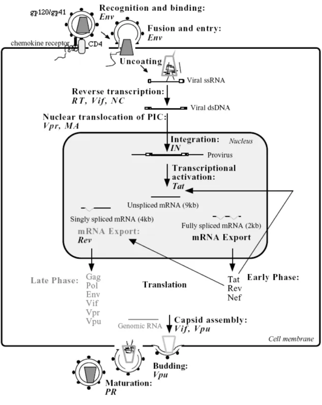
Figure 1.3. Replication cycle of HIV-1. Modified from Ceccherini-Silberstein, 2001
(http://edoc.ub.Muenchen.de/archive/00000533/01/Ceccherini-Silberstein_Francesca.pdf). Each fundamental step is presented in bold. Names in italic refer to viral gene products involved in the specific
steps. HIV-1 gene expression is stimulated by HIV-1 Tat and Rev, which act at transcriptional and post-transcriptional levels, respectively, and can be divided into two phases. The early phase is Rev-independent and the later phase is Rev-dependent (text in gray). Rev stabilizes and mediates export of singly spliced and unspliced RNA transcripts out of the nucleus into the cytoplasm.
1.1.4 Pathogenesis AIDS
HIV infection has been associated with the acquired immunodeficiency syndrome (AIDS). A diagnosis of AIDS is made whenever a person is HIV-positive and have:
• CD4+ T cell count below 200 cells/mms;
• CD4+ T cells account for fewer than 14% of all lymphocytes;
• Diagnosis with one or more of the 25 AIDS defining illness, including various opportunistic infection, brain and nerve disease, certain cancers, and wasting syndrome.
Approximately 10% of HIV-infected patients progress to AIDS within the first 2 to 3 years of infection, while for approximately 40% this progression is observed over a period of 10 years. 10% to 17% of HIV-infected patients may be AIDS free, some with no evidences of disease progression. These variations in responses may be due to differences in the degree of stimulation of the immune system by infection with the other pathogens as well as to viral factor, such as deletions in the nef gene or altered cell tropism (Kupfer et al., 1998).
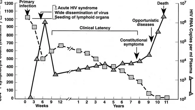
Course of infection
Schematically, the course of infection can be divided into an acute, an asymptomatic, and symptomatic phase (Fig. 1.4). The acute phase accounts for the first 5-10 weeks of infection and is characterized by high virus production, and activation of lymphocytes in lymphonodes. Up to 5x10³ infectious particles per ml of blood plasma may be found
steady rate, while virus replication remains constant at a low rate. The duration of the asymptomatic phase may last between 2 and 20 years. The end stage of disease, when the patient develops AIDS, is characterized by CD4+ cells count below 200 copies/ml and increased quantities of the virus. The number of CD8+ cytotoxic lymphocytes also decreases and lymphoid cells and tissues are damaged.
CD4+T cell depletion
The hypothesis that CD4+ cell depletion is caused the lysis of infected cells during viral replication has been supported by the observation of an immediate and large increase of CD4+ count after the initiation of antiretroviral therapy that blocks viral replication (Ho et al., 1995; Wei et al., 1995). This hypothesis has not withstood more detailed analyses of T cell dynamics (Roederer et al., 1998). In fact, it has been turned out that in HIV-infected patients all T cell subset are progressively destroyed, irrespective of CD4+ expression, and AIDS appear to be a disease of perturbed homeostasis. Many pathogenetic mechanisms have been proposed, including viral gene products, syncitium formation, direct virus killing of cell, apoptosis, autoimmunity, cytokine and chemokines expression, superantigens, virus directed cell mediated cytolysis and disruption of lymphoid architecture.
1.1.5 Epidemiology
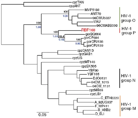
Several African primates harbour lentiviruses and HIV is believed to be entered the human population in Africa by zoonotic transmission of SIVcpz from chimpanzee population. The first cross species transfer has been estimated to have occurred between 1915 and 1941 (Korber et al., 2000). Two types of HIV are known: the most common HIV-1, which is responsible to the world-wide AIDS epidemic and the immunologically distinct HIV-2 (Clavel et al., 1986), which is much less common and less virulent (Ariyoshi et al., 2000), but produces clinical findings similar to HIV-1 (Wilkins et al., 1993). The HIV-1 type itself includes four groups M, N, O, P which have different geographic distributions but all produce similar clinical symptoms (Fig. 1.5). The M group is further divided into 9 pure subtypes (A, B, C, D, F, G, H, J, K), 4 sub-subtype (A1, A2, F1, F2) and 45 circulating recombinant forms on the basis of phylogenetic analysis. Almost all subtypes are present in Africa, while in Europe, North America, and Australia subtype B is more dominant, and subtype C is more common in Asia (McCutchan, 2000; Robertson et al., 2000).
At the end of 2009, 33.4 million adults and children have been estimated to live with HIV/AIDS, most of them in Sub-Saharan Africa and South East Asia (Fig. 1.6). Only a minority of HIV-infected individuals live in the industrialized countries and has access to the anti-HIV drugs and professional health care.
Figure 1.5. Phylogenetic relationship of primate lentiviruses. Phylogenetic tree derived from the
alignment of pol gene sequences of HIV-1 and SIV strain (SIVcpz and SIVgor).Reproduced from Plantier et al., 2009.
1.2 Drug resistance development
1.2.1 Antiretroviral treatment
Anti-retroviral (ARV) agents target different stages in the life cycle of HIV, the binding to cells expressing CD4 receptor, fusion (viral and cellular membrane fusion), reverse transcription of viral RNA to DNA, integration of viral DNA into cellular DNA, transcription of proviral DNA into viral RNA, protein synthesis from the HIV RNA, post translational modification of proteins through proteolytic cleavage, virus assembly and budding from cells. The reverse transcriptase and subsequently the protease enzymes have been the original targets of anti-HIV therapy with fusion inhibitors introduced as a reserve agent for highly treatment- experienced patients (enfuvirtide or T20 approved in March 2003). These drugs were followed by the introduction of integrase inhibitors such as raltegravir, that inhibits the strand transfer process, and the chemokine receptor antagonists, such as maraviroc, that disrupt binding of HIV-1 to either CCR5 or CXCR4 co-receptors, both approved in 2007.
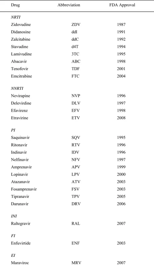
In table 1.2 all anti-HIV compounds currently approved for clinical use by the U.S. Food and Drug Administration (FDA) [Division of AIDS, National Institute of Allergy and Infectious Diseases, National Institutes of Health] are listed.
Table 1.1. Antiretroviral drugs in clinical use.
Drug Abbreviation FDA Approval
NRTI Zidovudine ZDV 1987 Didanosine ddI 1991 Zalcitabine ddC 1992 Stavudine d4T 1994 Lamivudine 3TC 1995 Abacavir ABC 1998 Tenofovir TDF 2001 Emcitrabine FTC 2004 NNRTI Nevirapine NVP 1996 Delevirdine DLV 1997 Efavirenz EFV 1998 Etravirine ETV 2008 PI Saquinavir SQV 1995 Ritonavir RTV 1996 Indinavir IDV 1996 Nelfinavir NFV 1997 Amprenavir APV 1999 Lopinavir LPV 2000 Atazanavir ATV 2003 Fosamprenavir FSV 2003 Tipranavir TPV 2005 Darunavir DRV 2006 INI Raltegravir RAL 2007 FI
1.2.2 Drug resistance
The development of resistance to the currently available drugs derived mainly by the interplay of three factors:
Replication. Due to the lack of proof-reading mechanism, reverse transcription is a
highly error-prone process. It has been estimated that the mutations rate of HIV is in the range of 10-4 to 10-6 substitution per base pair per replication cycle, and recombination rates are also estimated to be high with 4% per kilobase per replication cycle (Mansky et al., 1998). In blood plasma of drug-naïve patients, HIV may reach a titer ranging 10-3 to 10-6 copies/ml, in lymphonodes this concentration may be 2 to 3 order higher. Moreover, HIV is characterized by a short generation time (1-3 days). Thus, this high and erroneous turn over represents the driving force of viral evolution and variation within a single patients (Coffin, 1995).
Diversity. HIV exist in a single individual as a mixture of genetically different variants,
described as “quasispecies”, whose distribution reflects the relative fitness of the single virus (Holland et al., 1992). Thus, it is believed that drug resistant viruses pre-exist in the population especially if the genetic distance to the dominate wild-type virus is short.
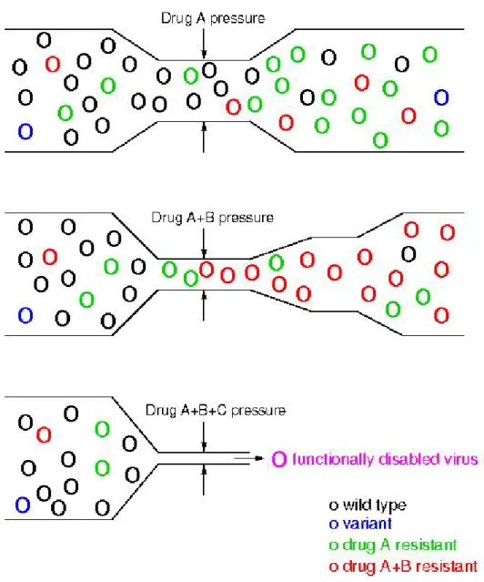
Selection. In presence of antiretroviral drugs, drug resistant variants may replicate
better than the wild-type. Thus, if therapy failed to completely suppress viral replication, drug resistant variants may compete with the wild-type and become dominant in the viral population (Fig. 1.20), thus leading to viral rebound. In addition to mutations that directly confer resistance, additional
mutations are selected during suboptimal therapy in order to rescue losses in fitness due to the presence of resistance-conferring mutations.
1.2.3 Mechanism of drug resistance PIs
Protease inhibitors (PI) prevent cleavage of the viral gag-pol polyprotein resulting in production of immature, non-infectious viral particles (Debouk et al., 1992 and Kaplan et al., 1993). Unfortunately, the early protease inhibitors had poor oral bio-availability and short half-lives due to limited oral absorption and rapid hepatic clearance through cytocrome P450 enzymes (Flexner, 1998). Consequently, patients were required to take a large number of pills at frequent dosing intervals, compromising therapy adherence. Due to these limitations, a viral replication persisted in presence of drug selection pressure and variants with decreased susceptibility to the drugs in HAART regimen were frequently selected. From 1997 onwards it became evident that a therapeutically suboptimal dose of ritonavir enhances the concentration of a administrated PI by inhibition of its hepatic cytochrome metabolism (Kempf et al., 1997). This approach effectively raises the genetic barrier to resistance.
Resistance to protease inhibitors is the consequence of amino acid substitutions that emerge either inside the substrate-binding domain of the enzyme or at distant sites (Condra et al., 1995; Kaplan et al, 1994; Molla et al., 1996). Directly or indirectly, these amino acid changes modify the number and the nature of the points of contact between the inhibitors and the protease, thereby reducing their affinity for the enzyme. As an example, the common resistance mutation V82A reduces the size of an amino acid residue in the protease that is more important for binding most inhibitors than for binding the natural viral protein substrate (Prabu-Jeyabalan et al., 2002). Protease inhibitors have been designed to bind the protease with maximal affinity and tend to occupy more space inside the active site cavity than do natural substrates. Unlike the
inhibitors, the natural substrates of the protease have a variable, but generally less tight, interaction with the catalytic site, a phenomenon that promotes the ordered sequential cleavage of the polyproteins required for proper assembly of the viral particle. Resistance mutations in the protease, which result in an overall enlargement of the catalytic site of the enzyme, would thus be predicted to have a greater effect on the binding of inhibitors than the natural templates. Some mutations are selected for only by certain PIs (Fig. 1.9), reflecting particularities in the chemical structure of the inhibitors that influence their interaction with the substrate-binding domain of the enzyme.
NRTIs
Nucleoside reverse trasnscriptase inhibitors (NRTI) were the first antiretroviral drugs that became available. The viral enzyme reverse transcriptase (RT) is responsible for copying a single-stranded viral RNA genome into double-stranded DNA. NRTIs compete with natural nucleosides for incorporation into pro-viral genome by RT. Since NRTIs lack the 3’-hydroxyl group, no additional nucleotides can be attached after incorporation of NRTIs in the proviral genome resulting in DNA-chian termination and interruption of HIV-replication (Mitsuya et al., 1985). Soon after the introduction of the thymidine analogue zidovudine in 1987 it became evident that, when used as monotherapy, the antiviral effect was transient and rapidly blunted by the selection of viral variants with decreased susceptibility (De Jong at al., 1996).
Resistance to NRTIs may be mediated by al least three different mechanisms:
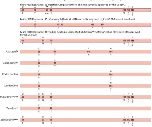
Impairment of Analogue Incorporation. Several mutations or groups of mutations in
reverse transcriptase can promote resistance by selectively impairing the ability of reverse transcriptase to incorporate an analogue into DNA. These mutations include M184V, K65R, L74V, Q151M, Y115F, and V75I (Fig 1.10).
Removal of the Analogue from the Terminated DNA Chain. Removal of the nucleoside
analogue from the terminated DNA chain is associated with a group of mutations commonly termed “thymidine analogue mutations” (Figure 1.10), most frequently selected for after the failure of drug combinations that include thymidine analogues, such as zidovudine and stavudine. These mutations promote resistance by fostering ATP- or pyrophosphate-mediated removal of nucleoside analogues from the 3' end of the terminated DNA strand (Arion et al., 1998; Meyer et al., 1999). ATP and pyrophosphate, which are abundant in normal lymphocytes, do not participate in the
DNA-polymerization reaction, but the structure of a reverse transcriptase expressing thymidine analogue mutations facilitates their entry into a site adjacent to the incorporated analogue (Boyer et al., 2001; Chamberlain et al., 2002). In this position, ATP or pyrophosphate can attack the phosphodiester bond that links the analogue to DNA, resulting in removal of the analogue. Interestingly, the efficiency of this process, also known as “primer rescue,” can be significantly decreased by the presence of other mutations in reverse transcriptase, a phenomenon that has been best described in the case of the M184V mutation (Larder et al., 1995), that slows the selection of thymidine analogue mutations by thymidine analogues (Picard et al., 2001) and may slightly increase the residual antiviral activity of some nucleoside analogues in spite of the presence of thymidine analogue mutations.
Increased reverse transcriptase packaging. Mutations in the p1-p6 region of HIV gag
have been described to increase the number of reverse transcriptase molecules per virion (Peters et al., 2001).
NNRTIs
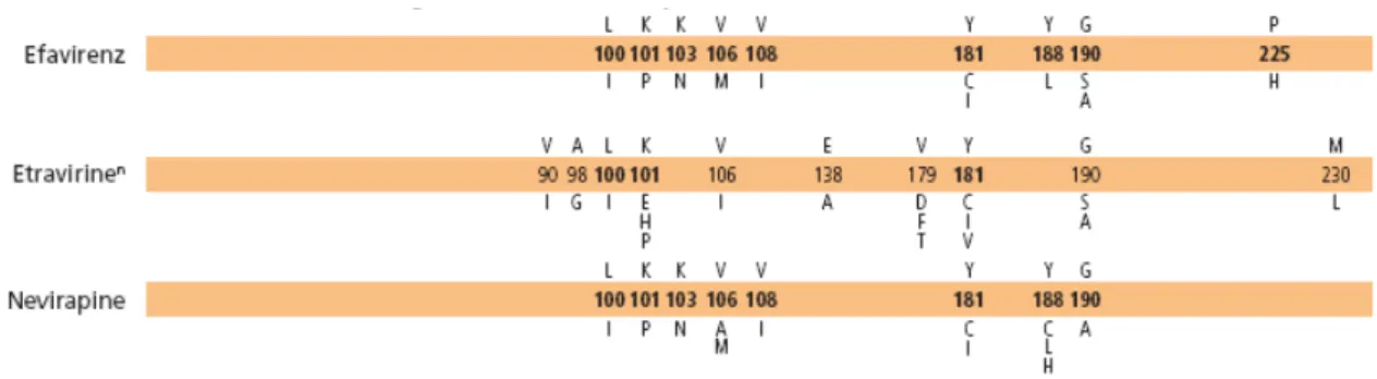
The introduction of non-nucleoside reverse transcriptase inhibitors (NNRTIs) in 1998 provided the opportunity for simpler and more tolerable drug-regimens. NNRTIs bind in a non-competitive manner to a hydrophobic pocket in close proximity to the active site of RT, resulting in inhibition of the catalytic step (De Clercq, 1994). In contrast to unboosted PIs, NNRTIs have an excellent bioavailability; unfortunately, they have a low threshold of resistance.
Some mutations associated with NNRTI resistance may also compromise virus replication by changing the conformation of the dNTP binding pocket or altering RNAse H activity. Moreover, mutations in the NNRTI binding pocket may interfere with NRTI resistance. The NNRTI resistance conferring mutations Y181C and L100I hypersensitize HIV-1 to Zidovudine (Larder et al., 1994), and the multi-nucleoside resistance phenotype can be partially reversed by NNRTI mutations (Van Leathem et al., 2001). See Figure 1.11.
INIs
Integrase catalyses the insertion of HIV-DNA into the genome of the host cell. The integrase strand transfer inhibitor Raltegravir inhibits the linkage of the viral DNA 3’ ends to the cellular target DNA. Unfortunately, this potent drug appears to have a low genetic barrier: just one mutation in the integrase gene can result in considerable resistance. Raltegravir failure is associated with integrase mutations in at least 3 distinct genetic pathways defined by 2 or more mutations including a) a signature (major) mutation a Q148H/K/R, N155H, or Y143R/H/C at the catalytic site of the Integrase and b) 1 or more additional minor mutations (Figure 1.12) (Hazuda et al., 2007).
FIs
Enfuvirtide is the first fusion inhibitor approved. It is a small syntetic peptide that mimics the HR2 region of gp41. Binding of enfuvirtide to the HR1 region of gp41 prevents interaction between HR1 and HR2 for formation of a harpin structure and subsequent fusion pore (Poveda et al., 2005). Resistance to Enfuvirtide is mediated by the appearance of mutations in the Enfuvirtide target region encompassing the residues 36-45 of the gp41 HR1 domain (Fig. 1.13) (Greenberg et al., 2004; Miller et al., 2004; Reeves et al., 2005).
CCR5 entry inhibitors
CCR5-antagonists target the interaction of the viral gp120 with the host cell surface CCR5 chemochine receptor, binding the human coreceptor. Virologic failure of these drugs frequently is associated with outgrowth of D/M or X4 virus from a preexisting
of a CCR5 antagonist. Most of these mutations are found in the V3 loop, the major determinant of viral tropism (Nolan et al., 2009). There is as yet no consensus on specific signature mutations for CCR5 antagonist resistance. Some CCR5 antagonist-resistant viruses selected in vitro have shown mutations in gp41 without mutations in V3; the clinical significance of such mutations is not yet known (Huang et al., 2009).
Figure 1.11. Mutations associated with NNRTI resistance (Johnson et al., 2009)
Figure 1.12. Mutations associated with Raltegravir resistance (Johnson et al., 2009)
1.2.4 Combination therapy
Currently, so called “highly active antiretroviral therapy (HAART) is recommended for the treatment of HIV-infected patients. HAART is defined as is defined as the reported use of three or more antiretroviral medications, one of which has to be a PI, an NNRTI, one of the NRTIs abacavir or tenofovir, an integrase inhibitor (e.g., raltegravir), or an entry inhibitor (e.g., Maraviroc or enfuvirtide) (DHHS/Kaiser Panel, 2008).
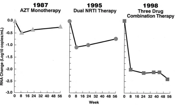
The introduction of the highly active antiretroviral therapy (HAART) has provided an extraordinary clinical benefit in HIV-infected patients in lowering morbidity and mortality (Palella et al., 1998; Valenti et al., 2001) (Fig. 1.19) nevertheless, toxicity profiles, limited tolerability and development of drug resistance reduce the durability of efficacy.
1.2.5 Detection of drug resistance
There are 2 general types of resistance assays used in clinical practice: genotypic assays (i.e., HIV-1 gene sequencing to detect mutations that confer HIV-1 drug resistance) and phenotypic assays (i.e., cell culture–based viral replication assays in the absence or presence of drugs). Both tests have shown their benefits in the clinical practice of treatment-experienced HIV-infected patients, and were become of clinical value for antiretroviral-naïve individuals too (Durant et al., 1999; Baxter et al., 2000).
Genotypic resistance tests were to date considered as the standard of care for monitoring the response to HAART in HIV-1 infected patients. This assay identifies nucleotide changes in the viral genome compared to a viral reference strain. The profile of mutations is interpreted using algorithms that correlate known mutational profiles to phenotypic susceptibility data and/ or clinical outcome. Genotypic tests may, therefore, identify mutations or genetic profiles that are not necessarily resulting in phenotypic resistance but may furnish evidence of archived drug resistance. This test can be performed with commercial assay kits or in-house protocols.
Phenotypic drug-susceptibility assays determine the concentration of an antiretroviral agent that is required to inhibit HIV-1 replication by 50% (IC50) or 90% (IC90). The results are compared to the concentration of drug necessary to inhibit a reference strain of susceptible virus. The difference in IC50 or IC90 defines the susceptibility of the virus to the different antiretroviral agents.
Phenotypic resistance tests and genotypic sequence analyses are generally capable of identifying viral populations that make up at least 20% of the quasispecies. Drug resistant variants, partly overgrown by or reverted to wild-type may, therefore, be missed, suggesting that these tests might underestimate the presence of drug resistant
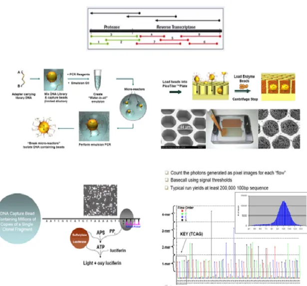
viruses. More sensitive resistance tests, such as allele-specific PCR, parallel-allele-specific sequencing, standard cloning, Ling Amp, or the Ultra deep pyrosequencing (UDPS), may be very useful for this indication. Originally designed for high throughput sequencing of mammal and bacterial genomes, the UDPS technique is particularly well-suited for an in depth analysis of a population of heterogeneous genomes like those of retroviruses. To date the parallel pyrosequencing technique can detect rare genetic variants constituting as little as 1% of the population. The procedure used for HIV-1 is reported in figure 1.14. Currently, the length of sequencing reads per clone is between 300 to 400 contiguous base pairs. This allows studying the genetic linkage of clinically relevant resistance mutations clusters, like mutations surrounding the NNRTI binding pocket, the integrase catalytic site, most mutations in the protease, and the complete V3 loop of gp120.
Figure 1.14. Schematic procedure for 454 in HIV. The target sequence is amplified with primers
tagged with a sample-specific identification. An water/oil emulsion is prepared so each droplet contains 1 bead and 1 amplicon; each amplicon is then amplified by emulsion PCR within the droplet. The emulsion is disrupted and 28uM beads are loaded in 44 uM wells, so, theoretically, there is only one bead per well. Sequencing enzymes including luciferase are included in each well. Pyrosequencing proceeds by series of flows of known nucleotides. When a nucleotide hibridizes with its complementary pyrophsfate is released and this results in the release of chemioluminescent signal reordered by a CCD camera and integrated in a computer. Vertical bars in the flowchart represented the number of bases. Source: 454 sequencing website (www.454.com)
1.3 Transmission of HIV-1 drug resistance
1.3.1 Evolution of drug resistance in treatment naïve patients
In treated patients carrying drug resistant HIV variants, cessation of the complete regimen usually results in a rapid reappearance of the original drug-sensitive wild-type virus (Devereux et al., 1999). However, resistant variants may persist as minority populations in plasma and are maintained as provirus in resting cells within lymphoid tissue. As soon as therapy is reintroduced, the resistant variants can be rapidly reselected (Karlsson et al., 1999).
The frequent detection of drug resistance among HIV-infected treatment failing patients can in turn increase the risk of new infections driven by drug resistant viral strains. In antiretroviral-naïve individuals, wild-type may dominate resistant variants with impaired fitness, if the infecting quasispecies contained both wildtype and resistant variants. If, however, through a founder effect, the diversity of infecting quasispecies is restricted to resistant variants, and no minority of wild-type variants is present, dominance by wild-type is not possible. In the absence of drug pressure, the transmitted drug resistant quasispecies may revert back to wild-type or mutate to other more fit variants. Viruses harbouring a single resistance-related point mutation, such as M184V in the reverse transcriptase (RT) gene, may revert more rapidly than variants that have evolved after multiple mutations (Brenner et al., 2002). Also, persistence of transmitted resistant variants for several months or years in the plasma of the newly infected subject
T215F, which need two nucleotide changes for reversion to wild-type (De Ronde et al., 2001; Garcia Lerma et al., 2001).
Viruses containing these partial revertants or atypical mutations are selected because they display an increased fitness compared to the resistant variants. In vitro experiments have shown that variants with the atypical mutation 215D, or partial revertant 215S, can replicate as efficiently as wildtype, which may explain their persistence in vivo. Although the partial revertants or atypical mutants do not, by themselves, confer phenotypic resistance, they are only one step away from the resistant variants 215Y or 215F, compared to the two mutational steps that are needed from wildtype to 215Y/F. This may indicate an increased risk for developing resistance under subsequent treatment with zidovudine or stavudine (Violin et al., 2008).
In the absence of selective drug pressure in the new host, resistant variants may completely revert to wild-type, leaving no evidence of transmitted resistance in the plasma. However, one might expect that the initial resistant quasispecies will still be present as archived provirus in resting cells or as minority variants in plasma of the untreated subject.
1.3.2 Mechanism of transmission of drug resistant virus
Individuals may be exposed to varying amounts of virus depending on the route of HIV-1 transmission. Sexual and vertical transmission generally involves smaller amounts of virus and infected cells than transmission due to direct blood-to blood contact as occurs during intravenous drug use or blood transfusion (Zhu et al., 1993; Overbaugh et al., 1999; Wolinsky et al., 1993).
proliferating in the blood of recently-infected recipients is generally more homogeneous than the markedly heterogeneous quasispecies in chronically-infected donors (Zhu et al., 1993; Delwart et al., 2002; Wolfs et al., 1992; Zhu et al., 1992). The restriction in quasispecies implies the occurrence of a bottleneck process during transmission and establishment of infection. This process is determined by the number of transmitted virions, by the diversity of the transmitted viral quasispecies and by host factors, such as the mucosal barrier, the density of target cells and the immune system (Quiñones-Mateu et al., 2002).
Recent studies suggest that multiple strains are frequently transmitted, resulting in more genomic variation in the recipient than previously reported, though still not as heterogeneous as the donor population (Overbaugh et al., 1991; Learn et al., 2002). Subsequently, variants among the transmitted population that are most capable of widespread dissemination might be positively selected. The viruses that succeed in establishing infection do not have to be the variants that displayed the highest fitness under drug pressure in the donor quasispecies –as demonstrated by the ability of minor variants from the donor quasispecies to accomplish infection in the recipient (Zhu et al., 1993). This finding might be explained by stochastic processes that influence the diversity of the viral variants in the inoculum. Another reason for a shift in quasispecies might be that characteristics preferred during chronic infection differ from viral properties selected during transmission and adaptation to the new host.
transmission of an HIV variant resistant to multiple RT and Protease inhibitors. Since then, transmission of virus resistant to antiretroviral drugs has been repeatedly documented through many infection routes: sexual contact, needle sharing among intravenous drug users, perinatal transmission and accidental exposure of health care workers (De Ronde et al., 1996; Siegrist et al., 1994; Conlon et al., 1994; Anonymous, 1993).
More recently, cohort studies have been published describing patterns of baseline drug resistance profiles in a significant percentage of therapy- naïve patients (Yerly et al., 1999; Boden et al., 1999; Brodine et al., 1999).
Multiple studies have reported impaired enzyme function and decreased viral fitness of HIV-1 isolates harboring drug resistant mutations, often resulting in a moderate or low viral load in the patient (Nijhuis et al., 2001). One might speculate that these strains are also less capable of accomplishing infection in a new host. On the other hand, if the transmitted quasispecies also contains drug-sensitive variants, resistant variants with impaired fitness may be at selective disadvantage during infection and/ or initial amplification in the new host.
Transmission of 1 is strongly influenced by the level of viremia in the HIV-infected donor. Consequently, it seems logical to relate the rate of transmission of HIV to the overall level of viremia in the HIV-infected population. In this perspective, access to HAART has, by reduction of HIV-RNA concentrations, the potential to decrease transmission of HIV on a population level (Quinn et al., 2000; Hosseinipour et al., 2002). In pregnant HIV-infected women, the use of HAART has indeed shown a profound reduction of HIV incidence in their off-spring. Despite this success in preventing vertical transmission, HAART is currently not effective as a “population
prevention measure”. In contrast, prolonged survival of infected patients, continuous injecting drug use and sexually related transmission, have resulted in an increased prevalence of HIV-infections in countries with access to HAART (HIV/AIDS Surveillance in Europe, 2006 and 2007).
The impact of HAART on the spread of HIV and its drug resistant variants may vary between different transmitters. For instance, the beneficial effect of therapy on HIV-RNA load will not reach individuals who are unaware of their infection status. Persons with primary infection among this population may be very infectious, because viremia in the plasma is extremely high during the acute phase of infection (Hubert et al., 2000). In contrast, the viremia in semen during acute infection appears not to be different from that during established infection, although this has only been studied to a limited extent (Dyer et al., 1997). Nevertheless, data from the Swiss cohort prescribe an important role of new diagnoses and primary infections on the spread of HIV-1. One third of acutely-infected patients in the cohort appeared to be acutely-infected by persons who had acquired HIV very recently themselves (Yerly et al., 2001). Although seroconverters are rarely assumed to be the source of drug resistant viruses, secondary transmission of resistant virus has been described in recent HIV infection and may become more frequent in the future.
Other possible transmitters are chronically-infected individuals that have been diagnosed with HIV, but not yet exposed to therapy. Toxicity and complexity of the current antiretroviral regimens have resulted in new guidelines favouring less aggressive
been described, this new strategy may lead to an increased risk of transmission at the population level (Yerley et al., 2009; Huè et al., 2009).
Antiretroviral-experienced individuals make up a third group of possible transmitters (Goudsmit et al., 2001). Ideally, persons taking antiretroviral therapy should be less efficient in transmitting HIV, at least if their viral load is successfully suppressed (Quinn et al., 2000). Unfortunately, in many individuals on HAART, the viral load is not below the detection of the commercially available assays. Furthermore, the limited amount of therapeutic options supports continued use of HAART regimens that, even though unsuccessful from a virology point of view, are capable of sustaining a prolonged immunologic response. Since patients experiencing viral rebound frequently harbour drug resistant variants, they are the most likely source of transmission of resistant viruses. However, it has been suggested that the frequency of transmission of drug resistant viruses is less than expected, due to their impaired viral fitness.
1.3.4 Estimation rate of transmission of drug resistant virus
To date, there is a growing literature about the rate of transmission of HIV-1 drug resistant virus. In the United States and in Europe, where there is a wide access to highly active antiretroviral therapy (HAART), the prevalence of HIV-1 drug resistant strains ranges between 3.3% and 14.0% in recently infected patients and between 6.1% and 12.5% in chronically infected ones (Novak et al., 2005; Wensing et al., 2005; Yerly et al., 2007; UK, 2007; SPREAD, 2008). These different rates may reflect variations in access to therapy, therapeutic strategies, adherence or risk-related behaviour, but first of all for a lack of uniformity in the definition of transmitted resistance. Only with a standard list of mutations is it possible to compare the prevalence of transmitted
resistance from different times and regions and facilitate meta-analyses of surveillance data collected by different groups at different times. Compiling such a standard list, however, is not simple because of the rapidly changing field of ARV therapy and the large numbers of mutations associated with ARV drug resistance (Bennett et al., 2009). Shafer at al., in 2007, provided a provisional list for identifying surveillance drug resistance mutations (SDRMs) on the basis of four criteria. The first criterion was that SDRMs should be recognized as causing or contributing to drug resistance – defined as being present on three or more of five expert lists of drug resistance mutations. The second criterion was that mutations should be non-polymorphic and should not occur at highly polymorphic positions. The third criterion was that the mutation list had to be applicable to the eight most common HIV-1 subtypes. The fourth criterion was that the list should be parsimonious, excluding mutations resulting exceedingly rarely from drug pressure.
The list of 2007 was uploaded in 2009, when new drug resistance mutations have been identified including mutations arising from the increased use of non-thymidine-analog containing regimens, the expanded use of two new protease inhibitors (PIs), and the recent approval of a new non-nucleoside RT inhibitor (NNRTI) (Bennet et al., 2009) (Table 1.2).
Position AA PI NRTI NNRTI Position AA PI NRTI NNRTI 41 L ● 23 I ● 65 R ● 24 I ● 67 N ● 30 N ● G ● 32 I ● E ● 46 I ● 69 D ● L ● ins ● 47 V ● 70 R ● A ● E ● 48 V ● 74 V ● M ● I ● 50 V ● 75 M ● L ● T ● 53 L ● A ● Y ● S ● 54 V ● 77 L ● L ● 115 F ● M ● 116 Y ● A ● 151 M ● T ● 184 V ● S ● I ● 73 S ● 210 W ● T ● 215 Y ● C ● F ● A ● I ● 76 V ● S ● 82 A ● C ● T ● D ● F ● V ● S ● E ● C ● 219 Q ● M ● E ● L ● N ● 83 D ● R ● 84 V ● 100 I ● A ● 101 E ● C ● P ● 85 V ● 103 N ● 88 D ● S ● S ● 106 M ● 90 M ● A ● 179 F ● 181 C ● I ● V ● 188 L ● H ● C ● 190 A ● S ● E ● 225 H ●
1.3.4 Clinical implications of transmitted drug resistance
Major concerns exist about the clinical implications of transmitted drug resistance. Although it might be expected that the response to antiretroviral therapy is diminished in the case of primary resistance, only limited data assessing clinical outcome are available at present. In a large North American cohort, therapeutic outcome for patients infected with HIV harboring major mutations was not different from patients infected with drug-susceptible viruses (Alexander et al., 2001).
However the majority of patients in this cohort were not prescribed drugs to which the virus they carried exhibited resistance. The small group of patients who did initiate therapy to which the virus exhibited possible resistance at baseline, displayed a relatively inferior virological outcome (Alexander et al., 2001).
Having transmitted drug resistance mutations seems not to be predictive of virological failure when patients received a fully active treatment also for a recent European study (Wittktop et al., 2010). HIV-1 infected patients enrolled in this project had a poor virological response only when the HAART regimen was based upon drugs to which the virus has lost susceptibility.
In three other studies from the United States and France, the time to achieve viral suppression was significantly prolonged in individuals carrying virus with major phenotypic or genotypic drug resistance at baseline compared to subjects with fully-susceptible virus at baseline (Little et al., 2002; Harzic et al., 2002; Grant et al., 2002; Peuchant et al., 2008).
revertants (Riva et a., 2002). These revertants have an increased ability to select the RT T215Y/F mutations that limit the efficacy of thymidine analogues. Of the 13 patients who carried the 215 mutants, 9 experienced virological failure.
Some groups report a lower baseline viral load (Harzic et al., 2002; Descamps et al., 2001) or a higher CD4 cell count (Grant et al., 2002) in antiretroviral-naïve individuals carrying primary drug resistant variants in comparison to individuals with drug-sensitive virus. These differences might reflect variations in duration of infection. Alternative interesting hypothesis suggested by the authors is a decreased viral replication capacity of the transmitted resistant viruses.
As described above, minority strains with transmitted drug resistance related mutations might not always be detected by conventional resistance testing, therefore a single mutation or a revertant might be an indicator that more extensive resistance has been transmitted. In a preliminary study in HIV-1 infected individuals experiencing failure of a first-line NNRTI-based regimen, a strong association between the presence of minority drug resistant variants and virological failure was observed (Paredes et al., 2010).
In this line, a recent case-control study found that 7% of individuals who experienced their first virologic failure had minority drug resistance mutations at baseline, and that minority resistance was rarely found in treatment successes (Johnson et al., 2008). These findings suggest that also the minority drug resistant HIV variants may have clinical consequences and that the presence of such variants in individuals who have not previously taken ART may reduce the efficacy of some ART regimens.
In summary, preliminary data show that transmission of resistance, even in minority species, can compromise therapy outcome. For these reasons, the new guidelines
recommend to perform the genotypic resistance testing in all drug-naïve patients, before beginning a first line antiretroviral regimen (Hirsch et al., 2008; Department of Health and Human Services, 2009).
1.4 Rationale of the work
The development of resistance to the currently available antiretroviral drugs against HIV-1 infection is one of the major limitations to the maintenance of a successful treatment. The frequent detection of resistance among HIV-infected treatment failing patients (Pillay et al., 2005; Wensing et al., 2005; Spread Programme, 2008) can in turn increase the risk of new infections driven by drug resistant viral strains (Wensing and Boucher, 2003). The prevalence of drug resistant HIV in newly diagnosed individuals naïve to antiretroviral therapy is likely to be influenced by the prevalence of drug resistance in the treated population, even if recent studies assumed that the transmission of drug resistant HIV-1 viruses reflects not only infection from drug-experienced patients, but also the circulation of resistant strains between drug-naïve individuals (Yerley et al., 2009; Huè et al., 2009).
Once transmitted, a drug resistant virus can either revert to wild-type or more frequently persist for months to years into the host (Barbour et al., 2004) as minority species, or stored in proviral DNA of infected cells. In drug naïve patients these minority drug resistant HIV-1 strains may become dominant at the time of HAART initiation, increasing the probability of virological failure to the first-line antiretroviral therapy (Little et al., 2002; Harzic et al., 2002; Grant et al., 2002; Peuchant et al., 2008; Roquebert et al., 2006).
In this light, the aim of this thesis was to characterize the distribution and the spread of drug resistant HIV-1 in Italy. The main objectives were to ascertain how many people naïve to the antiretroviral therapy are infected with drug resistant virus and which resistance profiles are circulating.
population dynamics of transmitted resistance and the potential contribution of untreated patients to the spread of antiretroviral resistance. The identification of potential epidemiological networks and sequence interrelationships between acute/early and chronic infections in both drug-naïve and drug-experienced individuals can provide new tools in prevention strategy as well as in management of treatment programs in a local, specific and well-characterized territory.
Finally, the last objective was to define new genetic markers in the pol gene that can predict the presence of a transmitted drug resistant minority species. Our attention has been focused on few selected mutations, T69S, L210M and K103R, found at 3 reverse transcriptase (RT) resistance positions and easily detected by standard population sequencing (GRT). Their prevalence in drug naïve patients is generally low (T69S, 2.4%; L210M, 0.9%; K103R, 1.7%, prevalence estimated in 2089 HIV-1 B subtype infected drug naïve patients) and no association with phenotypic resistance to RT inhibitors, in either clinical isolates or site-directed mutagenesis experiments was to date proved (Harrigan et al., 2005; Berkhout et al., 2006). Certainly, the analysis by UDPS of minority quasispecies, has allowed to better define the role of these mutations as potential markers of the existence of resistant minority variants, and to determine their impact on clinical virologic outcomes, thus enhancing the understanding and management of resistance in the future. This is even more relevant from the practical point of view, since the methods to detect minority quasispecies resistant to antivirals (such as UDPS, etc) are today used only for research purpose, and it is difficult to
2 Methods
In this chapter, dataset and methods used in each study described in this thesis were reported.
2.1
Characterization of the patterns of drug resistance mutations in
newly diagnosed HIV-1 infected patients naïve to the
antiretroviral
drugs
2.1.1 Study population
The study included 263 HIV-1 infected individuals enrolled between January 2004 and March 2007 in the SENDIH (Studio Epidemiologico Nuove Diagnosi Infezione HIV-1) programme, a multicenter study aimed to collect behavioural, virological and molecular data on persons with newly diagnosed HIV infection. Characteristics and methods of the study have been previously described by Orchi et al., 2008. Individuals with a first HIV-1 positive test performed in HIV-10 public Counselling and Testing centres (CTC) in Lazio Region, Italy, were invited to participate in the study. At the diagnosis, clinical and immunologic data, and blood sample have been collected from all participants to investigate the molecular characterization of the virus and to identify recently acquired infections.
Informed consent was obtained from participants and the ethics committee of the National Institute for Infectious Diseases L. Spallanzani, Rome approved the study. All of the information gathered during the study was analyzed in a completely anonymous way.