Review Article
18
F-labeled radiopharmaceuticals for the
molecular neuroimaging of amyloid
plaques in Alzheimer’s disease
Luca Filippi1, Agostino Chiaravalloti2,3, Oreste Bagni1, Orazio Schillaci2,3
1Department of Nuclear Medicine, Santa Maria Goretti Hospital, Via Canova 3, Latina 04100, Italy; 2Department of Biomedicine and Prevention, University Tor Vergata, Rome, Italy; 3IRCCS Neuromed, Pozzilli, Italy
Received May 20, 2018; Accepted August 19, 2018; Epub August 20, 2018; Published August 30, 2018
Abstract: Alzheimer’s disease (AD) is the most common cause of dementia in the elderly, with tremendous impact on the affected individuals and the society. Definitive diagnosis can be achieved only by post mortem examination. Clinical diagnosis criteria currently applied in clinical practice for AD often fail to accurately discriminate between AD and non-AD dementia with up to 40% of misdiagnosed patients. Several published papers demonstrated that the pre-clinical phase of AD is characterized by an early rise in beta-amyloid accumulation into inter-neuronal space, followed by a severe synaptic dysfunction. Thus, beta-amyloid protein, detected in the cerebrospinal fluid, has been considered a specific AD biomarker. Molecular imaging of beta-amyloid deposits, with positron emission tomogra-phy (PET) and 18F-labeled radiopharmaceuticals such as 18F-florbetapir, 18F-florbetaben, and 18F-flutemetamol, has emerged as potential powerful tool for aiding AD diagnosis. The aim of the present paper is to review the existing literature on the clinical use of these new amyloid tracers in order to delineate their diagnostic value and limitations. Keywords: PET, amyloid plaques, Alzheimer’s disease, neuroimaging
Introduction
Alzheimer’s disease (AD) represents the most common cause of dementia worldwide and its global burden is expected to grow further due to population’s aging. It is characterized by early deficits in memory which inevitably prog-ress to severe and generalized cognitive dete-rioration. The annual incidence of AD increases with age; it has been reported an incidence of 53 new cases per 1000 people aged 65 to 74, 170 new cases per 1000 people aged 75-84, and 230 new cases per 1000 people aged over 85 [1]. Thus, AD represents a critical pub-lic health issue in many countries around the world, with a tremendous impact on the so- ciety.
As concerns its etiopathology, in most of the cases AD occurs sporadically with late onset and multi-factorial in etiology [2]. Definitive diagnosis of AD is based on the post mortem examination. Therefore, the initial diagnosis is presumptive and made by clinical evaluation
and neurophycological testing. Conventional neuroimaging with magnetic resonance (MRI) presents some limitations, especially in terms of specificity, in the diagnosis of AD [3]. Fur- thermore, a variety of clinical conditions can mimic AD and, according to several published reports, between 12% and 23% of patients diagnosed with AD resulted to be misdiagnos- ed at the autoptic examination [4]. The most common features leading to definitive AD diag-nosis consist of general atrophy of the cortex, neuron and synapse loss, extracellular plaques composed of insoluble beta-amyloid (Abeta) and intraneuronal neurofibrillary tangles (NFTs). Several scientific evidences indicate that the pathogenesis of AD is driven by the progressive accumulation of Abeta peptide into the inter-neuronal space [5]. Although the pathogene- tic pathways leading to AD are certainly very complex involving several mechanisms such as the dysfunction in cholinergic neurons and the aberrant aggregation of hyperphosphorilated tau protein, it has been demonstrated that the
amyloid cascade plays a fundamental role [6]. Cerebrospinal fluid levels of beta-amyloid is clinically used as AD biomarker since altered levels of this peptide are highly associated with conversion of presymptomatic patients to AD [7].
It has to be pointed out that the Abeta peptide has represented also the most studied target for the neuroimaging of AD. The identification of amyloid deposits, with positron emission tomo- graphy (PET) and 18F-labeled radiopharmaceu-ticals such as 18F-florbetapir, 18F-florbetaben, and 18F-flutemetamol, has been recently intro-duced as potential powerful tool for aiding cli- nicians in AD diagnosis.
The purpose of this paper is to review the exist-ing literature on the clinical use of these new amyloid-tracers in order to delineate their diag-nostic value and limitations.
The amyloid cascade hypothesis in AD patho-genesis
The amyloid cascade hypothesis is based on the assumption that AD pathogenesis is due to a series of abnormalities in the production and in the secretion of the amyloid precursor
pro-tein (APP). APP is a transmembrane glycopro-tein with a still unclear role in cell function. APP is sequentially processed through a series of cleavages operated by beta- and gamma-se- cretases. In particular, gamma-secretase is a high molecular weight complex minimally com-posed of four components: presenilins (PSEN), nicastrin, anterior pharynx defective 1, and pre-senilin enhancer 2 [8]. Gamma-secretase cuts the gamma-site of carboxyl-terminal fragment of APP producing the 2 major Abeta isoforms: Abeta42 (42 residues long) and Abeta40 (40 residues long).
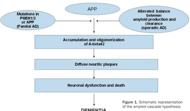
Concentrations of amyloid in brain are the con-sequence of the balance between production and clearance; in AD patients, a not fully under-stood mechanism leads to an abnormal accu-mulation of amyloid beta. Abeta42 isoform is the major component of amyloid plaques due to its low solubility and propensity to form ag- gregates with beta-pleated sheet structure [9]. Extracellular Abeta oligomers bind the cell sur-face, leading to functional disruption of a num-ber of receptors, thus producing dysfunction and neurodegeneration (Figure 1) [10].
The role and the importance of biomarkes for the correct framework of AD have been recently
Figure 1. Schematic representation of the amyloid cascade hypothesis.
underlined by the taskforce organized by the National Institutes of Aging and the Alzheimer’s Association (NIA-AA). It has been proposed, in fact, that the diagnosis of AD should be based on the presence or absence of a well defined biomarker system instead of clinical symptoms [11]. This biomarker system, termed AT (N), in- cludes three hallmarks typical of AD. The bio-markers in the A group are representative of the amyloid burden and can be evaluated by either PET imaging with amyloid tracers or mea-suring the levels of Abeta isoforms in the cere-brospinal fluid. The biomarkers in the T group reflect the aggregated tau proteins and can be determined by measuring hyperphosporilated tau proteins in cerebrospinal fluid and also with PET radioligands specifically binding to tau deposits in brain. Finally, the biomarkers in the N group are indicative of neurodegeneration and include atrophy on MRI and hypometabo-lism on 8F-Fluoro-deoxy-glucose (FDG) PET. It is worthy of note that by combining informa-tion from each of the three biomarker groups, clinicians can also obtain a staging of AD since the more biomarkers are abnormal, the more advanced the pathologic stage is. Another ad- vantage of this classification is its flexibility to incorporate new biomarkers for each category.
In this context molecular imaging with specific probes (i.e. with amyloid or tau radioligands) plays a fundamental role.
The pittsburgh compound B (PIB)
As the amyloid cascade hypothesis presents a high rate of acceptance by the scientific com-munity, the target for the molecular imaging of AD has been represented by a radioligand binding to the insoluble fibrillar forms of amy-loid peptides. The chemical structures of the mentioned radiotracers in the manuscript are reported in Figure 2.
11C-Pittsburgh Compound B (PIB) was the first radioligand developed for PET imaging. It was derived from a fluorescent amyloid dye (i.e. the thioflavin T) and developed at Pittsburgh University. It was found to present high affinity and specificity for fibrillar Abeta-aggregates [11]. The first study in humans with PIB was published in 2004 and included 16 patients with AD and 9 healthy volunteers. Compared with controls, AD patients typically showed ma- rked retention of PIB in areas of association cortex known to contain large amounts of amy-loid deposits [13]. Furthermore, Mintun et al. investigated whether abnormal binding of PIB
Figure 2. Chemical structures of thioflavin T and of the compounds applied for PET amyloid imaging.
in brain may occur in clinically normal individu-als, prior to the development of cognitive ch- anges. It has to be pointed out that PIB-PET was found able to detect amyloid deposits not only in AD patients, but also in some non-demented patients, thus suggesting that amy-loid imaging might be sensitive for the detec-tion of preclinical AD [14].
The relationship between PIB uptake and the topography of amyloid plaques at post mortem examination was investigated by Ikonomovic et al. [15]. The authors examined 28 clinically diagnosed and autopsy-confirmed Alzheimer’s disease subjects, including 1 AD patient who had undergone PIB-PET imaging 10 months prior to death. It was found out a direct correla-tion of in vivo PIB retencorrela-tion with the region-matched quantitative analyses of Abeta pla- ques in the same patient, thus supporting the validity of PIB-PET as a method for the evalua-tion of amyloid plaque burden.
Of note, PIB is labeled with 11C with a short 20 minutes half-life limiting the use of this radio-pharmaceutical only in PET-centers with on-site cyclotron and experience in 11C-radiochemistry. These drawbacks triggered the development of 18F-labeled radioligands for the imaging of amy-loid deposits in AD.
18F-florbetapir showed a clear separation betw- een cortical and cerebellar activity beginning around 30 min after injection, thus allowing starting brain PET scan at 30-50 minutes post injection. In a report from Wong et al. [17], 18F- florbetapir uptake in brain was visually evalu- ated and also analyzed by semiquantitative methods that confirmed significant elevations of tracer uptake in several brain regions of AD patients, compared with controls. Furthermore, results from phase III clinical trial showed a strong correlation between 18F-florbetapir PET images and the distribution of amyloid deposi-tions at post mortem examination. No serious adverse events were reported in any of the clini-cal trials of 18F-florbetapir [18].
18F-florbetaben: 18F-florbetaben (Neuraceq, Pira- mal Imaging) is an 18F-labeled derivative from stilbene and was approved by FDA in 2014 [19]. 18F-florbetaben was demonstrated to present nanomolar binding affinity to synthetic beta-amyloid fibrils and AD brain homogenate. 18F- florbetaben binding to amyloid plaques has been revealed in AD brain sections [20]. Worth of note, 18F-florbetaben was found not to bind to tau- or alpha-synuclein deposits thus show-ing high specificity for amyloid neuritic plaques. After injection, the compound binds to plasma proteins and is metabolized by several
cyto-Figure 3. A 65 year-old-male with cognitive impairment. PET with 18 F-florbeta-ben (axial slices) showed radioligand binding in the cortical regions, indica-tive of high amyloid burden.
The amyloid PET 18F-labeled
radiopharmaceuticals
18F-florbetapir: 18F-florbetapir (Amyvid, Eli Lilly/Avid Radio- pharmaceuticals), a derivative from stilbene, was the first 18F-labeled PET tracer devel-oped for the imaging of amy-loid plaques with PET technol-ogy [16]. It was approved by FDA in 2012. Preclinical stud-ies demonstrated high bind-ing affinity of 18F-florbetapir to Abeta fibrils and specific labeling of amyloid plaques in the cortical regions and hip-pocampus. After injection, the tracer diffuses through the blood-brain barrier with kinet-ics similar to those for PIB but faster than those for other 18F-labeled amyloid imaging agents. In patients with AD,
chrome enzymes. Preliminary studies in ani-mals supported the usefulness of 18F-florbeta- ben for the imaging of amyloid plaques as in a mouse over-expressing mutant beta-amyloid precursor protein the concentration of the trac-er was found significantly high [21]. In a study comparing 18F-florbetaben with PIB, both trac-ers were found equally accurate in discriminat-ing patients with AD from healthy controls with an excellent correlation at the semiquantita- tive analysis [22]. The recommended activity is 300 MBq, with scan duration of 20 min, start-ing 90 min after tracer administration. Accord- ing to the manufacture’s indications, PET imag-es should be displayed in the transaxial orien-tation using gray scale or inverse gray scale (Figure 3).
18F-flutemetamol: 18F-flutemetamol (GE Health-
care. VizamylTM) is another compound devel-oped for AD imaging. It is an 18F-labeled deriva-tive of PIB, approved by FDA in 2013 [23]. It
ould be displayed with a color scale providing progression of low to high intensity (Figure 4). Table 1 summarizes the main manuscripts on the use of 18F-labeled amyloid radiopharma-ceuticals in different clinical settings.
Amyloid-PET imaging in mild cognitive impairment
Mild Cognitive Impairment (MCI) is a clinical disorder characterized by a moderate impair-ment of the thinking abilities. MCI is worldwide considered as a “gray zone” between intact cognitive functions and dementia [28]. Accord- ing to the original classification of the Mayo Clinic [29], to be categorized as having MCI, a patient should present memory complaints in spite of preserved general cognitive function-ing, as well as the capability to perform daily life activities independently. MCI is considered a pre-dementia state, as MCI patients present
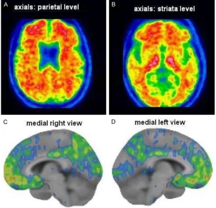
Figure 4. A 63 year-old-female patient with amnestic MCI (MMSE = 24/30). Amyloid PET with 18F-flutemetamol was positive showing tracer accumulation in the cortical regions of interest, as evident in the axial slices at the level of the parietal lobes (A) and of the striata (B). Abnormal radiopharmaceutical accumulation was also detected in the frontal lobe, in the posterior cyngulate cortex and in the precuneus at the quantitative analysis performed by the dedicated software Cortex ID Suite (GE Healthcare), as shown in the medial right (C) and left (D) volume rendering views.
was developed by General Electric Healthcare and pres-ents similar kinetic properties to those of PIB. Post mortem examination in AD brain ho- mogenates demonstrated a strong correlation between binding of 18F-flutemetamol and the localization of amy-loid deposits in different re- gions of the brain [24]. The cortical retention of PIB and 18F-flutemetamol closely ma- tched also in the first report in humans consisting of a dual tracer study in 1 pa- tient affected by AD and in 2 healthy controls [25]. A phase-III trial including 176 patients undergoing PET with 18F-flutemetamol demonstrat-ed that the compound was safe with high sensitivity and specificity for the in vivo de- tection of brain beta-amyloid plaque density [26, 27]. The recommended activity to be administered is 185 Mbq, the scan duration and the start-ing time are similar to those reported for 18F-florbetaben. For 18F-flutemetamol, all imag-es (axial, coronal, sagittal) sh-
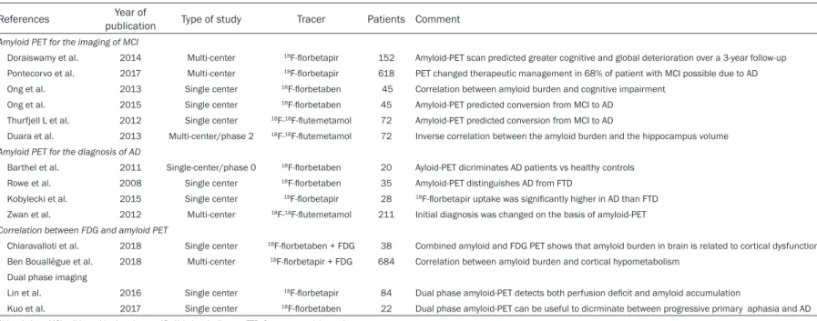
Table 1. Summary of the main manuscripts on the use of 18F-labeled amyloid radiopharmaceuticals in clinical setting
References publicationYear of Type of study Tracer Patients Comment
Amyloid PET for the imaging of MCI
Doraiswamy et al. 2014 Multi-center 18F-florbetapir 152 Amyloid-PET scan predicted greater cognitive and global deterioration over a 3-year follow-up Pontecorvo et al. 2017 Multi-center 18F-florbetapir 618 PET changed therapeutic management in 68% of patient with MCI possible due to AD Ong et al. 2013 Single center 18F-florbetaben 45 Correlation between amyloid burden and cognitive impairment
Ong et al. 2015 Single center 18F-florbetaben 45 Amyloid-PET predicted conversion from MCI to AD Thurfjell L et al. 2012 Single center 18F-18F-flutemetamol 72 Amyloid-PET predicted conversion from MCI to AD
Duara et al. 2013 Multi-center/phase 2 18F-18F-flutemetamol 72 Inverse correlation between the amyloid burden and the hippocampus volume
Amyloid PET for the diagnosis of AD
Barthel et al. 2011 Single-center/phase 0 18F-florbetaben 20 Ayloid-PET dicriminates AD patients vs healthy controls Rowe et al. 2008 Single center 18F-florbetaben 35 Amyloid-PET distinguishes AD from FTD
Kobylecki et al. 2015 Single center 18F-florbetapir 28 18F-florbetapir uptake was significantly higher in AD than FTD Zwan et al. 2012 Multi-center 18F-18F-flutemetamol 211 Initial diagnosis was changed on the basis of amyloid-PET
Correlation between FDG and amyloid PET
Chiaravalloti et al. 2018 Single center 18F-florbetaben + FDG 38 Combined amyloid and FDG PET shows that amyloid burden in brain is related to cortical dysfunction Ben Bouallègue et al. 2018 Multi-center 18F-florbetapir + FDG 684 Correlation between amyloid burden and cortical hypometabolism
Dual phase imaging
Lin et al. 2016 Single center 18F-florbetapir 84 Dual phase amyloid-PET detects both perfusion deficit and amyloid accumulation
Kuo et al. 2017 Single center 18F-florbetaben 22 Dual phase amyloid-PET can be useful to dicrminate between progressive primary aphasia and AD Abbreviations: MCI, mild cognitive impairment; AD, Alzheimer’s disease; FTD, frontotemporal dementia.
higher risk of evolving to dementia with about 10% MCI subjects converting to dementia per year.
Amyloid-PET imaging with 18F-labeled com-pounds has been applied for predicting the probability of conversion from MCI to AD. A prospective multicenter study [30] involved 69 cognitively normal controls, 52 with recently diagnosed MCI and 31 with probable AD to evaluate whether subjects with amyloid path- ology, detected using 18F-florbetapir PET, pre-sented greater cognitive decline than subjects without amyloid pathology. The authors found that all MCI subjects with positive amyloid-PET scan showed greater cognitive and global dete-rioration over a 3-year follow-up as compared with subjects with negative amyloid-PET scan. These results were subsequently confirmed in a larger series of 618 patients with MCI possi-bly due to AD in which a high percentage of sub-jects (i.e 68%) received a change in medication on the basis of amyloid-PET results [31]. As concerns the imaging of MCI patients, Ong et al. evaluated 45 MCI patients with PET and 18F-florbetaben [32]. The authors found high Abeta burden in 53% of MCI subjects. Of note, a quantitative approach was applied to calcu-late the standardized uptake value ratio (SUVR) in the cortical regions of interest using the cer-ebellar cortex as reference with a threshold ≥ 1.45 to discriminate high from low Abeta bur-den in the examined subjects. It has to be pointed out that regression analyses showed SUVR and hippocampal volume both contribut-ing to episodic memory impairment in indepen-dent fashion, thus suggesting that amyloid accumulation might have a direct effect on memory storage and retrieval. This lack of cor-relation between SUVR and hippocampal atro-phy is of great interest since the association between Abeta burden and memory is thought to be mediated by hippocampal atrophy [33]. The same author subsequently published a re- search [34] on a cohort of MCI patients (n=45) undergoing PET scan with 18F-florbetaben, MRI and neuropsychological assessment at base-line and at 2-year with an overall clinical follow-up of 4 years. Among these subjects, at base-line 24 showed amyloid deposits at 18F-florbe- taben PET while the remaining 21 were PET negative. Of note, during follow up 18 (i.e. 78%) of the 24 patients with positive PET progressed to AD, while only 2 patients with negative PET
showed progression to AD, yielding a predictive accuracy of 83% for this imaging modality. It is worth noting that at baseline the authors found a strong association between the entity of 18F- florbetaben uptake and the grade of memory defect, while over the following 2 years became stronger the association between the hippo-campal atrophy and the memory defects. On the basis of this evidence, it might be hypothe-sized that the amyloid deposition might trigger the neurodegenerative process leading to the morphostructural changes in the hippocampal region.
As regards the potential role of PET with 18F- flutemetamol for predicting the conversion of MCI to AD, this topic was investigated in a cohort of 27 AD patients, 25 healthy volunteers and 20 subjects with MCI, who underwent a 2 year follow-up for monitoring the progression of the disease [35]. The authors found out that, among the examined subjects, 9 patients with MCI were positive at the amyloid PET scan and, within this group, 8 progressed to AD during the follow up, thus suggesting a high predictive value for MCI conversion for this imaging mo- dality. Another interesting investigation about the role of amyloid-PET with 18F-flutemetamol in MCI was performed by Duara and colleagues [36]. The authors evaluated the additional value of the combination of structural MRI and 18F-flutemetamol PET for the correct classifica-tion of amnestic MCI (aMCI). Among aMCI sub-jects, 80% of patients showed both amyloid deposits at PET scan and medial temporal atro-phy at MRI with inverse correlation between the amyloid burden and the hippocampus volume. Furthermore, it was found that the amyloid load revealed by 18F-flutemetamol PET in aMCI was correlated with deficit in executive function and that temporal atrophy was primarily correlated with episodic memory performance and cate-gory fluency. Thus, the combination of MRI and 18F-flutemetamol PET was found to be of addic-tive value for the correct clinical classification of aMCI.
Amyloid-PET imaging for the diagnosis of Alzheimer’s disease
18F-labeled amyloid tracers have been widely investigated for assessing their added value for the correct diagnosis of AD. In this regard, Barthel et al. performed a phase 0 study in order to evaluate the capacity of 18F-florbeta-
ben in discriminating between AD and healthy controls: 10 patients with mild-moderate prob-able AD and 10 age-matched healthy controls were enrolled [37]. The subjects underwent PET scan with 18F-florbetaben and images were assessed by both visual and semiquantitative analysis. The authors found that 18F-florbetaben presented high accuracy in discriminating AD from healthy controls with good inter-observer agreement both for qualitative and quantitative assessment. Furthermore, amyloid-PET was found to have high accuracy for the differential diagnosis between AD and fronto-temporal dementia (FTD): although in a limited cohort of patients (n=35): 18F-florbetaben uptake in brain measured by the SUVR was significantly higher in AD patients than in those affected by FTD. Of note, this study confirmed visual and quantita-tive interpretation of the images equally sensi-tive and specific for the diagnosis of AD [38]. The clinical usefulness for amyloid-PET imaging for distinguishing AD from FTD was confirmed also in studies performed with 18F-florbetapir and 18F-flutemetamol. In this regard, it has been published a paper by Kobylecki and col-laborators: 10 AD patients, 8 FTD subjects and 10 healthy controls were carefully examined by neurophychological tests, MRI and genetic analysis of the apolipoprotein E status [39]. All participants underwent PET scans with 18F-flor- betapir and images were assessed by qualita-tive and quantitaqualita-tive evaluation. This study in- dicated that 18F-florbetapir uptake was signifi-cantly higher in AD than in FTD patients. It has to be pointed out that 1 patient with FTD but homozygous for apolipoprotein E presented high amyloid burden at PET scan.
Interesting results in this field were obtained in a prospective bi-center study recently pub-lished in which 211 patients were included. All subjects were divided in 4 groups according to the expected underlying etiology: 138 were ex- pected to have AD, 28 FTD, 18 other dementia diagnosis and the remaining 12 were consid-ered to present non-neurodegenerative dis-ease. All patients underwent PET scans with 18F-flutemetamol and, subsequently, initial dia- gnosis was revised on the basis of PET results. In 59 (28%) patients, the PET findings were in- consistent with expected PET results prior to scanning. In particular, a negative PET scan in patients with an initial diagnosis of AD led to a change in diagnosis in 26 cases while 4
patients with pre-PET diagnosis of FTD had a change in diagnosis to AD due to positive amy-loid imaging [40].
Correlation between FDG PET and amyloid-PET imaging
A solid amount of scientific data proved the usefulness of FDG PET for the imaging of meta-bolic activity in patients with dementia [41, 42]. The typical pattern of FDG uptake in AD patients consists in a regional hypometabolism in the temporo-parietal lobes. However, it has been described an involvement of the frontal cortex when the disease progresses, while other re- gions of the brain, such as striata and cerebel-lum are generally preserved [43]. Of note, FDG PET was found to be highly sensitive for the diagnosis of AD, but with low specificity in dif-ferentiating AD from other neurodegenerative diseases. Furthermore, it is worth mentioning that FDG PET resulted of great value for predict-ing patient’s outcome: a negative FDG PET in a subject with MCI was found to be indicative of poor probability of progression during the mean 3-year follow-up [44]. As concerns the sensitiv-ity for discriminating AD from healthy controls and other dementias, automated voxel-based analysis may be helpful: in a multi-center study including 548 subjects, this approach was able to correctly identify 95% AD, 92% DLB, 94% FTD, and 94% healthy controls [45].
Since FDG and 18F-labeled amyloid tracers pro-vide complementary information in patients with cognitive impairment, several papers have investigated the relationship between the met-abolic pattern and the distribution of amyloid deposits. In 2015, Frings and colleagues evalu-ated whether the asymmetric deposition of amyloid burden in brain was correlated with hypometabolism and clinical symptoms: 132 patients were submitted to both FDG and PIB PET [46]. A positive correlation between asym-metries of PIB binding and hypometabolism was detected in 6 of 25 brain regions: most interestingly, the hypometabolism was more pronounced on the side of greater amyloid deposition. These preliminary results were re- cently confirmed by other investigators with 18F-labeled amyloid compounds. In particular, a recent paper by Chiaravalloti et al. explored the relationship between FDG and 18F-florbeta- ben uptake in 38 patients: SPM analysis in AD patients demonstrated a significant negative
correlation between 18F-florbetaben and FDG uptake in temporal and parietal lobes bilate- rally, thus suggesting that the amyloid burden in AD might be related to the neuronal dysfunc-tion [47]. An even larger cohort of patients (n= 684) was evaluated in the study published by Ben Bouallègue et al. who performed PET with 18F-florbetapir and FDG in participants to the Alzheimer’s Disease Neuroimaging Initiative (ADNI) [48]. In such subjects, the correlation between regional amyloid and metabolic up- take was evaluated and the predictive value of PET concerning the conversion of MCI to AD was assessed. Of note, among these patients, the rate of five-year conversion was highest in subjects with both positive FDG and amyloid PET. Thus, the complementary assessment of metabolism and amyloid burden seems to be of value for predicting the conversion of MCI to AD.
In this respect, the possibility of acquiring in- formation of neuronal dysfunction and amyloid burden with a single imaging modality would be of great usefulness for the characterizat- ion of patients with cognitive impairment. This need triggered the development of the
so-and amyloid PET imaging. Furthermore, the dual phase imaging may entail lower medical costs and radiopharmaceutical expenses. How- ever, although promising, the dual phase amy-loid imaging needs further validation with larg-er cohorts of patients.
Appropriate use of amyloid-PET imaging and its limitations
Which is the correct place of the amyloid imag-ing with PET in the diagnostic workflow of de- mentia is a still debated issue. In 2013, it has been proposed by Vandenberghe and collea- gues [52] that the appropriate use of amyloid-PET should consider the clinical context, the health care system and how AD diagnosis is perceived in the society. In the same year, the society of Nuclear Medicine and Molecular Imaging and the Alzheimer’s Association jointly published a document focusing the appropriate use criteria (AUC) for amyloid imaging in clinical practice: 1) patients with persistent or progres-sive unexplained MCI; 2) patients satisfying core clinical criteria for possible AD because of unclear clinical presentation, either an atypical clinical course or an etiologically mixed
presen-Figure 5. A 66-year-old female with cognitive impairment (amnestic MCI, MMSE 22/30). Patient was submitted to dual phase amyloid PET scan with 18F-flutemetamol. Axial early image (first row, A) showed bilateral perfusion defect in the temporo-parietal regions (white arrows) as typically found in AD patients. Late phase (axial B) was positive for high amyloid burden. This case illustrates the potential usefulness of dual-phase amyloid imaging for the concomitant assessment of both amyloid burden and neuronal dysfunction.
called “dual-phase amyloid imaging” [49]. This approach consists of a first short (5- 6 minutes) image acquired, often as dynamic modality, immediately after the injec-tion of the compound followed by a late phase to assess the binding of the tracer to the amyloid deposits (Figure 5). The preliminary published re- ports indicate that the imag- es obtained in the first phase are very similar to the perfu-sion data obtained by the sin-gle photon emission tomogra-phy (SPECT) and to the meta-bolic pattern revealed by FDG PET [50, 51]. On the contrary, the late phase is able to de- tect the amyloid accumulat- ion in brain. This dual-time-point approach presents sev-eral doubtless advantages: first of all, it allows a reduct- ion of the overall dose deliv-ered to the patient, as com-pared to the sequential FDG
tation; 3) patients with progressive dementia and atypically early age of onset (usually defin- ed as 65 years or less in age) [53]. It is worth mentioning that the clinical impact of amyloid-PET resulted greater when it was clinically used according to the AUC: among 229 subjects with cognitive decline undergoing amyloid imaging, higher rates of change in management plans after scanning were found in patients fulfilling the AUC than in non-AUC cases [54].
Several limitations can be considered for the available 18F-labeled amyloid tracers. First of all, the clinical approach for the interpretation of an amyloid-PET scan is dichotomic: positive or negative for amyloid burden. This “binary” approach may result problematic in case of ambiguous PET scan. In such patients, the quantitative analysis of tracer binding in the cortical regions of interest may be helpful to gain a correct diagnosis. Furthermore, all 18F- labeled amyloid PET tracers present consistent non-specific binding to brain white matter that may hinder image interpretation. Of note, it has been demonstrated that cognitively normal in- dividuals may present amyloid deposits detect-able with PET. The prevalence of amyloid de- posits in healthy individuals has been shown to be age-related, since it rises from 10-15% at 65 years to about 50% at 85 years [55]. Furthermore, it Is not fully understood whether or not the 3 18F-labeled amyloid radiopharma-ceuticals are equally specific and sensitive in the differential diagnosis between AD and he- althy controls. In this regard, it worth mention-ing that Johnson et al. [56] reviewed the mo- st relevant studies performed with the 3 FDA approved radiopharmaceuticals using statis-tics model. The authors pointed out that every single tracer was validated through a compari-son with PIB. Since all these 18F-labeled amy-loid radioligands showed high correlation with PIB, it is reasonable to hypothesize that the 3 18F-labeled amyloid radiopharmaceuticals are quite similar to each other. All told, although these molecules are similar, it is unclear wheth-er they are fully intwheth-erchangeable. Only direct head-to-head comparison between the radioli-gands within the same patients can be of value to answer this important question.
It is worth mentioning that beyond the 18F-la- beled radiopharmaceuticals specifically cover- ed in the present paper, other new tracers for
PET imaging of amyloid burden have been de- veloped, although still not approved by FDA. Sudaram and colleagues have synthesized a radioligand (i.e. 18F-7B) showing a binding affin-ity for AD homogenate similar to that of 18F- florbetaben. This new radiopharmaceutical sh- owed a substantially higher retention in brain of transgenic mice at microPET imaging as compared to wild type animals [57]. More re- cently, Hogashi and collaborators investigated the potential usefulness of another tracer (i.e. 18F-FPYBF-2), which is a benzofuran derivative [58]. In the first study in humans, 61 healthy volunteers and 55 patients with suspected de- mentia were submitted to PET with 18F-FPYBF-2; among them, 16 subjects also underwent PET with PIB for comparative purpose. The authors found pathological 18F-FPYBF-2 uptake in pa- tients with AD with a good correlation between the results of this new tracer and those found with PIB. Further studies are needed to better define how much these new amyloid tracers will be helpful to supplement the diagnostic arse-nal for the imaging of AD.
Finally, it has to be underlined that amyloid plaques are not the only pathological hallmark of AD. New radiopharmaceuticals specifically addressing other potential surrogate “AD mark-ers” such as the pathological tau proteins are under evaluation to better understand whether these alternative PET probes may be useful for diagnosis and staging of AD [59].
Ethical considerations
The possibility of detecting in vivo amyloid de- posits with PET technology raises important ethical questions. The amyloid pathological “cascade”, underlying the cognitive impairme- nt and the neurodegenerative process, begins many years before the symptomatology is evi-dent. In other words, 18F-labeled amyloid radio-pharmaceuticals might be clinically used also to disclose AD in the preclinical stage. However, the concept of the “pre-clinical AD” is based on the postulation that all subjects with amyloid deposits in brain will develop symptomatic AD during their life. But this postulation is not true: autopsy demonstrated that one-third of the older adults die with cerebral amyloid deposi-tions without expressing a dementia syndrome [60]. Therefore, whether or not to disclose the results of amyloid-PET imaging to cognitive nor-mal individuals, it is a very debated argument
[61]. In this respect, Grill et al. [62] analyzed the pros and cons of disclosing this information to patients, taking into account the principle of
non nocere. As the authors correctly state in
their paper, there are several arguments aga- inst the disclosure: first of all, the pre-clinical significance of amyloid-PET is still to be fully defined, since some subjects with amyloid de- posits may present unknown protective fac- tors preventing the development of dementia. Secondly, the psychological implications of the disclosure on the life of patients and their rela-tives have not been adequately investigated. All told, the scientific community should respect the choice of cognitively intact subjects and their rights of being disclosed about their amy-loid status, also to make changes in their style of life to prevent or slowing the arise of symp-toms. In a recently published paper, it has been demonstrated that the disclosure of amyloid status in a group of cognitively normal subjects did not cause relevant mood disturbance than negative results in a short period of time [63]. Conclusions
Although the molecular etiopathogenesis of AD is complex and involves several mechanisms, the amyloid accumulation in brain plays a cru-cial role in triggering the neurodegenerative process. The introduction of three 18F-labeled radiopharmaceuticals (i.e. 18F-florbetapir, 18F- florbetaben and 18F-flutemetamol) has opened intriguing and unique possibilities for the in
vivo imaging of amyloid deposits. PET with
18F-labeled amyloid tracers has been shown accurate for discriminating AD from healthy controls and other forms of dementia. Furth- ermore, this innovative imaging modality has been demonstrated of predictive value for de- fining the risk of conversion of MCI to AD. The “dual phase amyloid PET” seems to be promis-ing for obtainpromis-ing information on the neuronal dysfunction and the amyloid status with a sin-gle imaging modality. Further studies with larg-er sample size are needed to bettlarg-er define the limitations of amyloid-PET and whether or not these three 18F-labeled compounds are fully interchangeable.
Disclosure of conflict of interest None.
Address correspondence to: Luca Filippi, Depart- ment of Nuclear Medicine, Santa Maria Goretti
Hospital, Via Canova 3, Latina 04100, Italy. Tel: 0039-0773-6553591; Fax: 0039-0773-6553593; E-mail: [email protected]; [email protected]
References
[1] Adlard PA, Tran BA, Finkelstein DI, Desmond PM, Johnston LA, Bush AI, Egan GF. A review of β-amyloid neuroimaging in Alzheimer’s dis-ease. Front Neurosci 2014; 8: 327.
[2] Minati L, Edginton T, Bruzzone MG, Giaccone G. Current concepts in Alzheimer’s disease: a multidisciplinary review. Am J Alzheimers Dis Other Demen 2009; 24: 95-121.
[3] Johnson KA, Fox NC, Sperling RA, Klunk WE. Brain imaging in Alzheimer disease. Cold Spr- ing Harb Perspect Med 2012; 4: a006213. [4] Gaugler JE, Ascher-Svanum H, Roth DL,
Fafo-wora T, Siderowf A, Beach TG. Characteristics of patients misdiagnosed with Alzheimer’s dis-ease and their medication use: an analysis of the NACC-UDS database. BMC Geriatrics 2013; 13: 137.
[5] Murphy MP, LeVine H. Alzheimer’s disease and the β-Amyloid peptide. J Alzheimers Dis 2010; 19: 311.
[6] Sanabria-Castro A, Alvarado-Echeverría I, Mo- nge-Bonilla C. Molecular pathogenesis of Al-zheimer’s disease: an update. Ann Neurosci 2017; 24: 46-54.
[7] Hu WT, Watts KD, Shaw LM, Howell JC, Tro-janowski JQ, Basra S, Glass JD, Lah JJ, Levey AI. CSF beta-amyloid 1-42-what are we mea-suring in Alzheimer’s disease? Ann Clin Transl Neurol 2015; 2: 131-139.
[8] Zhang X, Li Y, Xu H, Zhang Y. The γ-secretase complex: from structure to function. Front Cell Neurosci 2014; 8: 427.
[9] Gu L, Guo Z. Alzheimer’s Aβ42 and Aβ40 pep-tides form interlaced amyloid fibrils. J Neuro-chem 2013; 126: 305-311.
[10] Kayed R, Lasagna-Reeves CA. Molecular me- chanisms of amyloid oligomers toxicity. J Al-zheimers Dis 2013; 33: S67-78.
[11] Jack CR Jr, Bennett DA, Blennow K, Carrillo MC, Dunn B, Haeberlein SB, Holtzman DM, Jagust W, Jessen F, Karlawish J, Liu E, Molinuevo JL, Montine T, Phelps C, Rankin KP, Rowe CC, Scheltens P, Siemers E, Snyder HM, Sperling R; Contributors. Toward a biological definition of Alzheimer’s disease. Alzheimers Dement 2018; 14: 535-562.
[12] Mathis CA, Wang Y, Holt DP, Huang GF, Deb-nath ML, Klunk WE. Synthesis and evaluation of 11C-labeled 6-substituted 2-arylbenzothia-zoles as amyloid imaging agents. J Med Chem 2003; 46: 2740-2754.
[13] Klunk WE, Engler H, Nordberg A, Wang Y, Blo- mqvist G, Holt DP, Bergström M, Savitcheva I, Huang GF, Estrada S, Ausén B, Debnath ML,
Barletta J, Price JC, Sandell J, Lopresti BJ, Wall A, Koivisto P, Antoni G, Mathis CA, Långström B. Imaging brain amyloid in Alzheimer’s dis-ease with Pittsburgh Compound-B. Ann Neurol 2004; 55: 306-319.
[14] Mintun MA, Larossa GN, Sheline YI, Dence CS, Lee SY, Mach RH, Klunk WE, Mathis CA, De- Kosky ST, Morris JC. [11C]PIB in a nondement-ed population: potential antecnondement-edent marker of Alzheimer disease. Neurology 2006; 67: 446-452.
[15] Ikonomovic MD, Klunk WE, Abrahamson EE, Mathis CA, Price JC, Tsopelas ND, Lopresti BJ, Ziolko S, Bi W, Paljug WR, Debnath ML, Hope CE, Isanski BA, Hamilton RL, DeKosky ST. Post-mortem correlates of in vivo PiB-PET amyloid imaging in a typical case of Alzheimer’s dis-ease. Brain 2008; 131: 1630-1645.
[16] http://www.amyvid.com/Pages/index.aspx. [17] Wong DF, Rosenberg PB, Zhou Y, Kumar A,
Ray-mont V, Ravert HT, Dannals RF, Nandi A, Brasić JR, Ye W, Hilton J, Lyketsos C, Kung HF, Joshi AD, Skovronsky DM, Pontecorvo MJ. In vivo im-aging of amyloid deposition in Alzheimer dis-ease using the radioligand 18F-AV-45 (florbeta-pir F 18). J Nucl Med 2010; 51: 913-920. [18] Okamura N, Yanai K. 18F-florbetapir (18F), a
PET imaging agent that binds to amyloid plaques for the potential detection of Alzheim-er’s disease. Drugs 2010; 13: 890-899. [19] http://piramal.com/neuraceq/.
[20] Sabri O, Seibyl J, Rowe C, Barthel H. Beta-amy-loid imaging with florbetaben. Clin Transl Imag-ing 2015; 3: 13-26.
[21] Rominger A, Brendel M, Burgold S, Keppler K, Baumann K, Xiong G, Mille E, Gildehaus FJ, Carlsen J, Schlichtiger J, Niedermoser S, Wän-gler B, Cumming P, Steiner H, Herms J, Haass C, Bartenstein P. Longitudinal assessment of cerebral beta-amyloid deposition in mice over-expressing Swedish mutant beta-amyloid pre-cursor protein using 18F-florbetaben PET. J Nucl Med 2013; 54: 1127-1134.
[22] Villemagne VL, Mulligan RS, Pejoska S, Ong K, Jones G, O’Keefe G, Chan JG, Young K, Tochon-Danguy H, Masters CL, Rowe CC. Comparison of 11C-PiB and 18F-florbetaben for Abeta imag-ing in ageimag-ing and Alzheimer’s disease. Eur J Nucl Med Mol Imaging 2012; 39: 983-989. [23] http://www3.gehealthcare.com/en/Products/
Categories/Nuclear_Imaging_Agents/Vizamyl. [24] Mathis CA, Ikonomovic MD, Debnath ML,
Ham-ilton RL, DeKosky ST, Klunk WE. Comparison of the binding of 3’-F-PiB and PiB in human brain homogenates. Neuroimage 2008; 41 Suppl: T113-4.
[25] Mathis C, Lopresti B, Mason N, Price J, Flatt N, Bi W. Comparison of the amyloid imaging
agents [F-18]3’-F-PIB and [C-11]PIB in Alzhei- mer’s disease and control subjects. J Nucl Med 2007; 48: 56P.
[26] Curtis C, Gamez JE, Singh U, Sadowsky CH, Villena T, Sabbagh MN, Beach TG, Duara R, Fleisher AS, Frey KA, Walker Z, Hunjan A, Holmes C, Escovar YM, Vera CX, Agronin ME, Ross J, Bozoki A, Akinola M, Shi J, Vandenber-ghe R, Ikonomovic MD, Sherwin PF, Grachev ID, Farrar G, Smith AP, Buckley CJ, McLain R, Salloway S. Phase 3 trial of 18F-18F-flute-metamol labeled with radioactive fluorine 18 imaging and neuritic plaque density. JAMA Neurol 2015; 72: 287-294.
[27] Rabinovici GD. The translational journey of brain β-amyloid imaging: from positron emis-sion tomography to autopsy to clinic. JAMA Neurol 2015; 72: 265-266.
[28] Petersen RC, Caracciolo B, Brayne C, Gauthier S, Jelic V, Fratiglioni L. Mild cognitive impair-ment: a concept in evolution. J Intern Med 2014; 275: 214-228.
[29] Petersen RC, Smith GE, Waring SC, Ivnik RJ, Tangalos EG, Kokmen E. Mild cognitive impair-ment: clinical characterization and outcome. Arch Neurol 1999; 56: 303-308.
[30] Doraiswamy PM, Sperling RA, Johnson K, Rei-man EM, Wong TZ, Sabbagh MN, Sadowsky CH, Fleisher AS, Carpenter A, Joshi AD, Lu M, Grundman M, Mintun MA, Skovronsky DM, Pontecorvo MJ; AV45-A11 Study Group. 18F-florbetapir F 18 amyloid PET and 36-month cognitive decline: a prospective multicenter study. Mol Psychiatry 2014; 19: 1044-1051. [31] Siderowf A, Dubois B, Doraiswamy PM, Frisoni
GB, Grundman M, Nobili F, Sadowsky CH, Sal-loway S, Arora AK, Chevrette A, Deberdt W, Dell’Agnello G, Flitter M, Galante N, Lowrey MJ, Lu M, McGeehan A, Devous MD Sr, Mintun MA. Effectiveness of 18F-florbetapir PET imaging in changing patient management. Dement Geri-atr Cogn Disord 2017; 44: 129-143.
[32] Ong K, Villemagne VL, Bahar-Fuchs A, Lamb F, Chételat G, Raniga P, Mulligan RS, Salvado O, Putz B, Roth K, Masters CL, Reininger CB, Rowe CC. 18F-florbetaben Aβ imaging in mild cognitive impairment. Alzheimers Res Ther 2013; 5: 4.
[33] Mormino EC, Kluth JT, Madison CM, Rabinovici GD, Baker SL, Miller BL, Koeppe RA, Mathis CA, Weiner MW, Jagust WJ. Episodic memory loss is related to hippocampal-mediated beta-amyloid deposition in elderly subjects. Brain 2009; 132: 1310-1323.
[34] Ong KT, Villemagne VL, Bahar-Fuchs A, Lamb F, Langdon N, Catafau AM, Stephens AW, Seibyl J, Dinkelborg LM, Reininger CB, Putz B, Rohde B, Masters CL, Rowe CC. Aβ imaging with 18
F-florbetaben in prodromal Alzheimer’s disease: a prospective outcome study. J Neurol Neuro-surg Psychiatry 2015; 86: 431-436.
[35] Thurfjell L, Lötjönen J, Lundqvist R, Koikkalain-en J, SoininKoikkalain-en H, Waldemar G, Brooks DJ, Van-denberghe R. Combination of biomarkers: PET [18F]18F-18F-flutemetamol imaging and stru- ctural MRI in dementia and mild cognitive im-pairment. Neurodegener Dis 2012; 10: 246-249.
[36] Duara R, Loewenstein DA, Shen Q, Barker W, Potter E, Varon D, Heurlin K, Vandenberghe R, Buckley C. Amyloid positron emission tomogra-phy with 18F-flutemetamol and structural mag-netic resonance imaging in the classification of mild cognitive impairment and Alzheimer’s. Al-zheimers Dement 2013; 9: 295-301. [37] Barthel H, Luthardt J, Becker G, Patt M,
Ham-merstein E, Hartwig K, Eggers B, Sattler B, Schildan A, Hesse S, Meyer PM, Wolf H, Zim-mermann T, Reischl J, Rohde B, Gertz HJ, Reininger C, Sabri O. Individualized quantifica-tion of brain β-amyloid burden: results of a proof of mechanism phase 0 18F-florbetaben PET trial in patients with Alzheimer’s disease and healthy controls. Eur J Nucl Med Mol Imag-ing 2011; 38: 1702-1714.
[38] Rowe CC, Ackerman U, Browne W, Mulligan R, Pike KL, O’Keefe G, Tochon-Danguy H, Chan G, Berlangieri SU, Jones G, Dickinson-Rowe KL, Kung HP, Zhang W, Kung MP, Skovronsky D, Dyrks T, Holl G, Krause S, Friebe M, Lehman L, Lindemann S, Dinkelborg LM, Masters CL, Vil-lemagne VL. Imaging of amyloid beta in Al-zheimer’s disease with 18F-BAY94-9172, a novel PET tracer: proof of mechanism. Lancet Neurol 2008; 7: 129-135.
[39] Kobylecki C, Langheinrich T, Hinz R, Vardy ER, Brown G, Martino ME, Haense C, Richardson AM, Gerhard A, Anton-Rodriguez JM, Snowden JS, Neary D, Pontecorvo MJ, Herholz K. 18 F-flor-betapir PET in patients with frontotemporal de-mentia and Alzheimer disease. J Nucl Med 2015; 56: 386-391.
[40] Zwan MD, Bouwman FH, Konijnenberg E, van der Flier WM, Lammertsma AA, Verhey FR, Aalten P, van Berckel BN, Scheltens P. Diag- nostic impact of [18F]18F-18F-flutemetamol PET in early-onset dementia. Alzheimers Res Ther 2017; 9: 2.
[41] Shivamurthy VK, Tahari AK, Marcus C, Subra-maniam RM. Brain FDG PET and the diagnosis of dementia. AJR Am J Roentgenol 2015; 204: W76-85.
[42] O’Brien JT, Firbank MJ, Davison C, Barnett N, Bamford C, Donaldson C, Olsen K, Herholz K, Williams D, Lloyd J. 18F-FDG PET and perfu-sion SPECT in the diagnosis of Alzheimer and Lewy body dementias. J Nucl Med 2014; 55: 1959-1965.
[43] Mosconi L. Brain glucose metabolism in the early and specific diagnosis of Alzheimer’s dis-ease. Eur J Nucl Med 2005; 32: 486-510. [44] Silverman DH, Small GW, Chang CY, Lu CS,
Kung De Aburto MA, Chen W, Czernin J, Rapo-port SI, Pietrini P, Alexander GE, Schapiro MB, Jagust WJ, Hoffman JM, Welsh-Bohmer KA, Alavi A, Clark CM, Salmon E, de Leon MJ, Miel-ke R, Cummings JL, Kowell AP, Gambhir SS, Hoh CK, Phelps ME. Positron emission tomog-raphy in evaluation of dementia: regional brain metabolism and long-term outcome. JAMA 2001; 286: 2120-2127.
[45] Mosconi L, Tsui WH, Herholz K, Pupi A, Drzezga A, Lucignani G, Reiman EM, Holthoff V, Kalbe E, Sorbi S, Diehl-Schmid J, Perneczky R, Clerici F, Caselli R, Beuthien-Baumann B, Kurz A, Minoshima S, de Leon MJ. Multi-center stan-dardized FDG-PET diagnosis of mild cognitive impairment, Alzheimer’s disease and other de-mentias. J Nucl Med 2008; 49: 390-398. [46] Frings L, Hellwig S, Spehl TS, Bormann T,
Bu-chert R, Vach W, Minkova L, Heimbach B, Klöp-pel S, Meyer PT. Asymmetries of amyloid-β bur-den and neuronal dysfunction are positively correlated in Alzheimer’s disease. Brain 2015; 138: 3089-3099.
[47] Chiaravalloti A, Castellano AE, Ricci M, Bar-bagallo G, Sannino P, Ursini F, Karalis G, Schil-laci O. Coupled imaging with [18F]FBB and [18F] FDG in AD subjects show a selective associa-tion between amyloid burden and cortical dys-function in the brain. Mol Imaging Biol 2018; 20: 659-666.
[48] Ben Bouallègue F, Mariano-Goulart D, Payoux P; Alzheimer’s Disease Neuroimaging Initiative (ADNI). Joint assessment of quantitative 18 F-florbetapir and 18F-FDG regional uptake using baseline data from the ADNI. J Alzheimers Dis 2018; 62: 399-408.
[49] Garibotto V, Morbelli S, Pagani M. Erratum to: dual-phase amyloid PET: hitting two birds with one stone. Eur J Nucl Med Mol Imaging 2016; 43: 1747.
[50] Lin KJ, Hsiao IT, Hsu JL, Huang CC, Huang KL, Hsieh CJ, Wey SP, Yen TC. Imaging characteris-tic of dual-phase (18)F-18F-florbetapir (AV-45/ Amyvid) PET for the concomitant detection of perfusion deficits and beta-amyloid deposition in Alzheimer’s disease and mild cognitive im-pairment. Eur J Nucl Med Mol Imaging 2016; 43: 1304-1314.
[51] Kuo HC, Hsiao IT, Hsieh CJ, Huang CY, Huang KL, Wai YY, Chuang WL, Kung MP, Chu YC, Yen TC, Lin KJ, Huang CC. Dual-phase 18 F-florbeta-pir positron emission tomography in patients with primary progressive aphasia, Alzheimer’s disease, and healthy controls: a preliminary study. J Formos Med Assoc 2017; 116: 964-972.
[52] Vandenberghe R, Adamczuk K, Dupont P, Laere KV, Chételat G. Amyloid PET in clinical practice: its place in the multidimensional space of Alzheimer’s disease. Neuroimage Clin 2013; 2: 497-511.
[53] Johnson KA, Minoshima S, Bohnen NI, Dono-hoe KJ, Foster NL, Herscovitch P, Karlawish JH, Rowe CC, Carrillo MC, Hartley DM, Hedrick S, Pappas V, Thies WH. Appropriate use criteria for amyloid PET: a report of the amyloid imag-ing task force, the society of nuclear medicine and molecular imaging, and the Alzheimer’s association. J Nucl Med 2013; 54: 476-490. [54] Grundman M, Johnson KA, Lu M, Siderowf A,
Dell’Agnello G, Arora AK, Skovronsky DM, Min-tun MA, Pontecorvo MJ; 18F-AV-45-A17 Study Group. Effect of amyloid imaging on the diag-nosis and management of patients with cogni-tive decline: impact of appropriate use criteria. Dement Geriatr Cogn Disord 2016; 41: 80-92. [55] Rowe CC, Ellis KA, Rimajova M, Bourgeat P,
Pike KE, Jones G, Fripp J, Tochon-Danguy H, Morandeau L, O’Keefe G, Price R, Raniga P, Robins P, Acosta O, Lenzo N, Szoeke C, Salva-do O, Head R, Martins R, Masters CL, Ames D, Villemagne VL. Amyloid imaging results from the Australian Imaging, Biomarkers and Life-style (AIBL) study of aging. Neurobiol Aging 2010; 31: 1275-1283.
[56] Johnson, KA, Minoshima S, Bohnen NI, Dono-hoe KJ, Foster NL, Herscovitch P, Karlawish JH, Rowe CC, Carrillo MC, Hartley DM, Hedrick S, Pappas V, Thies WH; Alzheimer’s Association; Society of Nuclear Medicine and Molecular Im-aging; Amyloid Imaging Taskforce (2013). Ap-propriate use criteria for amyloid PET: a report of the Amyloid Imaging Task Force (AIT), the Society of Nuclear Medicine and Molecular Im-aging (SNMMI) and the Alzheimer Association (AA). Alzheimers Dement 2013; 9: e1-e16.
[57] Sundaram GS, Dhavale D, Prior JL, Sivapacki-am J, Laforest R, Kotzbauer P, Sharma V. Syn-thesis, characterization, and preclinical valida-tion of a PET radiopharmaceutical for inter- rogating Aβ (β-amyloid) plaques in Alzheimer’s disease. EJNMMI Res 2015; 5: 33.
[58] Higashi T, Nishii R, Kagawa S, Kishibe Y, Taka-hashi M, Okina T, Suzuki N, Hasegawa H, Na- gahama Y, Ishizu K, Oishi N, Kimura H, Wata-nabe H, Ono M, Saji H, Yamauchi H. 18F-FPYBF- 2, a new F-18-labelled amyloid imaging PET tracer: first experience in 61 volunteers and 55 patients with dementia. Ann Nucl Med 2018; 32: 206-216.
[59] James OG, Doraiswamy PM, Borges-Neto S. PET imaging of tau pathology in Alzheimer’s disease and tauopathies. Front Neurol 2015; 6: 38.
[60] Vlassenko AG, Benzinger TL, Morris JC. PET amyloid-beta imaging in preclinical Alzheim-er’s disease. Biochim Biophys Acta 2012; 1822: 370-379.
[61] Bennett DA, Schneider JA, Arvanitakis Z, Kelly JF, Aggarwal NT, Shah RC, Wilson RS. Neuropa-thology of older persons without cognitive im-pairment from two community-based studies. Neurology 2006; 66: 1837-1844.
[62] Grill JD, Johnson DK, Burns JM. Should we dis-close amyloid imaging results to cognitively normal individuals? Neurodegener Dis Manag 2013; 3: 43-51.
[63] Wake T, Tabuchi H, Funaki K, Ito D, Yamagata B, Yoshizaki T, Kameyama M, Nakahara T, Mu-rakami K, Jinzaki M, Mimura M. The psycho-logical impact of disclosing amyloid status to Japanese elderly: a preliminary study on as-ymptomatic patients with subjective cognitive decline. Int Psychogeriatr 2017; 2: 1-5.