Aging-related tau astrogliopathy (ARTAG): Not only tau phosphorylation in astrocytes
Isidro Ferrer MD, PhD1,2,3,4,CA , Meritxell Aguiló García1, Irene López González PhD3, Daniela Diaz Lucena PhD3, Aina Roig Villalonga1, Margarita Carmona Tech1,3, , Franc Llorens PhD3, Paula Garcia-Esparcia PhD2, Alejandra Martinez-Maldonado1, Margalida Frau Mendez1, Benjamín Torrejón Escribano5, Joan Josep Bech Serra PhD6, Eduard Sabido, PhD7,Carolina de la Torre Gómez PhD6, José Antonio del Rio PhD3,4,8
1Department of Pathology and Experimental Therapeutics, University of Barcelona; 2Senior Consultant, Bellvitge University Hospital, IDIBELL (Bellvitge Biomedical Research Centre); 3CIBERNED (Network Centre of Biomedical Research of Neurodegenerative Diseases), Institute of Health Carlos III, Ministry of Economy and Competitiveness; 4Institute of Neurosciences, University of Barcelona; 5Biology Unit, Scientific and Technical Services, University of Barcelona, Hospitalet de Llobregat; 6Proteomics platform, IDIBELL, Hospitalet de Llobregat; 7 Proteomics Unit, Centre de Regulació Genòmica, Barcelona Institute of Science and Technology, Barcelona; 8Molecular and Cellular Neurobiotechnology, Institute of Bioengineering of Catalonia (IBEC), Barcelona Institute for Science and Technology, Parc Científic de Barcelona, Barcelona; Spain
CA: Prof. I. Ferrer, Department of Pathology and Experimental Therapeutics, University
of Barcelona, Feixa Llarga sn, 08907 Hospitalet de Llobregat, Spain; email: [email protected]
Running title: ARTAG
Financial disclosure and conflict of interests
No relevant data.
Funding
This study was funded by Ministry of Economy and Competitiveness, Institute of Health Carlos III – Fondos FEDER, a way to build Europe: FIS PIE14/00034 and PI17/00809 to IF; and 13FIS037 and PT13/0001/0033 to IDIBELL Proteomics Unit, ProteoRed, PRB2-ISCIII; and Miguel Servet - CP16/00041 to FLl.
Abstract
Aging-related tau astrogliopathy (ARTAG) is defined by the presence of two types of tau-bearing astrocytes: thorn-shaped astrocytes (TSAs) and granular/fuzzy astrocytes in the brain of old-aged individuals. The present study is focused on TSAs in rare forms of ARTAG with no neuronal tau pathology or restricted to entorhinal and transentorhinal cortices, to avoid bias from associated tauopathies. TSAs show 4Rtau phosphorylation at several specific sites and abnormal tau conformation, but they lack ubiquitin and they are not immunostained with tau-C3 antibodies which recognize truncated tau at Asp421. Astrocytes in ARTAG have atrophic processes, reduced glial fibrilary acidic protein (GFAP), and increased superoxide dismutase 2 (SOD2) immunoreactivity. Gel electrophoresis and western blotting of sarkosyl-insoluble fractions reveal a pattern of phospho-tau in ARTAG characterized by two bands of 68kDa and 64kD, and several middle bands between 35kDa and 50kDa which differ from what is seen in AD. Phosphoproteomics of dissected vulnerable regions identifies an increase of phosphorylation marks in a large number of proteins in ARTAG compared with controls. GFAP, aquaporin 4, several serine-threonine kinases, microtubule associated proteins, and other neuronal proteins are among the differentially phosphorylated proteins in ARTAG thus suggesting a hyper-phosphorylation background that affects several molecules, including many kinases and proteins from several cell compartments and various cell types. Finally, present results show for the first time that tau seeding is produced in neurons of the hippocampal complex, astrocytes, oligodendroglia and along fibers of the corpus callosum, fimbria, and fornix following inoculation into the hippocampus of wild type mice of sarkosyl-insoluble fractions enriched in hyper-phosphorylated tau from selected ARTAG cases. These findings show astrocytes as crucial players of tau seeding in tauopathies.
Introduction
Aging-related tau astrogliopathy (ARTAG) is defined by the presence of two types of tau-bearing astrocytes: thorn-shaped astrocytes (TSAs) and granular/fuzzy astrocytes (GFAs) in the brain of old-aged individuals (28). TSAs are a variety of fibrillar astrocyte characterized by a thorn-like appearance, located in the subependymal and subpial regions, perivascular spaces, and in clusters in the frontal and temporal cortices, basal forebrain, and brain stem (16, 21, 22, 23, 30, 36, 37, 40, 49, 54). TSAs were first described in association with Alzheimer’s disease (AD) and argyrophilic grain disease (AGD) but they are also common in other tauopathies in the elderly (29, 31). GFAs are mainly located in the grey matter; firstly identified in a particular subgroup of patients with dementia in the elderly (30), they are also present in combination with other tauopathies in the elderly (14).
TSAs in advanced stages of AD show tau phosphorylation at several specific sites and abnormal tau conformation, but they lack ubiquitin and are not immunostained with tau-C3 antibodies which recognize truncated tau at Asp421 (37). Gel electrophoresis and western blotting to phospho-tau of sarkosyl-insoluble fractions from TSAs-containing white matter showed a pattern of two bands of 68kDa and 64 kDa typical of 4R-taupathies in contrast with the phospho-tau band pattern of AD characterized by three bands of 68kDa, 64kDa, and 60kDa, and a lower band of truncated tau of about 20 kDa obtained from the neurofibrillary tangle (NFT)-rich dissected hippocampus of the same cases (37). Therefore, TSAs in AD are 4Rtau astrocytes with immunohistochemical properties of pre-tangles (14).
Pioneering studies have demonstrated seeding and spreading of abnormal tau derived from brain homogenates of AD and other tauopathies inoculated into the brain of transgenic mice over-expressing human tau or mutated tau under the rationale that this background facilitates tau seeding and propagation (4, 7, 34). The characteristics of seeding differ depending on the type of tauopathy, thus suggesting that several types of tau species have particular properties (8, 41). Recent studies have shown seeding of human tau from homogenates of AD and tauopathy cases inoculated into the brain of wild-type mice, thereby indicating that a potentiating background is not mandatory to trigger this process (19, 41). All these experiments were performed using brain samples with tau pathology only in neurons, or in neurons and glial cells. None of these studies enabled the examination of glial tau as the source of abnormal tau seeding. This is an important aspect because neurodegenerative diseases are not restricted to neurons but rather involve neurons and glial cells (12, 44, 45, 58).
The present study is designed to delve further into the characteristics of tau deposition, and the properties and environment of TSAs in ARTAG cases with maximal tau deposition limited to the entorhinal and transentorhinal cortex (Braak and Braak stages I-II of NFT pathology) to avoid bias linked to associated tauopathies. The study is focused on four main aspects: i. biochemical characteristics of tau, ii. properties of TSAs, iii. assessment of the phosphoproteome and identification of phosphorylated proteins in TSA-enriched temporal white matter to characterize the biochemical environment in which TSAs develop, and iv. capacity of tau seeding of sarkosyl-insoluble fractions from ARTAG cases inoculated into the hippocampus of wild-type mice, and identification of cell targets of tau seeding.
Material and methods Brain samples
Brain tissue was obtained from the Institute of Neuropathology HUB-ICO-IDIBELL Biobank following the guidelines of Spanish legislation on this matter (Real Decreto de Biobancos 1716/2011) and approval of the local ethics committee. One hemisphere was immediately cut in coronal sections, 1cm thick, and selected areas of the encephalon were rapidly dissected, frozen on metal plates over dry ice, placed in individual air-tight plastic bags, and stored at -80ºC until use for biochemical studies. The other hemisphere was fixed by immersion in 4% buffered formalin for 3 weeks for morphological studies; sections from twenty representative brain regions were stained with hematoxylin and eosin, periodic acid-Schiff (PAS) and Klüver-Barrera, or processed for immunohistochemistry for microglia Iba1, GFAP, β-amyloid, Aβ40, Aβ42, phospho-tau AT8, α-synuclein, TDP-43, ubiquitin, p62C and p62N, using EnVision+ System peroxidase (Dako), and diaminobenzidine and H2O2. Details of the antibodies are shown in Table I.
NFT stages were categorized according to Braak and Braak modified for paraffin sections (5, 6). Since the present series of ARTAG was restricted to cases with no neuronal pathology or with NFTs and pre-tangles limited to the entorhinal and entorhinal cortex without β-amyloid deposition, the possibility that some cases had associated primary age-related tauopathy (PART) (9) cannot be excluded. Staging of argyrophilic grain disease (AGD) was established as reported elsewhere (16). Chronic traumatic encephalopathy (35) was not recorded in any case.
Control cases had not suffered from neurologic or psychiatric diseases, infections of the nervous system, brain neoplasms, or systemic and central immune diseases, and did not have abnormalities in the neuropathological examination. Cases with associated pathologies such as vascular diseases (excepting mild atherosclerosis and arteriolosclerosis), TDP-43 proteinopathy, metabolic syndrome, and hypoxia were excluded from the present study.
ARTAG cases were six men and two women aged 77 ± 8.6 years. ARTAG lesions were categorized as detailed elsewhere (31). Semi-quantitative assessment of optical microscopy sections was performed separately by at least two people. The abundance of THAs, as revealed in AT8-immunostained sections, was categorized as: ++ for large and + for small numbers of positive cells/inclusions, and – for no immunoreactivity. The characteristics of cases and the distribution of lesions in each case are summarized in Table II.
78) was used in the study of phospho-tau band patterns from sarkosyl-insoluble fractions. The cause of death in ARTAG and control cases was variable and included bronchopneumonia, respiratory failure, cardiac arrest, kidney failure, pulmonary thromboembolism, and metastatic carcinoma. Post-mortem delay between death and tissue processing was between 4h and 18h in ARTAG and control cases.
All cases were used for immunohistochemistry, immunofluorescence and confocal microscopy, and RT-qPC. Cases 3 (medulla oblongata, basal forebrain), 5, and 7 (basal forebrain and caudate) were used for the extraction of sarkosyl-insoluble fractions and western blotting studies of tau. Cases 2 (medulla oblongata), 3, and 6 (temporal white matter) were used for phosphoproteomics.
Sarkosyl-insoluble fractions used for inoculation were obtained from the medulla oblongata and basal forebrain of an 87-year-old women with PARTI (case 3); and from the basal forebrain and caudate of two men aged 75 and 70 years with neuropathological diagnoses of PART2 and Lewy body disease stage 3, respectively (cases 5 and 7) (Table II).
Importantly, all the samples used for inoculation studies were first checked for morphological changes in cryostat sections stained with anti-tau antibodies. Although the preservation of the material was suboptimal due to freezing, it was suitable to verify the presence of large numbers of TSAs and the lack of tau pathology in other cell types including neurons (as expected from the screening and selection of cases in the present series) and oligodendrocytes.
Control and ARTAG cases were processed in parallel in all assessments.
Immunohistochemistry
De-waxed sections, 4 microns thick, were processed for immunohistochemistry. The sections were boiled in citrate buffer (20min) to retrieve tau antigenicity. Endogenous peroxidases were blocked by incubation in 10% methanol-1% H2O2 solution (15min) followed by 3% normal horse serum solution. Then the sections were incubated at 4ºC overnight with one of the primary antibodies against 4Rtau, 3Rtau, amino acids 14-26 (antibody 499), amino acids 229-233 (antibody 229), amino acids 394-398), specific phospho-tau Thr181, Ser199, Thr231, Ser262 and Ser422, double-phosphorylation sites Ser202-Thr205 (clone AT8), Ser396-404 (PHF1) and Thr212-Ser214 (tau-100), conformational tau modifications at amino acids 5-15 (Alz50) and amino acids 312-322 (MC-1), and tau truncated at aspartic acid 421 (tau-C3). Other sections were incubated with one of the following antibodies against glial fibrillary acidic protein (GFAP): P-GFAP Ser8, phosphorylated tuberin (P-tuberin Ser939), superoxide dismutase 2 (SOD2), aquaporine 4 (AQP4), phosphorylated p38 (p38-P Thr180-Tyr182), phosphorylated protein kinase A α/β (PKA-P α/β Thr197), glutamate transporter solute
carrier family 1, member 2 (GLT-1/EAAT2), vimentin and YKL-40. The characteristics of the antibodies, dilutions, and suppliers are listed in Table I.
Following incubation with the primary antibody, the sections were incubated with EnVision + system peroxidase (Dako, DK) for 30 min at room temperature. The peroxidase reaction was visualized with diaminobenzidine and H2O2. Control of the immunostaining included omission of the primary antibody; no signal was obtained following incubation with only the secondary antibody.
Double-labeling immunofluorescence and confocal microscopy in human cases
De-waxed sections, 4 microns thick, were stained with a saturated solution of Sudan black B (Merck, DE) for 15 min to block autofluorescence of lipofuscin granules present in cell bodies, and then rinsed in 70% ethanol and washed in distilled water. The sections were boiled in citrate buffer to enhance antigenicity and blocked for 30 min at room temperature with 10% fetal bovine serum diluted in PBS. Then, the sections were incubated at 4ºC overnight with combinations of primary antibodies against phospho-tau Thr181, and antibodies 499 and 394; P-GFAP Ser8 and phospho-phospho-tau Thr181; and AT8 and SOD2, vimentin, YKL-40, P-tuberin, GLT-1, AQP4, p38-P Thr180-Tyr182, and P-PKA α/β Thr197.
The characteristics of the antibodies, dilutions, and suppliers are listed in Table I. After washing, the sections were incubated with Alexa488 or Alexa546 (1:400, Molecular Probes, USA) fluorescence secondary antibodies against the corresponding host species. Nuclei were stained with DRAQ5TM (1:2,000, Biostatus, GB). After washing, the sections were mounted in Immuno-Fluore mounting medium (ICN Biomedicals, USA), sealed, and dried overnight. Sections were examined with a Leica TCS-SL confocal microscope.
Image acquiring and analysis
Confocal images were acquired using a microscope Leica DMIRE2 and Leica confocal software. Quantification of GFAP positive cell area and size was performed using Fiji ImageJ software in three different sections containing between 25-100 cells per image.
Statistical Analysis
Statistical analysis was performed using GraphPad Prismv5 software. Differences between groups were analyzed by one-way ANOVA followed by the appropriate post-hoc test.
Normalization of antibody-based protein detection
Series of cases were processed in parallel to equalize the conditions of staining of a particular antibody in sections from different entities, and a given antibody was used in different series to minimize day-to-day variations. The estimation of co-localization of
microscope was assessed by counting the number of cells expressing both antigens in relation to the number of cells stained with each one of the antibodies in five selected fields per section at a magnification of x600 in every case. In most instances, the values were expressed as the percentage of the more abundant protein because the less abundant protein represented a subset of the former.
Western blotting of sarkosyl-insoluble fractions
Frozen samples of about 1g were lysed in 10 volumes (w/v) with cold suspension buffer (10mM Tris-HCl, pH 7.4, 0.8M NaCl, 1mM EGTA) supplemented with 10% sucrose, protease, and phosphatase inhibitors (Roche, GE). The homogenates were first centrifuged at 20,000×g for 20min (Ultracentrifuge Beckman with 70Ti rotor) and the supernatant (S1) was saved. The pellet was re-homogenized in 5 volumes of homogenization buffer and re-centrifuged at 20,000×g for 20min (Ultracentrifuge Beckman with 70Ti rotor). The two supernatants (S1 + S2) were then mixed and incubated with 0.1% N-lauroylsarkosynate (sarkosyl) for 1h at room temperature while being shaken. Samples were then centrifuged at 100,000×g for 1h (Ultracentrifuge Beckman with 70Ti rotor). Sarkosyl-insoluble pellets (P3) were re-suspended (0.2 ml/g) in 50mM Tris–HCl (pH 7.4). Protein concentrations were quantified with the bicinchoninic acid assay (BCA) assay (Pierce, Waltham, MA). Samples were mixed with loading sample buffer and heated at 95ºC for 5min. 60ug of protein was separated by electrophoresis in SDS-PAGE gels and transferred to nitrocellulose membranes (200mA per membrane, 90 min). The membranes were blocked for 1h at room temperature with 5% non-fat milk in TBS containing 0.2% Tween and were then incubated with one of the primary antibodies: anti-tau Ser422 (diluted 1:1000; Thermo Fisher (Waltham, MA, USA), or anti-4Rtau (diluted 1:1,000; Millipore). After washing with TBS-T, blots were incubated with the appropriate secondary antibody (anti-mouse/anti-rabbit IgG conjugated with horseradish peroxidase diluted at 1:2,000, DAKO, DE) for 45min at room temperature. Immune complexes were revealed by incubating the membranes with chemiluminescence reagent (Amersham, GE Healthcare, Buckinghamshire, UK).
Phosphoproteomics
Sample preparation, phosphopeptide enrichment, and LC-MSMS analysis
Three control and three ARTAG fresh brain samples were processed for protein extraction in 7M urea, 2M thiourea, and 2% SDS. After that, samples were quantified using the BCA method and 350μg of every sample condition was digested using a FASP (Filter-Aided Sample Preparation) approach. Briefly, proteins were reduced with dithiothreitol 10mM (60min, 32ºC) and alkylated with iodoacetamide 20mM (30min at 25ºC in the dark). Then, the samples were loaded onto an Amicon Ultra (filter 10KDa,
0.5mL, Millipore, Billerica, MA, USA) device to remove interfering agents with 2 rounds of centrifugations/washes with 100mM ammonium bicarbonate buffer (13,600g; 25min at room temperature). Digestion was carried out in two steps: first, samples were digested (1:50w sample/w enzyme) with Lys-C (Wako, Richmond, VA, USA) in 6M urea buffer for 3h at 35ºC, second, the samples were diluted 10-fold with 100mM ammonium bicarbonate buffer and digested with modified porcine trypsin (Promega-Gold, Madison, WI, USA) (1/25w sample/w enzyme) for 16h at 37ºC. The resulting peptide mixture was recovered by centrifuging the filter. Then, the filter was washed twice with 300μL of 50mM ammonium bicarbonate and once with 200μL of 20% acetonitrile/50mM ammonium bicarbonate (13,600g for 25min at room temperature). All the fractions were pooled, and the final peptide mixture was acidified with formic acid. Finally, the final volume of the acidified peptide solution was reduced on a SpeedVac vacuum system (Thermo Fisher Scientific, Barcelona, Spain), and the peptide solution was desalinated with a C18 spin column (Thermo Fisher Scientific) following the indications of the supplier. An aliquot of 200μg was separated and further processed for phosphopeptide enrichment.
Phosphopeptide enrichment was carried out with titanium dioxide (TiO2) magnetic beads following the specifications of the supplier (High-Select™TiO2 Phosphopeptide Enrichment Kit, Thermo Fisher Scientific). Briefly, peptides were re-suspended by vortexing in 150μL of binding/equilibration buffer at pH <3. Then, the phosphopeptides were enriched with the TiO2 beads and finally dried in a SpeedVacunder vacuum. Samples were analyzed in a Proxeon 1000 liquid chromatographer coupled to an Orbitrap Fusion Lumos (Thermo Fisher Scientific) mass spectrometer. Samples were re-suspended in 0.5% formic acid in water, and 4.5l were injected for LC-MSMS analysis. Peptides were trapped on an NTCC-360/75-3-123 LC column and separated using a C18 reverse phase LC column-Easy Spray (Thermo Fisher Scientific). The gradient used for the elution of the peptides was 1% to 35% in 90min followed by a gradient from 35% to 85% in 10min with 250nL/min flow rate. Eluted peptides were subjected to electrospray ionization in an emitter needle (PicoTipTM, New Objective, Scientific Instrument Services, Ringoes, NJ, USA) with an applied voltage of 2,000V. Peptide masses (m/z 300-1700) were analyzed in data-dependent mode where a full scan MS was acquired on the Orbitrap with a resolution of 60,000 FWHM at 400m/z. Up to the 10 most abundant peptides (minimum intensity of 500 counts) were selected from each MS scan and then fragmented using CID (collision-induced dissociation) in the linear ion trap using helium as collision gas with 38% normalized collision energy. Multistage activation was enabled to favor the detection of phosphopeptides. The scan
time settings were: full MS at 250ms and MSn at 120ms. Generated .raw data files were collected with Thermo Xcalibur (v.2.2) (Termo Fisher Scientific).
Database search
Thermo Proteome Discoverer (v. 2.0.0.802) was used to search the .raw data obtained in the MS analyses against a SwissProt/Uniprothuman database with the MASCOT search engine (v2.5). A target and decoy database was used in combination with the Percolator algorithm to assess the false discovery rate (FDR). The PhosphoRS node was used to provide a confidence measure for the localization of phosphorylation in the peptide sequences identified with this modification.
Phosphopeptides analysis
Before proceeding to the statistical analysis data were pre-processed in four sequential steps. (i) the phosphopeptide list was filtered in order to remove potential contaminants (i.e., residual peptides bound to the chromatographic column such as bovine serum proteins); (ii) phosphopeptides with the identical amino acid sequence as well as phosphosites were merged into a single phosphopeptide (i.e., methionine-oxidided and non-methionine oxidized phosphopeptides); (iii) the information on the multi-phosphorylated peptides was split so that each phosphosite appears in a single row; and (iv) the final list of phosphosites was filtered to retain only the sites with a probability site value (pRS) greater than 75% (0.75). After that, missing not at random areas (MNAR) were imputed using the minimal area value detected in every run by the mass spectrometer. Missing values were considered MNAR when three valid values appear in one experimental condition and only one or no valid values appear in the other condition.
Before the imputation step, the quantification values for every sample were log2-normalized against the corresponding median. In order to avoid potential bias introduced by the phosphopeptide enrichment, this value is calculated, for every sample, using the intensity values of the total peptides. Finally, a two-sided t-test was performed for every phosphosite and the p-value adjusted by False Discovery Rate using the Benjamini-Hochberg procedure.
Phosphomotif enrichment analysis
The enrichment of potential phosphorylation motifs was performed usingthe motif-X web tool (http://motif-x.med.harvard.edu/) (50). Previous to the analysis, the phosphosites of interest were centered, using the canonical sequence for every protein in the human uniprot database, so that a final 15-mer was obtained for every phosphosite. The list of 15-mers was analysed at motif-x. The searching parameters used were “centered”, “foreground format,” and “width” or number of total characters in the motif (in our samples, “width”=15). The rest of the parameters were set by default.
RNA purification, retrotranscription reaction and RT-qPCR for detection of 3R and 4R tau isoforms
Purification of RNA from the temporal white matter in control, AD and ARTAG cases was carried out using RNeasy Lipid Tissue Mini Kit (Qiagen, Hilden, Germany) following the protocol provided by the manufacturer combined with DNase digestion to avoid extraction and later amplification of genomic DNA. The concentration of each sample was obtained from A260 measurements with NanoDrop 2000 spectrophotometer (Thermo Scientific, Waltham, MA, USA). RNA integrity was tested using the Agilent 2100 BioAnalyzer (Agilent, Santa Clara, CA, USA). Retrotranscription reaction of RNA samples was carried out with the High-Capacity cDNA Archive kit (Applied Biosystems, Foster City, CA, USA) following the guidelines provided by the supplier, and using Gene Amp® 9700 PCR System thermocycler (Applied Biosystems). A parallel reaction for one RNA sample was processed in the absence of reverse transcriptase to rule out DNA contamination. Tau mRNA isoforms were assessed by using SYBR green quantitative RT-PCR; 1,000ng of total RNA was used as a template. cDNA samples obtained from the retrotranscription reaction were diluted 1:20 and duplicate SYBR green PCR assays for each gene were performed. For each reaction, 2.5µl of cDNA was mixed with 1.25µl of forward primer 10μM, 1.25µl reverse primer 10μM and 5µl of PowerUp™ SYBR® Green Master Mix (Applied Biosystems). The reactions were performed following the parameters: 50°C for 2 min, 95°C for 10 min, and 40 cycles at 95ºC for 15 sec and at 60°C for 1 min. SYBR green PCR data were captured using the Sequence Detection Software (SDS version 2.2, Applied Biosystems). Forward and reverse primer sequences for quantitative PCR were 3Rtau
forward: GTCCGTACTCCACCCAAGTC; 3Rtau reverse:
GTTTGTAGACTATTTGCACCTTC; 4Rtau forward:
GGCGGGAAGATGCAGATAATTAAT; 4Rtau reverse:
GTAGACTATTTGCACACTGCC. Parallel assays for each sample were carried out using primers for β-glucuronidase (GUS-β) forward: GTCTGCGGCATTTTGTCGG, reverse: CACACGATGGCATAGGAATGG; X-prolyl aminopeptidase P1 (XPNPEP1) forward: CTCATTCCTGTCAAGGAGAACC, reverse: ACCACATGACGTTCCTCTCAG; and glyceraldehyde-3-phosphate dehydrogenase (GADPH) forward: CGCTCTCTGCTCCTCCTGTT and reverse: CCATGGTGTCTGAGCGATGT; as endogenous controls. For the data analysis, threshold cycle (CT) values for each sample were processed to obtain the double delta CT (ΔΔCT) values. First, delta CT (ΔCT) values were calculated as the normalized CT values of each target gene in relation to the CT of endogenous controls GUS-β, XPNPEP1 and GADPH. Then,
population of control samples. The fold change was calculated using the equation 2^(-ΔΔCT). Mean fold-change values of each experimental group were analyzed by One Way ANOVA test with post-hoc Tukey by using GraphPad Prism version 5.01 (La Jolla, CA, USA) and Statgraphics Statistical Analysis and Data Visualization Software version.1 (Warrenton, VA, USA).
Animals and tissue processing
All animal procedures were carried out following the guidelines of the European Communities Council Directive 2010/63/EU and with the approval of the local ethical committee (University of Barcelona, Spain).
Wildtype C57BL/6 mice were injected into the hippocampus at the coordinates AP: -1.9, ML: ± 1.4 and DV: -1.5 for 10 min, followed by a waiting time of 5 min and removal of the needle 10 min later; or into the lateral ventricle at the coordinates AP: -0.6, ML: ± 1.2 and DV: -2.2 for 15 min, followed by a waiting time of 4 min and removal of the needle 15 min later. The quantity injected was 0.8μL in every case. Each mouse was injected with inoculum from a single ARTAG (or AD) case. Animals were killed under anaesthesia at the desired time-periods and the brains were rapidly fixed with paraformaldehyde in phosphate buffer and embedded in paraffin. Consecutive serial sections 4μm thick were obtained with a sliding microtome; sections were stained with haematoxylin and eosin or processed for immunohistochemistry using the antibody AT8), 4Rtau and 3Rtau. Following incubation with the primary antibody, the sections were incubated with EnVision + system peroxidase for 30 min at room temperature. The peroxidase reaction was visualized with diaminobenzidine and H2O2. Control of the immunostaining included omission of the primary antibody; no signal was obtained following incubation with only the secondary antibody.
Double-labeling immunofluorescence was carried out on de-waxed sections, 4μm thick, which were stained with a saturated solution of Sudan black B (Merck, DE) for 15 min to block autofluorescence of lipofuscin granules present in cell bodies, and then rinsed in 70% ethanol and washed in distilled water. The sections were boiled in citrate buffer to enhance antigenicity and blocked for 30 min at room temperature with 10% foetal bovine serum diluted in PBS. Then, the sections were incubated at 4ºC overnight with combinations of AT8 and one of the following primary antibodies: GFAP, Iba-1, Olig2 and p38-P Thr180-Tyr182. Other sections were immunostained with anti-phospho tauThr181 and anti-NeuN (see Table I for the characteristics of the antibodies). After washing, the sections were incubated with Alexa488 or Alexa546 fluorescence secondary antibodies against the corresponding host species. Nuclei were stained with DRAQ5TM. After washing, the sections were mounted in Immuno-Fluore mounting
medium, sealed, and dried overnight. Sections were examined with a Leica TCS-SL confocal microscope.
Results
TSAs were recognized using AT8 antibodies and 4Rtau immunoreactivity, but they were negative with anti-3Rtau antibodies. TSAs were localized in subpial and subependymal regions, perivascular areas, clusters in the temporal and frontal white matter, basal forebrain, caudate, amygdala (including white matter surrounding the amygdala), and medulla oblongata. The distribution of TSAs was subject to individual variations; its characteristics in every case are summarized in Table II.
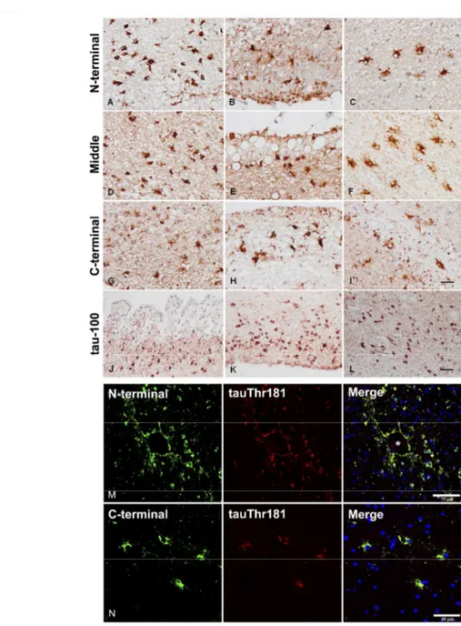
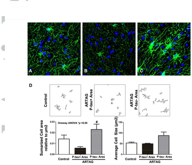
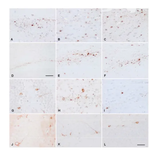
In agreement with our previous characterization of TSAs in AD (37), THAs in the present series were stained with specific tau antibodies against phospho-Thr181, phospho-Ser199, phospho-Thr231, phospho-Ser262, and phospho-Ser422, double-phosphorylation sites Ser202-Thr205 (clone AT8) and phospho-Ser396-404 (PHF1), conformational tau modifications at amino acids 5-15 (Alz50), and amino acids 312-322 (MC-1). TSAs were negative with antibodies against tau truncated at aspartic acid 421 (tau-C3). In addition, TSAs were stained with antibodies 499 (recognizing amino acids 14-26), 229 (amino acids 229-233), and 394 (amino acids 394-398), and with antibody anti-tau-100 (phospho-Thr212/Ser214) (Figure 1 A-L). Double-labeling immunofluorescence and confocal microscopy with antibody 499 and specific tau antibody against phospho-Thr181, and with antibody 394 and anti-tau phospho-Thr181, disclosed 499 and 394 immunoreactivity in all TSAs (Figure 1 M, N). TSAs, as revealed with GFAP immunohistochemistry, showed small size and reduced numbers of branches in frontal white matter, temporal white matter, subpial region, and subependymal region (Figure 2). This was in contrast not only with astrocytes in control, younger individuals but especially with neighbouring reactive GFAP-immunoreactive astrocytes in the same cases. Double-labeling immunofluorescence and confocal microscopy confirmed reduced GFAP immunoreactivity in TSAs (Figure 3).
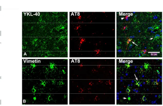
YKL-40, used as a marker of inflammatory astrocytes (38), and vimentin was expressed in a subpopulation of astrocytes in control and ARTAG cases. The number of vimentin and YKL-40 positive astrocytes was higher in ARTAG when compared with controls in agreement with reactive astrogliosis (12). However, double-labelling immunofluorescence and confocal microscopy revealed reduced size and decreased number of immunoreactive processes in TSAs (Figure 4)
Band pattern of tau in sarkosyl-insoluble fractions
Dissected basal forebrain and medulla oblongata from ARTAG cases with abundant TSAs were processed in parallel with samples of the frontal cortex from AD cases stage V. Three phospho-tau bands (anti-tau phopho-Ser422) of 68kDa, 64kDa, and 60kDa were seen in the frontal cortex of cases with AD. In addition, several bands of
molecular weight between 50kDa and 30kDa together with a lower band of about 23kDa, of variable intensity among cases, were found in AD. These corresponded to two bands of 4R tau between 68 and 60kDa, and several bands of lower molecular stained visualized with specific anti-4R antibodies, respectively. In contrast, the band pattern of ARTAG phospho-tau using the same antibody and processed in parallel with AD samples showed two bands of 68kDa and 64kDa, and several bands of lower molecular weight corresponding to truncated forms of tau. The lower phospho-tau band of about 23kDa was absent in ARTAG cases. Curiously, one sample of the medulla oblongata showed a very weak band of 68kDa and a weak double-band at 64 kDa. Parallel membranes blotted with 4Rtau antibodies showed a doublet of 64kDa (Figure 5).
3Rtau and 4Rtau ratios
Although not significant, 3Rtau and 4Rtau mRNA expression levels were reduced in AD and ARTAG cases when compared with controls. The ratio 3Rtau/4Rtau was also reduced in AD and ARTAG cases when compared with controls. Yet no differences were seen between AD and ARTAG (Supplementary Figure 1)
SOD2, AQP4 and GLT-1 immunoreactivity
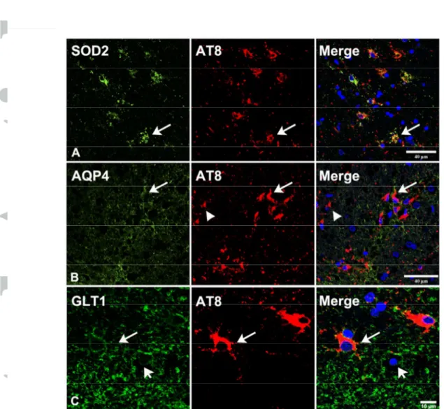
Single immunohistochemistry showed increased SOD2 immunoreactivity in subpial, subependymal, and some perivascular astrocytes, and in astrocytes of the white matter and basal forebrain in all cases with ARTAG when compared with middle-aged individuals. These findings were consistent with senescent modifications of astrocytes (18). Double-labeling immunofluorescence to SOD2 and phospho-tau clone AT8 revealed that TSAs and non-TSAs in the same tissue section were immunoreactive to SOD2 (Figure 6A).
AQP4 immunoreactivity was variable from one case to another, but in general terms it was more pronounced in the molecular layer, subependymal region, and basal forebrain in ARTAG in comparison with control samples. Individual variations did not permit a realistic quantitative validation in the present series. However, double-labeling immunofluorescence to AQP4 and tau AT8 in samples of the temporal white matter revealed AQP4 immunoreactivity at the cell membrane of TSAs. A similar pattern was seen in other glial cells in the temporal white matter with no phospho-tau deposition (Figure 6B).
GLT-1 immunoreactivity was present in astrocytes of the basal forebrain, temporal white matter, subpial region at the level of the medulla oblongata, and subependymal regions (the regions here assessed) in control and ARTAG cases. Double-labeling immunofluorescence and confocal microscopy to GLT-1 and hyper-phosphorylated tau
(clone AT8) identified GLT-1 immunoreactivity at the cell membrane of TSAs and non-TSAs in the same tissue section in ARTAG (Figure 6C).
Phosphoproteomics
Phosphosites differences between control and ARTAG cases, as seen in the heat map in Figure 7A, were imputated as MNAR. One-hundred twenty-two phosphosites showed higher abundance in ARTAG when compared to controls, and thirty-six phosphosites were more abundant in controls when compared to ARTAG (Supplementary Table 1). Since some proteins showed various sites of phosphorylation the total number of differentially phosphorylated proteins was 109 and 31, respectively, for ARTAG and controls (Table III). Several phosphorylated proteins with identification of phosphorylation positions are shown in Table IV. Among them, GFAP was found to be phosphorylated at positions 8, 14, 82, and 424, and aquaporin 4 was phosphorylated at position 273. Interestingly, many neuronal and glial proteins were differentially phosphorylated in ARTAG. These include proteins of the cytoskeleton, kinases, proteins linked to calcium/calmodulin signalling, cAMP signalling and DNA repair, nuclear and nucleolar regulators, proteins linked to tight junctions, proteins linked to proteolysis, and synaptic proteins, among others (Table III). A smaller number of proteins showed decreased levels of phosphorylated peptides in ARTAG when compared with controls (Table III). Regarding microtubule associated proteins (MAPs), phosphorylation of MAP-tau (as expected), MAP1A, MAP1AA, MAPK1S and MAP2 was greater in ARTAG, whereas phosphorylation of MAP1B, MAP4 and MAP6 was higher in controls.
Phosphomotif-enriched analysis revealed a motif enriched in ARTAG (SP). This motif is a phosphorylation site used for a wide number of kinases such as MAPK (including p38) and CDKs (Figure 7B).
Immunohistochemistry of selected phosphorylated proteins in ARTAG
Double-labeling immunofluorescence revealed that TSAs co-localized GFAP-P, PKA-P, and p38-P. GFAP-P was expressed in hyper-phosphorylated tau-bearing and non-hyper-phosphorylated tau-bearing astrocytes in the temporal white matter. TSAs co-localized active forms (phosphorylated at specific sites) of kinases p38 and PKA α/β. Tuberin-P was expressed in neurons of the hippocampus but not in tau-containing astrocytes (Figure 8).
Tau seeding in inoculated mice
Mice injected with AD fractions showed phospho-tau deposits in the fimbria, corpus callosum, and neurons of the hippocampus seven months after inoculation. The distribution and profile in the two mice inoculated with homogenates from two different AD cases was the same as the pattern already described by others (18) thus validating
those observations and serving as positive controls in the present series (data not shown).
The first group of six ARTAG-treated mice were injected at the age of seven months, three in the ventricle and three in the hippocampus, and killed at the age of ten months. No tau deposits were seen following intraventricular injection. However, intracellular deposits of hyper-phosphorylated tau were found in threads and in a few cells in the fimbria, lateral corpus callosum, and lateral part of the hippocampus in two of the three mice injected in the hippocampus (Figure 9A and B).
A second group of mice was injected into the hippocampus at the age of three months and killed at the age of ten months; four animals were inoculated with ARTAG and two with vehicle alone. All ARTAG-injected animals showed hyper-phosphorylated tau deposits in the fimbria, fornix, corpus callosum traversing the contralateral hemisphere, and the hippocampal complex including gyrus dentatus, CA1 and CA3 region of the hippocampus, and subiculum (Figure 9C-I). Curiously, cells were positive to 4Rtau (Figure 9J), as expected, but also to 3Rtau (Figure 9K and L). Double-labeling immunofluorescence and confocal microscopy disclosed hyper-phosphorylated tau deposition in neurons and astrocytes, as revealed with anti-NeuN and anti-GFAP antibodies, respectively, in animals with short interval between inoculation and examination (Figure 10A and B); microglia, as revealed with Iba-1 antibodies, did not contain hyper-phosphorylated tau (Figure 10C). Similar intracellular localization of hyper-phosphorylated tau was found in neurons and astrocytes in animals with long interval between inoculation and neuropathological examination, as revealed with the same antibodies; microglia did not contain hyper-phosphorylated tau deposits (Figure 11A, C, D, E and F). In addition, hyper-phosphorylated tau was found in oligodendroglia mainly in corpus callosum as revealed by double-labeling immunofluorescence using anti-Olig2 antibodies (Figure 11G and H). Finally, the possibility that hyper-phosphorylated tau deposition was an active process within seeds was sustained by co-localization of active p38 kinase (phosphorylated at Thr180-Tyr182) only in cells containing hyper-phosphorylated tau (Figure 11B).
Discussion
The morphological characteristics and distribution of TSAs in this ARTAG series with no tau deposition in neurons or with NFTs and pre-tangles restricted to the entorhinal and transentorhinal cortex are similar to those described in ARTAG associated with tauopathies (28, 29, 31, 36). The pattern of tau phosphorylation of TSAs identified here does not differ from TSAs associated with other tauopathies including AD and AGD (14, 37). Additional features of THAs include lack of evidence of truncated tau using specific antibodies against C-terminal and N-terminal regions, together with negativity to tau-C3 which recognizes truncated tau at Asp421. Gel electrophoresis and western blotting of sarkosyl-insoluble fractions show two bands of 68 kDa and 64 kDa (in some areas as a doublet) and several weaker bands of lower molecular weight. In no case are triple bands of 68kDa, 64kDa, and 60kDa and a lower band of truncated tau of about 20kDa, typical of AD, found in ARTAG. In conclusion, TSAs are composed of hyper-phosphorylated 4Rtau with features of pre-tangles lacking tau truncation at terminal regions.
Considering that TSAs are present in aged brains, further analysis was carried out to learn about categorization of these astrocytes and functional implications of this particular astrocytopathy (12). TSAs show reduced GFAP expression, reduced volume, and reduced numbers of branches. This was further recognized using anti-vimentin and YKL-40 antibodies. SOD2 immunoreactivity, which is augmented in senescent astrocytes (48), is increased in TSAs when compared with astrocytes of younger individuals. Yet these features are not restricted to TSAs but also apply to neighboring astrocytes with no hyper-phosphorylated tau deposition. Moreover, neighboring astrocytes show increased area coverage, and increased GFAP, vimentin and YKL-40 immunoreactivity. Therefore, TSAs are a subpopulation of astrocytes showing senescent features (18, 46, 48).
Astrocytes have specific functions mediated by selective molecules and pathways, among them the modulation of glutamate transport through astroglial glutamate transporter solute carrier family 1, member 2 (GLT-1/ EAAT2) (39, 42). Double-labeling immunofluorescence and confocal microscopy identifies GLT-1 immunoreactivity at the cell membrane of TSAs and non-TSAs in the same tissue sections. Decreased GLT-1 immunoreactivity in tau-containing astrocytes has been reported in a familial behavioral variant of frontotemporal dementia associated with astrocyte-predominant tauopathy, in which astrocytes show advanced stage of tangle formation with tau truncation and ubiquitination (13). GLT-1 expression is also altered in transgenic mice with astroglial tau phosphorylation (10); tau in transgenic mice is abnormally phosphorylated,
ubiquitinated, and filamentous; abnormal astrocytes are variably positive with thioflavine S (17). Therefore, relative preservation of GLT-1 in TSAs compared with astrocytes in the previous conditions may be related to the less advanced stage of cellular damage linked to tau phosphorylation and truncation in ARTAG.
Another important function of astrocytes is the regulation of water homeostasis through specific water channel aquaporin 4. AQP4 is expressed at the cell membrane of astrocytes, particularly at the perivascular, subpial, and subependymal interfaces (2). Double-labeling immunofluorescence to AQP4 and tau AT8 in samples of the temporal white matter shows AQP4 immunoreactivity in TSAs and in other astrocytes. Due to individual variations we are not able to conclude that AQP4 immunoreactivity is specifically altered in TSAs. However, this does not dismiss the possibility of blood-brain barrier dysfunction in ARTAG as suggested in other studies (33, 35).
The study of the phosphoproteome in relatively pure cases of ARTAG, although limited by the small number of cases, avoids possible contamination of altered tau in ARTAG associated with other tauopathies. Several proteins in ARTAG have differential phosphorylation marks when compared with controls, among them AQP4 at Thr273 and GFAP at positions 8, 14, 82, and 424. GFAP phosphorylation at Ser8 was further validated by immunohistochemistry and immunofluorescence and characterized by confocal microscopy. GFAP is phosphorylated at different sites of the amino-terminal, thus modulating filaments assembly (24, 43, 52). Several kinases phosphorylate GFAP at specific sites; Cdc2 kinase (cyclin dependent kinase 1) phosphorylates GFAP at Ser8 whereas Ca(2+)-CaM-dependent protein kinase II phosphorylates GFAP at Ser13, Ser17, Ser34, and Ser389 (56). The role of cAMP-dependent and Ca2+ -dependent protein kinases on GFAP phosphorylation has also been studied in digitonin-permeabilized astrocytes exposed to cAMP and Ca2+ which increases the phosphorylation state of GFAP (25). Despite these achievements, little is known about the effect of combined phosphorylation at different sites on GFAP filament assembly (52).
Phosphorylation of some aquaporins has been proposed as regulating their water permeability via gating of the channel itself. Certain protein kinases phosphorylate AQP4 thus facilitating AQP4 trafficking and water permeability (20, 26). However, studies in Xenopus oocytes have shown that phosphorylation at COOH-terminal residues Ser180, Ser276, Ser285, Ser315, Ser316, Ser321, and Ser322 does not modulate trafficking or channel gating (1, 3, 47). Thus, regulatory patterns and physiological roles for AQP4 remain to be fully explored (2); functional studies are needed to learn about the functional role of AQP4 phosphorylation at Thr273 and its
Phosphoproteomics has also evidenced increased phosphorylation marks in several kinases including calcium/calmodulin-dependent protein kinase type II subunit gamma, cAMP-dependent protein kinase catalytic subunit alpha, c-Jun-amino-terminal kinase-interacting protein 3, cyclin-dependent kinase 12, protein kinase C alpha type, and protein kinase C epsilon type. Since a typical protein kinase must distinguish one from among a few hundred bona fide phosphorylation sites in a background of approximately 700,000 potentially phosphorylatable residues (57), it is premature to advance which are the substrates of such activated kinases in the context of ARTAG. In spite of the large number of putative targets, double-labeling immunofluorescence has shown co-localization of active kinases p38 and PKA α/β with hyper-phosphorylated tau in TSAs in ARTAG. Moreover, many other proteins are phosphorylated at specific sites in ARTAG including MAP7 domain-containing protein 1, associated protein 1A, associated protein 1AA, microtubule-associated protein 1B, microtubule-microtubule-associated protein 1S, and microtubule-microtubule-associated protein 2, in addition to microtubule-associated protein tau. Furthermore, increased phosphorylation of microtubule-associated protein 1B, microtubule-associated protein 4, and microtubule-associated protein 6 is found in controls compared with ARTAG. Several neuronal proteins, such as synaptic proteins synapsin-1, synaptophysin, synaptopodin and synaptophilin; and tuberin, are also differentially phosphorylated in ARTAG, indicating that differences in the phosphorylation state of various cytoskeletal and non-cytoskeletal proteins are not limited to astrocytes but also compromise neurons. Little is known about functional implications of tuberin phosphorylation. However, since tuberin is linked to microtubule biology through ROCK2 signaling (15), it can be suggested that changes in tuberin phosphorylation modify microtubule organization.
Several studies in AD have located differentially phosphorylated proteins in the cerebral cortex and hippocampus using bi-dimensional gel electrophoresis and mass spectrometry. Most proteins are linked to energy metabolism, neuronal plasticity, signal transduction, and oxidative stress responses (11, 55, 59). These studies also identify GFAP as a phosphorylated protein in AD. More recently, phosphopeptide enrichment and LC-MS/MS assessment have identified more than one thousand phosphorylated proteins in AD (53). In another study, most of the core phosphoproteins are directly connected and form a functional network linked to synaptic spine formation. Systems biology analyses suggest that over-activated kinases including protein kinases C and calmodulin-dependent kinases initiate synapse pathology (51). The change of the core network starts at a preclinical stage even before histological β-amyloid deposition (51).
The study of Tagawa et al. (51) points to similarities between AD and ARTAG regarding phosphorylation of similar kinases and synaptic proteins. These modifications in AD are independent of β-amyloid and phosphor-tau deposition, and in ARTAG cases independent of neurofibrillary tangle pathology as well. In addition, GFAP phosphorylation is not restricted to ARTAG but also occurs in AD.
The present observations suggest that TSAs are just part of the modifications occurring in ARTAG. TSAs are developed in selected populations of senescent astrocytes in a context of increased protein phosphorylation among which are several kinases with the capacity to phosphorylate a large number of substrates. These observations in several regions not associated with neuronal tau pathology in ARTAG identify changes which are common to AD, and they may explain the frequency of ARTAG in AD and other tauopathies in the elderly.
Finally, the present study shows for the first time the capacity of tau seeding of sarkosyl-insoluble fractions enriched in hyper-phosphorylated tau from selected ARTAG cases in which TSAs are the only source of abnormal hyperphosphorylated tau. Therefore, certain subpopulations of astrocytes may be primary carriers for abnormal tau seeding. Moreover, tau seeding in our model occurred not only in host astrocytes but also in neurons and oligodendroglia, thus pointing out that neurons and oligodendrocytes can be seed targets of astrocyte-derived hyper-phosphorylated tau. This is important, as hyper-phosphorylated tau inclusions are frequent in oligodendrocytes in AGD and in other tauopathies (16, 27). Present observations support the hypothesis that TSAs may facilitate tau hyper-phosphorylation and deposition in neurons and other glial cells in human tauopathies, and they point to the cardinal role of astrocytopathy in the pathogenesis of neurodegenerative diseases with abnormal protein aggregates. The morphology of tau-containing astrocytes after ARTAG inoculation in mice does not have the morphology of TSAs. Its distribution and that of tau-containing neurons and oligodendrocytes in ARTAG-inoculated mice correlate with the inoculation site rather than with the distribution of TSAs in ARTAG. In fact, the same pattern is seen following the inoculation of AD homogenates, in agreement with previous observations with AD extracts (19).Therefore, additional factors must be examined in the future mainly those related with the age of the inoculated animals. ARTAG is a tauopathy related to aging and TSAs are senescent astrocytes. Inoculation of ARTAG (and AD) homogenates in old animals is mandatory to move closer to the environment occurring in old age.
Acknowledgements
We wish to thank the CRG–UPF proteomics unit, and Tom Yohannan for editorial assistance
References
1. Assentoft M, Kaptan S, Fenton RA, Hua SZ, de Groot BL, MacAulay (2013) Phosphorylation of rat aquaporin-4 at Ser(111) is not required for channel gating. Glia 61: 1101-1112.
2. Assentoft M, Larsen BR, MacAulay N (2015) Regulation and function of AQP4 in the central nervous system. Neurochem Res 40: 2615-2627.
3. Assentoft M, Larsen BR, Olesen ET, Fenton RA, MacAulay N (2014) AQP4 plasma membrane trafficking or channel gating is not significantly modulated by phosphorylation at COOH-terminal serine residues. Am J Physiol Cell Physiol 307: C957-65.
4. Boluda S, Iba M, Zhang B, Raible KM, Lee VM, Trojanowski JQ (2015) Differential induction and spread of tau pathology in young PS19 tau transgenic mice following intracerebral injections of pathological tau from Alzheimer’s disease or corticobasal degeneration brains. Acta Neuropathol 129:221–237.
5. Braak H, Alafuzoff I, Arzberger T, Kretzschmar H, Del Tredici K. Staging of Alzheimer disease-associated neurofibrillary pathology using paraffin sections and immunocytochemistry. Acta Neuropathol 2006; 112: 389-404.
6. Braak H, Braak E (1991) Neuropathological staging of Alzheimer-related changes. Acta Neuropathol 82: 239-259.
7. Clavaguera F, Bolmont T, Crowther RA, Abramowski D, Frank S, Probst A, Fraser G, Stalder AK, Beibel M, Staufenbiel M, Jucker M, Goedert M, Tolnay M (2009) Transmission and spreading of tauopathy in transgenic mouse brain. Nat Cell Biol 11:909-913.
8. Clavaguera F, Lavenir I, Falcon B, Frank S, Goedert M, Tolnay M (2013) “Prion-like” templated misfolding in tauopathies. Brain Pathol 23:342–349
9. Crary JF, Trojanowski JQ, Schneider JA, Abisambra JF, Abner EL, Alafuzoff I, Arnold SE, Attems J, Beach TG, Bigio EH, Cairns NJ, Dickson DW, Gearing M,
Kofler J, Kukull WA, Mackenzie IR, Masliah E, McKee A, Montine TJ, Murray ME, Neltner JH, Santa-Maria I, Seeley WW, Serrano-Pozo A, Shelanski ML, Stein T, Takao M, Thal DR, Toledo JB, Troncoso JC, Vonsattel JP, White CL 3rd, Wisniewski T, Woltjer RL, Yamada M, Nelson PT (2014) Primary age-related tauopathy (PART): a common pathology associated with human aging. Acta Neuropathol 128: 755-766. 10. Dabir DV, Robinson MB, Swanson E, Zhang B, Trojanowski JQ, Lee VM, Forman MS (2006) Impaired glutamate transport in a mouse model of tau pathology in astrocytes. J Neurosci 26: 644-654.
11. Di Domenico F, Sultana R, Barone E, Perluigi M, Cini C, Mancuso C, Cai J, Pierce WM, Butterfield DA (2011) Quantitative proteomics analysis of phosphorylated proteins in the hippocampus of Alzheimer's disease subjects. J Proteomics 74: 1091-1103. 12. Ferrer I (2017) Diversity of astroglial responses across human neurodegenerative disorders and brain aging. Brain Pathol 27: 645–674.
13. Ferrer I, Legati A, García-Monco JC, Gomez-Beldarrain M, Carmona M, Blanco R, Seeley WW, Coppola G (2015) Familial behavioral variant frontotemporal dementia associated with astrocyte-predominant tauopathy. J Neuropathol Exp Neurol 74: 370-379.
14. Ferrer I, López-González I, Carmona M, Arregui L, Dalfó E, Torrejón-Escribano B, Diehl R, Kovacs GG (2013) Glial and neuronal tau pathology in tauopathies: characterization of disease-specific phenotypes and tau pathology progression. J Neuropathol Exp Neurol 73: 81-97
15. Ferrer I, Mohan P, Chen H, Castellsague J, Gómez-Baldó L, Carmona M, García N, Aguilar H, Jiang J, Skowron M, Nellist M, Ampuero I, Russi A, Lázaro C, Maxwell CA, Pujana MA (2014) Tubers from patients with tuberous sclerosis complex are characterized by changes in microtubule biology through ROCK2 signalling. J Pathol 233: 247-257.
16. Ferrer I, Santpere G, van Leeuwen FW (2008) Argyrophilic grain disease. Brain 146: 1640-1651.
17. Forman MS, Lal D, Zhang B, Dabir DV, Swanson E, Lee VM, Trojanowski JQ (2005) Transgenic mouse model of tau pathology in astrocytes leading to nervous system degeneration. J Neurosci 25: 3539-3550.
18. García-Matas S, Gutierrez-Cuesta J, Coto-Montes A, Rubio-Acero R, Díez-Vives C, Camins A, Pallàs M, Sanfeliu C, Cristòfol R (2008) Dysfunction of astrocytes in senescence-accelerated mice SAMP8 reduces their neuroprotective capacity. Aging Cell 7: 630-640.
19. Guo JL, Narasimhan S, Changolkar L, He Z, Stieber A, Zhang B, Gathagan RJ, Iba M, McBride JD, Trojanowski JQ, Lee VMY (2016) Unique pathological tau conformers from Alzheimer’s brains transmit tau pathology in nontransgenic mice. J Exp Med 213: 2635-2654.
20. Han Z, Wax MB, Patil RV (1998) Regulation of aquaporin-4 water channels by phorbol ester-dependent protein phosphorylation. J Biol Chem 273: 6001-6004.
21. Hashimoto N, Takeuchi T, Ishihara R, Ukai K, Kobayashi H, Iwata K, Mizuno Y, Yamaguchi H, Shibayama H (2003) Glial fibrillary tangles in diffuse neurofibrillary tangles with calcification. Acta Neuropathol 106: 150-156.
22. Ikeda K, Akiyama H, Arai T, Nishimura T (1998) Glial tau pathology in neurodegenerative diseases: their nature and comparison with neuronal tangles. Neurobiol Aging 19 Suppl: S85-91.
23. Ikeda K, Akiyama H, Kondo H, Haga C, Tanno E, Tokuda T, Ikeda S (1995) Thorn-shaped astrocytes: possibly secondarily induced tau-positive glial fibrillary tangles. Acta Neuropathol 90: 620-625.
24. Inagaki M, Nakamura Y, Takeda M, Nishimura T, Inagaki N (1994) Glial fibrillary acidic protein: dynamic property and regulation by phosphorylation. Brain Pathol 4: 239-243.
25. Karla J, Goofried C, Tramontina F, Dunkley P, Rodnighta R, Gonçalves CA (2000) GFAP phosphorylation studied in digitonin-permeabilized astrocytes: standardization of conditions. Brain Res 853; 32-40.
26. Kitchen P, Day RE, Taylor LH, Salman MM, Bill RM, Conner MT (2015) Identification and molecular mechanisms of the rapid tonicity-induced relocalization of the aquaporin 4 channel. J Biol Chem 290: 16873-16881.
27. Kovacs GG (2015) Invited review: Neuropathology of tauopathies: principles and practice. Neuropathol Appl Neurobiol 41: 3-23.
28. Kovacs GG, Ferrer I, Grinberg LT, Alafuzoff I, Attems J, Budka H, Cairns NJ, Crary JF, Duyckaerts C, Ghetti B, Halliday GM, Ironside JW, Love S, Mackenzie IR, Munoz DG, Murray ME, Nelson PT, Takahashi H, Trojanowski JQ, Ansorge O, Arzberger T, Baborie A, Beach TG, Bieniek KF, Bigio EH, Bodi I, Dugger BN, Feany M, Gelpi E, Gentleman SM, Giaccone G, Hatanpaa KJ, Heale R, Hof PR, Hofer M, Hortobágyi T, Jellinger K, Jicha GA, Ince P, Kofler J, Kövari E, Kril JJ, Mann DM, Matej R, McKee AC, McLean C, Milenkovic I, Montine TJ, Murayama S, Lee EB, Rahimi J, Rodriguez RD, Rozemüller A, Schneider JA, Schultz C, Seeley W, Seilhean D, Smith C, Tagliavini F, Takao M, Thal DR, Toledo JB, Tolnay M, Troncoso JC, Vinters HV, Weis S, Wharton SB, White CL 3rd, Wisniewski T, Woulfe JM, Yamada M, Dickson DW (2016) Aging-related tau astrogliopathy (ARTAG): harmonized evaluation strategy. Acta Neuropathol 131: 87-102.
29. Kovacs GG, Lee VM, Trojanowski JQ (2017) Protein astrogliopathies in human neurodegenerative diseases and aging. Brain Pathol 27: 675-690.
30. Kovacs GG, Molnár K, László L, Ströbel T, Botond G, Hönigschnabl S, Reiner-Concin A, Palkovits M, Fischer P, Budka H (2011) A peculiar constellation of tau pathology defines a subset of dementia in the elderly. Acta Neuropathol 122: 205-222. 31. Kovacs GG, Robinson JL, Xie SX, Lee EB, Grossman M, Wolk DA, Irwin DJ, Weintraub D, Kim CF, Schuck T, Yousef A, Wagner ST, Suh E, Van Deerlin VM, Lee VM, Trojanowski JQ (2017) Evaluating the patterns of aging-related tau astrogliopathy unravels novel insights into brain aging and neurodegenerative diseases. J Neuropathol Exp Neurol 76: 270-288.
32. Kovacs GG, Xie SX, Lee EB, Robinson JL, Caswell C, Irwin DJ, Toledo JB, Johnson VE, Smith DH, Alafuzoff I, Attems J, Bencze J, Bieniek KF, Bigio EH, Bodi I, Budka H, Dickson DW, Dugger BN, Duyckaerts C, Ferrer I, Forrest SL, Gelpi E, Gentleman SM, Giaccone G, Grinberg LT, Halliday GM, Hatanpaa KJ, Hof PR, Hofer
M, Hortobágyi T, Ironside JW, King A, Kofler J, Kövari E, Kril JJ, Love S, Mackenzie IR, Mao Q, Matej R, McLean C, Munoz DG, Murray ME, Neltner J, Nelson PT, Ritchie D, Rodriguez RD, Rohan Z, Rozemuller A, Sakai K, Schultz C, Seilhean D, Smith V, Tacik P, Takahashi H, Takao M, Rudolf Thal D, Weis S, Wharton SB, White CL 3rd, Woulfe JM, Yamada M, Trojanowski JQ (2017) Multiple assessment of aging-related astrogliopathy. J Neuropathol Exp Neurol 76: 605-619.
33. Kovacs GC, Yousef A, Kaindl S, Lee VM, Trojanowski JQ (2017) Connexin -43 and aquaporin-4 are markers of ageing-related tau astrogliopathy (ARTAG)-related astroglial response. Neuropathol Appl Neurobiol Jul 29. Doi: 10.1111/nan.12427 [Epub ahead of print]
34. Lewis J, Dickson DW (2016) Propagation of tau pathology: hypotheses, discoveries, and yet unresolved questions from experimental and human brain studies. Acta Neuropathol 131: 27-48.
35. Ling H, Neal JW, Revesz T (2017) Evolving concepts of chronic traumatic encephalopathy as a neuropathological entity. Neuropathol Appl Neurobiol 43: 467-476.
36. Liu AKL, Goldfinger MH, Questari HE, Pearce RKB, Gentleman SM (2016) ARTAG in the basal forebrain: widening the constellation of astrocytic tau pathology. Acta Neuropathol Commun 4: 59.
37. López-González I, Carmona M, Blanco R, Luna-Muñoz J, Martínez-Mandonado A, Mena R, Ferrer I (2013) Characterization of thorn-shaped astrocytes in white matter of temporal lobe in Alzheimer's disease brains. Brain Pathol 23:144-153.
38. Llorens F, Thüne K, Tahir W, Kanata E, Diaz-Lucena D, Xanthopoulos K, Kovatsi E, Pleschka C, Garcia-Esparcia P, Schmitz M, Ozbay D, Correia S, Correia Â, Milosevic I, Andréoletti O, Fernández-Borges N, Vorberg IM, Glatzel M, Sklaviadis T, Torres JM, Krasemann S, Sánchez-Valle R, Ferrer I, Zerr I (2017) YKL-40 in the brain and cerebrospinal fluid of neurodegenerative dementias. Mol Neurodegener 12: 83. 39. Maragakis NJ, Rothstein JD (2006) Mechanisms of disease: astrocytes in neurodegenerative disease. Nat Clin Pract Neurol 2: 679-689.
40. Muñoz DG Woulfe J, Kertesz A (2007) Argyrophilic thorny astrocyte clusters in association with Alzheimer's disease pathology in possible primary progressive aphasia. Acta Neuropathol 114: 347-357.
41. Narasimhan S, Guo JL, Changolkar L, Stieber A, McBride JD, Silva LV, He Z, Zhang B, Gathagan RJ, Trojanowski JQ, Lee VMY (2017) Pathological tau strains from human brains recapitulate the diversity of tauopathies in non-transgenic mouse brain. J Neurosci 37: 11406-11423.
42. Nedergaard M, Takano T, Hansen AJ (2002) Beyond the role of glutamate as a neurotransmitter. Nat Rev Neurosci 3: 748-755.
43. Noetzel MJ (1990) Phosphorylation of the glial fibrillary acidic protein. J Neurosci Res 27: 184-192.
44. Osborn LM, KamphuisW, WadmanWJ, Hol EM (2016) Astrogliosis: an integral player in the pathogenesis of Alzheimer’s disease. Prog Neurobiol 144:121–141. 45. Pekny M, Pekna M, Messing A, Steinhäuser C, Lee JM, Parpura V, Hol M, Sofroniew MW, Verkhratsky A (2016) Astrocytes: a central element in neurological diseases. Acta Neuropathol 131:323–345.
46. Rodríguez JJ, Yeh CY, Terzieva S, Olabarria M, Kulijewicz-Nawrot M, Verkhratsky A (2014) Complex and region-specific changes in astroglial markers in the aging brain. Neurobiol Aging 35: 15-23.
47. Sachdeva R, Singh B (2014) Phosphorylation of Ser-180 of rat aquaporin-4 shows marginal affect on regulation of water permeability: molecular dynamics study. J Biomol Struct Dyn 32: 555-566.
48. Salminen A, Ojala J, Kaarniranta K, Haapasalo A, Hiltunen M, Soininen H (2011) Astrocytes in the aging brain express characteristics of senescence-associated secretory phenotype. Eur J Neurosci 34: 3-11.
49. Schultz C, Ghebremedhin E, Del Tredici K, Rüb U, Braak H (2004) High prevalence of thorn-shaped astrocytes in the aged human medial temporal lobe. Neurobiol Aging 25: 397-405.
50. Schwartz D, Gygi SP (2005) An iterative statistical approach to the identification of the protein phosphorylation motifs from large-scale data sets. Nature Biotech 23: 1391-1398.
51. Tagawa K, Homma H, Saito A, Fujita K, Chen X, Imoto S, Oka T, Ito H, Motoki K, Yoshida C, Hatsuta H, Murayama S, Iwatsubo T, Miyano S, Okazawa H (2015) Comprehensive phosphoproteome analysis unravels the core signaling network that initiates the earliest synapse pathology in preclinical Alzheimer's disease brain. Hum Mol Genet 24: 540-558.
52. Takemura M, Gomi H, Colucci-Guyon E, Itohara S (2002) Protective role of phosphorylation in turnover of glial fibrillary acidic protein in mice. J Neurosci 22: 6972-6979.
53. Tan H, Wu Z, Wang H, Bai B, Li Y, Wang X, Zhai B, Beach TG, Peng J (2015) Refined phosphopeptide enrichment by phosphate additive and the analysis of human brain phosphoproteome. Proteomics 15: 500-507.
54. Tolnay M, Braak H (2011) Argyrophilic grain disease. In: Neurodegeneration, the Molecular Pathology of Dementia and Molecular Disorders, Dickson DW, Weller RO (eds.), pp. 165-170, Wiley-Blackwell, Oxford.
55. Triplett JC, Swomley AM, Cai J, Klein JB, Butterfield DA (2016) Quantitative phosphoproteomic analyses of the inferior parietal lobule from three different pathological stages of Alzheimer's disease. J Alzheimers Dis 49: 45-62.
56. Tsujimura K, Tanaka J, Ando S, Matsuoka Y, Kusubata M, Sugiura H, Yamauchi T, Inagaki M (1994) Identification of phosphorylation sites on glial fibrillary acidic protein for cdc2 kinase and Ca(2+)-calmodulin-dependent protein kinase II. J Biochem 116: 426-434.
57. Ubersax JA, Ferrell JE (2007) Mechanisms of specificity in protein phosphorylation. Nature Rev Mol Cell Biol 8: 530-541.
59. Zahid S, Oellerich M, Asif AR, Ahmed N (2012) Phosphoproteome profiling of substantia nigra and cortex regions of Alzheimer's disease patients. J Neurochem 121: 954-963.
Table I: Antibodies used, origins, dilutions, and suppliers
Table II: Cases and distribution of lesions in the present series of pure ARTAG
Table III: Total list of differentially phosphorylated proteins in ARTAG compared with
controls, and in controls compared with ARTAG
Table IV: List of phosphorylated proteins in ARTAG and identification of
phosphorylation sites (probability site value, pRS, greater than 75% is highlighted in yellow)
Supplementary Table 1: Whole data of one-hundred twenty-two differentially occupied
phosphosites differentially expressed in ARTAG compared with controls, and 36 phosphosites differentially occupied in controls when compared with ARTAG
Figure legends
Figure 1: A-L: single immunohistochemistry with anti-tau antibodies directed to
N-terminal, middle, and C-terminal regions of tau. A, D, G, J: subependymal astrocytes; B, E, H, K: subpial astrocytes; C, F, I, L: temporal white matter. A-C: antibody 499 (amino acids 14-26; amino terminal); D-F: antibody 229 (amino acids 229-233, middle regioin); G-I: antibody 394 (amino acids 394-398, C-terminal ); J-L: antibody tau-100 (P-tau Thr212-Ser214). TSAs are stained with all these antibodies. Paraffin sections slightly counterstained with haematoxylin; A-I, bar in I = 25μm; J-L, bar in L = 100μm. M, N: double-labeling immunofluorescence and confocal microscopy using (M) antibody 499 (green) and P-tau Thr181 (red); and (N) antibody 394 (green) and P-tau Thr181 (red) in the vicinity of a blood vessel (M, asterisk) and in the temporal white matter. N-terminal and C-terminal tau segments are co-expressed with phosphorylated tau. Paraffin sections; nuclei (blue) are stained with DRAQ5TM; M, bar = 75μm; N, bar = 40μm.
Figure 2: GFAP-immunoreactive astrocytes in white matter (A-D) (A) and ARTAG
cases in TSAs-rich regions (B) and neighboring regions without TSAs astrocytes (C). GFAP-positive astrocytes in ARTAG regions without TSAs have larger branches when compared with controls. However, THAs have smaller size and reduced number of branches when compared with astrocytes in controls. Paraffin sections; nuclei (blue) are stained with DRAQ5 TM; bar = 50μm. Particle selection mascara of astrocytes is shown in white matter (D). Quantification of astrocytes areas per μm2 (left lower panels) and astrocyte average size (right lower panels). Graphs show mean ± SEM of three different sections. Quantifications analyzed by One-way ANOVA show significant difference among groups *p<0.05 in white matter astrocyte coverage area; Turkey post-hoc shows significant differences #p<0.05 in astrocyte coverage area between THAs (P-tau+) and neighboring P-tau negative (P-tau-) astrocytes in ARTAG.
Figure 3: Double-labeling immunofluorescence to GFAP (green) and AT8 (red)
showing the morphology of TSAs in the temporal white matter. Short arrow: cells only stained green; arrowhead: cells only stained red. Hyper-phosphorylated tau-containing astrocytes have reduced GFAP immunoreactivity. Paraffin sections; nuclei (blue) are stained with DRAQ5TM; bar = 50μm.
Figure 4: A: Double-labeling immunofluorescence to YKL-40 (green) and AT8 (red) in