TABLE OF CONTENTS
I. List of publications 3
II. List of abbreviations 4
III. Abstract 5
1. Introduction 6
1.1 Innate immune memory and trained immunity 6
1.2 Monocytes 8
1.3 GM-CSF and IL-3 10
1.3.1 GM-CSF and IL-3: role in inflammatory and autoimmune diseases 11
2. Aim of the study 13
3. Materials and Methods 14
3.1 Cell isolation and culture 14
3.2 ELISA and NAD quantification 14
3.3 Phospho-flow cytometry 15
3.4 Fluorescence, time-lapse and high-content microscopy 15
3.5 RNA isolation and Real Time RT-PCR 16
3.6 Statistical analysis 17
4. Results 18
4.1 GM-CSF and IL-3 priming enhances LPS-induced TNF-α production 18 4.2 GM-CSF and IL-3 priming modulate TNF-α production at post-transcriptional
level 23
4.3 NAD- dependent SIRT2, MAPKs ERK1/2 and P38 mediate the effect of GM-CSF
and IL-3 priming on TNF-α production 27
4.4 GM-CSF and IL-3 priming modulate monocyte renewal in a c-Myc dependent
5. Discussion 45
6. Conclusion 49
7. References 50
I. List of pubblications
The main body of the thesis is based upon the following publication:
• Borriello, F.*, Iannone, R.*, Di Somma, S.*, Loffredo, S., Scamardella, E., Galdiero, M.R., Varricchi, G., Granata, F., Portella, G., Marone, G. (2017) GM-CSF and IL-3 modulate human monocyte TNF-α production and renewal in in vitro models of trained immunity. Front Immunol. 7:680. doi: 10.3389/fimmu.2016.00680.
*First Author Other publications:
• Borriello, F.*, Iannone, R.*, Di Somma, S.*, Vastolo, V., Petrosino, G., Visconte, F., Raia, M., Scalia, G., Loffredo, S., Varricchi, G., Galdiero, M.R., Granata, F., Del Vecchio, L., Portella, G., Marone, G. (2017). Lipopolysaccharide-Elicited TSLPR Expression Enriches a Functionally Discrete Subset of Human CD14+ CD1c+ Monocytes. J Immunol. 198(9):3426-3435. doi: 10.4049/jimmunol.1601497.
*First Author
• Galdiero, M.R., Varricchi, G., Loffredo, S., Bellevicine, C., Lansione, T., Ferrara, A.L., Iannone, R., Di Somma, S., Borriello, F., Clery, E., Triassi, M., Troncone, G., Marone, G. (2018). Potential involvement of neutrophils in human thyroid cancer. PLoS One. 13(6):e0199740. doi: 10.1371/journal.pone.0199740.
• Passaro, C., Borriello, F., Vastolo, V., Di Somma, S., Scamardella, E., Gigantino, V., Franco, R., Marone, G., Portella, G. (2016). The oncolytic virus dl922-947 reduces IL- 8/CXCL8 and MCP-1/CCL2 expression and impairs angiogenesis and macrophage infiltration in anaplastic thyroid carcinoma. Oncotarget. 2016 Jan 12;7(2):1500-15. doi: 10.18632/oncotarget.6430.
II. List of abbreviations
CCL17/TARC: C-C Motif Chemokine Ligand 17/ T Cell-Directed CC Chemokine CCR2: C-C Motif Chemokine Receptor 2
CD14: Cluster of differentiation 14 CD16: Cluster of differentiation 16 CPT1: Carnitine palmitoyltransferase I HIF-1α: Hypoxia-inducible factor 1-alpha IFN- γ: Interferon gamma
IL-4: Interleukin 4 IL-13: Interleukin 13
JNK: c-Jun N-terminal kinase LPS: Lipopolysaccharide
M-CSF: macrophage colony- stimulating factor MEK1/2: Mitogen-activated protein kinase kinase ½ MTORC1: mammalian target of rapamycin complex 1 PHD: Prolyl hydroxylase
III. Abstract
GM-CSF and IL-3 are hematopoietic cytokines that modulate the effector functions of several immune cell subsets. In particular, GM-CSF and IL-3 exert a significant control on monocyte and macrophage effector functions, as assessed in experimental models of inflammatory and autoimmune diseases and in human studies. Here we sought to investigate the mechanisms and the extent to which GM-CSF and IL-3 modulate the pro-inflammatory, LPS-mediated, activation of human CD14+ monocytes taking into account the new concept of trained immunity (i.e. the priming stimulus modulates the response to subsequent stimuli mainly by enhancing immune activation status). We demonstrate that GM-CSF and IL-3 priming enhances TNF-α production upon subsequent LPS stimulation (short-term model of trained immunity) in a p38- and SIRT2-dependent manner without increasing TNF mRNA or primary transcript levels (a more direct measure of transcription), thus supporting a post-transcriptional regulation of TNF- α in primed monocytes. GM-CSF and IL-3 priming followed by 6 days of resting also results in increased TNF-α production upon LPS stimulation (long-term model of trained immunity). In this case, however, GM-CSF and IL-3 priming induces a c-Myc-dependent monocyte renewal and increase in cell number that is in turn responsible for heightened TNF-α production. Overall, our results provide insights to understand the biology of monocytes in health and disease conditions in which the hematopoietic cytokines GM-CSF and IL-3 play a role and also extend our knowledge of the cellular and molecular mechanisms of trained immunity.
1. Introduction
1.1 Innate immune memory and trained immunity
Every encounter between immune cells and a stimulus leaves a footprint, shaping the originally naive immune system, which will progressively learn to differentiate between harmless stimuli and potentially pathogenic threats [1]. Immunological memory has been classically described for the adaptive immune system, in which naive B and T lymphocytes develop antigen-specific, long-lasting memory cells after encountering a new antigen. However, accumulating evidence highlight that innate immune cells can learn from previous encounter and alter their function, demonstrating that immunological memory is not an exclusive peculiarity of lymphocytes. In mammals, cells of the innate immune compartments such as monocytes or natural killer (NK) cells, build up an innate memory upon a first challenge (priming) with an insult. Once these cells encounter a secondary stimulus, either similar or unrelated to the first insult, their response is altered, which could result both in a stronger or attenuated response, especially with regards to cytokine production. The overall capacity of innate immune cells to remember and alter their responses is referred as innate immune memory [2]. The induction of a non-specific memory, resulting in an enhanced immune status and enabling a robust cellular response to re- stimulation, is termed “trained immunity” [3].
Trained immunity has been reported in several organisms and with a variety of stimuli, including toll like receptor (TLR) agonists and cytokines. The in vitro and in vivo experimental approaches employed so far consist of either priming followed shortly after (e.g. the next day) by activation with the same or a different stimulus (short-term model) [4-9], or priming followed by prolonged resting (e.g. 5-6 days or several weeks) before stimulation (long-term model) [10-14]. Most of these models point to epigenetic and metabolic reprogramming as mechanisms to retain memory of the priming stimulus (Figure 1). Nevertheless, we still lack complete knowledge of the mediators and the cellular and molecular mechanisms of trained
immunity, as well as the way it could exert long-lasting effects in the organism since the cells of the innate immune system have a short lifespan in circulation.
The answer to these questions is poised to have broad implications for understanding the pathogenesis of and defining new therapeutic strategies for infectious and chronic inflammatory diseases as well as vaccine design [15-17].
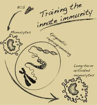
Figure 1. Trained Immunity: an innate immune memory program induced by a variety of stimuli, eg BCG, that results in a more active functional state of innate immune cells and confers broad non-specific protection against infections. From Netea, M.G. and van der Meer, J.W. (2017). Cell Host Microbe. 21(3):297-300.
1.2 Monocytes
Monocytes are innate immune cells that play distinct roles in tissue homeostasis and immunity. Monocytes are functionally characterized by the ability to phagocytose, produce cytokines, and present antigen; they are members of the mononuclear phagocyte system, a family of myeloid cells that comprises monocytes and two other major subtypes: namely, dendritic cells (DCs) and macrophages. Monocytes originate in the bone marrow and continuously enter the blood circulation, where they constitute ~10% of the total leukocyte population. Circulating monocytes represent a versatile and dynamic cell population, composed of multiple subsets which differ in phenotype, size, morphology, and transcriptional profiles [18].
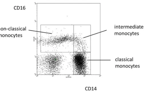
In human peripheral blood, three functionally different subsets of monocytes have been identified and characterized based on the differential expression of CD14 and CD16 cell surface markers (Figure 2). The major monocyte subset, accounting for approximately 90% of the total monocyte population, expresses high levels of CD14 and no CD16 (CD14++ CD16-), these cells are referred to as classical monocytes. Monocytes expressing CD16 can be further divided into two distinct subpopulations: intermediate monocytes (CD14+ CD16+) and non-classical monocytes expressing low levels of CD14 (CD14lowCD16++) [19].
The non-classical monocyte subset demonstrates patrolling behavior along blood vessel walls. Classical and intermediate monocytes, also known as inflammatory monocytes, are rapidly recruited to sites of infection [20] and injury [21], where, in response to environmental cues and molecular mediators, exhibit considerable functional plasticity [22] and can differentiate into monocyte-derived macrophages. Monocyte-derived macrophages are commonly classified into “classical” or “pro-inflammatory” (M1) and “alternative” or “anti-inflammatory” (M2) macrophages.
Most of the functional and molecular studies on monocyte and macrophage effector functions have relied on the activation of these cells by discrete stimuli, for example by LPS/IFN-γ for
M1 polarization, IL-4/IL-13 for M2 polarization [23]. However, it is now clear that monocytes and macrophages are exposed to a plethora of stimuli in vivo that eventually determines their functional specialization [24]. Moreover, these cells may be exposed to different and sometimes opposing stimuli in a sequential manner, and the priming stimulus may exert a significant control over the response to subsequent stimuli, leading to an enhanced activation status (trained immunity or trained monocytes). Recent studies have also shown that circulating monocytes and myeloid progenitor cells in the bone marrow are able to reprogram towards a long-term non-specific pro-inflammatory phenotype following initial exposure to microorganisms or microbial products [25]. Interestingly, trained immunity is not only induced by microbial products, but also by endogenous stimuli, such as cytokines [17;26]. Although beneficial in the context of resistance against reinfections, this mechanism might be detrimental in non- infectious chronic inflammatory conditions in which myeloid cells contribute to disease progression. Therefore, it is necessary to elucidate the mechanisms and the context in which trained immunity is driven.
Figure 2. Blood monocyte subsets in man. Illustration of the definition of human monocyte subsets in
health based on a typical distribution of events in a CD14 CD16 staining. From Ziegler-Heitbrock L. 2015. Front Immunol. 6:423.
1.3 GM-CSF and IL-3
Granulocyte–macrophage colony-stimulating factor (GM-CSF) and interleukin-3 (IL-3), are members of a small family of cytokines (also including IL-5), that regulates the production and activity of hematopoietic cells.
In contrast to many other cytokines, the biological activities of GM-CSF and IL-3, are largely not essential for the maintenance of steady-state functions [27]. Instead, the GM-CSF/IL-3 family of cytokines plays a key role in emergency hematopoiesis and immune response. They are produced by many immune and non immune cells, mainly by activated T cells, following infection, and serve to elevate leukocyte numbers and heighten their activation status, in particular stimulating the function of myeloid cells.
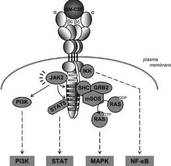
GM-CSF and IL-3 bind to heterodimeric cell surface receptors composed of a β common (βc) subunit (CD131), which is the principal signal transducing subunit, and a cytokine specific α subunits (CD116 and CD123, respectively) [28]. Receptors for the GM–CSF/IL-3 family of cytokines are expressed at very low level (< 1,500 receptors per cell) on hematopoietic cells. The binding of cytokine to its cognate receptor activates several signaling pathways (Figure 3) including: Janus kinase (JAK)/signal transducer and activator of transcription (STAT) pathway, the mitogen-activated protein kinase (MAPK) pathway, the phosphatidylinositol 3-kinase (PI3- K) pathway and the nuclear factor NF-κB pathway. These pathways collectively regulate cell survival, proliferation, and differentiation as well as effector functions of several mature immune cell subsets.
Figure 3. The GM-CSF receptor and initiation of signal transduction. From van de Laar, L., Paul J. Coffer, P.J., Woltman, M.A. (2012). Blood 119:3383-3393.
1.3.1 GM-CSF and IL-3: role in inflammatory and autoimmune diseases
GM-CSF and IL-3 modulate the effector functions of several mature immune cells including human and mouse mononuclear phagocytes (i.e. monocytes, macrophages and DCs), thereby exerting an important role in maintaining tissue homeostasis as well as in the pathogenesis of inflammatory and autoimmune diseases [29-33].
For example, GM-CSF deficiency leads to impaired alveolar macrophage differentiation and pulmonary alveolar proteinosis. GM-CSF promotes the maturation and effector functions of myeloid cells (e.g. monocytes and neutrophils) and plays a protective role in an experimental model of acute sepsis [34]. On the contrary, IL-3 exacerbates myelopoiesis and incites a cytokine storm (i.e. TNF-α, IL-1β and IL-6) that is detrimental to the host in the same sepsis model [35]. In an animal model of multiple sclerosis (experimental autoimmune encephalomyelitis) conditional deletion of Csf2rb (the mouse orthologue of CD131) in CCR2+ monocytes abrogates a pathogenetic signature that is required for disease development [30]. In patients with rheumatoid arthritis, elevated concentrations of GM−CSF and its receptor have been observed in tissue and synovial fluid [36] and recombinant GM−CSF administration
exacerbates RA disease activity. Of note, an anti-GM-CSF receptor α monoclonal antibody has been shown to reduce disease activity in patients with rheumatoid arthritis [37].
We have recently shown that GM-CSF and IL-3 synergize with IL-4 to enhance the production of the IL-4-responsive chemokine CCL17/TARC by human CD14+ monocytes [38]. Thus, GM- CSF and IL-3 exert a significant control on monocyte effector functions. The mechanisms and the extent to which GM-CSF and IL-3 modulate the pro-inflammatory (e.g. LPS-mediated) activation of human CD14+ monocytes are still unclear.
2. Aim of the study
Monocytes and macrophages are exposed to a plethora of stimuli in vivo that eventually determines their functional specialization [24]. Moreover, these cells may be exposed to different and sometimes opposing stimuli in a sequential manner, and the priming stimulus may exert a significant control over the response to subsequent stimuli. This phenomenon, which is referred to as innate immune memory or trained immunity, has been reported in several organisms and with a variety of stimuli, including toll-like receptor agonists and cytokines [17]. GM-CSF and IL-3 are hematopoietic cytokines produced by many immune and non immune cells and are important mediators of emergency myelopoiesis [27]. GM-CSF and IL-3 modulate the effector functions of several immune cell subsets and exert a significant control on monocyte effector function in inflammatory and autoimmune diseases [29-33].
We have recently shown that GM-CSF and IL-3 synergize with IL-4 to enhance the production of the IL-4-responsive chemokine CCL17/TARC by human CD14+ monocytes [38]. However, the mechanisms and the extent to which GM-CSF and IL-3 modulate the pro-inflammatory (e.g. LPS-mediated) activation of human CD14+ monocytes are still unclear. Here we sought to investigate whether GM-CSF and IL-3 modulate the pro-inflammatory, LPS-mediated, activation of human CD14+ monocytes taking into account the new concept of trained immunity.
3. Materials and methods 3.1 Cell isolation and culture
The study protocol involving the use of human blood cells was approved by the Ethics Committee of the University of Naples Federico II. Cells were isolated from buffy coats of healthy donors. Blood was layered onto Histopaque-1077 (Sigma-Aldrich) and mononuclear cells were collected at the interface. Monocytes were further purified with anti-CD14 Microbeads (Miltenyi Biotec). Purity of cell preparations was > 95% as assessed by flow cytometry. Cells were cultured in cIMDM-5 (IMDM, 5% FCS, 1x non essential amino acids, 1x UltraGlutamine, 25 mM HEPES, 5 μg/mL gentamicin [Lonza]) in 96-well flat-bottom plates (105 monocytes/well) in a final volume of 250 μL. For experiments involving flow cytometry, cells were cultured in suspension (1.5 mL tubes) in cIMDM-5 at a concentration not greater than 2*106 cells/mL, then spun down and collected for subsequent experiments.
Cells were treated with different combinations of: LPS (E. coli 026:B6) 10 ng/mL (Sigma Aldrich), IL-3 5 ng/mL (Peprotech), M-CSF 25 ng/mL, GM-CSF 5 ng/mL (Miltenyi Biotec), P3CSK4 10 ng/mL, Poly(I:C) 1 µg/mL, flagellin 10 ng/mL, imiquimod 1 µg/mL, ODN2006 1 µM (Invivogen), BAY11-7082 1 µM, SP600125 2 µM, rapamycin 50 and 250 nM, torin1 10 and 50 nM, AGK2 10 µM, APO866 0.1, 1 and 10 nM, TG101348 125, 250 and 500 nM (Selleckchem), UO126 2 µM, LY294002 10 µM, SB203580 2 µM (Cell Signaling Technology), 10058-F4 40 µM, EX-527 500 nM (Tocris Bioscience), 2-Deoxy-D-Glucose 1 mM, Etomoxir 40 µM, BAY 85-3934 1 µM, CAY-10585 10 µM (Cayman Chemical), nicotinic acid 10 µM (Sigma Aldrich), Pyridone 6 100 nM (BioVision), Trichostatin A 5 nM (Calbiochem).
Cytokine concentrations in cell-free supernatants or total protein lysates (0.1% Triton X-100) were measured using ELISA kits for TNF-α (R&D Systems), IL-1β and IL-6 (eBioscience). Standard curves were generated with a Four Parametric Logistic curve fit and data were analysed using MyAssays Analysis Software Solutions (www.myassays.com). Cytokine levels in protein lysates were normalized on total protein concentrations as determined by Bradford protein assay (Biorad) and expressed as pg of cytokine/μg of total protein. Intracellular NAD levels were measured using the colorimetric assay NAD/NADH Quantitation assay (Sigma Aldrich) according to the manufacturer’s protocol and expressed as fold over control.
3.3 Phospho-flow cytometry
Monocytes were collected, rested in cIMDM-0.5 (0.5% FCS) for 1 hour and stimulated with LPS for the indicated times. Then, cells were fixed with 1.5% paraformaldehyde (EM-grade, Electron Microscopy Sciences) and permeabilized with absolute ice-cold methanol. Cells were stained (60 minutes at room temperature) in PBS + 10% human AB serum (Lonza) + 0.05% NaN3 with the following antibodies: anti-human-phospho-p38 PE (T180/Y182) (3D7, dilution 1:50), anti-human-phospho-p44/42 (ERK1/2) PE (T202/Y204) (19762, dilution 1:50) (Cell Signaling Technology). Samples were acquired on MACSQuant Analyzer 10 (Miltenyi Biotec) and analyzed using FlowJo v10. Doublets and debris were excluded from the analysis. Data are expressed as percentage of positive cells and median fluorescence intensity (MFI).
3.4 Fluorescence, time-lapse and high-content microscopy
Microscopy experiments were performed with the Operetta High-Content Imaging System (PerkinElmer). Monocytes were cultured in 96-well black CellCarrier plates (PerkinElmer). For fluorescence microscopy, cells were fixed with 1% paraformaldehyde (EM-grade, Electron Microscopy Sciences) and permeabilized with PBS + 0.1% Triton X-100. Then, actin was
stained with rhodamin phalloidin and nuclei with Hoechst 33342 (ThermoFishcer Scientific) according to manufacturer’s protocol. Representative images were taken with a 60x objective. For time-lapse microscopy, monocytes were cultured for 6 days at controlled temperature (37°C) and CO2 (5%). Over this time window, digital phase contrast images of 15 fields/well were taken every 60 minutes with a 20x objective. To quantify cell confluency, brightfield images of 15 fields/well were taken on days 1, 4, 7 and 8 with a 20x objective. PhenoLOGIC (PerkinElmer) was employed for image segmentation and to calculate the cell-free area (b) and the area covered by cells (a). Cell confluency was calculated as a proportion of cell area over total cell area using the following formula: (a/(a+b))*100.
3.5 RNA isolation and real time RT-PCR
RNA was extracted using TRIzol (ThermoFischer Scientific) and reverse-transcribed (500 ng) using SuperScript III Reverse Transcriptase (ThermoFischer Scientific). Real time RT-PCR was performed by using Universal SYBR Green Supermix (Bio-Rad) on CFX96 Real Time detection system (Bio-Rad). Relative quantification of gene expression was calculated by the ΔCt (relative expression x 104) method. Each Ct value was normalized to the respective ubiquitin C (UBC) Ct value. The following primer pairs were used: TNF mRNA forward 5’-
tctccttcctgatcgtggca-3’; TNF mRNA reverse 5’-cagcttgagggtttgctacaac-3’; TNF primary transcript forward 5’-taagggtgactccctcgatgt-3’; TNF primary transcript reverse 5’- ccaaacccaaacccagaatta-3’; SIRT2 forward 5’-cccaaccccagcaaatctctaa-3’; SIRT2 reverse 5’- ggctggtagagatgcctgtt-3’; MYC forward 5’-attctctgctctcctcgacg-3’; MYC reverse 5’- agcctgcctcttttccaca-3’; MAFB forward 5’-gtatgaaccgcatggtgcttg-3’; MAFB reverse 5’- ctgttgcggcaggtttgatt-3’; UBC forward 5’-ggtcgcagttcttgtttg-3’; UBC reverse 5’-
3.6 Statistical analysis
Statistical analysis was performed with Prism 6 (GraphPad Software). P values were calculated as indicated in figure legends. P < .05 was considered significant.
4. Results
4.1 GM-CSF and IL-3 priming enhances LPS-induced TNF-α production
In order to evaluate the effect of GM-CSF and IL-3 on the pro-inflammatory LPS-mediated activation of human CD14+ monocytes, we simultaneously treated these cells with LPS and GM-CSF or IL-3, and then assessed the production of TNF-α, IL-1β and IL-6. Surprisingly, neither GM-CSF (Figure 4A) nor IL-3 (Figure 4B) modulated cytokine production in response to LPS.
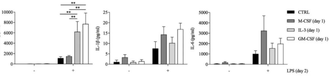
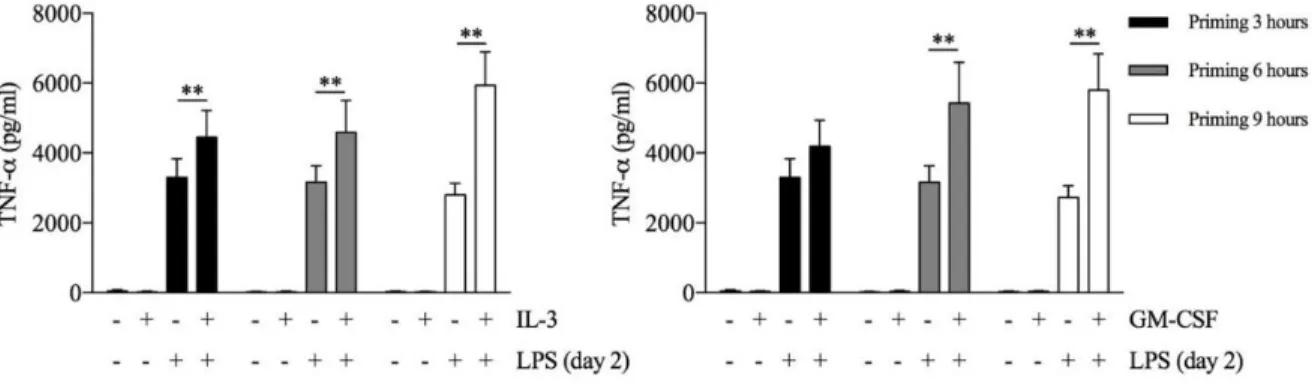
Since several stimuli may act as priming stimuli [17;26], we reasoned that GM-CSF and IL-3 could prime monocytes for increased responsiveness to LPS. Monocytes were left untreated (CTRL) or treated for 18 hours with M-CSF, IL-3 or GM-CSF, washed and then stimulated with LPS for additional 16-18 hours (short-term model of trained immunity). Interestingly, priming with GM-CSF or IL-3 increased LPS-induced TNF-α production, while IL-1β and IL- 6 were marginally affected (Figure 5). We also evaluated phagocytic activity of primed monocyte, GM-CSF and IL-3 priming did not enhance Escherichia coli phagocytosis.
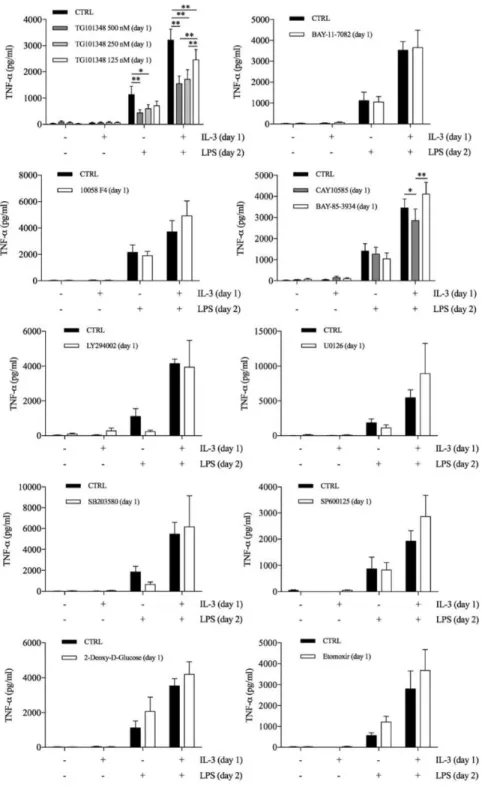
To verify whether the priming effect was shared among different TLRs, we stimulated primed monocytes with LPS (TLR4 agonist), P3CSK4 (TLR2 agonist), Poly(I:C) (TLR3 agonist), flagellin (TLR5 agonist), imiquimod (TLR7 agonist) or ODN 2006 (TLR9 agonist). The priming effect was more pronounced when monocytes were stimulated with LPS, although a trend toward an increase in TNF-α production was also observed with P3CSK4 (Figure 6). Finally, a time-course analysis revealed that at least 3 (for IL-3) or 6 (for GM-CSF) hours of priming were required to observe increased TNF-α secretion upon LPS stimulation (Figure 7). To gain insights into the molecular mechanisms of monocyte priming, we selectively treated monocytes during the priming phase with inhibitors of pathways that are known to modulate monocyte and macrophage activation: JAK2 (TG101348), NF-κB (BAY 11-7082), c-Myc (10058 F4), HIF-1α (CAY10585), PHD (BAY 85-3934), PI3K (LY294002), MEK1/2 (U0126),
p38 (SB203580), JNK (SP600125), hexokinase (2-Deoxy-D-Glucose, inhibitor of glycolysis), CPT1 (etomoxir, inhibitor of fatty acid β-oxidation). The readout of these experiments was the modulation of TNF-α production by primed monocytes without affecting unprimed cells. Most of the inhibitors had no or negligible effects on primed cells, while TG101348 reduced TNF-α production by both primed and unprimed cells (Figure 8).
Figure 4. (A, B) Human CD14+ monocytes were stimulated with LPS and GM-CSF (A) or IL-3 (B) for
16-18 hours. TNF-α, IL-1β and IL-6 levels were assessed by ELISA in cell-free supernatants. Data are shown as mean + SEM of 5 (A) or 6 (B) independent experiments and were analyzed by repeated measure one-way ANOVA with Tukey’s post hoc test.
Figure 5. Human CD14+ monocytes were left unprimed (CTRL) or primed with M-CSF, IL-3 and GM-
CSF for 18 hours (day 1), then washed and left untreated or stimulated with LPS for 16-18 hours (day 2). TNF-α, IL-1β and IL-6 levels were assessed by ELISA in cell-free supernatants. Data are shown as mean + SEM of 7 independent experiments. ** P < .01 determined by repeated measure two-way ANOVA with Sidak’s post hoc test.
Figure 6. Human CD14+ monocytes were left unprimed (CTRL) or primed with IL-3 for 18 hours (day
1), then washed and stimulated with several TLR agonists for 16-18 hours (day 2). TNF-α levels were assessed by ELISA in cell-free supernatants. Data are shown as mean + SEM of 4 independent experiments. ** P < .01 determined by repeated measure two-way ANOVA with Sidak’s post hoc test.
Figure 7. Human CD14+ monocytes were primed with IL-3 (left panel) or GM-CSF (right panel) for
different times as indicated in the figure, then washed and left untreated or stimulated with LPS for 16- 18 hours (day 2). TNF-α levels were assessed by ELISA in cell-free supernatants. Data are shown as mean + SEM of 8 independent experiments. ** P < .01 determined by repeated measure two-way ANOVA with Tukey’s post hoc test.
Figure 8. Human CD14+ monocytes were primed with IL-3 and the indicated compounds for 18 hours
(day 1), then washed and left untreated or stimulated with LPS for 16-18 hours (day 2). TNF-α levels were assessed by ELISA in cell-free supernatants. Data are shown as mean + SEM of 4-10 independent experiments. * P < .05, ** P < .01 determined by repeated measure two-way ANOVA with Tukey’s or Sidak’s post hoc test.
4.2 GM-CSF and IL-3 priming modulate TNF-α production at the post- transcriptional level
To obtain some clue on the modulation of TNF-α in GM-CSF and IL-3 primed monocytes, we performed a time-course analysis of TNF-α intracellular protein, mRNA and primary transcript levels (which is a more direct measure of transcription and is detected by using intronic PCR primers) [39]. GM-CSF and IL-3 priming resulted in the highest TNF-α intracellular protein levels as early as after 1 hour of LPS stimulation (Figure 9A). After 4 hours of LPS stimulation TNF-α intracellular protein levels were even more increased in primed monocytes (Figure 9B). The same pattern of TNF-α production was observed in cell-free supernatants. Unexpectedly,
TNF mRNA (Figure 10A) and primary transcript (Figure 11A) levels were either not modified
or only slightly increased by GM-CSF and IL-3 priming after 1 hour of LPS stimulation. After 4 hours, IL-3 priming increased TNF mRNA levels (Figure 10B) whereas TNF primary transcript levels were unchanged compared to unprimed cells (Figure 11B), suggesting that IL- 3 priming increases TNF mRNA stability. Thus, GM-CSF and IL-3 priming uncouples TNF-α protein and primary transcript levels upon LPS stimulation, thereby suggesting that TNF-α expression is modulated at the post-transcriptional level in primed monocytes.
Figure 9. (A-B) Human CD14+ monocytes were primed with GM-CSF and IL-3 for 18 hours (day 1),
then washed and stimulated with LPS (day 2) for 1 (A) or 4 (B) hours. TNF-α protein abundance (A, B) was measured in protein lysates by ELISA. Data are shown as the median, the 25th and 75th percentiles
(boxes) and the 5th and 95th percentiles (whiskers) of 8 independent experiments. * P < .05, ** P < .01
Figure 10. (A-B) Human CD14+ monocytes were primed with GM-CSF and IL-3 for 18 hours (day 1),
then washed and stimulated with LPS (day 2) for 1 (A) or 4 (B) hours. TNF mRNA measured by real time RT-PCR. Data are shown as the median, the 25th and 75th percentiles (boxes) and the 5th and 95th
percentiles (whiskers) of 8 independent experiments. * P < .05, ** P < .01 determined by Friedman test with Dunn’s post hoc test.
Figure 11. (A-B) Human CD14+ monocytes were primed with GM-CSF and IL-3 for 18 hours (day 1),
then washed and stimulated with LPS (day 2) for 1 (A) or 4 (B) hours. TNF primary transcript was measured by real time RT-PCR. Data are shown as the median, the 25th and 75th percentiles (boxes) and
the 5th and 95th percentiles (whiskers) of 8 independent experiments. * P < .05, ** P < .01 determined
4.3 NAD-dependent SIRT2 and MAPKs ERK1/2 and p38 mediate the effect of GM- CSF and IL-3 priming on TNF-α production
The expression of several genes with immune function (including TNF-α) can be regulated at the translational level, thereby modulating protein abundance independently of de novo transcription [40-42]. A central (albeit not exclusive) role in the modulation of mRNA translation is played by the translation-initiation factor eIF4E, whose activity is regulated by mTORC1 (a multiprotein complex that includes the serine/threonine kinase mTOR and raptor) and the MAPKs ERK1/2 and p38 [41].
TNF-α protein levels are also regulated by metabolic cues. Depletion of the intracellular pool of nicotinamide adenine dinucleotide (NAD) by the nicotinamide phosphoribosyltransferase (Nampt) inhibitor APO866 inhibits TNF-α protein production in response to LPS without modifying TNF mRNA levels. The effect of APO866 is reverted by the addition of nicotinic acid (NA, a substrate for Nampt-independent NAD synthesis), and is mimicked by inhibitors of the NAD-dependent histone deacetylases sirtuin (SIRT) 1 and 2 [43;44]. Thus, we reasoned that these pathways could contribute to the modulation of TNF-α protein levels in GM-CSF and IL-3 primed cells and proceeded to investigate this hypothesis.
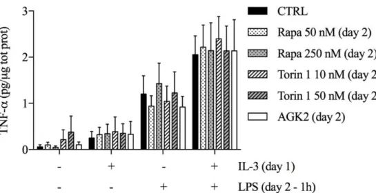
First, we assessed the contribution of mTORC1 by employing 2 different mTOR inhibitors (rapamycin and torin 1) and also an inhibitor of SIRT2 (which is involved in the regulation of the mTOR-activated translational machinery) [45]. We added the inhibitors along with LPS and evaluated TNF-α intracellular protein levels after 1 hour of stimulations. None of these inhibitors modulated TNF-α production in both unprimed and primed monocytes (Figure 12).
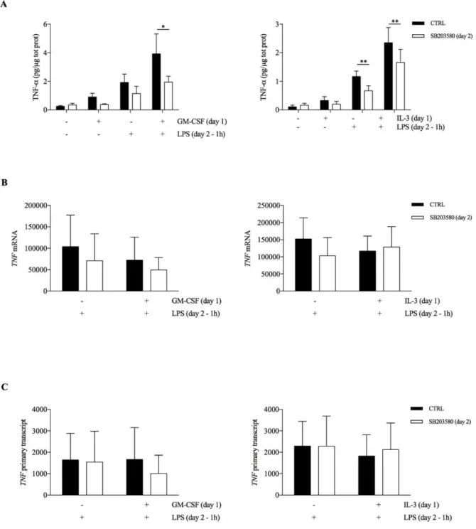
Then, we employed the same approach using inhibitors of MAPKs (i.e. ERK1/2, p38, JNK). The p38 inhibitor SB203580 reduced TNF-α intracellular protein in both unprimed and GM- CSF or IL-3 primed cells (Figure 13A), while ERK1/2 inhibition with the MEK1/2 inhibitor U0126 was effective only in GM-CSF primed cells (Figure 14A). No effect for the JNK
inhibitor SP600125 could be observed (data not shown). Importantly, p38 (Figures 13B, C) and ERK1/2 inhibition (Figures 14B, C) did not significantly modulate TNF RNA levels.
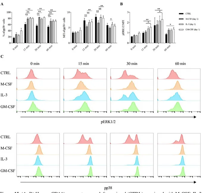
As early as after 15 minutes of stimulation GM-CSF and IL-3 primed cells exhibited the highest phosphorylation of p38, as assessed by the percentage (compared to unprimed cells) and the median fluorescence intensity (compared to M-CSF primed cells) of phospho-p38+ cells (Figure 15A). On the contrary, only GM-CSF primed cells exhibited increased ERK1/2 phosphorylation compared to unprimed or M-CSF primed cells (Figure 15B).
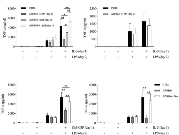
TNF-α production can also be regulated by metabolic cue, we used APO866 and NA to evaluate whether intracellular NAD levels contributed to the priming effect of GM-CSF and IL-3. We found that APO866 inhibited TNF-α secretion only when administered during priming (Figure 16A), moreover in a dose dependent manner. Importantly, the addition of NA reverted the effect of APO866 (Figure 16B).
We verified that APO866 reduced while NA addition restored intracellular NAD levels (Figure 17A). Interestingly, GM-CSF and IL-3 priming increased intracellular NAD levels compared to unprimed cells. However, the same effect was observed with M-CSF priming (Figure 17B), which does not modulate TNF-α secretion. Thus, the increase in intracellular NAD levels is a condition required but not sufficient for the priming effect of GM-CSF and IL-3.
We then evaluated the involvement of the NAD-dependent histone deacetylases SIRT1 and SIRT2 in GM-CSF and IL-3 priming by using the specific inhibitors EX-527 and AGK2, respectively (Figures 18A-B).
SIRT2 inhibition during priming selectively reduced TNF-α secretion and intracellular protein levels (Figure 19A) without significantly modulating TNF RNA (Figures 19B-C), although the effect was more pronounced for IL-3 rather than GM-CSF primed cells. We confirmed the specificity of SIRT2 involvement by showing that the inhibitor of NAD-independent histone deacetylases trichostatin A (TSA) had no effect on GM-CSF and IL-3 priming (Figure 20).
Figure 12. Human CD14+ monocytes were primed with IL-3 and the indicated compounds for 18 hours
(day 1), then washed and left untreated or stimulated with LPS for 1 hour (day 2). TNF-α levels were assessed by ELISA in total protein lysates. Data are shown as mean + SEM of 4 independent experiments. Data were analyzed by repeated measure two-way ANOVA with Tukey’s post hoc test.
Figure 13. (A-B-C) Human CD14+ monocytes were primed with GM-CSF and IL-3 for 18 hours (day
1), then washed and left untreated or stimulated with LPS for 1 hour in the presence or in the absence of the p38 inhibitor SB203580 (day 2). TNF-α levels were assessed by ELISA in total protein lysates (A). Alternatively, cells were harvested for RNA extraction and evaluation of mRNA (B) and TNF primary transcript (C) levels by real time RT-PCR. Data are shown as mean + SEM of 4 (A) or 6 (B-C) independent experiments. * P < .05, ** P < .01 determined by repeated measure two-way ANOVA with Sidak’s post hoc test.
Figure 14. (A-F) Human CD14+ monocytes were primed with GM-CSF and IL-3 for 18 hours (day 1),
then washed and left untreated or stimulated with LPS for 1 hour in the presence or in the absence of the MEK1/2 inhibitor U0126 (day 2). TNF-α levels were assessed by ELISA in total protein lysates (A). Alternatively, cells were harvested for RNA extraction and evaluation of mRNA (B) and TNF primary transcript (C) levels by real time RT-PCR. Data are shown as mean + SEM of 4 (A) or 6 (B-C) independent experiments. * P < .05, ** P < .01 determined by repeated measure two-way ANOVA with Sidak’s post hoc test.
Figure 15. (A, B) Human CD14+ monocytes were left unprimed (CTRL) or primed with M-CSF, IL-3
and GM-CSF for 18 hours (day 1), then washed and left untreated (0 min) or stimulated with LPS for different time points (15 min, 30 min, 60 min). (A) p38 phosphorylation was assessed by flow cytometry as percentage (left panel) and MFI (right panel) of phospho-p38+ cells. (B) ERK1/2 phosphorylation
was assessed by flow cytometry as MFI. (C) Representative histograms of p38 and ERK1/2 phosphorylation. Data are shown as mean + SEM of 8 independent experiments. * P < .05, ** P < .01 determined by repeated measure two-way ANOVA with Tukey’s post hoc test.
Figure 16. (A) Human CD14+ monocytes were primed with IL-3 in the presence (left panel) or absence
(right panel) of the Nampt inhibitor APO866 for 18 hours (day 1), then washed and stimulated with LPS in the absence (left panel) or presence (right panel) of APO866 for 16-18 hours (day 2). TNF-α levels were assessed by ELISA in cell-free supernatants. (B) Cells were stimulated with GM-CSF or IL-3 in the presence of APO866 and nicotinic acid (NA) for 18 hours (day1), then washed and stimulated with LPS for 16-18 hours (day 2). Data are shown as mean + SEM of 6 independent experiments. * P < .05, ** P < .01 determined by repeated measure two-way ANOVA with Tukey’s or Sidak’s post hoc test (A, respectively left and right panel) or repeated measure one-way ANOVA with Tukey’s post hoc test (B).
Figure 17. Human CD14+ monocytes were left untreated (CTRL), stimulated with IL-3 in the presence
of APO866 and nicotinic acid (NA) (A), or with M-CSF, IL-3 and GM-CSF (B) for 18 hours. Intracellular NAD levels were measured in cellular lysates and expressed as fold over CTRL (A, B). Data are shown as mean + SEM of 6 independent experiments. * P < .05, ** P < .01 determined by repeated measure one-way ANOVA with Tukey’s post hoc test.
Figure 18. Human CD14+ monocytes were primed with GM-CSF and IL-3 for 18 hours in the presence
of SIRT1 inhibitor EX-527 (A) or SIRT2 inhibitor AGK2 (B) (day 1), then washed and stimulated with LPS for 16-18 hours (day 2). TNF-α levels were assessed by ELISA in cell-free supernatants. Data are shown as mean + SEM of 7 independent experiments. ** P < .01 determined by repeated measure two- way ANOVA with Tukey’s (A) or Sidak’s (B-D) post hoc test.
Figure 19. (A-C) Human CD14+ monocytes were primed with GM-CSF and IL-3 for 18 hours in the
presence or absence of the SIRT2 inhibitor AGK2 (day 1), then washed and left untreated or stimulated with LPS for 1 hour (day 2). TNF-α levels were assessed by ELISA in total protein lysates (A). Alternatively, cells were harvested for RNA extraction and evaluation of TNF mRNA (B) and primary transcript (C) levels by real time RT-PCR. Data are shown as mean + SEM of 4 (A) or 6 (B, C) independent experiments. * P < .05, ** P < .01 determined by repeated measure two-way ANOVA with Sidak’s post hoc test.
Figure 20. Human CD14+ monocytes were primed with GM-CSF and IL-3 for 18 hours in the presence
of trichostatin A (TSA), an inhibitor of NAD-independent histone deacetylases (day 1), then washed and stimulated with LPS for 16-18 hours (day 2). TNF-α levels were assessed by ELISA in cell-free supernatants. Data are shown as mean + SEM of 7 independent experiments. ** P < .01 determined by repeated measure two-way ANOVA with Tukey’s (A) or Sidak’s (B-D) post hoc test.
4.4 GM-CSF and IL-3 priming modulate monocyte renewal in a c-Myc-dependent manner
The results obtained so far have characterized the role of GM-CSF and IL-3 in a short-term model of trained immunity. Since the effects of training can be retained by human monocytes for several days even in the absence of the training stimulus (e.g. β-glucan) [10;13], we investigated the possible roles of GM-CSF and IL-3 in a long-term model of trained immunity. Monocytes were primed with GM-CSF, IL-3, M-CSF and LPS for 16-18 hours, washed out and rested in complete medium until day 7 when they were stimulated with LPS. In accordance with the results obtained with β-glucan, cells primed with GM-CSF or IL-3 secreted higher levels of TNF-α compared to those primed with M-CSF and LPS (Figure 21A). Although these results could suggest stable chromatin remodeling events in response to GM-CSF and IL-3 priming (similar to what has been demonstrated for β-glucan), we also found that GM-CSF and IL-3 priming increased total cellular protein content (Figure 21B) and that monocytes primed with GM-CSF, IL-3 and M-CSF expressed comparable levels of TNF RNA upon LPS stimulation (Figure 21C-D).
Thus, we reasoned that GM-CSF and IL-3 priming could induce monocyte renewal and increase the number of cells, which in turn would result in higher TNF-α secretion upon LPS stimulation. We investigated this hypothesis by evaluating the degree of cell confluency (assessed as the proportion of cell area to total area). Monocytes primed with GM-CSF and IL-3 reached remarkable higher cell confluency on day 7 compared to monocyte primed with M-CSF or LPS (Figure 22A). Comparable results were obtained when we expressed cell confluency on days 4, 7 and 8, as fold increase over day 1, to account for possible differences in cell detachment after the washing step (Figure 22B). We also obtained evidence of increased cell number following GM-CSF and IL-3 priming by fluorescence (Figure 22C) and time-lapse microscopy. Thus,
GM-CSF and IL-3 priming induces monocyte renewal in a long-term model of trained immunity.
Maintenance of the pool of tissue-resident macrophages is supported by self-renewal without the contribution of hematopoietic stem cell in several experimental models [46-49]. Interestingly, macrophage renewal is sustained by a gene network that includes the transcription factor c-Myc and requires downregulation of the transcription factor MafB [50;51]. In keeping with these data, GM-CSF and IL-3 priming increased the MYC/MAFB mRNA ratio compared to M-CSF priming (Figure 23). Moreover, the c-Myc inhibitor 10058 F4 restrained monocyte renewal induced by GM-CSF and IL-3 priming (Figure 24).
To better characterize the pathways required for GM-CSF- and IL-3-induced monocyte renewal, we employed metabolic inhibitors (2-Deoxy-D-Glucose, etomoxir, APO866) and inhibitors of the signaling pathway activated upon GM-CSF/IL-3 receptor stimulation. Inhibitors were added along with GM-CSF or IL-3 and then cell confluency was evaluated as indicated above. Metabolic inhibitors as well the JNK inhibitor SP600125 (Figure 25) had no effect on cell confluency. The selective JAK2 inhibitor TG101348 (Figure 26A), the pan-JAK inhibitor Pyridone 6 (Figure 26B) and the p38 inhibitor SB203580 (Figure 26C) restrained only IL-3 priming-induced monocyte expansion. Finally, the PI3K inhibitor LY294002 (Figure 27A) and the MEK1/2 inhibitor U0126 (which in turn inhibits ERK1/2 activity) (Figure 27B) reduced monocyte expansion in response to both GM-CSF and IL-3 priming, although IL-3 priming was affected to a greater degree.
Figure 21. Human CD14+ monocytes were primed with IL-3, GM-CSF, M-CSF or LPS for 16-18 hours,
washed out and rested in complete medium until day 7. On day 7 cells were stimulated with LPS for 16- 18 hours (A, B, C) or 1 hour (D). TNF-α levels were assessed by ELISA in cell-free supernatants (A). Cells were harvested for total protein quantification (B) or RNA extraction and evaluation of TNF primary transcript and mRNA levels by real time RT-PCR (C, D). Data are shown as mean + SEM of 10 (A, B, C) or 5 (D) independent experiments. * P < .05, ** P < .01 determined by repeated measure one-way (B-D) with Tukey’s post hoc test.
Figure 22. (A-C) Human CD14+ monocytes were primed with IL-3, GM-CSF, M-CSF or LPS for 16-
18 hours, washed out and rested in complete medium until day 7. On day 7 cells were stimulated with LPS for 16-18 hours. Brightfield images (15 fields/well) were obtained at the indicated time points with a 20x objective. The percentage of cell area/total area was calculated and expressed as absolute values (A) or fold increase over day 1 (B). On day 8 representative fluorescence microscopy images were also obtained with a 60x objective. Data are shown as mean + SEM of 8 independent experiments. Orange, actin; blue, nuclei (C). ** P < .01 determined by repeated measure two-way (A, B) ANOVA with Tukey’s post hoc test.
Figure 23. Human CD14+ monocytes were primed with IL-3, M-CSF or GM-CSF for 18 hours. Then,
cells were harvested for RNA isolation and MYC and MAFB mRNA levels were evaluated by real time RT-PCR. Data are shown as mean + SEM of 7 independent experiments. * P < .05, ** P < .01 determined by repeated measure one-way (A) ANOVA with Tukey’s post hoc test.
Figure 24. Human CD14+ monocytes were primed with IL-3, GM-CSF, M-CSF or LPS for 16-18
washed out and rested in complete medium until day 7. On day 7 cells were stimulated with LPS for 16- 18 hours. Cells were also treated with the c-Myc inhibitor 10058 F4 on day 1 (upper panels), day 1 and 2 (middle panels), day 1, 2 and 4 (lower panels). Analysis was performed as in Fig. 22B. Data are shown as mean + SEM of 8 independent experiments. * P < .05, ** P < .01 determined by repeated measure two-way (B) ANOVA with Tukey’s post hoc test.
Figure 25. Human CD14+ monocytes were primed with GM-CSF (upper panels), IL-3 (lower panels)
and M-CSF for 16-18, washed out and rested in complete medium until day 7. On day 7 cells were stimulated with LPS for 16-18 hours. Cells were also treated with different inhibitors during the priming phase. Analysis was performed as in Fig. 22B. Data are shown as mean + SEM of 8 independent experiments. Data were analysed by repeated measure two-way ANOVA with Tukey’s post hoc test.
Figure 26. (A-C) Human CD14+ monocytes were primed with IL-3, GM-CSF, M-CSF for 16-18 hours,
washed out and rested in complete medium until day 7. On day 7 cells were stimulated with LPS for 16- 18 hours. Cells were also treated different inhibitors during the priming phase. Analysis was performed as in Fig. 22B. Data are shown as mean + SEM of 8 independent experiments. * P < .05, ** P < .01 determined by repeated measure two-way ANOVA with Tukey’s post hoc test.
Figure 27. (A-B) Human CD14+ monocytes were primed with IL-3, GM-CSF, M-CSF for 16-18 hours,
washed out and rested in complete medium until day 7. On day 7 cells were stimulated with LPS for 16- 18 hours. Cells were also treated with with the PI3K inhibitor LY294002 (A) and the MEK1/2 inhibitor U0126 (B) during the priming phase. Analysis was performed as in Fig. 22B. Data are shown as mean + SEM of 8 independent experiments. * P < .05, ** P < .01 determined by repeated measure two-way ANOVA with Tukey’s post hoc test.
5. Discussion
Monocytes and macrophages are exposed to a plethora of stimuli in vivo that eventually determines their functional specialization [24]. These cells may be exposed to different and sometimes opposing stimuli in a sequential manner, and the priming stimulus may exert a significant control over the response to subsequent stimuli, leading to an enhanced activation status. The ability of innate immune cells to switch their reactivity in a stimulus specific manner and to build up immunological memory is termed “trained immunity” [3]. Trained immunity has been reported in several organisms and with a variety of stimuli, including toll like receptor (TLR) agonists and endogenous stimuli, such as cytokines [17;26]. Although beneficial in the context of resistance against reinfections, this mechanism might be detrimental in non- infectious/chronic inflammatory conditions in which myeloid cells contribute to disease progression.
GM-CSF and IL-3 are hematopoietic cytokines that modulate the effector functions of several immune cell subsets. In particular, GM-CSF and IL-3 exert a significant control on monocyte and macrophage effector functions, and play important roles in maintaining tissue homeostasis as well in the pathogenesis of inflammatory and autoimmune disease. Here we show that GM-CSF and IL-3 modulate the activation of human CD14+ monocyte in response to LPS stimulation in short- and long-term models of trained immunity. Employing a short- term model of trained immunity, we demonstrated that GM-CSF and IL-3 priming of human monocytes increases TNF-α production in response to LPS stimulation. To understand the regulation of TNF-α production in GM-CSF and IL-3 primed monocytes, we looked at intracellular TNF-α protein levels, TNF mRNA and primary transcript levels upon LPS stimulation for 1 and 4 hours. GM-CSF and IL-3 priming increases TNF-α intracellular protein levels as early as after 1 hour of LPS stimulation. Unexpectedly, TNF mRNA and primary transcript levels are modestly modified by GM-CSF and IL-3 priming. Although in several
experimental models priming of monocytes and macrophages results in chromatin remodeling [6-14], our data suggest a post-transcriptional regulation of TNF-α production in primed monocytes. Post-transcriptional mechanisms, including modulation of mRNA stability and translation [40-42], are increasingly recognized as critical regulators of gene and protein expression also in immune cells [9;10;39;43;44]. A central, albeit not exclusive role, in the modulation of mRNA translation is played by the MAPKs p38 and ERK1/2 [41]. Indeed, the inhibition of p38 and ERK1/2 (only for GM-CSF) during LPS stimulation reduces intracellular TNF-α protein levels without significantly modulating TNF RNA levels. In keeping with these data, GM-CSF priming increases phosphorylation of both ERK1/2 and p38 upon LPS stimulation, while IL-3 priming enhances only p38 phosphorylation. TNF-α protein levels are also regulated by metabolic cues. Depletion of the intracellular pool of NAD, inhibits TNF-α protein production in response to LPS without modifying TNF mRNA levels, moreover the same effect has been observed employing inhibitors of the NAD-dependent histone deacetylases sirtuin (SIRT) 1 and 2 [43;44]. Our results demonstrate that inhibition of SIRT2 during priming reduces intracellular TNF-α protein levels without significantly modulating
TNF RNA levels. However, it is still unclear how SIRT2 inhibition during the priming phase
modulates TNF-α protein production. Nevertheless, the phenotype of GM-CSF- and IL-3- primed monocytes may be regulated by pathways not investigated in this study. For example, several mRNAs encoding immune-regulatory proteins, including TNF-α, have AU-rich elements (AREs) in their 3′ untranslated region (UTR) [55] that generally determine mRNA instability. AREs serve as binding sites for a number of regulatory proteins that either destabilize or stabilize their targets mRNA. Among ARE binding protein Tristetraprolin (TTP, ZFP36), a LPS inducible protein, binds to AREs of TNF-α and promotes a rapid mRNA decay [56]. Moreover, small regulatory micro (mi)-RNAs represent another system that enhances the degradation of several mRNAs encoding important components of signal transduction cascades
that are activated during adaptive and innate immune responses [55]. Apart from the molecular mechanisms that may also reveal subtle differences in monocyte responsiveness to GM-CSF and IL-3 priming, genome-wide approaches aimed at evaluating the translatome or at comparing the transcriptome and the proteome may provide critical insights into the modulation of LPS-induced monocyte response by GM-CSF and IL-3 priming.
In order to fully characterize the priming effect of GM-CSF and IL-3, we also employed a long-term model of trained immunity. Using this model, it was shown that human monocytes primed with β-glucan undergo extensive chromatin remodeling that modulates their response to subsequent microbial challenges (e.g. increased TNF-α production upon LPS stimulation) [10;13]. Our results demonstrate that GM-CSF and IL-3 priming increases LPS-induced TNF- α production in this model, but the mechanism is independent of increased TNF gene expression (and likely chromatin remodeling). Instead, GM-CSF and IL-3 priming induces an increase of cell number and monocyte renewal, responsible for heightened TNF-α production. Macrophage renewal is sustained by a gene network that includes the transcription factor c-Myc and requires downregulation of the transcription factor MafB. Our results demonstrate that GM-CSF and IL- 3 priming induces monocyte renewal by increasing the MYC/MAFB mRNA ratio. In keeping with these data, the c-Myc inhibitor 10058 F4 restrained monocyte renewal induced by GM- CSF and IL-3 priming. Interestingly, proliferation of monocyte-derived cells (either macrophages or dendritic cells) has been observed in several models of inflammation [25;49;50]. Although we found similarities as well as differences between GM-CSF and IL-3 priming in the pathways required for monocyte renewal, it is noteworthy that GM-CSF priming induces a greater monocyte expansion than IL-3 priming. The reason for this difference probably relies in the constitutive expression of CD116 (the α subunit of GM-CSF receptor) by human monocytes [52], while CD123 (the α subunit of IL-3 receptor) expression is inducible [38;53]. Nevertheless, bone marrow CD14+ monocytes express CD123 [54], thus it is tempting
to speculate that IL-3 preferentially acts on bone marrow-resident monocytes, while GM-CSF exerts its effect on peripheral blood monocytes. Moreover, since priming with both cytokines induces monocyte renewal and priming also affects the biology of hematopoietic precursors [25,57-59], it would be interesting to assess whether GM-CSF and IL-3 priming can modulate the functions of hematopoietic precursors and their progeny. Further studies are required to address these questions.
6. Conclusion
We characterize the activation of human monocytes in response to the hematopoietic cytokines GM-CSF and IL-3 using in vitro models of trained immunity. Our results point to mechanisms of GM-CSF and IL-3 priming that are independent of chromatin remodeling, but unexpectedly rely on post-transcriptional regulation of TNF-α production and c-Myc-dependent monocyte renewal. Given the central role of GM-CSF and IL-3 in inflammatory and autoimmune diseases [30-33], our results provide important information to understand mechanisms of monocyte activation in such disorders and prompt their investigation in vivo. We also demonstrate that GM-CSF and IL-3 activate monocytes through shared as well as distinct pathways. Such differences may become even more evident in vivo, since it is conceivable that GM-CSF and IL-3 are produced with a different timing and/or act on different target populations (e.g. bone marrow vs peripheral cells). Thus, GM-CSF and IL-3 may differentially impact on the immune response due to distinct effects at the cellular and the system levels. Understanding such complexity is poised to unravel distinct as well as overlapping roles for GM-CSF and IL-3 in health and disease.
7. References
1. Dominguez-Andres, J., Netea, M.G. (2018). Long-term reprogramming of the innate immune system. J Leukoc Biol. doi: 10.1002/JLB.MR0318-104R.
2. Hamon, M.A., Quintin, J. (2016). Innate immune memory in mammals. Semin Immunol. (4):351-8. doi: 10.1016/j.smim.2016.05.003.
3. Netea, M.G., Quintin, J., van der Meer, J.W. (2011). Trained immunity: a memory for innate host defense. Cell Host Microbe. 9(5):355-61. doi: 10.1016/j.chom.2011.04.006. 4. Foster, S.L., Hargreaves, D.C., Medzhitov, R. (2007). Gene-specific control of inflammation by TLR-induced chromatin modifications. Nature. 447(7147):972-978. doi: 10.1038/nature05836.
5. Hu, X., Paik, P.K., Chen, J., Yarilina, A., Kockeritz, L., Lu, T.T., et al. (2006). IFN- gamma suppresses IL-10 production and synergizes with TLR2 by regulating GSK3 and CREB/AP-1 proteins. Immunity. 24(5):563-574. doi: 10.1016/j.immuni.2006.02.014.
6. Ostuni, R., Piccolo, V., Barozzi, I., Polletti, S., Termanini, A., Bonifacio, S., et al. (2013). Latent enhancers activated by stimulation in differentiated cells. Cell. 152(1- 2):157-171. doi: 10.1016/j.cell.2012.12.018.
7. Park, S.H., Park-Min, K.H., Chen, J., Hu, X., Ivashkiv, L.B. (2011). Tumor necrosis factor induces GSK3 kinase-mediated cross-tolerance to endotoxin in macrophages. Nat
Immunol. 12(7):607-615. doi: 10.1038/ni.2043.
8. Qiao, Y., Giannopoulou, E.G., Chan, C.H., Park, S.H., Gong, S., Chen, J., et al. (2013). Synergistic activation of inflammatory cytokine genes by interferon-gamma-induced chromatin remodeling and toll-like receptor signaling. Immunity. 39(3):454-469. doi: 10.1016/j.immuni.2013.08.009.
9. Su, X., Yu, Y., Zhong, Y., Giannopoulou, E.G., Hu, X., Liu, H., et al. (2015). Interferon- gamma regulates cellular metabolism and mRNA translation to potentiate macrophage activation. Nat Immunol. 16(8):838-849. doi: 10.1038/ni.3205.
10. Cheng, S.C., Quintin, J., Cramer, R.A., Shepardson, K.M., Saeed, S., Kumar, V., et al. (2014). mTOR- and HIF-1alpha-mediated aerobic glycolysis as metabolic basis for trained immunity. Science. 345(6204):1250684. doi: 10.1126/science.1250684.
11. Kleinnijenhuis, J., Quintin, J., Preijers, F., Joosten, L.A., Ifrim, D.C., Saeed, S., et al. (2012). Bacille Calmette-Guerin induces NOD2-dependent nonspecific protection from reinfection via epigenetic reprogramming of monocytes. Proc Natl Acad Sci U S A. 109(43):17537-17542. doi: 10.1073/pnas.1202870109.
12. Quintin, J., Saeed, S., Martens, J.H., Giamarellos-Bourboulis, E.J., Ifrim, D.C., Logie, C., et al. (2012). Candida albicans infection affords protection against reinfection via functional reprogramming of monocytes. Cell Host Microbe. 12(2):223-232. doi: 10.1016/j.chom.2012.06.006.
13. Saeed, S., Quintin, J., Kerstens, H.H., Rao, N.A., Aghajanirefah, A., Matarese, F., et al. (2014). Epigenetic programming of monocyte-to-macrophage differentiation and trained innate immunity. Science. 345(6204):1251086. doi: 10.1126/science.1251086. 14. Yoshida, K., Maekawa, T., Zhu, Y., Renard-Guillet, C., Chatton, B., Inoue, K., et al.
(2015). The transcription factor ATF7 mediates lipopolysaccharide-induced epigenetic changes in macrophages involved in innate immunological memory. Nat Immunol. 16(10):1034-1043. doi: 10.1038/ni.3257.
15. Blok, B.A., Arts, R.J., van Crevel, R., Benn, C.S., Netea, M.G. (2015). Trained innate immunity as underlying mechanism for the long-term, nonspecific effects of vaccines.
16. Levy, O., Netea, M.G. (2014). Innate immune memory: implications for development of pediatric immunomodulatory agents and adjuvanted vaccines. Pediatr Res. 75(1- 2):184-188. doi: 10.1038/pr.2013.214.
17. Netea, M.G., Joosten, L.A., Latz, E., Mills, K.H., Natoli, G., Stunnenberg, H.G., et al. (2016). Trained immunity: A program of innate immune memory in health and disease.
Science. 352(6284):aaf1098. doi: 10.1126/science.aaf1098.
18. Geissmann, F., Jung, S., Littman, D.R. (2003). Blood monocytes consist of two principal subsets with distinct migratory properties. Immunity. 19(1):71–82. https://doi.org/10.1016/S1074-7613(03)00174-2.
19. Ziegler-Heitbrock L. (2015). Blood Monocytes and Their Subsets: Established Features and Open Questions. Front Immunol. 6:423. doi: 10.3389/fimmu.2015.00423.
20. Liao, C.T., R. Andrews, L.E. Wallace, M.W. Khan, A. Kift-Morgan, N. Topley, D.J. Fraser, and P.R. Taylor. (2017). Peritoneal macrophage heterogeneity is associated with different peritoneal dialysis outcomes. Kidney Int. 91(5):1088–1103. doi: 10.1016/j.kint.2016.10.030.
21. Zigmond, E., S. Samia-Grinberg, M. Pasmanik-Chor, E. Brazowski, O. Shibolet, Z. Halpern, and C. Varol. (2014). Infiltrating monocyte-derived macrophages and resident Kupffer cells display different ontogeny and functions in acute liver injury. J Immunol. 193:344–353. http ://dx .doi.org /10 .4049 /jimmunol .1400574
22. Sica, A., Mantovani, A. Macrophage plasticity and polarization: in vivo veritas. J Clin Invest. 122 (3): 787–795. doi: 10.1172/JCI59643.
23. Martinez, F.O., Sica, A., Mantovani, A., Locati, M. (2008). Macrophage activation and polarization, Front Biosci. 13:453–461.
24. Ginhoux, F., Schultze, J.L., Murray, P.J., Ochando, J., Biswas, S.K. (2016). New insights into the multidimensional concept of macrophage ontogeny, activation and function. Nat Immunol. 17(1):34-40. doi: 10.1038/ni.3324.
25. Mitroulis, I., Ruppova, K., Wang, B., Chen, LS., Grzybek, M., Grinenko, T., et al. (2018). Modulation of Myelopoiesis Progenitors Is an Integral Component of Trained Immunity. Cell. 172(1-2):147-161.e12. doi: 10.1016/j.cell.2017.11.034. 26. Glass, C.K., Natoli, G. (2016). Molecular control of activation and priming in
macrophages. Nat Immunol. 17(1):26-33. doi: 10.1038/ni.3306.
27. Broughton, S.E., Dhagat, U., Hercus, T.R., Nero, T.L., Grimbaldeston, M.A., Bonder, C.S., et al. (2012). The GM-CSF/IL-3/IL-5 cytokine receptor family: from ligand recognition to initiation of signaling. Immunol Rev. 250(1):277-302. doi: 10.1111/j.1600-065X.2012.01164.x.
28. Metcalf, D. (2008). Hematopoietic cytokines. Blood. 111(2):485-491. doi: 10.1182/blood-2007-03-079681.
29. Croxford, A.L., Lanzinger, M., Hartmann, F.J., Schreiner, B., Mair, F., Pelczar, P., et al. (2015). The Cytokine GM-CSF Drives the Inflammatory Signature of CCR2+ Monocytes and Licenses Autoimmunity. Immunity. 43(3):502-514. doi: 10.1016/j.immuni.2015.08.010.
30. Croxford, A.L., Spath, S., Becher, B. (2015). GM-CSF in Neuroinflammation: Licensing Myeloid Cells for Tissue Damage. Trends Immunol. 36(10):651-662. doi: 10.1016/j.it.2015.08.004.
31. Dabritz, J. (2015). GM-CSF and the role of myeloid regulatory cells in the pathogenesis and treatment of Crohn's disease. Mol Cell Pediatr. 2(1):12. doi: 10.1186/s40348-015- 0024-4.
32. Hamilton, J.A. (2015). GM-CSF as a target in inflammatory/autoimmune disease: current evidence and future therapeutic potential. Expert Rev Clin Immunol. 11(4):457- 465. doi: 10.1586/1744666X.2015.1024110.
33. Mathias, B., Szpila, B.E., Moore, F.A., Efron, P.A., Moldawer, L.L. (2015). A Review of GM-CSF Therapy in Sepsis. Medicine (Baltimore). 94(50):e2044. doi: 10.1097/MD.0000000000002044.
34. Rauch, P.J., Chudnovskiy, A., Robbins, C.S., Weber, G.F., Etzrodt, M., Hilgendorf, I., et al. (2012). Innate response activator B cells protect against microbial sepsis. Science. 335(6068):597-601. doi: 10.1126/science.1215173.
35. Weber, G.F., Chousterman, B.G., He, S., Fenn, A.M., Nairz, M., Anzai, A., et al. (2015). Interleukin-3 amplifies acute inflammation and is a potential therapeutic target in sepsis.
Science. 347(6227):1260-1265. doi: 10.1126/science.aaa4268.
36. Bell, A.L., Magill, M.K., McKane, W.R., Kirk, F., Irvine, A.E. (1995). Measurement of colony-stimulating factors in synovial fluid: potential clinical value. Rheumatol Int. 14(5):177–82. https://doi.org/10.1007/BF00262295.
37. Burmester, G.R., McInnes, I.B., Kremer, J., Miranda, P., Korkosz, M., Vencovsky, J., et al. (2017). A randomised phase IIb study of mavrilimumab, a novel GM–CSF receptor alpha monoclonal antibody, in the treatment of rheumatoid arthritis. Ann
Rheum Dis. 76(6):1020-1030. doi: 10.1136/annrheumdis-2016-210624.
38. Borriello, F., Longo, M., Spinelli, R., Pecoraro, A., Granata, F., Staiano, R.I., et al. (2015). IL-3 synergises with basophil-derived IL-4 and IL-13 to promote the alternative activation of human monocytes. Eur J Immunol. 45(7):2042-2051. doi: 10.1002/eji.201445303.
39. Qiao, Y., Giannopoulou, E.G., Chan, C.H., Park, S.H., Gong, S., Chen, J., et al. (2013). Synergistic activation of inflammatory cytokine genes by interferon-gamma-induced
chromatin remodeling and toll-like receptor signaling. Immunity. 39(3):454-469. doi: 10.1016/j.immuni.2013.08.009.
40. Kafasla, P., Skliris, A., Kontoyiannis, D.L. (2014). Post-transcriptional coordination of immunological responses by RNA-binding proteins. Nat Immunol. 15(6):492-502. doi: 10.1038/ni.2884.
41. Piccirillo, C.A., Bjur, E., Topisirovic, I., Sonenberg, N., Larsson, O. (2014). Translational control of immune responses: from transcripts to translatomes. Nat
Immunol. 15(6):503-511. doi: 10.1038/ni.2891.
42. Turner, M., Galloway, A., Vigorito, E. (2014). Noncoding RNA and its associated proteins as regulatory elements of the immune system. Nat Immunol. 15(6):484-491. doi: 10.1038/ni.2887.
43. Preyat, N., Leo, O. (2013). Sirtuin deacylases: a molecular link between metabolism and immunity. J Leukoc Biol. 93(5):669-680. doi: 10.1189/jlb.1112557.
44. Van Gool, F., Galli, M., Gueydan, C., Kruys, V., Prevot, P.P., Bedalov, A., et al. (2009). Intracellular NAD levels regulate tumor necrosis factor protein synthesis in a sirtuin- dependent manner. Nat Med. 15(2):206-210. doi: 10.1038/nm.1906
45. Hong, S., Zhao, B., Lombard, D.B., Fingar, D.C., Inoki, K. (2014). Cross-talk between sirtuin and mammalian target of rapamycin complex 1 (mTORC1) signaling in the regulation of S6 kinase 1 (S6K1) phosphorylation. J Biol Chem. 289(19):13132-13141. doi: 10.1074/jbc.M113.520734.
46. Ensan, S., Li, A., Besla, R., Degousee, N., Cosme, J., Roufaiel, M., et al. (2016). Self- renewing resident arterial macrophages arise from embryonic CX3CR1(+) precursors and circulating monocytes immediately after birth. Nat Immunol. 17(2):159-168. doi: 10.1038/ni.3343.
47. Sieweke, M.H., Allen, J.E. (2013). Beyond stem cells: self-renewal of differentiated macrophages. Science. 342(6161):1242974. doi: 10.1126/science.1242974.
48. Davies, L.C., Rosas, M., Smith, P.J., Fraser, D.J., Jones, S.A., Taylor, P.R. (2011). A quantifiable proliferative burst of tissue macrophages restores homeostatic macrophage populations after acute inflammation. Eur J Immunol. 41(8):2155-2164. doi: 10.1002/eji.201141817.
49. Yona, S., Kim, K.W., Wolf, Y., Mildner, A., Varol, D., Breker, M., et al. (2013). Fate mapping reveals origins and dynamics of monocytes and tissue macrophages under homeostasis. Immunity. 38(1):79-91. doi: 10.1016/j.immuni.2012.12.001.
50. Aziz, A., Soucie, E., Sarrazin, S., Sieweke, M.H. (2009). MafB/c-Maf deficiency enables self-renewal of differentiated functional macrophages. Science. 326(5954):867- 871. doi: 10.1126/science.1176056.
51. Soucie, E.L., Weng, Z., Geirsdottir, L., Molawi, K., Maurizio, J., Fenouil, R., et al. (2016). Lineage-specific enhancers activate self-renewal genes in macrophages and embryonic stem cells. Science. 351(6274):aad5510. doi: 10.1126/science.aad5510. 52. Rissoan, M.C., Soumelis, V., Kadowaki, N., Grouard, G., Briere, F., de Waal Malefyt,
R., et al. (1999). Reciprocal control of T helper cell and dendritic cell differentiation.
Science. 283(5405):1183-1186. doi:10.1126/science.283.5405.1183.
53. Leveque, C., Grafte, S., Paysant, J., Soutif, A., Lenormand, B., Vasse, M., et al. (1998). Regulation of interleukin 3 receptor alpha chain (IL-3R alpha) on human monocytes by interleukin (IL)-4, IL-10, IL-13, and transforming growth factor beta (TGF-beta).
Cytokine. 10(7):487-494. doi: 10.1006/cyto.1997.0324
54. Silbermann, R., Bolzoni, M., Storti, P., Guasco, D., Bonomini, S., Zhou, D., et al. (2014). Bone marrow monocyte-/macrophage-derived activin A mediates the