DOCTORAL SCHOOL IN BIOLOGY
SECTION “BIOMOLECULAR AND CELLULAR SCIENCES” XXVIII CYCLE
“INVESTIGATING THE OXIDATIVE/NITROSATIVE STRESS RESPONSE INDUCED BY HIV-TAT PROTEIN IN GLIAL AND
NEURONAL CELLS”
“STUDIO DELLA RISPOSTA ALLO STRESS
OSSIDATIVO/NITROSATIVO INDOTTO DALLA PROTEINA TAT DEL VIRUS HIV IN CELLULE GLIALI E NEURONALI”
PhD STUDENT: Roberta Mastrantonio
Tutor: Prof.ssa Tiziana Persichini
Coordinator: Prof. Marco Alberto Bologna
1
INDEXAbstract...1
Sintesi………..4
1) Introduction I. Oxidative/nitrosative stress and neurodegenerative disorders...7
II. Oxidative/nitrosative stress in HAND...8
i. Neurotoxicity induced by HIV-Tat...13
ii. Neurotoxicity induced by HIV-gp120...16
III. The antioxidant cell response: the p62/Keap1/Nrf2/ARE pathway...17
2) Aim of the work...21
3) Results and discussion I. HIV-Tat Induces the Nrf2/ARE Pathway through NMDA Receptor-Elicited Spermine Oxidase Activation in Human Neuroblastoma Cells...23
II. The role of nitric oxide in SMO-mediated activation of Nrf2 by HIV-Tat in human neuronal cells...31
III. Analysis of autophagy markers in neuronal cells: the role of p62...34
IV. The effects of Tat and gp120 HIV proteins on astroglial cells: induction of proinflammatory and antioxidant response...40
Proinflammatory response in astrocytes: the two HIV proteins, gp120 and Tat, induce NFkB activation via cPLA2/AA pathway...41
Antioxidant response in astrocytes: activation of Nrf2/ARE pathway...46
V. Effects of astrocyte-secreted Tat on Golgi complex and viability of co-cultured neuronal cells...54
4) Conclusion...59
Acknowledgment ...61
References...62
Material and methods………...71
Appendix...78
1
ABSTRACT
In the last years, evidence has been accumulated suggesting that oxidative stress plays a major role in the HIV associated neuropathogenesis. Oxidative stress is defined as an imbalance between the pro-oxidant and the anti-oxidant systems, with the shift towards the pro-oxidant system. To balance ROS levels and counteract their toxic effects, cells employ several enzymatic and non-enzymatic antioxidant systems. The transcription factor Nrf2, is an important regulator of cell survival and adaptive mechanisms, in conditions of elevated oxidative stress, translocates into the nucleus, binds to the promoter regions of many phase II detoxifying and antioxidant genes, the antioxidant-response elements.
As previously demonstrated in our lab, the neurotoxic effects of the HIV protein Tat are associated with the stimulation of the NMDA receptors that in turn induce an increased spermine oxidase activity and a consequent ROS accumulation. Since in many cell types ROS are also able to induce an antioxidant response, we analyzed the effect of Tat-induced spermine oxidase activation on the Nrf2/ARE pathway in SH-SY5Y human neuroblastoma cells. We found out that Tat was able to induce Nrf2 activation, and this effect was reverted to control levels by chlorexidine, a strong competitive inhibitor of spermine oxidase. Next we evaluated, by RT-qPCR analysis, the expression of some ARE genes in Tat-treated cells. The results indicate a significant up-regulation of all the genes analyzed
(
HO-1, SOD 1, SOD 2, NQO1 and CAT) at 4h post-treatment. This effect was reverted to control levels by chlorexidine pre-treatment. Since Tat-induced spermine oxidase activation is mediated by the stimulation of NMDAR, we analyzed Nrf2 activation and ARE genes expression in Tat-stimulated cells pretreated with MK801, a specific NMDAR antagonist. We found out that MK801 completely prevented Tat-induced Nrf2 activation and ARE genes expression thus indicating the involvement of NMDAR in this pathway. The results strongly suggest a role for this receptor and for spermine oxidase in Tat-induced antioxidant response in human neuronal cells.Since in neurons the activation of NMDAR leads to enhancement of NO production by the calcium-dependent neuronal NOS, and NO is an endogenous inducer of Nrf2-dependent phase-2 enzymes both in vitro and in vivo, we also analyzed the role of NO in Nrf2-ARE pathway activation in Tat-stimulated neuronal cells. The results showed that L-NAME, a specific NOS inhibitor, was able to significantly reduce the nuclear translocation of Nrf2 induced by Tat. Next, we studied the involvement of NO in Tat-induced SMO activation. Our data indicate that the pre-treatment of SH-SY5Y cells with L-NAME significantly reduced Tat-induced spermine oxidase activation. Therefore, we wondered whether Tat could directly affect the
2
enzyme activity by S-nitrosylation. To this aim we carried out biotin-switch assay on protein extracts of SH-SY5Y cells treated with Tat and found out that, although the treatment with NO donors induced SMO S-nitrosylation, the treatment with Tat was not able to induce this protein modification. Thus, further studies are needed to deeply understand the effects of NO in Tat-elicited SMO activation that could be due to modifications of other targets. Moreover, since oxidative stress leads to protein misfolding and aggregation, we evaluated the induction of p62-mediated selective autophagy in SH-SY5Y cells treated with Tat. We found out that Tat was able to induce a 2/3-fold increase of p62 expression at 8h, 16, 24h post-treatment. Conversely we didn’t observe any induction of LC3 in the same experimental conditions. Next, we evaluated the presence of Tat in both cytosol and nucleus compartments at different time points and we found out that Tat was present in the cytosol at 4h and 8h post-treatment. Tat levels strongly decreased at 16h and 24h when p62 protein increased, thus suggesting a role for p62 in Tat degradation. Based on this consideration, we evaluated Tat/p62 interaction by co-immunoprecipitation experiments and we found that p62 partially co-precipitated with Tat.Besides the study on neuronal cells we analyzed also the effect of HIV proteins on astroglial cells. Astrocytes play a critical role in mediating neuronal toxicity or neuronal rescue. In neurodegeneration associated with HIV-1, chronic inflammation and oxidative stress play a crucial role and these conditions are often related. As an example, excessive amount of NO, as produced by inducible NO synthase upon the exposure of activated astrocytes to cytokines and/or viral proteins, is assumed to contribute to neuronal dysfunction associated to HIV infection. We observed that gp120 and Tat are able to induce a cPLA2-dependent arachidonic acid production, this response being critical for allowing activation of the transcriptional factor NF-kB and subsequent iNOS and interleukin-1β transcription in astroglial cells. Tat and gp120 effects were evaluated in the absence and presence of a cPLA2 inhibitor and/or arachidonic acid. The results demonstrate that treatment of cells with these two HIV proteins was able to activate NF-κB, this activation being inhibited by pre-treatment with cPLA2 inhibitor and restored by pre-treatment with arachidonic acid. Since NF-kB is involved in the transcription of a variety of pro-inflammatory genes, including iNOS and IL1β, we have analyzed the role of the cPLA2-AA pathway in the regulation of iNOS and IL1β transcription. Tat and gp120 induced in a dose-dependent manner both iNOS and IL1β mRNA levels. The pre-treatment with the cPLA2 inhibitor restored mRNA levels of iNOS and IL1β to control levels. Altogether, these results suggest that HIV proteins induce an early arachidonic acid production that seems to act as an upstream proinflammatory effector.
3
Moreover, we analyzed the effect of Tat on the antioxidant response of astroglial cells. In particular, we demonstrated that Tat was able to induce Nrf2 and Nrf2-driven gene expression in U373 cells. The activation of Nrf2 was also evaluated in U373 cells transfected with Tat. Also in this model system, endogenously produced Tat was able to induce an antioxidant response as indicated by Nrf2 nuclear translocation and ARE gene expression(
GCLC, GPX, SOD1, SOD2, CAT, NQO1). In particular, we found out increased levels of SystemXc, an amino acid transporter that transports cystine into the cell in exchange for glutamate. SystemXc plays a crucial role in the regulation of extracellular glutamate and the maintenance of glutathione levels therefore it is involved in both excitotoxicity and antioxidant response. Based on the above considerations, we performed co-culture experiments to evaluate neuronal viability and Nrf2/ARE pathway activation in the presence of stably transfected astrocytes expressing Tat. Here, we reported a 20% reduction of viability of SY5Y cells co-cultured with Tat-expressing astrocytes. In addition, Tat also led to Golgi dispersal in neuronal cells. Besides this detrimental actions, we also demonstrated the induction of an antioxidant response in neuronal cells as elicited by astrocyte-released Tat, this effect being due to Nrf2 activation. It should be reminded that Tat-induced ROS/RNS generation may play crucial role in the canonical pathway of Nrf2 activation since they can directly modify the stress sensor protein Keap1.In summary, our findings provide evidence of an antioxidant response activation and may help our understanding of the mechanism by which Nrf2 can mediate protection against neurodegenerative diseases associated with HIV infection.
4
SINTESI
Negli ultimi anni, è stato evidenziato come lo stress ossidativo svolga un ruolo centrale nella patogenesi dei disturbi neurocognitivi associati all’infezione da HIV. Lo stress ossidativo è definito come uno sbilanciamento tra i sistemi pro-ossidanti e antiossidanti, con uno spostamento verso il sistema pro-ossidante. Per bilanciare i livelli di ROS e neutralizzare i loro effetti tossici, le cellule impiegano diversi sistemi antiossidanti, enzimatici e non-enzimatici. Il fattore di trascrizione Nrf2 è un importante regolatore della sopravvivenza cellulare e dei meccanismi adattativi messi in atto dalle cellule. In condizioni di elevato stress ossidativo, Nrf2 trasloca nel nucleo e si lega agli elementi ARE presenti nel promotore dei geni detossificanti e antiossidanti.
Come dimostrato precedentemente nel nostro laboratorio, l’effetto neurotossico di Tat è associato alla stimolazione dei recettori NMDA, che a sua volta induce un incremento dell’attività della spermina ossidasi e il conseguente accumulo di ROS. Poiché in alcuni tipi cellulari la produzione di ROS è in grado di indurre una risposta antiossidante, abbiamo analizzato l’effetto dell’attivazione della spermina ossidasi indotta da Tat nella via Nrf2/ARE in cellule di neuroblastoma umano SH-SY5Y. I risultati di questo lavoro mostrano che Tat è in grado di indurre l’attivazione di Nrf2 e che questo effetto può essere riportato ai livelli del controllo, pre-trattando le cellule con clorexidina, un potente inibitore competitivo della spermina ossidasi. Inoltre, l’attivazione di Nrf2 indotta da Tat provoca l’aumento di espressione dei geni HO-1, SOD1, SOD2, NQO1, CAT come indicato dagli esperimenti di RT-qPCR. In particolare abbiamo osservato un incremento significativo di tutti i geni analizzati dopo 4h di trattamento, tale aumento viene ricondotto ai livelli del controllo pretrattando le cellule con clorexidina. Poiché l’attivazione della spermina ossidasi indotta da Tat è mediata dalla stimolazione del recettore NMDA, abbiamo studiato il coinvolgimento di questo recettore nell’attivazione di Nrf2 in cellule SH-SY5Y trattate con Tat. Come riportato nella tesi, il trattamento con MK801, l’antagonista specifico del recettore NMDA, previene completamente sia l’attivazione di Nrf2 sia l’espressione di alcuni geni ARE nelle cellule trattate con Tat. Nell’insieme questi risultati indicano il coinvolgimento del recettore NMDA nella via di attivazione Nrf2/ARE, e suggeriscono un ruolo per questo recettore nella risposta antiossidante indotta da Tat in cellule neuronali umane.
Dal momento che nei neuroni l’attivazione del recettore NMDA porta all’aumento della produzione dell’NO da parte della NOS neuronale e che l’NO è un induttore endogeno degli enzimi di fase II dipendenti da Nrf2 sia in vitro che in vivo, abbiamo analizzato il ruolo dell’NO nell’ attivazione della via NRF2/ARE nelle stesse cellule neuronali stimolate con Tat. I risultati
5
mostrano che l’inibitore specifico delle NOS, L-NAME, è in grado di ridurre significativamente la traslocazione nucleare di Nrf2 indotta da Tat. Successivamente abbiamo osservato che il pretrattamento delle cellule SH-SY5Y con L-NAME riduce significativamente l’attivazione della spermina ossidasi indotta da Tat indicando il coinvolgimento dell’NO nell’attivazione dell’enzima. Inoltre, è stata analizzata l’eventuale S-nitrosilazione della spermina ossidasi tramite la metodica di biotin-switch. Benché i risultati mostrino come i donatori di NO siano in grado di indurre questa modificazione nell’enzima, il trattamento con Tat non è in grado di svolgere lo stesso effetto. Questo dato suggerisce che gli effetti dell’NO nell’attivazione della spermina ossidasi indotta da Tat potrebbero essere dovuti alla modificazione di altri target.Lo stress ossidativo è correlato a disfunzioni nel ripiegamento delle proteine e ad aggregazione proteica. La cellula è in grado di contrastare queste condizioni attraverso meccanismi di degradazione selettiva tramite la proteina p62. L’espressione di tale proteina è regolata da Nrf2. Pertanto abbiamo analizzato i livelli di p62 nelle cellule SH-SY5Y trattate con Tat. I risultati mostrano che Tat è in grado di indurre un incremento dell’espressione di p62 a 8, 16 e 24 h; al contrario, la proteina LC3, coinvolta nell’autofagia, non viene modulata nelle stesse condizioni sperimentali. Inoltre, abbiamo valutato la presenza di Tat in entrambi i compartimenti, citosolico e nucleare, a differenti tempi ed abbiamo osservato che i livelli di Tat diminuiscono fortemente a 16 e 24 h in corrispondenza dell’aumento di p62. Nell’ipotesi che p62 potesse avere un ruolo nella degradazione di Tat, abbiamo valutato l’interazione tra Tat e p62 attraverso esperimenti di co-immunoprecipitazione e abbiamo dimostrato che le due proteine interagiscono parzialmente.
Accanto allo studio delle cellule neuronali, abbiamo valutato l’effetto delle proteine del virus HIV su cellule astrogliali. Infatti, gli astrociti svolgono un ruolo critico nel mediare sia la protezione sia la tossicità neuronale. Nella neurodegenerazione associata ad HIV-1 l’infiammazione cronica e lo stress ossidativo giocano un ruolo cruciale e queste condizioni sono spesso correlate. Ad esempio, un eccesso di NO prodotto dalla NOS inducibile da parte di astrociti attivati esposti a citochine o proteine virali (come Tat e gp120 di HIV) può contribuire alla disfunzione neuronale. In questo lavoro è stato dimostrato come Tat e gp120 siano in grado di indurre la produzione di acido arachidonico da parte della cPLA2. Questa risposta permette l’attivazione del fattore di trascrizione NF-kB e l’espressione dei geni da esso attivati in cellule astrogliali, come iNOS e interleuchina 1β. I risultati ottenuti suggeriscono che le proteine dell’HIV inducono una produzione precoce di acido arachidonico che sembra agire precocemente come effettore pro-infiammatorio.
6
In seguito, abbiamo analizzato l’effetto della proteina Tat sulla risposta antiossidante in cellule astrogliali. In particolare abbiamo dimostrato che Tat è in grado di indurre l’espressione di Nrf2 e dei geni da esso regolati in cellule U373. La traslocazione di Nrf2 è stata valutata anche in cellule U373 trasfettate con Tat; anche in questo modello, Tat è in grado di indurre una risposta antiossidante come indicato dall’induzione della traslocazione di Nrf2 e dell’espressione dei geni ARE. In particolare, abbiamo focalizzato la nostra attenzione sulla sovra-regolazione del SystemXc, un trasportatore amminoacidico che trasporta cistina nelle cellule in cambio di glutammato. Il SystemXc gioca un ruolo cruciale nella regolazione del glutammato extracellulare e nel mantenimento dei livelli di glutatione, essendo così coinvolto sia nella eccitotossicità che nella risposta antiossidante. Basandoci su queste considerazioni, abbiamo effettuato esperimenti in co-cultura per valutare la vitalità delle cellule neuronali e l’attivazione della risposta antiossidante in presenza di astrociti stabilmente trasfettati esprimenti Tat. I risultati indicano una riduzione significativa della vitalità delle cellule neuronali in co-cultura con U373-Tat, nonostante una leggera attivazione della risposta antiossidante in cellule neuronali. Inoltre abbiamo dimostrato che sia il trattamento con Tat che Tat secreta dagli astrociti sono in grado di indurre la dispersione del Golgi in cellule neuronali. Complessivamente i risultati di questo lavoro mostrano come Tat sia in grado, da una parte, di mediare degli effetti neurotossici e dall’altra parte, di indurre una risposta antiossidante in cellule neuronali umane. Infine viene messo in evidenza il ruolo delle cellule astrogliali nella modulazione degli effetti protettivi e/o dannosi anche in base alle funzioni peculiari di questa popolazione cellulare.7
1) INTRODUCTION
Oxidative/nitrosative stress and neurodegenerative disorders
Oxidative/nitrosative stress can be defined as an imbalance between the production of free radicals and the antioxidant cell systems, with the shift towards free radicals generation. Living organisms possess finely regulated systems to maintain very low ROS/RNS levels, i.e. their production and elimination are well balanced resulting in a steady-state ROS/RNS level. However, under certain circumstances this balance can be disturbed. There are several reasons for that: increased level of endogenous and exogenous compounds, depletion of reserves of antioxidants, inactivation of antioxidant enzymes, decrease in production of antioxidant enzymes and, finally, combinations of two or more of the listed above factors.
Molecular entities or molecular fragments which contain one or more unpaired electrons in an atomic orbital or molecular orbital is referred as free radical (Halliwell, 1999). These unpaired electron(s) usually give a considerable degree of reactivity to the free radical. The most important class of radical species generated in living systems are radicals derived from oxygen species. Molecular oxygen has a unique electronic configuration and is itself a radical. The addition of one electron to O2 forms the superoxide
anion radical (O2-) (Miller and Buettner, 1990). Superoxide anion arising
either through metabolic processes or following oxygen “activation” by physical irradiation, is considered the primary ROS, and can further interact with other molecules to generate secondary ROS, either directly or prevalently through enzyme or metal-catalyzed processes (Valko and Morris, 2005). RNS are another class of free radicals including nitric oxide (NO) and its derivates. NO is generated in biological tissues by specific nitric oxide synthases (NOS) (Ghafourifar, 2005). NO is a reactive radical that acts as an important biological signaling molecule in a large variety of diverse physiological processes like neurotransmission, defence mechanisms, smooth muscle relaxation, blood pressure regulation, and immune regulation (Bergendi, 1999). Overproduction of RNS is called nitrosative stress (Ridnour et al., 2004) and it occurs when in a system the generation of RNS exceeds the system’s ability to eliminate and neutralize them. Nitrosative stress leads to post-translational protein modifications that can inhibit the normal function of many proteins.
Several pathologies such as cancer (Leinone et al., 2014) diabetes mellitus (Yan 2014), cardiovascular (Mei et al., 2014) and neurodegenerative diseases (Ahmad et al., 2014) are linked to chronic oxidative stress. However, the cause-effect relationship is still not completely understood. In many cases, the occurrence of such events depends on the possibility of living organisms to adapt their defense systems to enhanced ROS/RNS generation. These
8
systems usually operate at the level of expression of specific genes encoding antioxidant and associated enzymes, or enzymes responsible for production of antioxidants (Volodymyr, 2014).In neurodegenerative diseases, oxidative stress leads to protein misfolding and upon polyubiquitination the misfolded proteins accumulate in cytoplasmic and intracellular inclusions forming protein aggregates (Damme et al., 2015). Neurons are long-lived, terminally-differentiated cells that do not undergo renewal. Due to their extreme polarization, size and post-mitotic nature, they are uniquely sensitive to the accumulation of misfolded proteins, dysfunctional organelles and protein aggregates, because they cannot rely on the dilution of cellular waste occurring during cell division. An example of neurodegenerative disease associated with the accumulation of aggregates is the Parkinson disease (PD), where α-synuclein and parkin represent the major protein components of the inclusion bodies found in the brain (Goedert 2001). Other examples of protein aggregates include Amyloid plaques and neurofibrillary tangles in Alzheimer disease (AD), Lewy bodies in PD, Mallory bodies (MBs) in steatohepatitis, and intracytoplasmic hyaline bodies in hepatocellular carcinoma (HCC) (Kuusisto et al. 2001). Cellular homeostasis requires a constant balance between biosynthetic and catabolic processes. In particular, protein turnover is essential, both for maintaining the pool of amino acids required for continued protein synthesis and for removing defective proteins that are translated or folded incorrectly. Furthermore, many essential cellular functions, such as cell division, transcription and signal transduction, are regulated by the modulation of protein levels accomplished by altering the balance of protein synthesis and degradation (Nedelsky et al., 2008).
The two major intracellular protein degradation pathways are the ubiquitin proteasome system (UPS) and autophagy. The term “autophagy” refers to a range of processes, including chaperone-mediated autophagy, microautophagy and macroautophagy, the latter being the major and best-characterized subtype of autophagy (Frake et al., 2015).
Oxidative/nitrosative stress in HAND
The world has observed the development of HIV-related diseases across countries as a severe global health problem, with the number of people living with HIV infection reaching an estimated 34.0 million and an estimated 2.5 million new HIV infections occurring in 2011 (UNAIDS 2012). It is generally assumed that in HIV-1-infected patients, the virus not only destroy the immune system and leads to acquired immunodeficiency syndrome (AIDS), but also penetrates the central nervous system (CNS) soon after it infects target peripheral immune cells, presumably via infiltration of HIV-1-infected macrophages and lymphocytes, leading to several neurological
9
disorders, collectively known as HAND (HIV-1 associated neurological disorders). The American Academy of Neurology (AAN) modified the research diagnostic criteria of HAND in 2007 by recognizing three major categories: asymptomatic neurocognitive impairment (ANI), HIV-associated mild neurocognitive disorder (MND), and HIV-associated dementia (HAD) as the most severe form of neurocognitive impairment (Jianxun et al., 2013). The development of HAD is one of the most devastating consequences of HIV-1 infection in CNS and it involves a variety of neuropathological complications directly triggered by HIV-1, including peripheral neuropathies, vacuolar myelopathy, and a syndrome of cognitive and motor dysfunction (Kaul et al., 2001 and 2005). In fact, HAD is characterized by neurocognitive impairment (forgetfulness, slowing of thought and poor concentration), emotional disturbance (apathy and social withdrawal), and motor abnormalities (weakness, ataxia, clumsy gait, and tremor) (Yadav and Collman, 2009; del Palacio et al., 2012). The introduction of highly active antiretroviral therapy (HAART) has successfully increased life expectancies by reducing morbidity and mortality of patients infected with HIV-1 and has dramatically decreased incidence of HAD to as low as 10.5%. In addition, improved control of peripheral viral load and the treatment of opportunistic infections continue to prolong survival time. Nonetheless, HAART is insufficient to provide protection from HAD, or to reverse the disease in most cases, because it is unable to prevent the entry of HIV-1 into the CNS (Kaul et al., 2006; Xia et al., 2011). Consequently, as the incidence of dementia (estimated in the early 1990s as high as 20-30%) has declined to a current 10% in individuals with low CD4 T cell counts and advanced HIV disease (McArthur et al., 1993), as many as 40% of HIV-positive patients still suffer from HAND (Lindl et al., 2010). Indeed, with the longer lifespan of patients with HIV-1 infection and AIDS in recent years, the prevalence of HAND is on the rise even in patients with well-controlled symptoms (McArthur et al., 2003).The CNS is susceptible to infection by retroviruses of various species and by members of the lentivirus family, in particular. The specific requirements for entry to the brain and the many cell types in the CNS increase the complexity of virus-cell interactions in the brain. In theory, five main cell types (astrocytes, oligodendrocytes, neurons, perivascular macrophages, and microglia) are susceptible to retroviral infection, but of these five, the latter two are the most commonly infected by HIV-1 (Kramer-Hammerle et al., 2005). HIV-1 enters the brain early in the course of infection, presumably via infected macrophages and lymphocytes, and then persists primarily in perivascular macrophages and microglia (Koenig et al., 1986). Three pathways have been proposed for viral entry into the brain: (i) carriage of HIV-1 by infected leukocytes (“Trojan horse” hypothesis); (ii) passage of
10
cell-free virus into the brain; and (iii) release of virus into the brain by infected endothelial cells (Kramer-Hammerle et al., 2005) (Figure 1).Fig 1: HIV-1 neuroinvasion. According to the " Trojan Horse hypothesis" entry of HIV-1 into the brain takes place by the migration of infected monocytes, which differentiate into perivascular macrophage. These cells can release cytokine and chemokines, HIV virus and viral particles that lead to astrocytes activation. All of these pathways increase the glutamate and neurotoxins rates, leading to neuronal damage and death.
This can occur within 1–2 weeks after the virus enters into the systemic circulation (Gray et al., 1993). An important player may be the blood brain barrier (BBB), which separates the CNS from the periphery and supposedly controls the traffic of low-molecular-weight nutrients, peptides, proteins, and cells in and out of the brain (Banks et al., 2006). Thus, the condition of the BBB may potentially determine continuing or repeated neuroinvasion during the course of HIV disease. Following extravasation, infected monocytes/perivascular macrophages begin to actively produce and release HIV, as well as a variety of proinflammatory mediators that impacts the function of surrounding cells and enhances further monocyte recruitment (Librizzi et al., 2006). Among others, neuronal and glial cells are susceptible to the virus presence in the CNS; most studies have found a lack of infection of neurons, there are some reports of HIV-1 DNA and protein expression in these cells and it has previously shown that neuroblastoma cells are susceptible to HIV-1 infection (AlvarezLosada et al., 2002). The possibility of neuronal infection certainly would be an important factor in neuropathology, because infected neurons, like it happens in astrocytes, may provide a reservoir of virus with the capacity for reactivation. Because approximately 70% of the cells in brain are astrocytes, they are a significant
11
reservoir of latent HIV-1 DNA, being a significant factor in HIV-1-mediated neuropathogenesis (del Palacio et al., 2011). Although the low number of infected cells in the brain cannot explain the extent of damage observed in HIV-1 encephalopathy, it is widely accepted that viral proteins shed by infected cells, as well as a variety of toxic products secreted by activated cells (infected or uninfected), are the major factors involved in the underlying neuropathology (Gendelman et a., 1994). Excessive production of inflammatory biomolecules or mediators of inflammation produced by different cell types of CNS may induce neurotoxicity. Monocytes, lymphocytes, and activated macrophages after entering into CNS release various pro-inflammatory, inflammatory cytokines, reactive oxygen, and other biomolecules with high neurotoxic potential. These mediators individually, additively, or synergistically disrupt normal functioning of cells of CNS by inducing neurotoxicity (Munoz-Fernandez and Fresno, 1998). Among these NO is produced by microvascular endothelial cells, macrophages, and neurons which may result in N-methyl- D-aspartate (NMDA) type glutamate-associated neurotoxicity. Elevated levels of NOS have been reported in the brain of HAD patients, whereas a 40-fold increase in expression of NOS has been described in neurons of drug addict HIV-1 adults (Minagar et al., 2002). (Figure 2).Fig 2: Mechanism of neuropathogenesis. The two main components of this mechanism are the direct effect of the HIV-1 infection, including HIV-1 proteins, and the indirect consequence of infection comprising the secretion of cytokines and neurotoxins. The infected macrophages and microglia participate actively in the neurodegeneration shedding viral proteins and releasing significant amount of cytokines and neurotoxins into the CNS. The alteration of astrocytes function results in an increase in the level of neurotoxicity in the brain. Neurotoxins released from several sources lead to neuronal injury
12
Glutamate excitotoxicity is thought to be one of several mechanisms by which HIV exerts neurotoxicity that culminates in HAND. The amino acid glutamate is the principal excitatory neurotransmitter in mammalian CNS where it is synthesized and stored in the neuronal cytosol in synaptic vesicles in millimolar concentrations (Nedergaard et al. 2002). Extracellular concentrations of glutamate in the synaptic cleft are kept low (nanomolar ranges) by excitatory amino acid transporters (EAATs). These are glutamate transporters which are located mainly on astrocytes and function in removing excess glutamate from the synaptic cleft after the completion of a signaling event, returning it to homeostatic levels.The presence of excess glutamate in the synaptic clefts activates glutamate gated ion channels and results in high levels of ion influx into neuronal cells allowing the over activation of downstream calcium ion-dependent effectors and signaling pathways, culminating in neuronal damage. Neuronal damage then causes further release of intracellular glutamate into the extracellular space affecting nearby neurons. Most acute and chronic neuronal diseases, including HAD, have implicated this type of bystander pathology of excitotoxicity (Potter et al., 2013).
In the CNS, cystine–glutamate exchange is critical for the maintenance of extracellular glutamate concentrations as well as supplying cystine to astrocytes for glutathione production (Moran et al., 2005). The systemXC, an abundant amino acid transporter, transports intracellular cystine in exchange for extracellular glutamate (Ye and Sontheimer, 1999). SystemXC is a heterodimeric protein complex consisting of a catalytic light chain (xCT) and a regulatory heavy chain (4F2hc) (Sato et al., 1999), which is essential for membrane localization of the transporter (Bassi et al., 2001). It is highly expressed in glioma cells in which it exists in two splice variants, hxCTa and hxCTb, both of which are upregulated after oxidative stress (Kim et al., 2001). SystemXC-mediated glutamate release has been implicated in a number of conditions in which excitotoxic cell injury occurs. These include inflammation (Barger and Basile, 2001), virally induced encephalopathy (Espey et al., 1998), and periventricular leukomalacia (Oka et al., 1993). Specifically, cystine is an essential precursor for the biosynthesis of cellular glutathione (GSH), which is a key regulator of the redox status of the cells (Jefferies et al., 2003); therefore, glutamate release from glioma cells is an obligatory by-product of cystine uptake by glioma cells, which is necessary to maintain the high synthetic rates of glutathione biosynthesis in condition of oxidative and inflammatory stress. (Chung et al., 2005).
13
i. Neurotoxicity induced by HIV-1 Tat
HIV-1 genome contains three structural genes (gag, pol and env), four accessory (vif, vpr, vpu and nef) and two regulatory (tat and rev) genes, the products of which are responsible for establishing sophisticated interactions between the virus and human host (da Silva et al., 2006; Zuo et al., 2006). HIV-1 Tat is well-known as a transactivator protein that contributes to transactivation of viral and cellular genes (Ju et al., 2009; Mahlknecht et al., 2008; Nekhai et al., 2007). Tat is an early regulatory protein that has a variable length of 86–104 aa, encoded by two exons.
The first exon encodes the first 72 aa (Schwarze et al., 1999). Due to the variable length of Tat, its weight varies from 14 to 16 kDa. Incomplete forms of this viral protein (from 58 to 72 aa) may also be able to induce the biological effects of the full-length protein. A double splicing mechanism occurs after the transcription of Tat mRNA. This is a post-transcriptional modification that consists of cutting of the Tat mRNA and removal of unnecessary sequences. The process is followed by the joining together of nucleic acid sequences (Fanales-Belasio et al., 2009). The extracellular form of Tat, which is released from productively infected cells, is also able to enter target cells and induce its effects (Zheng et al., 2005). Studies of Tat-derived peptides have demonstrated that residues 48–60 from the basic domain (the protein transduction domain or PTD) account for the functional internalization into cells (Futaki et al., 2001) and that cellular heparan sulfate proteoglycans act as low-affinity cellular receptors for extracellular Tat (Tyagi et al., 2001). Mutational analysis of HIV-1 Tat has identified two important functional domains: an activation domain that mediates its interactions with cellular machinery and an arginine-rich region that is required for binding to the transactivation responsive element (TAR) RNA (Hwang et al., 2003). Functions proposed for HIV-1 Tat include chromatin remodelling, phosphorylation of RNA polymerase II that is involved in the transcription of the full-length viral mRNAs, transactivation of viral genes and binding to a specific structure of HIV-1 mRNAs (Richman et al., 2009). The thorough study of Tat suggests all of these functions are sequentially triggered as a cascade for a single purpose, namely, HIV-1 gene expression. In fact, HIV expression is limited by cellular barriers which inhibit effective mRNA transcription. The HIV-1 provirus overcomes these barriers through the action of its own activator, Tat (Pumfery et al., 2003). Tat recruits cellular proteins to relieve the repression of the viral long-terminal repeat (LTR), and thereby the viral promoter can induce the expression of viral genes (Richman et al., 2009).
In the absence of any stimulation, the integrated HIV-1 provirus remains silent in latently infected cells such as T cells, monocytes and macrophages. Expression of the viral genome is regulated by the enhancer and promoter
14
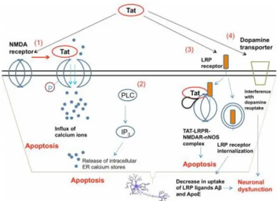
elements contained within the HIV LTR located at the 5′end of the integrated HIV provirus (Rohr et al., 2003). HIV-1 Tat interacts with several chromatin modifying complexes and histone modifying enzymes to relieve the virus LTR.The results of several studies suggest that Tat is a potential contributor to HIV-1 dementia (Wallace et al., 2005). Tat increases intracellular Ca2+ in neurons, followed by mitochondrial calcium uptake which results in the generation of ROS, activation of caspases and eventually apoptosis of neurons. Tat can be transported efficiently across the intact BBB. The mRNA levels for Tat have been reported to be elevated in the brain extracts from HAD patients. Tat plays an important role during neuropathogenesis, both as an intracellular and extracellular mediator of neurotoxicity (Fig 3) (Wong et al., 2005).
Fig 3. Mechanisms of Tat neurotoxicity. (1) Tat binds to the NMDA receptor and drives the phosphorylation of an intracellular NMDAR subunit, causing excess opening of cation channels and toxic accumulation of calcium. (2) When applied to neurons, Tat is able to induce the activation of PLC and drive the IP3-mediated release of intracellular calcium from ER stores, further contributing to calcium toxicity and apoptosis. (3) Tat can bind to LRP receptors, and be taken up as part of a macromolecular complex including NMDAR and neuronal nitric oxide synthase (nNOS) that induces cellular apoptosis [65]. Tat can also drive the internalization of the LRP receptor, reducing the uptake of LRP receptor ligands amyloid-β peptide and Apolipoprotein E, which may contribute to systemic neuronal dysfunction. (4) Tat interferes with the activity of dopamine transporter, diminishing the reuptake of dopamine by pre-synaptic neurons and interfering with signal transmission.
Tat also binds the low density lipoprotein receptor related protein (LRP) in neurons, causing LRP internalization and a decrease in uptake of natural LRP ligands such as amyloid-β peptide and Apolipoprotein E (Liu et al., 2000) (Fig 3). The interaction of Tat with LRP can lead to the formation of an apoptosis-promoting complex including postsynaptic density protein-95 (PSD- 95), NMDA receptors and neuronal nitric oxide synthase (nNOS) (Eugenin et al., 2007). Tat has been found to interfere with the expression of
15
miRNAs in neurons, increasing the levels of CREB-targeting miR-34a and leading to neuronal dysfunction (Chang et al., 2011). Tat can also interfere with the ability of dopamine transporter to reuptake dopamine (Zhu et al., 2011). This likely contributes to the particularly severe damage rendered to dopaminergic-rich regions in the brains of patients with severe HAND (Chang et al., 2008).It is noteworthy that, Tat is able to activate the NMDAR, leading to excitotoxicity and neuronal apoptosis, as a result of perturbed cellular calcium homeostasis and mitochondrial alterations (Self et al., 2004). It has been demonstrated that Tat causes apoptotic neuronal death, which can be antagonized by NMDAR blockers, indicating that Tat-induced neuronal injury is due to persistent activation of NMDAR (Li et al., 2009). Evidence suggests that Tat is an important neurotoxic effector, being able to interact with and activate NMDAR, both directly and indirectly (Eugenin e al., 2007). In 2013 Capone et al. provided clear evidence that the origin of ROS generation is related to Spm catabolism and Tat is able to induce SMO enzyme activity in neuroblastoma cells through the stimulation of NMDAR. The pivotal role of SMO as the primary source of the cytotoxic H2O2 has
already been demonstrated in a human breast cancer model, using a stable, short hairpin RNA knockdown strategy (Pledgie et al., 2005). Regarding the involvement of NMDAR in this pathway, it has been demostrated that the inhibition of the polyamine metabolism by the SMO-specific inhibitor CHL completely prevented NMDA-induced cell death as well as ROS production. In agreement with the stimulatory effect of Tat on SMO activity, it was observed a significant decrease in the intracellular content of Spm, that may have a detrimental effect on cell viability for two main reasons. On one hand, Spm is responsible for intrinsic gating of ion channels by direct plugging of the channel pore. In particular, intracellular Spm can block the channel pore of NMDAR (Igarashi and Kashiwagi, 2010). Therefore, the reduction of Spm concentration induced by Tat treatment may cause hyperactivity of NMDAR thus leading to excitotoxicity. On the other hand, the depletion of Spm also makes cells more sensitive to ROS damage, because Spm has been demonstrated to act as a free radical scavenger that can protect DNA from H2O2-induced damage (Casero and Pegg, 2009). In this respect, it has been
also observed a marked depletion of GSH content in neuroblastoma cells treated with Tat, which may have dramatic consequences on cell viability. In the literature, a reduction in GSH levels has been associated with physiological processes such as aging (Finkel and Holbrook, 2000) and neurological disorders such as Alzheimer and Parkinson diseases (Gu et al., 1998); in fact the GSH content in the brain of such patients has been stated to decrease by 40–50%, compared to controls (Sofic et al., 1992).
16
ii. Neurotoxicity induced by HIV-gp120
The process of HIV-1 infection begins by HIV-1 binding to CD4 receptor on the target cell surface, through the viral envelope protein gp120 (Matthews et al., 1987). Both monomeric and oligomeric gp120 have neurotoxic capabilities (Bardi et al., 2006), and transgenic mice expressing gp120 have a spectrum of neuronal and glial changes resembling abnormalities in brains of HIV-1-infected humans (Toggas et al., 1994). HIV-1 gp120 directly binds NMDAR on human embryonic neurons and can cause a lethal influx of calcium ions (Lannuzel et al., 2005). HIV-1 gp120 can bind to either CCR5 or CXCR4 and induce death in neuroblastoma cells (Catani et al., 2000) (Figure 4). This apoptosis apparently takes place through a p38-MAPK-mediated signaling cascade (Kaul et al., 1999). Cognitive testing of gp120 transgenic mice showed age-dependent deficits in open field activity and spatial reference memory tests (D’Hooge et al., 1999). The natural ligands of both CCR5 (eg. CCL5, CCL3) and CXCR4 (CXCL12) were found to be neuroprotective against gp120 neurotoxicity (Khan et al., 2003). However, CXCL12 displays neurotoxicity after the N-terminal cleavage of a tetrapeptide in CXCL12 by MMP-2 (Zhang et al., 2003). Another factor up-regulated by the interaction of gp120 with CXCR4 is the neuronal nicotinic receptor α7, which increases cellular permeability to [Ca2+] influx and contributes to cell death (Ballester et al., 2010).
Fig 4. Mechanisms of gp120 neurotoxicity. (1) gp120 can bind to the NMDA receptor and lead to excessive opening of NMDAR-gated cation channels, allowing the influx of calcium ions to toxic levels. (2) gp120 can directly bind to either CCR5 or CXCR4, activating an
p38-MAPK mediated signaling cascade that leads to neuronal apoptosis. The gp120-CXCR4 binding also up-regulates the expression of the nicotinic receptor α7, which increases cellular permeability to [Ca2+] influx and contributes to cell death.
17
Several studies indicate that HIV proteins (e.g.,gp120), which are assumed to contribute to neuronal abnormalities in HAND (Alfahad and Nat, 2013; Schouten et al., 2011), can induce NF-κB and relative target genes (Genis et al., 1992), as well as arise in intracellular calcium levels (Medina et al., 1999). In this contest, the cytosolic phospholipase A2 (cPLA2), a calcium-dependent enzyme, hydrolyzes membrane phospholipids to release arachidonic acid (AA) (Burke et al., 2009). Its metabolites play a number of functions in the organism, among them the regulation of the activity of several kinases, including protein-tyrosine kinases (Joubert et al., 2001), these being able to elicit constitutive NOS phosphorylation and inactivation (Colasanti et al., 1999). NO is synthesized by at least three distinct isoforms of NOS: NOS-I or neuronal NOS (nNOS), NOS-II or inducible NOS (iNOS), and NOS-III or endothelial NOS. NOS-I and NOS-III are both calcium/calmodulin-dependent, are constitutively expressed (so that they are both called cNOS), and release physiological concentrations of NO. In contrast, iNOS can produce a greater amount of NO after induction by bacterial lipopolysaccharide (LPS), cytokines, and/or viral proteins (Colasanti and Suzuki, 2000). The existence of a crosstalk between cNOS and iNOS has been widely reported; in fact under physiological conditions, low levels of NO produced by cNOS suppress the transcription factor NF-κB, which is involved in the transcription of several pro inflammatory genes, including iNOS (Colasanti and Persichini, 2000). After a pathological stimulus (e.g.,LPS and/or cytokines), cNOS is phosphorylated early on a tyrosine residue, thus leading to enzymatic inhibition and subsequent reduction of physiological NO levels (Colasanti et al., 1999). Under these conditions, NF-κB is freed from the inhibitory action of NO; it enters the nucleus and participates in the transcription of genes, including iNOS and interleukin-1β (IL-1β) (Colasanti and Suzuki, 2000). Note that NF-κB, in turn, is able to induce cPLA2 mRNA expression with a subsequent AA release, thus establishing a positive feedback (Hernandez et al., 1999). The expression of these genes promotes a wide production of inflammatory effectors (e.g.,NO,AA,cytokines), which could contribute to HAND pathogenesis.The antioxidant cell response: the p62/keap1/Nrf2/ARE pathway
In response to intrinsic and extrinsic stimuli, cells activate various adaptive mechanisms to promote ROS/RNS detoxification. The cap’n’collar (CNC) family proteins are transcription factors, which contain basic leucine zipper (bZIP) and CNC domains. By regulating various antioxidant genes and phase II detoxifying enzymes, which are required for metabolic detoxification of xenobiotics, they play a pivotal role in the cellular response to oxidative or electrophilic stresses (Motohashi and Yamamoto, 2007). The CNC family
18
consists of nuclear factor erythroid-derived 2 (NF-E2), NF-E2-related-1 [NRF1 or NF-E2-like 1 (NFE2L1)], NRF2 (NFE2L2), NRF3 (NFE2L3), and distantly related broad complex–tramtrack–bric-a-brac (BTB) and CNC homology 1 (BACH1) and BACH2. Functionally, the CNC transcription factors form heterodimers with small MAF proteins (Kannan et al., 2012). These heterodimers regulate genes containing the antioxidant response element (ARE) or the MAF recognition element (MARE) such as heme oxygenase 1 (HO-1), NADP(H):quinone oxidoreductase 1 (NQO-1), glutamylcysteine ligase (GCL), peroxiredoxin (PRDX), superoxide dismutase (SOD), catalase (CAT), glutathione peroxidase (GPx), sulfiredoxin (Srx), thioredoxin reductase (Txnrd) and glutathione S-transferase (GST), which are involved in detoxification and drug metabolism (Blank, 2008; Eychene et al., 2008). ARE core sequence, 5′-RGTGA(C/G)NNNGC-3′, acts as a cis-acting enhancer and shows significant homology to the MARE enhancer, 5′-TGCTGAG(C)TCAGCA-3′ (Nerland, 2007). Interestingly, many studies demonstrate that among CNC members, NRF2 is heavily involved in the regulation of antioxidant genes (Zhang et al., 2008).NRF2 is a soluble protein primarily localized to the cytoplasm. It is highly conserved across species and contains seven functional NRF2-ECH homology (Neh) domains (Figure 5) (Itoh et al., 1999), responsible for heterodimerization and ARE binding, interacting with the transcriptional coactivators (Nioi et al., 2005) and for stabilization by recruiting an ubiquitin ligase complex (McMahon et al., 2004). Moreover, there is a binding site for KEAP1 (Kelch-like erythroid cell-derived protein with CNC homology ECH-associated protein 1) to regulate the ubiquitination (Wang et al., 2013).
Fig 5. Structural domains of NRF2 and KEAP1. NRF2 includes the CNC-bZIP domain and functional Neh domains. The CNC-bZIP domain including Neh1 is required for DNA binding and interaction with ubiquitin conjugating enzymes. The transactivation domain contains Neh4 and Neh5. Neh2 is involved in Keap1 binding. KEAP1 contains the BTB domain for KEAP1 homodimerization and interaction with Cul3. Six kelch repeats in the Kelch/DGR domain interact with the Neh2 domain of NRF2 for binding. The IVR domain links the BTB and Kelch/DGR domains and has several critical cysteine residues for KEAP1 activation and NRF2 repression.
19
Under basal conditions, NRF2 is localized in the cytoplasm through a direct interaction with the KEAP1 protein that leads to ubiquitination and proteasomal degradation (Itoh et al., 1999). KEAP1 contains three functional domains, a BTB domain, an intervening region (IVR), and a double glycine repeat (DGR) or a Kelch motif, which contains six copies of the conserved kelch repeat that form a β-propeller structure. The BTB domain is required for KEAP1 homodimerization and the interaction with Cul3, which regulates KEAP1 through ubiquitination and proteasomal degradation. Twenty-seven cysteines within the IVR domain residues act as active stress sensors in human KEAP1 (Dinkova-Kostova et al., 2002). Among these, Cys151, Cys257, Cys273, Cys288, and Cys297 are known to be highly reactive toward oxidative and electrophilic stresses (Sekhar et a., 2010). In response to stress, these cysteine residues become oxidized and form disulfide bonds or covalent adducts. The cysteine modifications cause a conformational change in KEAP1 to prevent NRF2 ubiquitination (Zhang and Hannink , 2003). Free NRF2 then translocates into the nucleus, heterodimerizes with small MAFs, and binds to ARE sequences in promoter regions of antioxidant genes (Moon and Giaccia, 2015).In 2010, it was discovered that the antioxidant transcription factor Nrf2 can be activated by p62/SQSTM1 (sequestosome 1) by a ‘non-canonical’ pathway. The underlying mechanism is completely redox independent, and involves the recruitment of Keap1 that functions as an adapter protein of the Cul3-ubiquitin E3 ligase complex responsible for degrading Nrf2 (Komatsu et al., 2010). In agreement with this model, p62 binds to aggregates of ubiquitylated proteins and increases its affinity for Keap1 when phosphorylated at Ser351 (Ichimura et al., 2013) This event induces Keap1 degradation via autophagy (Taguchi et al., 2012) and leaves Nrf2 free to accumulate and translocate in the nucleus (Filomeni et al., 2014).
The p62 protein was originally identified because it bound to the tyrosine kinase Lck (Joung et al., 1996). Subsequently, p62 was found to bind atypical protein kinase C (Sanchez et al., 1998) and shown to act as a scaffold or adaptor protein in NFkB signaling pathways following activation of tumor necrosis factor-α (TNF- α) (Sanz et al., 1999), interleukin-1 (Sanz et al., 2000), and nerve growth factor (Wooten et al., 2000) receptors. In addition, p62 is involved in activation of caspase-8 upon stimulation of cell death receptors (Jin et al., 2009). p62 is required for Ras-induced tumorigenesis in vitro and in vivo and is up-regulated in different human tumors (Duran et al., 2008). Because p62 binds to ubiquitin and to LC3, it is both a selective autophagy substrate and a cargo receptor for autophagic degradation of ubiquitinated targets (Ichimura et al., 2008; Komatsu et al., 2007; Lamark et al., 2009). During the evolutionary conserved process of autophagy, p62 forms cytosolic inclusion bodies, which contain ubiquitinated protein
20
aggregates that subsequently can be degraded by autophagy (Bjørkøy et al., 2005). Isolation membranes embrace and envelop part of the cytoplasm, sequestering the content into a double-membrane vesicle called an autophagosome, that in turn, fuse with lysosomes, and their contents are degraded by lysosomal hydrolases (Mizushima et al., 2008). The expression of the p62 gene is induced by NRF2 upon exposure to electrophiles, ROS and NO (Kosaka et al, 2010). Furthermore, p62 protein has been reported to stimulate expression of genes containing ARE sequence in their promoter regions (Liu et al., 2007). A positive inter-relationship of some sort exists between NRF2 and p62, which influences the ARE-gene battery. In 2010, Jain et al. showed that NRF2 can induce p62 expression by binding directly to a conserved ARE in its promoter/enhancer. Noteworthy p62 protein is able to establish a positive feedback loop responsible for maintaining its high expression levels. Indeed, p62 is able to stimulate NRF2 activity by binding to KEAP1, sequestering it, and directing its degradation by autophagy. This association between p62 and KEAP1 leads to stabilization of NRF2, enabling the transcription factor to induce ARE-driven gene expression (Figure 6). The p62 ARE is conserved in mammals, and it contains the 5’ flanking region TGC-, and the core sequence -TGAGTCA- classifying it as a MAF-like recognition element (MARE). The NRF2 responsive p62 MARE contains a GC to CG substitution in the 3’ flanking region compared with the consensus MARE (Motohashi et al., 2002). It has been reported that MARE-like sequences with a G to C substitution in the 3’ flanking region preferentially bind to small MAF:NRF2 heterodimers (Jain et al., 2010).Fig 6. Under normal conditions, NRF2 is bound to KEAP1 and is inactivated as a transcription factor for antioxidant-defence genes by proteasome-mediated degradation. p62 is degraded through autophagy under normal conditions. In the presence of oxidative stress, KEAP1 is either modified so that it can no longer bind NRF2 or it is sequestered by p62, the expression of which is increased in response to oxidative stress. This displaces KEAP1 from NRF2 so that NRF2 can activate antioxidant-defence genes and promote survival. Oxidative stress also activates nuclear factor-κB (NF-κB) as a result of p62 upregulation and tumour necrosis factor receptor-associated factor 6 (TRAF6) complex formation, or by other mechanisms, to turn on antioxidant-defence gene expression. From “Deconvoluting the context-dependent role for autophagy in cancer” Eileen White Nature Reviews Cancer 12, 401-410 (June 2012)
21
2) AIMS OF THE WORK
HIV-1 infection is associated with neurological complications, recognized as disorders of various degree, known as HAND. Resident cells in the CNS, including microglia and astrocytes, can be infected and/or activated by HIV and subsequently release a variety of neurotoxins, including viral particles, such as Tat and gp120 in the brain microenvironment. Tat and gp120, more than other HIV proteins, play a pivotal role in HAND neuropathogenesis. The major factors involved in HAND are inflammation and oxidative/nitrosative stress. Excessive production of inflammatory biomolecules or mediators of oxidative stress produced by different cell types of CNS may induce neurotoxicity.
This PhD project deals with three topics, all linked to a major context. It is already known that Tat-induced neuronal injury is due to persistent NMDAR activation. Recently, our group provided clear evidence that the origin of ROS generation was related to Spm catabolism and that Tat was able to induce SMO enzyme activity in neuroblastoma cells through the stimulation of NMDAR.
As ROS also function as signaling molecules able to trigger many pathways in the cell, the first aim of this PhD work was to evaluate Tat-mediated induction of antioxidant response in neuronal cells. Here, we wanted to assess the activation of Nrf2/ARE pathway and the role of NO in SH-SY5Y neuroblastoma cells taken as cellular model. Moreover, since protein aggregation is a common marker of many neurodegenerative diseases, the involvement of p62 protein in Nrf2/ARE pathway and its role in the degradation of Tat will be analyzed.
Besides the study on neuronal cells we will also analyze the effect of HIV proteins on astroglial cells. Astrocytes are indeed the brain cells that profoundly affect neuronal functions and, in a variety of neurological disorders, are thought to play a critical role in mediating neuronal toxicity or neuronal rescue. In neurodegeneration associated with HIV-1, both chronic inflammation and oxidative stress play a crucial role and these conditions are often related. Thus, the second aim was to evaluate the role of cPLA2-AA pathway in the pro-inflammatory response induced by Tat and gp120, and then we wondered whether Tat was able to activate the antioxidant response in astroglial cells, evaluating Nrf2/ARE pathway induction and the involvement of NO. In particular, we planned to focus our attention on the pivotal role of SystemXc, a cystine/glutamate antiporter that allows astrocytes to maintain the high synthetic rates of glutathione to counteract ROS accumulation. Finally, we wanted to analyze the neurotoxic effect of Tat, performing neuronal-astroglial cell co-culture experiments. Since is noteworthy that during oxidative stress, the steady-state structure and proper
22
physiological function of the Golgi apparatus were often affected, we analyzed GA structure change in neurons treated with Tat and co-cultured with Tat-secreting astrocytes. Another consequence of the neurotoxicity induced by Tat could be the reduced viability of neuronal cells, or the simultaneous activation of the antioxidant response.23
3) RESULTS AND DISCUSSION
HIV-Tat Induces the Nrf2/ARE Pathway through NMDA Receptor-Elicited Spermine Oxidase Activation in Human Neuroblastoma Cells.
The HIV-Tat protein is known as a significant promoter of neurotoxicity (Kaul et al., 2001; Jin et al., 2012). Several mechanisms underline these toxic effect; Tat can freely penetrate neuronal cell membranes and induce lipid peroxidation (Haughey et al., 2004), leading to ROS generation such as superoxide and hydrogen peroxide. In addition, Tat can trigger iNOS expression thus increasing the production of NO, which binds superoxide anion yielding the highly reactive molecule peroxynitrite (ONOO-) (Nicotera et al., 1995) which can have deleterious CNS effects (Nakhostin-Roohi et al., 2013).
Moreover, it is noteworthy that HIV proteins, such as Tat, are able to activate NMDAR, leading to excitotoxicity and neuronal apoptosis, as a result of perturbed cellular calcium homeostasis and mitochondrial alterations (Self et al., 2004).
Here, we found out that the treatment of human SH-SY5Y neuroblastoma cells with 200 ng/ml recombinant Tat for 4, 8, and 24 h was able to increase the mRNA expression of antioxidant enzymes, such as NQO1, CAT, SOD1, SOD2 and HO-1. Tat concentration was chosen based on previous experiments (data not shown). As shown in figure 7, we have observed an increase of mRNA expression of all the genes analyzed reaching the maximum at 4 h (8 h for HO-1) after Tat treatment. At 24h post-treatment, gene expression was reverted to nearly control levels.
24
Fig 7: Effects of Tat on ARE-driven gene expression in SH-SY5Y cells. SH-SY5Y were treated with Tat (200 ng/ml) for 4, 8, and 24 h. After incubation at 37 °C, the cells were homogenized and total RNA has been purified to assess mRNA levels of several genes (NQO1, CAT, SOD1, SOD2, HO-1) by RT-qPCR. Data are calculated relative to the internal housekeeping gene (GAPDH) and are expressed as the mean fold change compared with control. Each value represents the mean ± SEM of three independent experiments. One-way ANOVA, followed by Bonferroni's test, was used to determine significant differences.NQO1: * p≤0.01 vs CTRL, ** p≤0.01 vs CTRL; CAT: * p≤0.01 vs CTRL, ** p≤0.05 vs CTRL; SOD1: * p≤0.01 vs CTRL, ** p≤0.05 vs CTRL; SOD2: * p≤0.01 vs CTRL, ** p≤0.05 vs CTRL; HO-1: * p≤0.05 vs CTRL, ** p≤0.01 vs CTRL.
Recently, another HIV-1 protein, reverse transcriptase (RT), was shown to induce an antioxidant response (Isaguliants et al., 2013). In particular, RT increased the transcription of the phase II detoxifying enzymes NQO1 and HO-1 in human embryonic kidney cells, revealing a direct link between the propensity of the viral proteins to induce oxidative stress and their immunogenicity (Isaguliants et al., 2013). Furthermore, it has been reported that gp120 significantly upregulates HO-1 and NQO1 in human astrocytes, suggesting a possible role of the antioxidant defense mechanism in promoting cell survival (Reddy et al., 2012). Notably, a progressive increase in serum catalase activity has been detected in advancing HIV infection, reflecting and/or compensating for systemic glutathione and other antioxidant deficiencies in HIV-infected individuals (Leff et al., 1992).
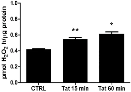
Normally, the expression of antioxidant enzymes is induced in response to oxidative stimuli, including ROS production. As reported elsewhere, the treatment of SH-SY5Y cells with Tat for 1 and 4 h was able to induce ROS generation by the upregulation of the SMO enzyme activity (Capone et al., 2013). Here, we hypothesize that Tat can elicit an antioxidant response in neuronal cells through a SMO-dependent activity. To address this question,
25
we first assessed the ability of Tat to upregulate the activity of SMO at an early time point (i.e., 15 min). Therefore, a chemiluminescence analysis was performed to measure H2O2 production in extracts from cells treated with Tat(200 ng/ml) for 15and 60 min. As shown in figure 8, the SMO activity was already increased at 15 min post-Tat treatment; this effect was maintained for up to 60 min.
Fig 8: Effects of Tat on spermine oxidase activity. SH-SY5Y cells were treated with Tat (200 ng/ml) for 15 and 60 min. At the end of incubation cell extracts were analyzed for SMO activity. The data shown are the means of three independent experiments. One-way ANOVA, followed by Bonferroni's test, was used to determine significant differences. ** p≤0.05 vs CTRL, * p≤0.01 vs CTRL.
Next, we wanted to determine whether the expression of detoxifying and antioxidant genes in response to Tat treatment was elicited by the SMO-induced activity. Therefore, we treated SH-SY5Y cells with 10 nM chlorhexidine digluconate (CHL), a strong competitive inhibitor of SMO (Cervelli et al., 2013), for 16 h before treatment with Tat (200 ng/ml). As expected, CHL completely prevented Tat-induced up-regulation of all the genes analyzed (Fig 9), thus suggesting that Tat may elicit an antioxidant response in neuronal cells through the activation of SMO.
26
Fig 9: Effects of SMO inhibition on Tat-induced gene expression in SH-SY5Y cells. Cells were overnight pretreated with CHL (10 nM) or medium alone before the addition of Tat (200 ng/ml) for 4 h. After incubation at 37 °C, the cells were homogenized and total RNA has been purified to assess mRNA levels of NQO1, CAT, SOD1, SOD2, HO-1 genes by RT-qPCR. Data are calculated relative to GAPDH and are expressed as the mean fold change compared with control. Each value represents the mean ± SEM of three independent experiments. One-way ANOVA, followed by Bonferroni's test, was used to determine significant differences. NQO1: * p≤0.01 vs CTRL, ^ p≤0.01 vs Tat; CAT: * p≤0.01 vs CTRL, ^ p≤0.01 vs Tat; SOD1: * p≤0.01 vs CTRL, ^ p≤0.01 vs Tat; SOD2: * p≤0.01 vs CTRL, ^ p≤0.01 vs Tat; HO-1: * p≤0.01 vs CTRL, ^ p≤0.01 vs Tat;Considering that ARE genes are mainly regulated by Nrf2, we investigated whether Tat was able to activate this transcription factor in human neuroblastoma cells. To this aim, we examined the Nrf2 translocation into the nucleus, after treatment with Tat (200 ng/ml) for 15 minutes, 2 hours and 16 hours. Nrf2 levels were measured in nuclear extracts by using Western blot analysis. As shown in figure 10, Tat induced a 2.68-fold increase of the nuclear Nrf2 levels already at 15 min post-treatment, which was maintained up to 16h.
27
Fig 10: Effects of Tat on Nrf2 nuclear translocation in SH-SY5Y cells. Cells were treated with Tat (200 ng/ml) for the indicated time points. After incubation at 37 °C, cells were mechanically harvested, and the nuclear extracts were prepared as specified in the Materials and Methods section to assess Nrf2 levels by western blot analysis with anti-Nrf2 1:1000 (Abcam) and anti-laminB 1:4000 (Abcam). The histogram shows the densitometric analysis of the western blots for each sample. Values are calculated relative to the nuclear Lamin B content and are the means ± SEM from three separate experiments, each performed in duplicate. One-way ANOVA, followed by Bonferroni's test, was used to determine significant differences. ** p≤0.05 vs CTRL, * p≤0.01 vs CTRL.The effect of Tat on Nrf2 activation was also confirmed by a TransAm kit based on ELISA method (see below in figure 12).
We found, for the first time, that Tat activates Nrf2 in neuronal cells, and our results are consistent with data reporting that Tat enhances the cellular expression of Nrf2 at the transcriptional and protein levels in MAGI cells (Zhang HS et al., 2009). Moreover, it has been reported that Nrf2 is also up regulated in response to gp120 in primary astrocytes, thereby suggesting a possible protective role of gp120-induced Nrf2 in regulating the levels of pro-oxidative and pro-inflammatory molecules in HANDs (Reddy et al., 2012). As described above (see figure 2), Tat is able to upregulated the activity of SMO at an early time point (i.e., 15 min). To investigate on the role of SMO in Tat-elicited Nrf2 activation, SH-SY5Y cells were exposed overnight to 10 nM CHL before treatment with Tat (200 ng/ml) for 15 min. Then, a western blot analysis was performed on nuclear extracts using an anti-Nrf2 specific antibody. As shown in figure 11, the pretreatment of SH-SY5Y cells with CHL completely prevented Tat-induced Nrf2 nuclear translocation, thereby suggesting an involvement of SMO in this mechanism.
28
Fig 11: Effects of SMO inhibition on Tat-induced Nrf2 activation in SH-SY5Y cells. Cells were overnight pretreated with CHL (10 nM) or medium alone before the addition of Tat (200 ng/ml) for 15 min. After incubation at 37 °C, cells were mechanically harvested, and the nuclear extracts were prepared as specified in Methods to assess Nrf2 levels by western blot analysis with anti-Nrf2 1:1000 (Abcam) and anti-laminB 1:4000 (Abcam). The histogram shows the densitometric analysis of the western blots for each sample. Values are calculated relative to the nuclear Lamin B content and are the means ± SEM from three separate experiments, each performed in duplicate. One-way ANOVA, followed by Bonferroni's test, was used tom determine significant differences. * p≤0.01 vs CTRL, ^ p≤0.01 vs Tat, § Not significant vs Tat + CHL.Previously, it has been reported that Tat was able to induce ROS production through the stimulation of SMO activity in neuroblastoma cells (Capone et al., 2013). Here, we evaluated the role of SMO-dependent ROS generation in Tat-induced Nrf2 activation by pretreating SH-SY5Y cells with the antioxidant NAC, which is able to prevent Tat-induced ROS generation (Capone et al., 2013). As shown in Fig 12A, the pre-treatment of cells with NAC (2 mM) for 1 h before treatment with Tat (200 ng/ml) for 15 min inhibited Nrf2 activation.
As we observed elsewhere, Tat induces SMO activity and ROS production through the stimulation of NMDAR in neuroblastoma cells (Capone et al., 2013). To investigate whether NMDAR was involved in Tat-induced Nrf2 activation, we pretreated SH-SY5Y cells for 2 h with the NMDAR antagonist MK-801 (10 M) and then treated with Tat (200 ng/ml) for 15 min. Figure 12A shows the strong inhibitory effect of MK-801 on the activation of Tat-induced Nrf2. Consistently, NMDA Tat-induced Nrf2 activation, clearly indicating that the stimulation of NMDAR can be responsible for the observed Nrf2 activation in neuroblastoma SH-SY5Y cells. As expected, Tat-induced mRNA expression of the two ARE genes, NQO1 and CAT was prevented by MK-801 pretreatment (Fig 12B). Altogether, these results indicate that Tat induces the Nrf2 pathway through NMDAR-elicited SMO activation in human neuroblastoma cells.
29
Fig 12: Role of NMDA receptor in Tat-induced Nrf2 activation. (A) SH-SY5Y cells were pretreated for 2 h with MK-801 (10 µM), NAC (2 mM), or medium alone before the addition of Tat (200 ng/ml) for 15 min. After incubation at 37 °C, the cells were homogenized, and Nrf2 activation was quantified by TransAM assay as detailed in the Materials and Methods section. Data points are the means ± S.E.M. from 3 separate experiments, each performed in duplicate. One-way ANOVA, followed by Bonferroni's test, was used to determine significant differences. * p≤0.01 vs CTRL, ^ p≤0.01 vs Tat, § p≤0.01 vs CTRL. (B) Cells were pretreated for 2 h with MK-801 (10 µM) or medium alone before the addition of Tat (200 ng/ml) for 4 h. After incubation at 37 °C, the cells were homogenized and total RNA has been purified to assess mRNA levels of NQO1 and CAT genes by RT-qPCR. Data are calculated relative to GAPDH and are expressed as the mean fold change compared with control. Each value represents the mean ± SEM of two independent experiments. One-way ANOVA, followed by Bonferroni's test, was used to determine significant differences. NQO1: * p≤0.05 vs Tat, ** p≤0.01 vs CTRL; CAT: * p≤0.05 vs Tat, ** p≤0.01 vs CTRL;The question now is whether the modulation of NMDAR/SMO/ROS/Nrf2/ARE pathways is sufficient for protection against Tat-induced oxidative stress (see figure 13 for a schematic overview). As we described elsewhere, NMDAR/SMO/ROS activation leads to a weak (approximately 30%) neurotoxicity in Tat-treated SH-SY5Y cells (Capone et al., 2013), so, cell death occurs (although weakly) despite the activation of Nrf2/ARE pathway. A similar paradox has been reported by Akay et al. (2014) in a study on the effects of ARV drugs in the central nervous system. In particular, the neuronal damage and death that occur following exposure to ARV drugs, despite the endogenous antioxidant response, suggest that this response may be insufficient or too delayed to protect cells from Tat toxicity.