EURHINODELPHINIDS FROM THE EARLY MIOCENE OF PERU:
FIRST UNAMBIGUOUS RECORDS OF THESE HYPER-LONGIROSTRINE DOLPHINS OUTSIDE THE NORTH ATLANTIC REALM
OLIVIER LAMBERT1*, CHRISTIAN DE MUIZON2, RAFAEL M. VARAS-MALCA3, MARIO URBINA3 & GIOVANNI BIANUCCI4
1*Corresponding author. Direction Opérationnelle Terre et Histoire de la Vie, Institut Royal des Sciences Naturelles de Belgique, rue Vautier 29,
1000 Brussels, Belgium. E-mail: [email protected]
2 CR2P UMR 7207, (MNHN, CNRS, UPMC, Sorbonne-Université), Muséum national d’Histoire naturelle, Département Origines et Evolution,
rue Cuvier 57, 75231 Paris, France.
3 Departamento de Paleontología de Vertebrados, Museo de Historia Natural – UNMSM, Avenida Arenales 1256, 15072 Lima, Peru. 4 Dipartimento di Scienze della Terra, Università di Pisa, Via S. Maria 53, 56126 Pisa, Italy.
To cite this article: Lambert O., Muizon C. de, Varas-Malca R.M., Urbina M. & Bianucci G. (2021) - Eurhinodelphinids from the early Miocene of Peru: first unambiguous records of these hyper-longirostrine dolphins outside the North Atlantic realm. Riv. It. Paleontol. Strat., 127(1): 17-32.
Abstract. Among the many hyper-longirostrine dolphins (Odontoceti) from the Miocene, members of the
family Eurhinodelphinidae bear two highly distinctive cranial features: a long and edentulous premaxillary portion of the rostrum and a mandible that is significantly shorter than the rostrum. Until now, unambiguously attributed members of this clade were only recorded from early to middle Miocene deposits of the North Atlantic realm (east coast U.S.A., North Sea Basin, and Mediterranean). In this work we describe and compare two partial skulls of longi-rostrine dolphins from late early Miocene (Burdigalian, 19.25-18 Ma) marine deposits of the Chilcatay Formation, in the East Pisco Basin (southern coast of Peru), preserving rostral and mandibular material, as well as ear bones. Based on these specimens we report diagnostic remains attributable to this family for the first time for the whole Southern Hemisphere and the whole Pacific Ocean. This major expansion of eurhinodelphinids’ palaeogeographic distribution contrasts with their proposed shallow-water, coastal environments; it suggests a new dispersal route for members of the family across the Central American Seaway; and it further highlights the similarities between the odontocete faunas of the southeastern Pacific and North Atlantic realm during the Miocene. Better-preserved eurhinodelphinid speci-mens from the odontocete-rich Chilcatay Formation will allow for a more detailed comparison with North Atlantic members of the family.
Received: September 3, 2020; accepted: December 23, 2020
Keywords: Cetacea; Odontoceti; Eurhinodelphinidae; Chilcatay Formation; Burdigalian; southeastern Pacific.
I
ntroductIonEurhinodelphinidae are a moderately diver-sified family of hyper-longirostrine dolphins (sen-su McCurry & Pyenson 2019) characterized by the unique combination of an elongated, edentulous premaxillary part of the rostrum with mandibles
that are significantly shorter than the rostrum (Abel 1901; Kellogg 1925). Never observed in any oth-er cetacean group, the resulting extreme ‘ovoth-erbite’ has been variously interpreted as a probing tool to detect prey in soft sediment of the seafloor or as a contacting or stunning device used higher in the water column, as in istiophorid and xiphiid billfish (Myrick 1979; Lambert 2005a; McCurry & Pyenson 2019). Together with their long snout, the long and relatively flexible neck of eurhinodelphinids
(as seen for example in Mycteriacetus and Xiphiace-tus, see Pilleri 1985; Lambert 2005a) suggests that
these small to medium-size odontocetes were slow swimmers, possibly living in shallow-water coastal environments (Lambert 2005a; Marx et al. 2016), a hypothesis that should be further tested with stable isotope analyses (see preliminary work by Barrick et al. 1992).
Although fossil odontocete remains from various regions of the world, including Patagonia, Peru, Caucasus, and South Australia, and from de-posits dated from the late Oligocene to the late Mi-ocene have been tentatively attributed to the family Eurhinodelphinidae (in some cases named Rhab-dosteidae; Lydekker 1893; Cabrera 1926; Mchedlidze 1976; Fordyce 1983; Pilleri 1989), their state of pres-ervation does not allow for the unambiguous recog-nition of the two apomorphic features listed above (Lambert 2005a; Marx et al. 2016; Supplementary Table 1). Therefore, based on specimens either at least showing one of these two synapomophies or robustly assigned to species or genera known from better-preserved specimens in other localities, the fossil record of the family is for now restricted to the genera Eurhinodelphis, Mycteriacetus, Schizodel-phis, Xiphiacetus, and ZiphiodelSchizodel-phis, from the western
North Atlantic, North Sea, and Mediterranean (to-gether grouped here in the North Atlantic realm), in deposits ranging from the early to middle (and possi-bly up to early late) Miocene (Abel 1901, 1902; Kel-logg 1925; Pilleri 1985; Muizon 1988a; Bianucci et al. 1994; Bianucci & Landini 2002; Lambert 2005a,b; Whitmore & Kaltenbach 2008; Marx et al. 2016).
Recently, lower Miocene (Burdigalian) de-posits of the Chilcatay Formation, in the East Pis-co Basin (southern Pis-coast of Peru), yielded a rich odontocete assemblage, including an heterodont longirostrine odontocete (Inticetus vertizi), two
phy-seteroids, several platanistoids (a platanistid, four squalodelphinids, and two other platanistoids), a longirostrine homodont dolphin (Chilcacetus cavirhi-nus), and a kentriodontid (Lambert et al. 2015, 2018,
2020; Bianucci et al. 2018a,b, 2020; Di Celma et al. 2018, 2019). Early field observations lead us to sug-gest in a previous work (Lambert et al. 2014) that a eurhinodelphinid closely related to Ziphiodelphis was
present in the Chilcatay Formation. However, a de-tailed study of collected material lead to the referral of this material to Chilcacetus, in another clade of
longirostrine odontocetes (Lambert et al. 2015;
Bia-nucci et al. 2018). In the present work we describe and compare two new skulls of longirostrine ho-modont odontocetes from the Chilcatay Formation in the fossil-rich locality of Zamaca, revealing the presence of eurhinodelphinids in the southeastern Pacific during the early Miocene.
MaterIalandMethods
Institutional abbreviations. CMM, Calvert Marine
Muse-um, Solomons, U.S.A.; IRSNB, Institut royal des Sciences naturelles
de Belgique, Brussels, Belgium; LACM, Natural History Museum of
Los Angeles County, Los Angeles, U.S.A.; MGP, Museo di Geologia
e Paleontologia, Padova, Italy; MLP, Museo de La Plata, La Plata,
Ar-gentina; MUSM, Museo de Historia Natural, Universidad Nacional
Mayor de San Marcos, Lima, Peru; OU, University of Otago,
Dun-edin, New Zealand; USNM, National Museum of Natural History,
Smithsonian Institution, Washington D.C., U.S.A.
Anatomical terminology. The terminology for skull
ana-tomical features follows Mead & Fordyce (2009), except where ex-plicitly stated.
s
ysteMatIcpalaeontologyCETACEA Brisson, 1762 Pelagiceti Uhen, 2008 Neoceti Fordyce & Muizon, 2001
Odontoceti Flower, 1867
Eurhinodelphinidae Abel, 1901 Eurhinodelphinidae indet.
Figs. 1-5
Referred specimens: MUSM 632, a
frag-mentary skull including large portions of the ros-trum, part of the bony nares region, part of the basicranium (mainly the basisphenoid, basioccipital, right exoccipital, and right squamosal), and a large part of the mandibles, together with a rib fragment; MUSM 3944, a partial cranium including the
prox-imal portion of the rostrum, part of the facial re-gion, and most of the basicranium, including the two in situ periotics and the two detached tympanic bullae.
Localities: Both specimens were discovered
in the odontocete-rich locality of Zamaca (see Lam-bert et al. 2018, 2020; Di Celma et al. 2019; Bianucci et al. 2020), East Pisco Basin. The exact locality of MUSM 632 is unknown, whereas the geographic
coordinates of MUSM 3944 are the following: S 14°37’34.95” - W 75°38’24.35”. MUSM 3944 was reported in the Zamaca fossil map of Di Celma et al. (2019) with the field number ZM 25, and tenta-tively referred there to “Eurhinodelphinidae indet.”
Horizon: Both specimens were collected in
the Chilcatay Formation, which is widely exposed in the locality of Zamaca. The precise stratigraphical level of MUSM 632 is unknown, whereas MUSM 3944 was collected at 32.4 m above the contact with the underlying Otuma Formation, in the allomem-ber Ct1a, together with other odontocete
(Odonto-ceti indet.) and marine turtle (Testudines indet.) re-mains, as well as shark teeth (Di Celma et al. 2019). The allomember Ct1a also yielded a rich odontocete
fauna including kentriodontids, physeteroids, and squalodelphinids (Di Celma et al. 2019; Bianucci et al. 2020; Lambert et al. 2020). In Zamaca and the nearby Ullujaya locality the cetacean-bearing layers of this formation have been dated through biostra-tigraphy, 40Ar/39Ar and 87Sr/86Sr radiometric analy-ses from a 19.25-18.02 Ma interval (Burdigalian, late early Miocene; Di Celma et al. 2018, 2019; Bosio et al. 2020a,b).
Remarks: In addition to the large amount
of similarities with known eurhinodelphinids (see below), both specimens described here are referred to the family Eurhinodelphinidae based on two syn-apomorphies: the presence of an extensive premax-illary portion of the rostrum and, for MUSM 632, the mandibles being significantly shorter than the rostrum (see details below). None of these char-acters is observed in any other odontocete clade. Similarities in cranial dimensions and morphology indicate that the two specimens could belong to the same species; however, the lack of a well-preserved facial region in both specimens and the absence of ear bones in MUSM 632 refrain us from providing a definitive conclusion on that matter. More complete specimens will also be needed for a more detailed comparison with eurhinodelphinid species from the North Atlantic realm.
Description
MUSM 632: The fragmentarily preserved
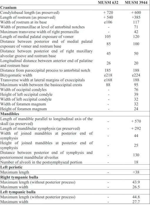
cra-nium of MUSM 632 has a minimum rostrum length of 540 mm (apex of the premaxillae is missing for an unknown length, see below) and a width of the rostrum at its base estimated at 106 mm; the bizy-gomatic width is estimated at 218 mm (see Table
1 for additional measurements), in the range of
Xiphiacetus cristatus, for instance (Lambert 2005a).
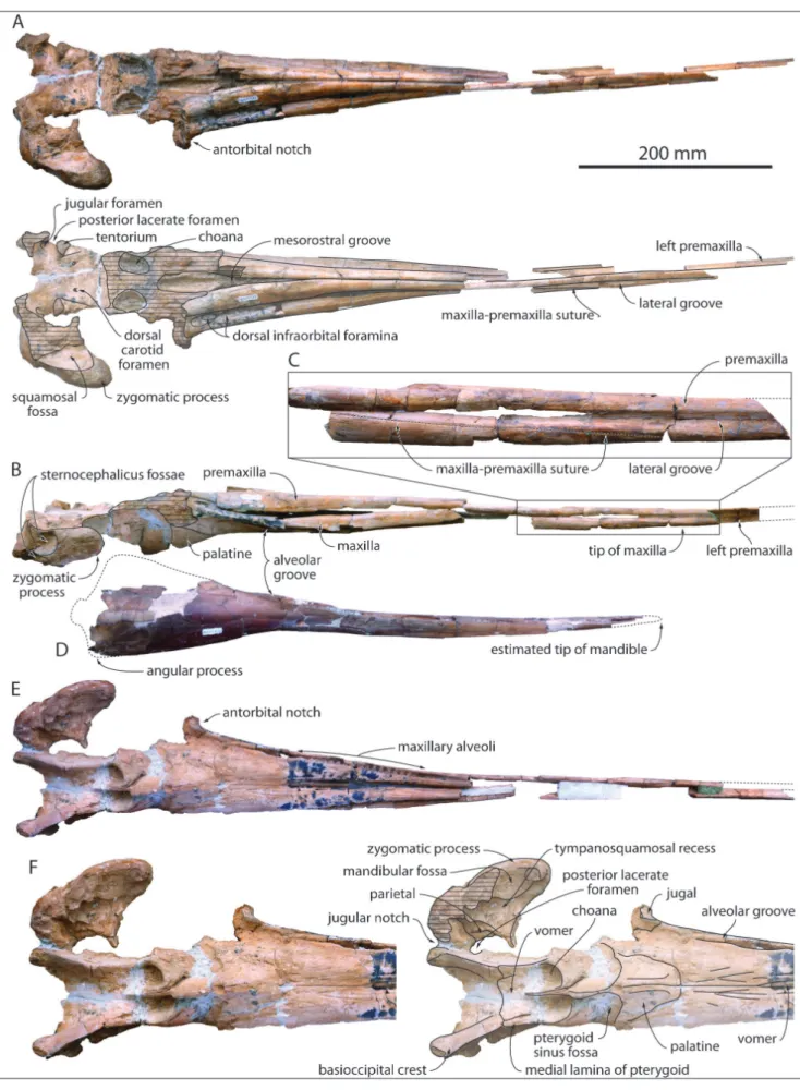
The lateral suture between the maxilla and the premaxilla on the rostrum is visible on both sides in lateral view; from a level about 255 mm an-terior to the antorbital notch, it starts descending anteroventrally from the lateral groove with a steep slope, reaching the ventral margin of the rostrum 490 mm anterior to the antorbital notch (tip of the maxilla) (Fig. 1B-C). The ratio between length of the maxilla on the rostrum and bizygomatic width is thus estimated at 2.25 in MUSM 632; this is sig-nificantly greater than in the type specimens of Eu-rhinodelphis cocheteuxi and E. longirostris (1.88 and 1.86,
respectively; Lambert 2004, 2005b). Seventy millim-eters anterior to the anterior tip of the maxilla, the dorsoventral height of the preserved section of left premaxilla is 16 mm, indicating that the premaxilla was originally considerably anteriorly longer than the maxilla, a diagnostic feature for Eurhinodelphi-nidae. In dorsal view the premaxillae are narrow for most of the rostrum length, with a convex trans-verse section, and they contact each other dorso-medially. They more significantly widen from a level
145 mm anterior to the antorbital notch and diverge posterolaterally from a level 120 mm anterior to the notch, gradually opening the mesorostral groove dorsally. At the level of the antorbital notch the dorsal surface of the premaxilla is flattened. At this level the premaxilla is proportionally much wider (compared to the maxilla) in Mycteriacetus and Ziphio-delphis. Only part of the right anteromedial sulcus is
preserved; the premaxillary foramen was posterior to the level of the antorbital notch.
In dorsal view the maxilla is exposed lateral to the premaxilla for its whole length on the ros-trum, with an exposure gradually increasing poste-riorly (Fig. 1A). The surface of the lateral groove is damaged in many parts of the rostrum; neverthe-less, this groove most likely occupied most of the rostrum length, as indicated by a finely preserved region dorsal and anterodorsal to the anterior tip of the maxilla. Level with the antorbital notch the dor-sal width of the maxilla is slightly narrower than the width of the premaxilla. Several medium to small-size dorsal infraorbital foramina (at least two on the right side) are present along the maxilla-premaxilla suture anterior to the antorbital notch. The largest posterior foramen marks the start of the lateral groove.
In ventral view, the alveolar groove ends posteriorly 45 mm anterior to the antorbital notch (Fig. 1E-F). Small, closely spaced, circular maxil-lary alveoli are observed, with diameters ranging between 5.0 and 5.5 mm and interalveolar septa ranging between 2.5 and 4.0 mm. The walls of the alveolar groove are too damaged or not preserved in the anterior part of the maxilla to allow for the identification of individual alveoli. Similarly, the preserved portion of the premaxillary part of the rostrum does not allow for a detailed description of the alveolar groove; no clue for premaxillary al-veoli could be found. On the palate, each maxilla is marked by a series of long, anteriorly, anteromedi-ally, and anterolaterally directed sulci, starting in the area of the major palatine foramen. A similar con-dition is observed in multiple specimens of Schizo-delphis and Xiphiacetus from the USNM collection.
A broad ventral exposure of the vomer is observed between the maxillae from a level a few centimeters anterior to the start of the alveolar grooves and for a length of 105 mm.
Together, the palatine-maxilla sutures draw a U shape, reaching a level 26 mm anterior to the antorbital notch, with no significant wedge of the maxillae between the palatines (Fig. 1F). The ante-rior limit of the transversely broad pterygoid sinus fossa is posterior (20-25 mm) to the level of the antorbital notch. Part of the lacrimojugal complex is preserved along the floor of the right antorbital notch, including the base of the styliform process of the jugal.
Due to the loss of the vertex and occipital shield, the ventral surface of the brain cavity is ex-posed, revealing small dorsal carotid foramina in the basisphenoid, the jugular foramina and the medial edge of the posterior lacerate foramina in the basi-occipital, just posterior to the tentorium (Fig. 1A). Right and left basioccipital crests diverge moderate-ly posterolateralmoderate-ly, drawing an angle of about 45°. Each crest thickens significantly transversely for its last posterior centimeters, whereas its ventral edge remains thin along its whole length (Fig. 1E-F). The distance between the lateralmost margins of the ba-sioccipital crests is 88 mm.
In lateral view the moderately long zygo-matic process of the squamosal is robust (maxi-mum height = 32 mm), with a convex, transversely thick dorsal margin, a rounded anterior end, and a rectilinear to slightly concave, transversely thinner
ventral edge (Fig. 1B). Two deep sternocephalicus fossae are present; the largest, dorsal fossa extends farther anteriorly, partly excavating the lateral sur-face of the zygomatic process. The postglenoid process is lost. In dorsal view the long axis of the zygomatic process is oblique, directed anterolat-erally. In ventral view the mandibular fossa is vast (transverse width = 32 mm) and anteroposteriorly concave. The tympanosquamosal recess is deep and anteriorly long, reaching the apex of the zygomat-ic process medial to a prominent tubercle for the short articulation surface of the styliform process of the jugal.
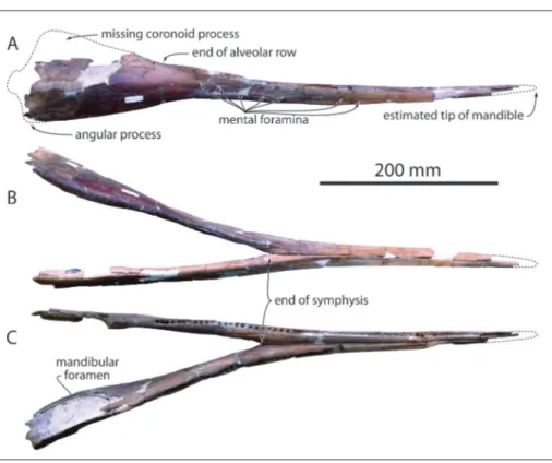
On the mandibles, the anterior tip of each dentary is missing, as well as the coronoid process-es, the condylprocess-es, and part of the angular processes (Fig. 2). Based on the gradual anterior narrowing and lowering of the slender anterior part of the mandibles we estimate that no more than 20-30 mm are missing anteriorly. Taking account of the missing mandibular condyles, the slope of the an-terior part of the coronoid process, and the cor-responding slope of the ventrolateral margin of the rostrum, we could position the mandibles in an approximately anatomically correct position along the anteroposterior axis of the rostrum (Fig. 1B). In such a position, the dorsoventral height of the preserved anterior section of the mandible is mark-edly lower than the corresponding section on the rostrum. Furthermore, the reconstructed tip of the mandible reaches an anterior level where the ros-trum is still dorsoventrally high. Together, these features suggest that the mandible was originally significantly shorter than the rostrum in MUSM 632, a derived character similarly observed or de-duced from preserved sections of the rostrum and mandible in the genera Mycteriacetus, Schizodelphis, Xiphiacetus, and Ziphiodelphis (Kellogg 1925; Myrick
1979; Pilleri 1985; Lambert 2005a). The tooth-bear-ing part of the mandible is slender (dorsoventrally low), with a slightly anterodorsally recurved outline (Fig. 2A). Posterior to the alveolar groove the dor-sal edge of the ramus becomes concave in lateral view, raising abruptly towards the coronoid process and possibly indicating the presence of a precoro-noid crest (sensu Fordyce et al. 2002), whereas in the same region the ventral margin is slightly convex and directed posteroventrally. The resulting abrupt increase of the height of the ramus posterior to the alveolar groove is similarly observed in Mycteriacetus,
Fig. 1 - Skull of MUSM 632, Eurhinodelphinidae indet. from the early Miocene of the Chilcatay Formation, East Pisco Basin, Peru. A) cranium in dorsal view (photo and interpretive drawing); B) cranium in right lateral view; C) detail of the anterior part of the rostrum in right lateral view, showing the descent of the maxilla-premaxilla suture; D) mandibles in right lateral view; E) cranium in ventral view; F) neurocranium and rostrum base in ventral view (photo and interpretive drawing).
Schizodelphis, and Xiphiacetus (Kellogg 1925; Myrick
1979; Pilleri 1985). The partly ankylosed mandib-ular symphysis (Fig. 2B-C) is long (more than 292 mm) with a triangular cross section, as seen in Myc-teriacetus, Schizodelphis, Xiphiacetus, and Ziphiodelphis.
Transverse width of the joined mandibles and dor-soventral height at the posterior end of the symph-ysis are 44 and 25 mm, respectively. In the anterior part of the mandibles, the alveolar groove bears small circular alveoli with a diameter of 4.5 mm and interalveolar septa of about 2 mm (Fig. 2C). At the posterior end of the symphysis alveoli retain a di-ameter of about 4.6 mm and septa ranging from 3.0 to 3.5 mm. 18 post-symphyseal alveoli are counted on a length of 120 mm on the left side. The lateral surface of the mandible is pierced by a series of obliquely organized mental foramina (at least seven on the right side), extended anteriorly by long sulci. No conspicuous lateral groove is observed on the mandibles. Deep longitudinal sulci mark the dorso-medial surface of each dentary in the region of the end of the symphysis. The large mandibular fora-men has a rounded anterior margin located 175 mm posterior to the symphysis.
MUSM 3944: Though missing a longer
an-terior part of the rostrum, including the premax-illary portion, this cranium is better preserved than MUSM 632 for the proximal part of the rostrum,
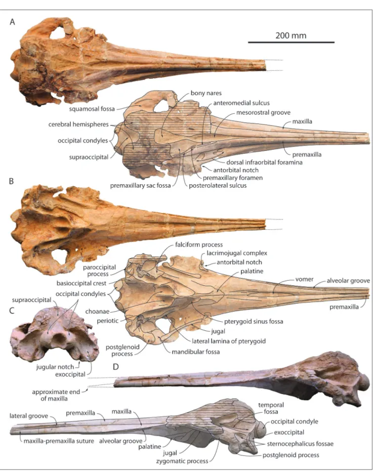
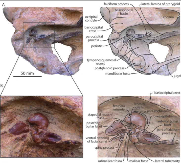
the right side of the facial region, and most of the basicranium, including the two in situ periotics (Figs. 3-4). The dorsal surface of the neurocranium is unfortunately heavily abraded, especially on the left side of the facial region, the vertex, and most of the occipital shield (Fig. 3A).
The bizygomatic width is estimated at 224 mm, only slightly greater than in MUSM 632, where-as the width of the rostrum at its bwhere-ase is estimated at 117 mm, also slightly greater than in the latter. In general, dimensions do not differ significantly from MUSM 632 (Table 1).
At the preserved anterior end of the rostrum, 385 mm anterior to its base, as in MUSM 632 the premaxillae are narrow in dorsal view and contact each other. Their medial margins similarly diverge posterolaterally, but the maximum dorsal opening of the mesorostral groove is somewhat more ante-rior than in MUSM 632, 20 mm anteante-rior to the level of the antorbital notch. Better preserved in that re-gion, MUSM 632 displays an abrupt narrowing of the opening of the mesorostral groove towards the bony nares, in a way reminiscent of squaloziphiids (Muizon 1991; Lambert et al. 2019). The dorsal opening of the mesorostral groove is much more reduced in Mycteriacetus and Ziphiodelphis. The
anteri-or outline of the bony nares is V-shaped. Preserved on both sides the premaxillary foramen is posterior to the level of the antorbital notch (more so than in
Fig. 2 - Mandibles of MUSM 632, Eurhinodelphinidae indet. from the early Miocene of the Chilcatay Formation, East Pisco Basin, Peru. A) right lateral view; B) ventral view; C) dorsal view.
Eurhinodelphis cocheteuxi and Mycteriacetus), followed
anteriorly by a medium-length anteromedial sulcus and posteriorly by a well-defined posterolateral sul-cus, reaching at least the level of bony nares’ mid-length. The right premaxillary sac fossa is moder-ately transversely concave.
In lateral view, the lateral groove becomes conspicuous about 200 mm anterior to the level of the antorbital notch (Fig. 3D); although this region is not as well preserved in MUSM 632, the lateral groove may have started more posteriorly in the lat-ter. The maxilla-premaxilla suture leaves the ventral floor of the lateral groove about 250 mm anterior to the antorbital notch, displaying an anteroventral slope similar to MUSM 632. The anterior tip of the
maxilla being lost, there is no direct evidence for the presence of an extended premaxillary portion of the rostrum. However, the strong similarities with MUSM 632 for the shape of the premaxillae on the rostrum and the slope of the suture, as well as the height of the maxilla at its preserved ante-rior section (ratio between this height and the to-tal height of the rostrum at that level equals 0.4 in MUSM 3944) strongly suggest that the premaxillae were similarly significantly longer than the maxillae on the rostrum in MUSM 3944.
Related to the proportionally broader ros-trum base compared to MUSM 632, the dorsal ex-posure of the maxilla at this level is wider than in the latter. Three dorsal infraorbital foramina are
vis-MUSM 632 vis-MUSM 3944 Cranium
Condylobasal length (as preserved) + 720 + 600 Length of rostrum (as preserved) + 540 +385
Width of rostrum at its base e106 117
Width of premaxillae at level of antorbital notches - 71 Maximum transverse width of right premaxilla - 42 Length of medial palatal exposure of vomer 105 120 Distance between posterior end of medial palatal
exposure of vomer and rostrum base 85 100 Distance between posterior end of right maxillary
alveolar groove and rostrum base 45 50 Longitudinal distance between anterior end of palatine
and rostrum base 26 20
Distance from paroccipital process to antorbital notch 185 188
Bizygomatic width e218 e224
Transverse width at lateral margins of exoccipitals e168 188 Maximum width between the basioccipital crests 88 95
Width of occipital condyles - 76
Height of left occipital condyle - 39 Width of left occipital condyle - 32
Width of foramen magnum - 32
Height of foramen magnum - 24
Mandibles
Length of mandible parallel to longitudinal axis of the
skull (as preserved) - + 570
Length of mandibular symphysis (as preserved) - + 292 Width of joined mandibles at posterior end of
symphysis - 44
Height of joined mandibles at posterior end of
symphysis - 25
Distance between posterior end of symphysis and
posteriormost mandibular alveolus - 130 Number of alveoli in the postsymphyseal portion - 18
Left periotic
Maximum length - +38
Right tympanic bulla
Maximum length (without posterior process) - 43.9
Maximum width - 26.5
Left tympanic bulla
Maximum length (without posterior process) - 44.8
Maximum width - 27.7
Tab. 1 - Measurements (in mm) on the skulls MUSM 632 and MUSM 3944, Eurhinodel-phinidae indet. from the early Miocene of the Chil-catay Formation, East Pisco Basin, Peru. +, incomplete; e, estimate; -, missing data.
ible in the same region as in MUSM 632, medial and
anteromedial to the antorbital notch (Fig. 3A). abraded in MUSM 3944 to allow for the measure- The walls of the alveolar groove are too
Fig. 3 - Cranium of MUSM 3944, Eurhinodelphinidae indet. from the early Miocene of the Chilcatay Formation, East Pisco Basin, Peru. A, dorsal view (photo and interpretive drawing); B, ventral view (photo and interpretive drawing); C, posterior view; D, left lateral view (photo and interpretive drawing).
ment of individual alveoli. Nevertheless, the width and orientation of the groove in the maxilla are similar to MUSM 632. Also, the ventral exposure of the vomer between the maxillae occurs in the same region as in the latter (for a slightly greater length of 120 mm) and long sulci are similarly ob-served in the ventral surface of the maxilla anterior to the palatine (Fig. 3B). The palatine-maxilla su-ture is also U-shaped and the pterygoid sinus fossa does not reach anteriorly the level of the antorbital notch. Better preserved in that region, MUSM 3944 displays on the lateral side of both pterygoid sinus fossae an elongated plate reaching the falciform process of the squamosal; it is identified here as the lateral lamina of the pterygoid. Such a feature is observed in several eurhinodelphinids (e.g., mul-tiple specimens of Schizodelphis and Xiphiacetus from
the USNM collection; Kellogg 1925; Muizon 1988a; Lambert 2005a), and was proposed to be absent in
Chilcacetus cavirhinus, the only other non-platanistoid
homodont and longirostrine medium-size dolphin recorded from the Chilcatay Formation (Lambert et al. 2015). We cannot exclude the possibility that part of this lateral lamina is made of the palatine, as proposed for Eurhinodelphis cocheteuxi (Lambert
2005b); better-preserved specimens would be need-ed to test this hypothesis. Slightly shiftneed-ed from its original position (more so on the right side), the styliform process of the jugal is slender and it con-tacts the ventral side of the zygomatic process of the squamosal at the level of the prominent tuber-cle described in MUSM 632 (Figs. 3B, 4A). As in the latter, the zygomatic process is transversely broad in dorsal view and directed anterolaterally. As in MUSM 632, two deep sternocephalicus fossae are present, with the dorsalmost being anteriorly longer (Fig. 3D). The squamosal fossa is widely open an-terolaterally, ventrally limiting an anteroposteriorly long and most likely dorsoventrally low temporal fossa (as suggested by the low position of the anter-omedial portion of the fossa’s roof; Fig. 3D). Not preserved in MUSM 632, the postglenoid process is anteroposteriorly thick and transversely wide in MUSM 3944 (Figs. 3B, D, 4A), in a way similar to other eurhinodelphinids, Eoplatanista, and at least
some members of the Chilcacetus clade (e.g., ‘Argy-rocetus’ joaquinensis) (Kellogg 1932; Pilleri 1985;
Lam-bert et al. 2015), but it is anteroposteriorly thinner and ventrally shorter than in squaloziphiids (Lam-bert et al. 2019), not reaching farther ventrally than
the paroccipital process of the exoccipital.
The preserved posteromedial part of the occipital shield displays a broad medial groove sep-arating the artificially exposed, posterodorsally in-flated cerebral hemispheres (Fig. 3A, C). In ventral view the basioccipital crests are thickened posteri-orly, as in MUSM 632. The large occipital condyles are prominent, reaching father posteriorly than the exoccipitals; they project slightly posteroventrally.
Both periotics of MUSM 3944 display tight contacts with the spiny process and posteromedi-al edge of the fposteromedi-alciform process of the squamosposteromedi-al (Fig. 3B, 4). The left periotic has a complete length of more than 38 mm (anterior tip of anterior pro-cess partly hidden by the falciform propro-cess). The anterior process is long, making more than 96 per cent of the length of the pars cochlearis (measured until the anteroventral margin of the fenestra ro-tunda), as in other eurhinodelphinids. Its ventral surface is excavated by a deep and long anterior bullar facet with ventrally high medial and lateral walls (Fig. 4B). No accessory ossicle is preserved in the anteroposteriorly short fovea epitubaria; the accessory ossicle is fused to the anterior process in some eurhinodelphinids (e.g. Eurhinodelphis cocheteuxi
IRSNB M.1856). The medium-size lateral tuberosi-ty extends anterolaterally, bounding with the lateral edge of the posterior process a deep and broadly open hiatus epitympanicus. The large mallear fos-sa faces posteroventrally. A small fosfos-sa (submallear fossa sensu Tanaka & Fordyce 2017; additional small fossa sensu Lambert 2005b) is wedged between the mallear fossa and the ventral opening for the facial canal. Such a fossa is observed in many other odon-tocetes, including eurhinodelphinids, physeteroids, ziphiids, Inticetus, Waipatia, and several related forms
(Muizon 1984; Lambert 2005b; Fordyce 1994; Tanaka & Fordyce 2017; Lambert et al. 2018). The posterior process is longer than the pars cochlearis (more so than in part of the specimens of Schizo-delphis sp. and Xiphiacetus cristatus at the USNM; see
Lambert 2005a) and directed posterolaterally. The posterior bullar facet displays a slightly undulating surface along the mediolateral axis. The facial crest is ventromedially prominent; it gives the medial margin of the process a highly convex outline, as in other eurhinodelphinids. The area where the ar-ticular rim could have been located is hidden by the spiny process. The pars cochlearis has a rounded outline in ventral view, with the anteromedial region
being slightly more angular. The fenestra rotunda is bounded dorsally by a small tuberosity, the lat-ter being medially and lalat-terally defined by a narrow groove leaving from the fenestra. The lateral caudal tympanic process is not prominent.
Although no clear apomorphic feature of the family Eurhinodelphinidae could be found on these bones, the periotics of MUSM 3944 do not display any significant difference with previously figured eurhinodelphinid periotics from the North Atlantic realm (see Muizon 1988a; Bianucci et al. 1994; Lambert 2004, 2005a,b; Benoit et al. 2011). In Eoplatanista, the anterior process of the periotic
is shorter and more pointed (Muizon 1988a). For
a detailed comparison with platanistoid periotics, the dorsal and lateral surfaces of the bone, which are not available in these in situ periotics, would be needed.
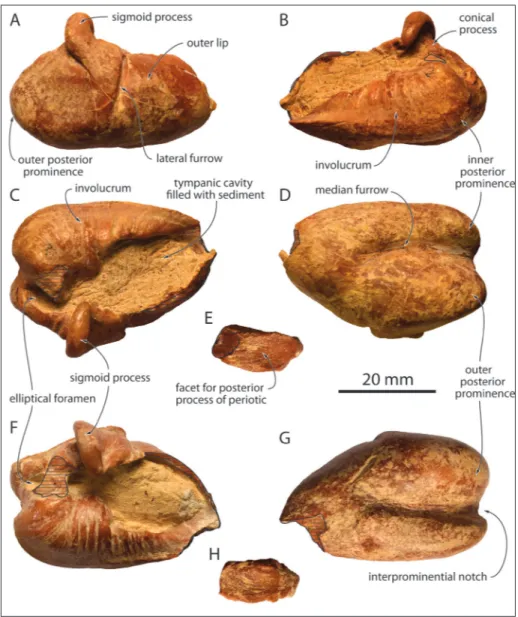
The two tympanic bullae were detached from the basicranium during preparation (Fig. 5); the accessory ossicle is lost on both tympanics; each posterior process is separated from the rest of the bone (Fig. 5E, H); and each tympanic is missing part of its anterior tip. The tympanic is proportionally broad in dorsal/ventral view (Table 1), with a max-imum width at about mid-length. The inner poste-rior prominence is transversely narrower than the outer posterior prominence, but reaches nearly the
Fig. 4 - Cranium of MUSM 3944, Eurhinodelphinidae indet. from the early Miocene of the Chilcatay Formation, East Pisco Basin, Peru. A) detail of the left side of the basicranium in ventrolateral view (photo and interpretive drawing); B) left periotic in ventral view (photo and interpretive drawing).
same posterior level. The interprominential notch is wide and shallow, followed on the ventral surface of the bone by a well-defined median furrow, ex-tending forwards for two thirds of the tympanic’s preserved length. Differing from platanistids, squal-odelphinids, and the closely related Ensidelphis, the
median furrow does not reach the tapering anterior part of the bone; on the other hand, Eoplatanista
lacks any median furrow (Muizon 1988a; Bianucci et al. 2020). Due to the incompleteness of the an-terior tip for the two tympanics, the presence and degree of development of an anterior spine can-not be assessed in MUSM 3944. In dorsal view the sigmoid process is slightly oblique, being directed medially and slightly anteriorly. In lateral view the vertical posterior margin of the sigmoid process gradually takes an anteroventral direction, lacking the posterior projection seen in most ziphiids and being ventrally shorter than in the latter (see
Lam-bert et al. 2013). The deep lateral furrow occupies three quarters of the dorsoventral height of the outer lip. As preserved the conical process is low. In dorsal view the dorsal edge of the involucrum is cut at mid-length by a major indentation, more pronounced on the left tympanic; this indentation is less conspicuous in medial view, with the dorsal margin of the involucrum more gradually lowering in an anteroventral direction. Whereas a somewhat similar condition is observed in Eoplatanista and
ziphiids (e.g. Muizon 1988a; Lambert et al. 2013), there is no marked indentation in several other clades of longirostrine to hyper-longirostrine odon-tocetes (e.g. Muizon 1987; Fordyce 1994; Tanaka & Fordyce 2014; Kimura & Barnes 2016; Bianucci et al. 2020). The ventral margin of the involucrum is ventrally bulging at two thirds of its length, before raising anterodorsally towards its dorsoventrally thin anterior tip. The whole morphology of the
in-Fig. 5 - Tympanic bullae of MUSM 3944, Eurhinodelphinidae indet. from the early Mio-cene of the Chilcatay For-mation, East Pisco Basin, Peru. Right tympanic in lat-eral (A), medial (B), dorsal (C), and ventral (D) views; E, right posterior process in dorsal view; left tympanic in dorsal (F) and ventral (G) views; H) left posterior pro-cess in dorsal view. Hatching highlights main break sur-faces.
volucrum closely matches the condition observed in several eurhinodelphinids, for example Eurhi-nodelphis cocheteuxi IRSNB M.1856, Xiphiacetus bossi
USNM 167629, X. cristatus IRSNB M.1902, and Ziphiodelphis abeli MGP 26194. A moderate size
el-liptical foramen is visible on both tympanics. The detached posterior process is narrow, and propor-tionally longer on the right tympanic.
Discussion
Comparison. In addition to general similarities
with other eurhinodelphinids as mentioned above, MUSM 632 is attributed to the family Eurhinodel-phinidae based on (1) the presence of an extensive premaxillary portion of the rostrum and (2) the mandibles being significantly shorter than the ros-trum; these two derived features are absent in other clades of longirostrine to hyper-longirostrine odon-tocetes (e.g. Kellogg 1924; Muizon 1988a; Fordyce 1994; Kimura & Barnes 2016; Bianucci et al. 2020). Similarities at the level of the rostrum suggest that MUSM 3944 was also characterized by an exten-sive premaxillary portion of the rostrum. The latter shares multiple characters with other known eurhi-nodelphinids (at the level of the rostrum, palate, lateral lamina of the pterygoid, postglenoid process of the squamosal, periotic, and tympanic), but each of these features can be individually found to some degree in other odontocete clades and cannot be considered as synapomorphic for Eurhinodelphini-dae (see comparison above).
Although displaying a series of minor dif-ferences, these two specimens roughly have the same size and share many anatomical similarities (see above). The lack of most of the facial region in both crania and of the periotic in MUSM 632 makes their referral to the same species only tenta-tive. Their fragmentary state of preservation further refrains us either from referring them to a known eurhinodelphinid genus or species from the North Atlantic realm, or from naming any new taxon. Among the available morphological features, these two specimens depart from Mycteriacetus bellunensis, Ziphiodelphis abeli, and Z. sigmoideus in the
mesoros-tral groove being more widely dorsally open and in the premaxillae being significantly narrower at ros-trum base. The maxillary portion of the rosros-trum of MUSM 632 is proportionally longer than in Eurhi-nodelphis cocheteuxi and E. longirostris, and the
premax-illary foramen is more posteriorly located than in E.
cocheteuxi and M. bellunensis. More complete crania,
including the highly diagnostic vertex, will be need-ed for a detailneed-ed comparison with species of the genera Schizodelphis and Xiphiacetus, not displaying
obvious differences with MUSM 632 and MUSM 3944 at the level of preserved parts (including the periotic and tympanic).
The preservation state of the two specimens does not allow to precisely quantify the portion of the rostrum that was only occupied by the premax-illae and to assess the degree of development of premaxillary alveoli. Nevertheless, their attribution to the family Eurhinodelphinidae suggests that, as in other eurhinodelphinids, premaxillary teeth were either vestigial (not held in distinct alveoli) or com-pletely absent.
The only other longirostrine dolphin from the Miocene of the whole Southern Hemisphere that is known from a reasonable part of the ros-trum and mandible and that has been previously referred to the family Eurhinodelphinidae is Argy-rocetus patagonicus, from the early Miocene of
Argen-tina (Lydekker 1893; Cabrera 1926; Muizon 1988a). The poorly preserved type specimen displays a very long mandible; much longer than reconstructed in MUSM 632, it actually reaches forward much farther than the preserved tip of the rostrum. The anterior part of the mandible being apparently edentulous, Cabrera (1926) suggested that the missing anterior part of the rostrum was also edentulous and would thus correspond to the edentulous premaxillary portion of the rostrum as observed in eurhinodel-phinids. If correct, this relatively hypothetical inter-pretation would mean that the mandibles were not shorter than the rostrum in A. patagonicus, a major
difference with MUSM 632 (and at least part of the other eurhinodelphinids). In addition, the mandible of A. patagonicus bears a distinct lateral groove, a
feature absent in MUSM 632, and it displays a low-er numblow-er of post-symphyseal alveoli. Although the postglenoid process of the squamosal is anter-oposteriorly thick in A. patagonicus, this character is
not exclusively observed in eurhinodelphinids (see above; Muizon 1991; Lambert et al. 2015, 2019), and this process reaches farther ventrally in A. pa-tagonicus compared to MUSM 3944. Other parts of
the skull are either too poorly preserved in the type of A. patagonicus or lost in MUSM 632 and MUSM
3944 (e.g. the nasals, typically projecting anterodor-sally in A. patagonicus). The observed differences are
nevertheless sufficient to conclude that the Peruvi-an specimens belong to a different taxon Peruvi-and that more complete specimens would be needed to fur-ther test the affinities of A. patagonicus with
eurhino-delphinids. In a previous phylogenetic analysis, A. patagonicus was recovered outside
Eurhinodelphini-dae, in a clade also including a series of early Mio-cene, northeastern and southeastern Pacific longi-rostrine odontocetes (Chilcacetus clade; Lambert et
al. 2015).
It is worth clarifying here another tenta-tive eurhinodelphinid record from South America, as the studied remains originate from Peru. Con-tradicting an earlier comment on the observed ab-sence of eurhinodelphinids in the Pisco Formation (Muizon 1988b), Pilleri (1989) suggested that two fossils found in layers of this unit in the locality of Aguada de Lomas, Sacaco Basin, could be referred to this family. The first specimen is a highly damaged odontocete neurocranium, only illustrated in dorsal view (Pilleri 1989, pl. 1). The U-shaped anterior margin of the bony nares and the abrupt posterior narrowing of the left premaxilla on the anterolateral corner of the corresponding bony naris are major differences with known eurhinodelphinids. These features are more reminiscent of delphinidans, and among those, more specifically of phocoenids. The latter are especially common in the upper part of the Pisco Formation (see Muizon 1984, 1988b) and this specimen is identified here as Phocoenidae indet. The second specimen figured by Pilleri (1989, pl. 2) is an isolated, highly abraded humerus, whose state of preservation does probably not allow for a more precise attribution than Cetacea indet.
B
IogeographyThe two new Peruvian specimens described here constitute the only unambiguous eurhinodel-phinid records, based on diagnostic rostral and man-dibular material, outside the western coast of the North Atlantic, North Sea, and Mediterranean (= North Atlantic realm), and thus the first clear records for the whole Pacific and the whole Southern Hem-isphere (for a revision and comments on more frag-mentary remains, see Muizon 1988a; Lambert 2005a, and Marx et al. 2016; Supplementary Table 1). As such, these new specimens from the East Pisco Ba-sin greatly expand the biogeographic distribution of
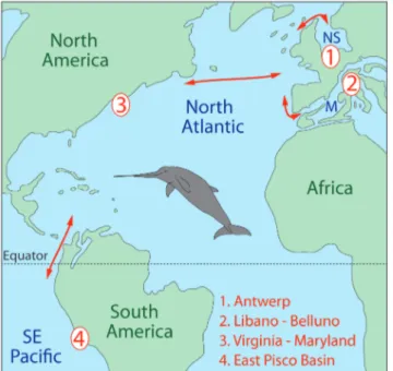
the family. In addition to the crossing of the North Atlantic and of the Mediterranean-Atlantic gateways (see Flecker et al. 2015 and references therein), as well as a dispersal via the northern part of the North Sea (strait of Dover was closed from the late Oligo-cene to the Tortonian; Van Vliet-Lanoë et al. 2010), a new dispersal route for members of the family can thus be proposed, during the early Miocene or earli-er, across the Central American Seaway (Fig. 6); the latter remained fully open at least until the early mid-dle Miocene, before a Pliocene final closure (O’Dea et al. 2016; Jaramillo et al. 2017). Previous reports of marine mammals crossing this seaway during the Miocene (e.g. Bianucci et al. 2010, 2016; Uhen et al. 2010) further support this migration route. These records from the southeastern Pacific also poten-tially indicate that early Miocene eurhinodelphinids from other Southern Hemisphere or North Pacific localities are still awaiting discovery or proper iden-tification.
Fig. 6 - Palaeogeographic map of the Atlantic and southeastern Pacific during the early Miocene (ca. 20 Ma) showing the main eurhinodelphinid localities (red numbers) from the North Sea Basin (Berchem Formation, Antwerp, Belgium), Mediterranean (Libano Sandstone, Libano and Belluno, Italy), Atlantic Coastal Plain (Calvert Formation, Virginia and Maryland), and East Pisco Basin (Chilcatay Forma-tion, Peru). M, Mediterranean; NS, North Sea. Red arrows indicate probable dispersal routes of eurhinodelphinids across the Central American Seaway, the North Atlantic, the Mediterranean-Atlantic gateways, and the northern part of the North Sea. Map redrawn from an original map by R.C. Blakey (available at deeptimemaps.com). Reconstruction of the body shape for the eurhinodelphinid Xiphiacetus bossi
re-drawn from original art by T. Scheirer (courtesy of Calvert Marine Museum).
The addition of the family Eurhinodelphini-dae to the taxonomic list of the Chilcatay Formation further increases the similarities noted between the East Pisco Basin and early to middle Miocene locali-ties of the North Atlantic realm. This early Burdiga-lian odontocete assemblage now shares five higher rank clades (Eurhinodelphinidae, Kentriodontidae, Physeteroidea, Platanistidae, and Squalodelphinidae) with the Aquitanian to Langhian of the Calvert For-mation, Atlantic Coastal Plain (Virginia and Mary-land, U.S.A.; Gottfried et al. 1994), three (Eurhino-delphinidae, Physeteroidea, and Squalodelphinidae) with the late Aquitanian to early Burdigalian Libano Sandstone (Libano and Belluno, Italy; Pilleri 1985; Bianucci & Landini 2002), and at least three (Ken-triodontidae, Physeteroidea, and Squalodelphinidae, including the species Notocetus vanbenedeni, see above
for the affinities of Argyrocetus patagonicus) with the
Aquitanian to early Burdigalian Monte León and Gaiman formations (eastern Patagonia, Argentina; Viglino et al. 2018; Cuitiño et al. 2019; Paolucci et al. 2019). Similarities with early Miocene assemblag-es from the North Atlantic realm were recently fur-ther supported by the tentative report of inticetids in France, North Carolina (U.S.A.), and Italy (Lam-bert et al. 2018; Boessenecker 2019; Peri et al. 2019). Additional work on the rich cetacean assemblages of the Chilcatay Formation and the lower part of the Pisco Formation will most likely reveal further con-nections, probably also at lower taxonomic ranks, be-tween the southeastern Pacific and the North Atlan-tic realm for both the odontocetes and mysAtlan-ticetes, as alluded to in earlier works (Muizon & DeVries 1985; Bianucci et al. 2010, 2019; Marx et al. 2019).
Acknowledgements: We wish to warmly thank W. Aguirre and
R. Salas-Gismondi (MUSM) for the preparation of the specimens described here and for their help during our successive visits at the MUSM, D.J. Bohaska, J.G. Mead, C. Potter, and N.D. Pyenson (USNM), S. Bruaux, G. Lenglet, and O. Pauwels (IRSNB), L.G. Barnes and V.R. Rhue (LACM), R.E. Fordyce (OU), L. del Favero and M. Fornasiero (MGP), and Z. Gasparini and L.H. Pomi (MLP) for facilitating the access to the collections of modern and fossil ceta-ceans under their care, M.D. Uhen for his tremendous contribution to the cetacean part of the Paleobiology Database (paleobiodb.org), T. Scheirer and S.J. Godfrey (CMM) for allowing us to modify the recon-struction of Xiphiacetus bossi prepared by the former, and Y. Tanaka, an
anonymous reviewer, and the editors for their constructive comments. This research was supported by funding from the Univer-sity of Pisa (PRA_2017_0032) to G.B., a grant from the Italian Min-istero dell’Istruzione, dell’Università e della Ricerca (MIUR) (PRIN Project, 2012YJSBMK EAR- 9317031) to G.B., and a National Geo-graphic Society Committee for Research Exploration grant (GEFNE 177-16) to O.L.
RefeRences
Abel O. (1901) - Les dauphins longirostres du Boldérien (Mi-ocène supérieur) des environs d’Anvers. I. Mémoires du Musée Royal d’Histoire Naturelle de Belgique, 1: 1-95.
Abel O. (1902) - Les dauphins longirostres du Boldérien (Mi-ocène supérieur) des environs d’Anvers. II. Mémoires du Musée Royal d’Histoire Naturelle de Belgique, 2: 99-190.
Barrick R.E., Fischer A.G., Kolodny Y., Luz B. & Bohaska D. (1992) - Cetacean bone isotopes as proxies for Miocene ocean composition and glaciation. Palaios, 7: 521-531.
Benoit J., Adnet S., Welcomme J.-L. & Fabre P.-H. (2011) - New skull of Schizodelphis sulcatus Gervais, 1861
(Mam-malia, Odontoceti, Eurhinodelphinidae) from the Lower Miocene of Pignan (Hérault, France) and its implica-tions for systematics of Eurhinodelphinidae. Geobios,
44: 323-334.
Bianucci G. & Landini W. (2002) - Change in diversity, ecologi-cal significance and biogeographiecologi-cal relationships of the Mediterranean Miocene toothed whale fauna. Geobios,
35: 19-28.
Bianucci G., Landini W. & Varola A. (1994) - New remains of Cetacea Odontoceti from the «Pietra leccese» (Apulia, Italy). Bollettino della Società Paleontologica Italiana, 33:
215-230.
Bianucci G., Lambert O. & Post K. (2010) - High concentra-tion of long-snouted beaked whales (genus Messapicetus)
from the Miocene of Peru. Palaeontology, 53: 1077-1098.
Bianucci G., Collareta A., Post K., Varola A. & Lambert O. (2016) - A new record of Messapicetus from the Pietra
leccese (Late Miocene, Southern Italy): antitropical dis-tribution in a fossil beaked whale (Cetacea, Ziphiidae).
Rivista Italiana di Paleontologia e Stratigrafia, 122: 63-74.
Bianucci G., Collareta A., Bosio G., Landini W., Gariboldi K., Gioncada A., Lambert O., Malinverno E., Muizon C. de, Varas-Malca R., Villa I.M., Coletti G., Urbina M. & Di Celma C. (2018a) - Taphonomy and palaeoecology of the lower Miocene marine vertebrate assemblage of Ul-lujaya (Chilcatay Formation, East Pisco Basin, southern Peru). Palaeogeography, Palaeoclimatology, Palaeoecology, 511:
256-279.
Bianucci G., Bosio G., Malinverno E., Muizon C. de, Villa I.M., Urbina M. & Lambert O. (2018b) - A new large squalodelphinid (Cetacea, Odontoceti) from Peru sheds light on the Early Miocene platanistoid disparity and ecology. Royal Society Open Science, 5: 172302.
Bianucci G., Marx F.G., Collareta A., Di Stefano A., Landini W., Morigi C. & Varola A. (2019) - Rise of the titans: baleen whales became giants earlier than thought. Biology Letters, 15: 20190175.
Bianucci G., Muizon C. de, Urbina M. & Lambert O. (2020) - Extensive diversity and disparity of the Early Miocene platanistoids (Cetacea, Odontoceti) in the southeastern Pacific (Chilcatay Formation, Peru). Life, 10: 27.
Boessenecker R.W. (2019) - Problematic archaic whale Phoc-ocetus (Cetacea: Odontoceti) from the Lee Creek Mine,
geochronol-ogy of the Pungo River Formation. PalZ, 93: 93-103.
Bosio G., Malinverno E., Collareta A., Di Celma C., Gioncada A., Parente M., Berra F., Marx F. G., Vertino A., Urbina M. & Bianucci G. (2020a) - Strontium Isotope Stratigra-phy and the thermophilic fossil fauna from the middle Miocene of the East Pisco Basin (Peru). Journal of South American Earth Sciences, 97: 102399.
Bosio G., Malinverno E., Villa I.M., Di Celma C., Gariboldi K., Gioncada A., Barberini V., Urbina M. & Bianucci G. (2020b) - Tephrochronology and chronostratigraphy of the Miocene Chilcatay and Pisco formations (East Pisco Basin, Peru). Newsletters on Stratigraphy, 53: 213-247.
Brisson M.-J. (1762) - Regnum Animale in classes IX distribu-tum, sine synopsis methodica. Theodorum Haak, Paris, 296 pp.
Cabrera A. (1926) - Cetaceos fosiles del Museo de La Plata.
Revista del Museo de La Plata, 29: 363-411.
Cuitiño J.I., Buono M.R., Viglino M., Farroni N.D. & Bessone S. (2019) - Factors affecting the preservation and distri-bution of cetaceans in the lower Miocene Gaiman For-mation of Patagonia, Argentina. Palaeogeography, Palaeocli-matology, Palaeoecology, 526: 110-125.
Di Celma C., Malinverno E., Collareta A., Bosio G., Gariboldi K., Lambert O., Landini W., Pierantoni P.P., Gioncada A., Villa I.M., Coletti G., Muizon C. de, Urbina M. & Bianucci G. (2018) - Facies analysis, stratigraphy and ma-rine vertebrate assemblage of the lower Miocene Chil-catay Formation at Ullujaya (Pisco basin, Peru). Journal of Maps, 14: 257-268.
Di Celma C., Pierantoni P.P., Malinverno E., Collareta A., Lambert O., Landini W., Bosio G., Gariboldi K., Gion-cada A., Muizon C. de, Molli G., Marx F.G., Varas-Malca R.M., Urbina M. & Bianucci G. (2019) - Allostratigraphy and paleontology of the lower Miocene Chilcatay For-mation in the Zamaca area, East Pisco basin, southern Peru. Journal of Maps, 15: 393-405.
Flecker R., Krijgsman W., Capella W., de Castro Martíns C., Dmitrieva E., Mayser J.P., Marzocchi A., Modestou S., Ochoa D. & Simon D. (2015) - Evolution of the Late Miocene Mediterranean–Atlantic gateways and their impact on regional and global environmental change.
Earth-Science Reviews, 150: 365-392.
Flower W.H. (1867) - Description of the skeleton of Inia geof-frensis and the skull of Pontoporia blainvillii, with remarks
on the systematic position of these animals in the Order Cetacea. Transactions of the Zoological Society of London, 6:
87-116.
Fordyce R.E. (1983) - Rhabdosteid dolphins (Mammalia: Cetacea) from the Middle Miocene, Lake Frome area, South Australia. Alcheringa, 7: 27-40.
Fordyce R.E. (1994) - Waipatia maerewhenua, new genus and
new species (Waipatiidae, new family), an archaic late Oligocene dolphin from New Zealand. In: Berta A. & Deméré T.A. (Eds) - Contributions in marine mammal paleontology honoring Frank C. Whitmore, Jr: 147-178.
Proceedings of the San Diego Society of Natural History, 29.
Fordyce R.E. & Muizon C. de. (2001) - Evolutionary history of cetaceans: a review. In: Mazin J.-M. & Buffrénil V. de
(Eds) - Secondary adaptation of tetrapods to life in wa-ter: 169-233. Verlag Dr. Friedrich Pfeil, München. Fordyce R.E., Quilty P.G. & Daniels J. (2002) - Australodelphis
mirus, a bizarre new toothless ziphiid-like fossil dolphin
(Cetacea: Delphinidae) from the Pliocene of Vestfold Hills, East Antarctica. Antarctic Science, 14: 37-54.
Gottfried M.D., Bohaska D.J. & Whitmore F. C. Jr. (1994) - Miocene cetaceans of the Chesapeake Group. In: Ber-ta A. & Deméré T.A. (Eds) - Contributions in marine mammal paleontology honoring Frank C. Whitmore, Jr.: 229-238. Proceedings of the San Diego Society of Natural History, 29.
Jaramillo C.A., Montes C., Cardona A., Silvestro D., Antonelli A. & Bacon C.D. (2017) - Comment on “Formation of the Isthmus of Panama” by O’Dea et al. Science Advances,
3: e1602321.
Kellogg R. (1924) - A fossil porpoise from the Calvert Forma-tion of Maryland. Proceedings of the United States National Museum, 63: 1-39.
Kellogg R. (1925) - On the occurrence of remains of fossil porpoises of the genus Eurhinodelphis in North America. Proceedings of the United States National Museum, 66: 1-40.
Kellogg R. (1932) - A Miocene long-beaked porpoise from California. Smithsonian Miscellaneous Collections, 87: 1-11.
Kimura T. & Barnes L.G. (2016) - New Miocene fossil Allo-delphinidae (Cetacea, Odontoceti, Platanistoidea) from the North Pacific Ocean. Bulletin of the Gunma Museum of Natural History, 20: 1-58.
Lambert O. (2004) - Systematic revision of the Miocene long-snouted dolphin Eurhinodelphis longirostris du Bus,
1872 (Cetacea, Odontoceti, Eurhinodelphinidae). Bulle-tin de l’Institut Royal des Sciences Naturelles de Belgique, Sciences de la Terre, 74: 147-174.
Lambert O. (2005a) - Review of the Miocene long-snouted dolphin Priscodelphinus cristatus du Bus, 1872 (Cetacea,
Odontoceti) and phylogeny among eurhinodelphinids.
Bulletin de l’Institut Royal des Sciences Naturelles de Belgique, Sciences de la Terre, 75: 211-235.
Lambert O. (2005b) - Phylogenetic affinities of the long-snout-ed dolphin Eurhinodelphis (Cetacea, Odontoceti) from
the Miocene of Antwerp. Palaeontology, 48: 653-679.
Lambert, O., Muizon C. de & Bianucci G. (2013) - The most basal beaked whale Ninoziphius platyrostris Muizon, 1983:
clues on the evolutionary history of the family Ziphiidae (Cetacea: Odontoceti). Zoological Journal of the Linnean So-ciety, 167: 569-598.
Lambert O., Bianucci G. & Urbina M. (2014) - Huaridelphis rai-mondii, a new early Miocene Squalodelphinidae (Cetacea,
Odontoceti) from the Chilcatay Formation, Peru. Journal of Vertebrate Paleontology, 34: 987-1004.
Lambert O., Muizon C. de & Bianucci G. (2015) - A new ar-chaic homodont toothed whale (Mammalia, Cetacea, Odontoceti) from the early Miocene of Peru. Geodiversi-tas, 37: 79-108.
Lambert O., Muizon C. de, Malinverno E., Di Celma C., Urbina M. & Bianucci G. (2018) - A new odontocete (toothed cetacean) from the Early Miocene of Peru ex-pands the morphological disparity of extinct heterodont
dolphins. Journal of Systematic Palaeontology, 16: 981-1016.
Lambert O., Godfrey S.J. & Fitzgerald E.M.G. (2019) -
Yaquinacetus meadi, a new latest Oligocene–early Miocene
dolphin (Cetacea, Odontoceti, Squaloziphiidae, fam. nov.) from the Nye Mudstone (Oregon, USA). Journal of Vertebrate Paleontology, 38: e1559174.
Lambert O., Muizon C. de, Urbina M. & Bianucci G. (2020) - A new longirostrine sperm whale (Cetacea, Physe-teroidea) from the early Miocene of the Pisco Basin (southern coast of Peru). Journal of Systematic Palaeontolo-gy, 18: 1707-1742.
Lydekker R. (1893) - Contribution to the knowledge of the fossil vertebrates of Argentina. Part II. Cetacean skulls from Patagonia. Anales del Museo de La Plata, 1893: 1-14.
Marx F.G., Lambert O. & Uhen M.D. (2016) - Cetacean paleo-biology. John Wiley & Sons, Chichester, 319 pp.
Marx F.G., Collareta A., Lambert O., Muizon C. de, Urbina M. & Bianucci G. (2019) - Late Miocene baleen whales from the Peruvian desert. Society of Vertebrate Paleontology 79th annual meeting, Brisbane, Australia, Program and Ab-stracts: 150A.
McCurry M.R. & Pyenson N.D. (2019) - Hyper-longirostry and kinematic disparity in extinct toothed whales. Paleo-biology, 45: 21-29.
Mchedlidze G.A. (1976) - General features of the palaeobi-ological evolution of cetacea. {Osnovnye Cherty Pale-obiologicheskoi Istorii Kitoobraznykh}. Metsniereba Publishers, Tbilisi, 139 pp. [translated from Russian in 1984 by Amerind Publishing Co. Pvt. Ltd., New Delhi] Mead J.G. & Fordyce R.E. (2009) - The therian skull: a lexicon
with emphasis on the odontocetes. Smithsonian Contribu-tions to Zoology, 627: 1-248.
Montes C., Cardona A., Jaramillo C., Pardo A., Silva J., Va-lencia V., Ayala C., Pérez-Angel L., Rodriguez-Parra L. & Ramirez V. (2015) - Middle Miocene closure of the Central American seaway. Science, 348: 226-229.
Muizon C. de (1984) - Les vertébrés de la Formation Pisco (Pérou). Deuxième partie: Les Odontocètes (Cetacea, Mammalia) du Pliocène inférieur de Sud-Sacaco. Travaux de l’Institut Français d’Etudes Andines, 27: 1-188.
Muizon C. de. (1987) - The affinities of Notocetus vanbenedeni,
an Early Miocene platanistoid (Cetacea Mammalia) from Patagonia, southern Argentina. American Museum Novi-tates, 2904: 1-27.
Muizon C. de (1988a) - Le polyphylétisme des Acrodelphidae, odontocètes longirostres du Miocène européen. Bulletin du Muséum National d’Histoire Naturelle, Paris, 10: 31-88.
Muizon C. de (1988b) - Les vertébrés fossiles de la Formation Pisco (Pérou). Troisième partie: Les Odontocètes (Ceta-cea, Mammalia) du Miocène. Travaux de l’Institut Français d’Etudes Andines, 42: 1-244.
Muizon C. de (1991) - A new Ziphiidae (Cetacea) from the Early Miocene of Washington State (USA) and phy-logenetic analysis of the major groups of odontocetes.
Bulletin du Muséum National d’Histoire Naturelle, Paris, 12:
279-326.
Muizon C. de & DeVries T.J. (1985) - Geology and paleontol-ogy of late Cenozoic marine deposits in the Sacaco area (Peru). Geologische Rundschau, 74: 547-563.
Myrick A.C. (1979) - Variation, taphonomy and adaptation of the Rhabdosteidae (= Eurhinodelphidae) (Odontoceti, Mammalia) from the Calvert Formation of Maryland and Virginia. Unpublished PhD thesis, University of California, Los Angeles. University Microfilms Interna-tional Ann Arbor, Michigan, 347 pp.
O’Dea A., Lessios H.A., Coates A.G., Eytan R.I., Restre-po-Moreno S.A., Cione A.L., Collins L.S., De Queiroz A., Farris D.W., Norris, R.D. et al. (2016) - Formation of the Isthmus of Panama. Science Advances, 2: e1600883.
Peri E., Collareta A., Insacco G. & Bianucci G. (2019) - An In-ticetus-like (Cetacea: Odontoceti) postcanine tooth from
the Pietra leccese (Miocene, southeastern Italy) and its palaeobiogeographical implications. Neues Jahrbuch für Geologie und Paläontologie, 291: 221-228.
Pilleri G. (1985) - The Miocene Cetacea of the Belluno sand-stones (eastern southern Alps). Memorie di Scienze Geolog-iche, 36: 1-87.
Pilleri G. (1989) - Vorkommen von Eurhinodelphinidae (Ce-tacea: Odontoceti) in der Pisco-Formation Perus? In Pilleri G. (Ed) - Beiträge zur paläontologie der cetaceen Perus: 221-222. Hirnanatomisches Institut der Universi-tät Bern, Bern.
Tanaka Y. & Fordyce R.E. (2014) - Fossil dolphin Otekaikea marplesi (latest Oligocene, New Zealand) expands the
morphological and taxonomic diversity of Oligocene cetaceans. PLoS ONE, 9: e107972.
Tanaka Y. & Fordyce R.E. (2017) - Awamokoa tokarahi, a new
basal dolphin in the Platanistoidea (late Oligocene, New Zealand). Journal of Systematic Palaeontology, 15: 365-386.
Uhen M.D. (2008) - New protocetid whales from Alabama and Mississippi, and a new cetacean clade, Pelagiceti.
Journal of Vertebrate Paleontology, 28: 589-593.
Uhen M.D., Coates A.G., Jaramillo C.A., Montes C., Pimiento C., Rincon A., Strong N. & Velez-Juarbe J. (2010) - Ma-rine mammals from the Miocene of Panama. Journal of South American Earth Sciences, 30: 167-175.
Van Vliet-Lanoë B., Gosselin G., Mansy J.-L., Bourdillon C., Meurisse-Fort M., Henriet J.-P., Le Roy P. & Trentesaux A. (2010) - A renewed Cenozoic story of the Strait of Dover. Annales de la Société Géologique du Nord, 17 :59-80.
Viglino M., Buono M.R., Gutstein C.S., Cozzuol M.A. & Cu-itiño J.I. (2018) - A new dolphin from the early Miocene of Patagonia, Argentina: Insights into the evolution of Platanistoidea in the Southern Hemisphere. Acta Palae-ontologica Polonica, 63: 261-277.
Whitmore F.C., Jr. & Kaltenbach J.A. (2008) - Neogene Ceta-cea of the Lee Creek Phosphate Mine, North Carolina.
Virginia Museum of Natural History Special Publication, 14: