ISSN: 2155-9929
The International Open Access
Journal of Molecular Biomarkers & Diagnosis
Special Issue Title:
DNA Diagnosis
Handling Editors
George J. Netto
Johns Hopkins University, USA
Yang Yongliang
Harvard University, USA
Shahrokh Francois Shariat
Cornell University, USA
T
his article was originally published in a journal by OMICS
Publishing Group, and the attached copy is provided by OMICS
Publishing Group for the author’s benefi t and for the benefi t of
the author’s institution, for commercial/research/educational use
including without limitation use in instruction at your institution,
sending it to specifi c colleagues that you know, and providing a copy
to your institution’s administrator.
All other uses, reproduction and distribution, including without
limitation commercial reprints, selling or licensing copies or access,
or posting on open internet sites, your personal or institution’s
website or repository, are requested to cite properly.
*Corresponding author: Roberta Zappacosta, Department of Experimental
and Clinical Sciences, G. d’Annunzio University of Chieti-Pescara, Italy, E-mail:
Received March 02, 2013; Accepted March 30, 2013; Published April 01, 2013 Citation: Zappacosta R, Caraceni D, Melatti G, Gatta DMP, Capanna S, et al.
(2013) HPV Model: From Gender Medicine to Healthcare of the Couple. J Mol Biomark Diagn S5: 003. doi:10.4172/2155-9929.S5-003
Copyright: © 2013 Zappacosta R, et al. This is an open-access article distributed
under the terms of the Creative Commons Attribution License, which permits unrestricted use, distribution, and reproduction in any medium, provided the original author and source are credited
Keywords:
HPV in men; Heterosexual couples; HPV concordance; Sexual partnersIntroduction
Her name was Henrietta Lacks, but scientists know her as “HeLa”. Henrietta Lacks is the woman of the immortal “HeLa” cells. HeLa cells, obtained from cervical cancer of Henrietta, were placed onto culture and grew wildly [1,2]. Previous efforts to grow cervical cancer in culture proved elusive [3]. HeLa cells have been essential in many of the great scientific discoveries of our time. They are immortal due to telomerase gene mutation induced by Papillomavirus type 18 that has been found to be integrated into four specific chromosomes [4]. Henrietta, absolutely monogamous, acquired HPV from her “disloyal” husband. More frequently male issues are, de facto, linked to women’s health. Even more so in Sexual Transmitted Diseases (STDs), men and women health are intertwined. HPV model would represent the best example.
Papilloma viruses represent the most sexually transmitted viruses [5]. Infection with high Human Papilloma virus (HPV), which is the major cause of cervical cancer, may also been linked to male anogenital lesions, including penile cancer and penile warts [6]. Penile HPV infection is very common; its incidence remains high throughout a wide range of age [7]. However, HPV infections have generally limited pathological consequences for healthy men. In male infection is frequently subclinical and cytological change distinctive of viral infection have low frequency [8]. Due to these diagnostic difficulties, usually male HPV infections escape from clinical observation. The asymptomatic nature of male HPV infection is believed to be
responsible for the transmission of the virus to female partners. As a consequence, knowledge and control of HPV infection in men would have a significant impact on cervical disease. Epidemiological studies showed that more than half of male partner of women with Cervical Intraepithelial Neoplasia (CIN) had penile HPV infection [9,10]. In 2006, the quadrivalent HPV vaccine (HPV serotypes 6,11,16,18) was licensed for use in women aging 9 to 26 [11]. In 2009, quadrivalent vaccination was extended to boys and men, aging 16 to 26, for prevention of genital warts [12]. Studies for the possible role of bivalent vaccine (HPV serotypes 16 and 18) in men are in progress [13]. Whether the same preventive tools may be used for both men and women, why we should not use on men the same validated diagnostic tools which are currently used on women, optimizing them according to gender? In female population, FDA-approved liquid-based cytology (LBC) is widely used to detect cervical abnormalities and, together with HPV-DNA test, has proven to be effective in reducing the burden of
HPV Model: From Gender Medicine to Healthcare of the Couple
Roberta Zappacosta1*, Donatella Caraceni2, Giovanni Melatti2, Daniela MP Gatta1, Serena Capanna1, Chiara D’Angelo1, Valentina Amato1,
Barbara Zappacosta1, Giuseppe Pizzicanella3 and Sandra Rosini1
1Experimental and Clinical Sciences Department, G. d’Annunzio University of Chieti-Pescara, Italy 2Cytopathology Unit, “F. Renzetti” Hospital of Lanciano, Italy
3Histopathology Unit, “F Renzetti Hospital” of Lanciano, Italy
Abstract
Background: Human Papilloma virus (HPV) infection is strongly related to cervical cancer and its precursors,
but the natural history of HPV infection in men is still unknown. In this field, more information is needed for preventive strategies, especially in view of the role that men would play in transmitting the virus to their female sexual partners. Male HPV infection is frequently subclinical and cytologic changes distinctive of viral infection have low frequency. Moreover, currently there is no FDA-approved molecular test to detect HPV in men.
The goals of this prospective study were to assess a reliable method to collect cells from penis, to investigate the prevalence of HPV infection in asymptomatic male partners of women affected by cervical abnormalities, and to evaluate HPV-types concordance between sexual partners.
Methods: 217 asymptomatic men, who were sexual partners of women with cervical lesions, were enrolled.
Exfoliated cells from different penile areas were both cytologically and molecularly evaluated to detect HPV infection and viral oncogenic expression.
Results: Cytologic signs of penile HPV infection were founded in 13.8% of the cases. No inadequate samples
have been found at β-globin analysis. 62.7% of men and 78.8% of female partners were found to be HPV-positive. Concordant HPV status has been achieved in 50% of the cases. 32% of the couple, where both partners tested positive, harboured the same HPV genotype(s). HPV oncogenic expression did not correlate with the grade of infection status in men.
Conclusions: Our data would suggest the high level of reliability and the high rate of adequacy reached by
sampling multiple penile areas. HPV infection demonstrated more prevalent in asymptomatic male partner of women with cervical lesions. Molecular testing on penile brushing would represent the more accurate tool to diagnose HPV infection, in view to limit the spread of the virus within the couple.
Citation: Zappacosta R, Caraceni D, Melatti G, Gatta DMP, Capanna S, et al. (2013) HPV Model: From Gender Medicine to Healthcare of the Couple.
J Mol Biomark Diagn S5: 003. doi:10.4172/2155-9929.S5-003
Page 2 of 7 cervical cancer disease [14]. Moreover, E6/E7 mRNA testing, with its
high level of specificity, is able to prevent overtreatment by stratifying the risk [14]. Routine screening of sexually active males is hampered by the lack of a simple, convenient and effective screening technique. This may be probably due to the small number of studies, as well as to the differences in both sampling methods and molecular technique [9,10]. Most of these studies especially focused on immunocompromised or homosexual populations. Very few data still exist regarding asymptomatic and immunocompetent men.
The goals of this prospective study were: (i) to assess a reliable penile sampling method, (ii) to evaluate the diagnostic performances that molecular tests would have in male management, and (iii) to investigate both the prevalence of HPV infections among asymptomatic male partners of women affected by cervical abnormalities, and HPV-type(s) concordance within the couples.
Material and Methods
Study population and sample collection
This study was performed in agreement with the standards of the ethics review board of “F Renzetti” Hospital, and was approved by the Ethical Committees of “G. d’Annunzio” University, in accordance with the principles outlined in the Declaration of Helsinki of 1975. Between January 2009 and December 2012, four hundreds and twenty-four consecutive women, referred to Obstetric and Gynaecological Department of “Renzetti” Hospital - Lanciano, because of previous cytological diagnosis of cervical abnormalities. At the time of cytological diagnosis, all women underwent to HC2, to HPV typing and mRNA testing. Patients referred to clinicians to undergo colposcopic-directed biopsy, without any confirmatory HPV test. Biopsies specimens were evaluated by at least two experienced pathologists. Histopathologic diagnosis was reported according to the World Health Organization nomenclature and criteria.
These women were asked to bring their husband or current stable (at least 6 months of regular sexual intercourse as monogamous couple) male partner for visits.
Male partners were eligible for this study if they: 1) were immunocompetent; 2) acknowledged no history of genital warts or penile or anal cancers; 3) had no penile discharge, no pain during urination, or no current sexually transmitted infection (STI) at time of enrolment.; 4) were un-circumcised. Basing on these criteria, two hundreds and seventeen men, aging 20-72 years (mean age 37.7 years, median 36 years) were enrolled. All the men provided written consent. At enrolment, male partners were instructed not to wash their genitals for at least 2 hours prior to the examination, and to be sexually abstinent for three day.
A simple naked eye inspection and examination under magnification of the male genitalia was first performed. After inspection, each man underwent to cytologic smear collection. To maximize sampling, by
use of Cytobrush(Qiagen, Mississauga, Ontario, Canada) and Dacron
(Digene, Gaithersburg, MD, USA) swabs, three separate specimens were collected from dorsal and ventral surface of penile shaft, inner part of the foreskin, coronal sulcus, frenulum, glans, scrotus, and distal urethra (2 to 3 cm deep). These smears were pooled together through dispersion into a vial containing PreservCyt cytology medium (Cytyc Corporation, Boxborough, MA), and stored. The same vial for each subject has been used for both liquid-based cytology and molecular tests. Cytological slides were prepared by Thin Prep technology (Cytyc
Corporation, Boxborough, MA) and were next stained by Papanicolaou procedure. The smears were screened for the presence of the classic cytomorphologic signs of HPV infection, koilocytosis, iperkeratosis, parakeratosis and dyskeratosis, dysplastic changes, or cancer. Cytologic slides were evaluated by two expert cytopathologists, without previous knowledge of the molecular findings.
HPV-DNA detection and typing
Cytological samples from men were checked for DNA quality by amplification of the β-globin gene. Only β-globin positive samples were tested for HPV presence.
Male specimens were evaluated for the presence of HPV by using Hybrid Capture 2 technology (HC2, Qiagen, Gaithersburg, MD), in accordance to manufacturers protocol. HC2 detects 13 oncogenic HPV types (16, 18, 31, 33, 35, 39, 45, 51, 52, 56, 58, 59 and 68) and 5 non-oncogenic HPV types (6, 11, 42, 43, 44). HC2 reactions as chemiluminescent signal were read by the offline luminometer system (Digene Microplate Luminometer DML 2000), which provided relative quantification of each individual sample compared to the mean of a series of positive controls containing 1 pg/ml of HPV DNA (corresponding to ~100,000 HPV-16 genomes/ml or 5,000 HPV copies per reaction). The cut-off of 1 relative light unit (RLU) was used to classify a specimen as positive or negative.
A 5 mL volume of each residual male cytological sample was transferred into a fresh 10 mL tube for nucleic acids extraction. After centrifugation, the supernatant was removed and the sample was transferred into a tube containing 2 mL Nuclisens Lysis Buffer (BioMèrieux, France). A next, magnetized silica dioxide particle was added to the lysate to initiate the nucleic acids isolation process. Finally, nucleic acids were eluted from the solid phase in 55 μL of elution buffer and stored at 20°C if not further processed immediately after extraction. 10 μL of male specimens were then tested by GP5+/6+ PCR (Ampliquality HPV HS BIO, variant Single Step; Ab Analitica, Padua, Italy) that amplify a broad spectrum of HPV genotypes by targeting a 150-bp fragment within the L1 open reading frame (ORF) of the HPV genome. All PCR products were genotyped, regardless of gel result, by reverse-line blot hybridization for the detection of 12 high-risk HPV types (16, 18, 31, 33, 35, 39, 45, 51, 52, 56, 58, and 59), 7 uncertain-risk HPV types (26, 53, 66, 68, 70, 73, and 82), and 10 low-risk HPV types (6, 11, 40, 42, 43, 44, 54, 61, 72, and 81) according to the manufacturer’s instructions (Ampliquality HPV type; Ab Analitica, Padua, Italy). PCR result defined HPV-DNA status for each patient. Moreover, genotyping result was defined by a positive test result for at least one of the twenty-nine HPV genotypes comprises in the test.
HPV E6/E7 mRNA detection and typing
For each male partner, 15 μL of nucleic acids was used to perform mRNA testing (Nuclisens EasyQ HPV, BioMèrieux, France), in accordance with the manufacturer’s instructions.
The mRNA testing is based on real-time Nucleic Acid Sequence Based Amplification (NASBA) procedure, which utilizes molecular beacon probes labelled with FAM and Texas Red fluorochromes at an isothermal temperature of 41°C. The test identifies full-length E6/ E7 mRNA from five high risk carcinogenic HPV types (16, 18, 31, 33, and 45). A fluorescent analyzer measured in real-time the emission of the fluorescence from molecular beacon hybridized with amplified mRNA. As a performance control, a primer set and probe directed against the human U1 small nuclear ribonucleoprotein (snRNP)-specific A protein (U1A mRNA) included in the kit, has been used.
Negative control reactions, consisting of all reagents except RNA, were performed at each run.
The classification of any HPV types was defined as positive test result for at least one of the five HPV genotypes detected by the test.
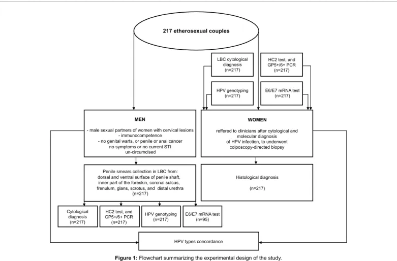
Figure 1 summarizes the informations regarding patient’s enrollment criterial, material collection, and test methods applied.
Statistical Analysis
By standard method, authors calculated the prevalence of morphological abnormalities and the prevalence of HPV-DNA and mRNA positive results, in both male and female study population. Data were then analysed by chi-square test, with continuity correction or Fisher’s exact test when appropriate. Concurrence of HPV infection in heterosexual couples was defined as the presence of infection in both of sexual partners, independently of whether or not there was HPV type’s concordance. For couples where both partners presented with at least one type of HPV infection, percentage for type-specific positive concordance was obtained. Concordance was evaluated based on contingency tables using the Kappa statistic. Interpretation of K was based on guidelines by Landis and Koch (1977): K<0, poor agreement; K=0-0.20, slight agreement; K=0.21-0.40, fair agreement; K=0.41-0.60, moderate agreement; K=0.61-0.80, substantial agreement; K=0.80-1.00, almost perfect agreement. p-values <0.05 was regarded as statistically significant. Statistical analyses were done using SPSS software (SPSS for Windows, Inc, Chicago, IL), version 19.0.
Results
HPV detection in male and female partner’s populations
The mean age of female sexual partners of the 217 men enrolled in the study was 34 years (range 18-63 years, median 33 years). No one received HPV vaccination. Following histological examination of biopsy specimens, a total of 184 (84.8%) women showed cervical lesions, as being either of low- (N=131) or of high-grade (N=84). The final histological diagnosis was based on the highest CIN grade present in the tissue. For the purpose of the study, histological results were grouped into: negative for dysplasia (including inflammatory and reactive findings), CIN2- (including CIN1), and CIN2-or-greater (CIN2+, including CIN2, CIN3 and squamous cell carcinoma).
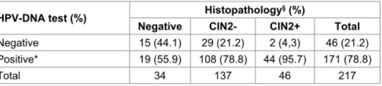
Association between histological diagnosis and HPV-DNA results is summarized in table 1. DNA-positivity has been found in 78.8% of the cases, particularly in 56% of negative cytology, in 78.8% of CIN2-, and in 95.7% of CIN2+ (Cochran–Armitage test for trend p<0.01). E6/E7 mRNA test has been performed on 95 women. Among those, 65.3% (N=63) tested negative, and 34.7 (N=32) tested positive. Table 2 exemplifies the distribution of mRNA test results in relation to histological diagnosis. Noteworthy, the proportion of mRNA positivity increases with the severity of the lesion (Cochran–Armitage test for trend p<0.01). The most frequent HPV types found by mRNA test was HPV-16, present in 21.1% of the cases. Among the 95 cases in which all molecular test have been performed, significant differences between HPV-DNA and mRNA testing results were found, according to histological diagnosis,
217 etherosexual couples MEN WOMEN HPV types concordance Cytological diagnosis (n=217) HC2 test, and GP5+/6+ PCR (n=217) HPV genotyping
(n=217) E6/E7 mRNA test(n=95)
Histological diagnosis (n=217)
reffered to clinicians after cytological and molecular diagnosis of HPV infection, to underwent colposcopy-directed biopsy LBC cytological diagnosis (n=217) HC2 test, and GP5+/6+ PCR (n=217) HPV genotyping
(n=217) E6/E7 mRNA test(n=217)
Penile smears collection in LBC from: dorsal and ventral surface of penile shaft,
inner part of the foreskin, coronal sulcus, frenulum, glans, scrotus, and distal urethra
(n=217)
- male sexual partners of women with cervical lesions - immunocompetence
- no genital warts, or penile or anal cancer no symptoms or no current STI
un-circumcised
Citation: Zappacosta R, Caraceni D, Melatti G, Gatta DMP, Capanna S, et al. (2013) HPV Model: From Gender Medicine to Healthcare of the Couple.
J Mol Biomark Diagn S5: 003. doi:10.4172/2155-9929.S5-003
Page 4 of 7
as shown in figure 2 (p<0.05). The mRNA test improved specificity (72.7% versus 50.6%) and Positive Predictive Value-PPV (36.4% versus 29.6%) of DNA test (p<0.05).
Among male population, evaluation of cytological samples according to the criteria mentioned in the studies of Boon et al. and Gupta et al. [15,16] gave the following results: negativity, 31% (N=67/217); inflammatory features not otherwise specified (NOS), 43.3% (N=94/217); classical signs indicative of HPV infection (such as iperkeratosis, parakeratosis, dyskeratosis), 22 (N=48/217); dysplastic changes, 3.7% (N=8/217). No koilocytosis has been found (Figure 3). Positive results for β-globin gene were obtained in 100% of male specimens. Considering results from HC2 test, 33.2% (N=72/217) of the specimens showed positivity, while 66.8% (N=145) gave negative result. Following application of PCR technology, 37.3% of samples (N=81/217) resulted positive, versus 62.7% (136/217) which tested negative. Relationship between cytological diagnosis and DNA test results is shown in table 3. Among the 48 smears which demonstrated cytological signs of HPV infection, 13 (27.1%) were found to be DNA negative. 100% of cases with dysplastic changes showed HPV-DNA positivity (McNemar test, p<0.01).
Among men who had cytological signs of HPV infection, 84% (N=47/56) were partners of women with cervical abnormalities (p<0.05). Figure 4 shows the distribution of female lesions in relation to male cytological diagnosis. Particularly, CIN2+ were associated in 18.6% of the cases with male negative/inflammatory NOS diagnosis, and in 28.6% of the cases with male cytological features pathognomonic of HPV infection (p<0.05). Correlation between male HPV-DNA results and female histological diagnosis is summarizes in table 4. Interestingly, 47.8% of CIN2+ were associated to male HPV-DNA positivity (OR=0.23, 95% CI: 0-1.41; p<0.05). Among 95 men who
performed mRNA test, negativity has been found in 94.7% (N=90) of the cases and positivity in 5.3% (N=5). Figure 5 shows the association between cytological diagnosis and mRNA test results. Molecular test result did not correlate with the grade of cytological lesion (p>0.05). Among the five mRNA positive results, HPV types included in the test were equally prevalent.
Molecular tests within the couple
Molecular status of HPV infection was matched in the 217 couple and described in table 5. Among those, concordance has been found in 50% (N=109) of cases. 34% (N=37/109) showed negative concordance,
HPV-DNA test (%) Negative HistopathologyCIN2- CIN2+§ (%) Total
Negative 15 (44.1) 29 (21.2) 2 (4,3) 46 (21.2)
Positive* 19 (55.9) 108 (78.8) 44 (95.7) 171 (78.8)
Total 34 137 46 217
*HPV-DNA positive result: positivity for one or more HPV genotypes [high risk (16, 18, 31, 33, 35, 39, 45, 51, 52, 56, 58, and 59), uncertain-risk (26, 53, 66, 68, 70, 73, and 82), and low-risk (6, 11, 40, 42, 43, 44, 54, 61, 72, and 81)] detected by GP5+/6+ PCR
§The final histological diagnosis was based on the highest CIN grade present in the
tissue. For the purpose of the study, histological results were grouped into: negative for dysplasia (including inflammatory and reactive findings), CIN2- (including CIN1), and CIN2-or-greater (CIN2+, including CIN2, CIN3 and squamous cell carcinoma) (p<0.01)
Table 1: Relationship between histological diagnosis and HPV-DNA test results, in
female partners of men included in the study.
*E6/E7 mRNA positive result: positivity for one or more HPV genotypes (16,18,31,33,35) included in the test
§The final histological diagnosis was based on the highest CIN grade present in the
tissue. For the purpose of the study, histological results were grouped into: CIN2-or-greater (CIN2+, including CIN2, CIN3 and squamous cell carcinoma), CIN2- (including CIN1), and negative for dysplasia (including inflammatory and reactive findings)
(p<0.01)
Table 2: Relationship between histological diagnosis and E6/E7 mRNA test
results, in female population.
E6/E7 HPV mRNA test (%) Negative HistopathologyCIN2- CIN2+§ (%) Total
Negative 17 (85) 39 (68.4) 6 (33.3) 62 (65.3) Positive* 3 (15) 18 (31.6) 12 (44) 33 (34.7) Total 20 57 18 95 Molecular tes ts r esults
N° of female sexual partners
0 5 10 15 20 25 30 35 40 45 HPV-mRNA + HPV mRNA -HPV-DNA + HPVDNA -CIN2+ CIN2-Negative
Figure 2: Correlation between molecular tests results and histological
diagnosis among the 95 women in which all molecular test have been performed. Significant differences between HPV-DNA and mRNA testing results were found, according to histological diagnosis (Chi square test, with continuity correction, p<0.05).
Figure 3: Penile smear showing cytological changes suggestive of atypia
(arrow). Papanicolaou stain, 40 x magnifications).
HPV-DNA test result (%)
Cytological diagnosis (%)
Negative Inflammatory NOS § Signs of HPV infection ° Dysplastic changes Total
Negative 51 (76.1) 72 (76.6) 13 (27.1) 0 136 (62.7)
Positive* 16 (23.9) 22 (23.4) 35 (72.9) 8 (100) 81 (37.3)
Total 67 94 48 8 217
*HPV-DNA positive result: positivity for one or more HPV genotypes [high risk (16, 18, 31, 33, 35, 39, 45, 51, 52, 56, 58, and 59), uncertain-risk (26, 53, 66, 68, 70, 73, and 82), and low-risk (6, 11, 40, 42, 43, 44, 54, 61, 72, and 81)] detected by GP5+/6+ PCR
§NOS, not otherwise specified
°Signs of HPV infection included: koilocytosis, iperkeratosis, parakeratosis, dyskeratosis
(p<0.01)
Table 3: Relationship between cytological diagnosis and DNA test results, in male
while 66% (N=72/109) demonstrated positive concordance. In contrast, 99 negative men had their female partner as positive (in five women, multiple HPV types have been found), while 9 HPV-DNA positive men had their sexual partner as negative. Overall percent agreement was 51%. It corresponded to a poor concordance (K=0.14), but to a statistically significant value (p<0.01).
Type-specific concordance (for one or most HPV genotype) occurred in 32% (N=23/72) of the couples. The most commonly shared HPV type was type 16, found in eight couples (34.8%), followed by 31, 45 and 66 found in three couples each (13%) (Figure 6). Within eight couples, male partners showed multiple HPV infection. All these men were cytologically negative or showed signs of HPV infection. Considering E6/E7 expression within the 95 couples in which mRNA test has been performed, concordance has been found in 66.3% (N=63/95) of the cases. Vice versa, 30 negative men had their female partners testing positive, while 2 positive men were associated to mRNA-negative partners. Overall percent agreement was poor (34%, K=0.15) and not significant (p=0.2), even if there were 100% agreement between E6/E7 mRNA genotypes. Moreover, 100% of female partners of men showing multiple infections including at least one of HPV types tested mRNA positive.
Discussion
Human Papilloma virus is responsible for the most sexually transmitted viral infection [17]. Characteristics of HPV infection in women have been widely investigated [18], as well as the causal link between oncogenic HPV types and high grade cervical intraepithelial lesions [19]. The close relation of HPV infection with cervical cancer and its venereal nature strongly suggests the role of male sexual partner as vector of the virus. The tendency of genital male HPV infection to be clinically unapparent explains why this association was not recognized until recently. On the other hand, controlled preventive and treatment measures need to be developed and effective strategies are needed to reach also in male population. Diagnosis of HPV infection in men could lead to decrease in the reservoir of the virus responsible for female genital neoplasia. This study was performed in view of potential benefits that can be achieved by regularly screening male asymptomatic partners of women having cervical abnormalities. In order to correctly diagnose HPV infection in asymptomatic male population, it’s really important to assess the most accurate method of penile sampling. To date, only a few studies evaluate penile sampling procedures [19-23].
In this study we collected cells from different penile areas, to improve both cytological diagnosis and molecular viral detection. CIN2+
CIN2-negative
Male cytological diagnosis
100 90 80 70 60 50 40 30 20 10 0
Negative Inflammatory features
NOS Signs of HPV infection
Dysplastic changes
Figure 4: Distribution of female lesions in relation to male cytological
diagnosis. CIN2+ lesions has been mainly found in association with male cytological features pathognomonic of HPV infection (Chi square test, with continuity correction, p<0.05). N° of men mRNA-mRNA+ E6/E7 mRNA tests results Dysplastic changes Signs of HPV infection Inflammatory, NOS Negative 0 10 20 30 40 50 60
Figure 5: Relationship between cytological diagnosis and E6/E7 mRNA test
results, among the 95 men who underwent mRNA test. Molecular test result did not correlate with the grade of cytological lesion (p>0.05).
9 8 7 6 5 4 3 2 1 0 16 31 45 66 33 52 53 58 62 HPV types N° of c ouples
Figure 6: Prevalence of HPV genotypes within the 217 sexual couples in
which concordance has been found. HPV-16 was detected in 34.8% of the couples.
*HPV-DNA positive result: positivity for one or more HPV genotypes [high risk (16, 18, 31, 33, 35, 39, 45, 51, 52, 56, 58, and 59), uncertain-risk (26, 53, 66, 68, 70, 73, and 82), and low-risk (6, 11, 40, 42, 43, 44, 54, 61, 72, and 81)] detected by GP5+/6+ PCR
§The final histological diagnosis was based on the highest CIN grade present in the
tissue For the purpose of the study, histological results were grouped into: CIN2-or-greater (CIN2+, including CIN2, CIN3 and squamous cell carcinoma), CIN2- (including CIN1), and negative for dysplasia (including inflammatory and reactive findings)
(p<0.05)
Table 4: Relationship between male HPV status and female histological diagnosis. Male HPV-DNA test
result (%) Female histological diagnosis
§ (%)
Negative CIN2- CIN2+ Total
Negative 19 (55.9) 93 (67.9) 24 (52.2) 136 (62.7)
Positive* 15 (44.1) 44 (32.1) 22 (47.8) 81 (37.3)
Total 34 137 46 217
*HPV-DNA positive result: positivity for one or more HPV genotypes [high risk (16, 18, 31, 33, 35, 39, 45, 51, 52, 56, 58, and 59), uncertain-risk (26, 53, 66, 68, 70, 73, and 82), and low-risk (6, 11, 40, 42, 43, 44, 54, 61, 72, and 81)] detected by GP5+/6+ PCR
(p<0.01)
Table 5: HPV-DNA test concordance between sexual partners of the 217 couple. MOLECULAR STATUS
Men (%) Negative Women (%)Positive Total
Negative 37 (80.4) 99 (57.9) 136 (62.7)
Positive* 9 (19.6) 72 (42.1) 81 (37.3)
Citation: Zappacosta R, Caraceni D, Melatti G, Gatta DMP, Capanna S, et al. (2013) HPV Model: From Gender Medicine to Healthcare of the Couple.
J Mol Biomark Diagn S5: 003. doi:10.4172/2155-9929.S5-003
Page 6 of 7 Sampling included penile shaft because this was considered the
anatomic site with the highest overall HPV prevalence [24]. Cells from sperm and urine were not collected, because controversial data have been published regarding their usefulness [25]. In our series, 100% of penile specimens were deemed adequate by the presence of β-globin gene. To our knowledge, this is the higher percentage of adequacy ever reached [20]. Sampling from these areas would then combine a high sensitivity in HPV detection with reliability and no tenderness.
Regarding cytological evaluation of penile smears, various features (koilocytosis, iperkeratosis, parakeratosis and dyskeratosis) have been described as indicative of HPV infection. Except for koilocytosis, they are individually not specific for HPV infection [15]. Only koilocytosis alone or a combination of two or more other cytological features may be diagnostic for viral infection [26,27]. In our series, koilocytosis has never been seen. The reason for the absence of koilocytes among HPV-DNA positive smears may be due to a thick layer of keratinised cells covering the koilocytes [15]. Another reason can be ascribed to the transitional epithelial lining of the urethra that more often undergo to squamous metaplasia. HPV infection of the metaplastic epithelium will usually not result in the production of the characteristic koilocytes [15]. Other reasons which could account for the absence of koilocytes in male specimens are that penile smear usually contains only the superficial layer of the epidermis in which there are very low frequencies of koilocytes. Aynaud et al. [27] say that koilocytosis is not always considered as a necessary changes for the diagnosis of HPV infection in penile smear, and that HPV infections are cytologically evident only in 15% of men without lesions. Our results are in agreement with those reported by Aynaud et al. [27]. In our series we also found a relatively small incidence (3.7%) of dysplastic changes, in comparison to the high detection rate (21.2%) of CIN2+ detected in female partners. This fact probably derives from the absence of squamous-columnar junction in penile areas (which is vice versa present within the cervix and in the anal canal) that makes the attachment of the virus more difficult [28].
In relation to HPV-DNA detection in men, we chose to analyze the reliability of both HC2 test and PCR technology. We decided to test HC2 performances in male population, since this method is widely used in women, particularly in the triage of cervical cytological abnormalities, in post-treatment follow-up and in primary screening [14]. GP5+/6+-based PCR are actually considered the most sensitive tool to detect HPV infection [14]. In our series, nine HC2-negative samples showed PCR positivity. Following genotyping, all HC2-negative/PCR-positive cases showed HPV infection by types not comprised in HC2 test, which however belong to intermediate and low-risk HPV groups.
The high clinical accuracy of E6/E7 mRNA test in female population has been attested by large cross-sectional study [29-31], and confirmed by the present findings. To date, there are no data regarding the possible use of E6/E7 mRNA in male population, except for Silling et al. [32], which evaluate the diagnostic accuracy of mRNA testing in anal sample. In our series, mRNA test results did not correlate with the grade of cytological findings in men, even if 40% of mRNA-positivities were associated with female CIN2+ lesions. Silling et al. on male anal specimens, found a positive correlation between the grade of the lesions and E6/E7 expression, similarly to that observed in CIN [32]. However, anal epithelium is more susceptible to transformation than epithelium of the penis, because of the presence of the squamocolumnar junction, which is the primary target of HPV infection.
Since E6/E7 expression mostly occurs during persistent infection, defined as infection which continues for six months or plus [33], only
follow-up study would clarify the real role of E6/E7 expression as predictive marker of penile abnormalities.
To satisfy the second aim of this study, we examined the prevalence of penile HPV infection and HPV type’s concordance between asymptomatic men and their female sexual partners. Until now, only a few studies analyzed the characteristics of HPV infection in men and within heterosexual couples [8,34]. Irrespective of the population studied, the results of all analyses shows high rates of acquisition of HPV infection in men, ranging from 35% to 76% [9,16,35]. This high variability is often due to the criteria of enrolment. Authors who found higher prevalence included in their analyses both asymptomatic and symptomatic men. Analyses of HPV concordance in sexual partners need improvement from other experiences, since studies which addresses this question achieved more often conflicting results. Some authors [36,37] found a high rate (up to 65%) of concordance, while others [35,38,39] show a poor agreement, ranging from 2% to 37%. One variability could be due to the technology used to detect HPV infection. A further variable could also be linked to the quality and reliability of penile sample. Regarding this point it should be underlined that HPV infection in men is frequently multifocal and that multiple sampling would be necessary. In the present study, where only asymptomatic subjects were enrolled, 62.7% of the men have been found to be HPV-positive.
Concordance in HPV status has been found in 50% of the couples. Among couples in which both partners were HPV-positive, type’s concordance was observed in 32% of the cases. The most commonly shared type was HPV-16. Multiple infection infections are most often seen in men than in women.
In summary, the results of this study would suggest that high reliability and adequacy is observed in collecting multiple penile areas. Moreover, molecular testing, achieved by GP5+/6+ PCR, would represent a more accurate tool than cytology in the diagnosis of male HPV infection. Our findings would also confirm that, in asymptomatic male population, there is a group that could represent an important reservoir for HPV infection or re-infection of the female partners. In this view, male partners which have been found HPV-positive in penile area should be advised to undergo to clinical follow-up and condom use, in order to limit the spread of the virus within the couple. Actually, only 62% of clinicians encourage women having either abnormal Pap test and/or positive HPV-DNA result to tell their sexual partner to underwent to HPV-DNA test [40].
In conclusion, an open mind toward the prevention of HPV infection should bypass the concept of sex to embrace the concept that the health of the men and women, impacting each other, is of major importance.
References
1. Skloot R (2010) The immortal life of Henrietta Lacks. Crown Publishers, New York.
2. Lucey BP, Nelson-Rees WA, Hutchins GM (2009) Henrietta Lacks, HeLa Cells, and Cell Culture Contamination. Arch Pathol Lab Med 133: 1463-1467. 3. Jones HW Jr, McKusick VA, Harper PS, Wuu KD, Gey JO (1971) George Otto
Gey. (1899-1970). The HeLa cell and a reappraisal of its origin. Obstet Gynecol 38: 945-949.
4. Hsu SH, Schacter BZ, Delaney NL, Miller TB, McKusick VA, et al. (1976) Genetic characteristics of the HeLa cell. Science 30: 392-394.
5. Georgieva S, Iordanov V, Sergieva S (2009) Nature of cervical cancer and other HPV - associated cancers. J BUON 143: 391–398.
This article was originally published in a special issue, DNA Diagnosis handled by Editor(s). Dr. George J. Netto, Johns Hopkins University, USA; Dr. Yang Yongliang, Harvard University, USA; Dr. Shahrokh Francois Shariat, Cornell University, USA
Citation: Zappacosta R, Caraceni D, Melatti G, Gatta DMP, Capanna S, et al.
(2013) HPV Model: From Gender Medicine to Healthcare of the Couple. J Mol Biomark Diagn S5: 003. doi:10.4172/2155-9929.S5-003
papillomavirus type distribution in invasive cervical cancer and high-grade cervical lesions: a meta-analysis update. Int J Cancer 21: 621–632.
7. Ferenczy A, Coutlée F, Franco E, Hankins C (2003) Human papillomavirus and HIV coinfection and the risk of neoplasias of the lower genital tract: a review of recent developments. CMAJ 169: 431-434.
8. Benevolo M, Mottolese M, Marandino F, Carosi M, Diodoro MG, et al. (2008) HPV prevalence among healthy Italian male sexual partners of women with cervical HPV infection. J Mol Virol 80: 1275-1281.
9. Bleeker MC, Hogewoning CJ, Van Den Brule AJ, Voorhorst FJ, Van Andel RE, et al. (2002) Penile lesions and human papillomavirus in male sexual partners of women with cervical intraepithelial neoplasia. J Am Acad Dermatol 47: 351– 357.
10. Bosch FX, Castellsague X, Munoz N, de Sanjose S, Ghaffari AM, et al. (1996) Male sexual behavior and human papillomavirus DNA: Key risk factors for cervical cancer in Spain. J Natl Cancer Inst 88: 1060–1067.
11. Brankovi I, Verdonk P, Klinge I (2013) Applying a gender lens on human papillomavirus infection: cervical cancer screening, HPV DNA testing, and HPV vaccination. Int J Equity in Health 12: 14.
12. Hillman RJ, Giuliano AR, Palefsky JM, Goldstone S, Moreira ED Jr, et al. (2012) Immunogenicity of the quadrivalent human papillomavirus (type 6/11/16/18) vaccine in males 16 to 26 years old. Clin Vaccine Immunol 19: 261-267. 13. Reisinger KS, Block SL, Lazcano-Ponce E (2007) Safety and persistent
immunogenicity of a quadrivalent human papillomavirus types 6, 11, 16, 18 L1 virus-like particle vaccine in preadolescents and adolescents: a randomized controlled trial. Pediatr Infect Dis J 26: 201–209.
14. Rosini S, Zappacosta R (2011) Overview on molecular markers to improve cervical cancer prevention: challenges and perspectives. Vanden Broeck, Croatia.
15. Boon ME, Schneider A, Hogewoning CJ, Kwast H, Bolhuis P, et al. (1988) Penile studies and heterosexual partners. Penioscopy, cytology, histology and immunocytochemistry. Cancer 61: 652-659.
16. Gupta JW, Gupta P, Shah K (1987) Detection of human papilloma virus in cervical smears. A comparison of in situ hybridization, immunocytochemistry and cytopathology. Acta Cytol 31: 387-396.
17. Martin CM, O’Leary JJ (2011) Histology of cervical intraepithelial neoplasia and the role of biomarkers. Best Pract Res Clin Obstet Gynaecol 25: 605-615. 18. Jordan J, Arbyn M, Martin-Hirsch P, Schenck U, Baldauf JJ (2008) European
guidelines for quality assurance in cervical cancer screening: recommendations for clinical management of abnormal cervical cytology, part 1. Cytopathology 19: 342-354.
19. Zhao C, Kalposi-Novak P, Austin RM (2011) Follow-up findings in young females with high-grade squamous intraepithelial lesion papanicolaou test results. Arch Pathol Lab Med 135: 361-364.
20. Giuliano AR, Nielson CM, Flores R, Dunne EF, Abrahamsen M, et al. (2007) The optimal anatomic sites for sampling heterosexual men for human papillomavirus (HPV) detection: the HPV detection in men study. J Infect Dis 196: 1146-1152.
21. Daniel RW, Ahdieh L, Hayden D, Cu-Uvin S, Shah KV (2000) Intra-laboratory reproducibility of human papillomavirus identification in cervical specimens by a polymerase chain reaction-based assay. J Clin Virol 19: 187–193.
22. Gravitt PE, Peyton CL, Apple RJ, Wheeler CM (1998) Genotyping of 27 human papillomavirus types by using L1 consensus PCR products by a single-hybridization, reverse line blot detection method. J Clin Microbiol 36: 3020– 3027.
23. Weaver BA, Feng Q, Holmes KK, Kiviat N, Lee SK, et al. (2004) Evaluation of genital sites and sampling techniques for detection of human papillomavirus DNA in men. J Infect Dis 189: 677–685.
24. Nielson CM, Flores R, Harris RB, Abrahamsen M, Papenfuss MR, et al. (2007) Human papillomavirus prevalence and type distribution in male anogenital sites and semen. Cancer Epidemiol Biomarkers Prev 16: 1107–1114.
25. Lazcano-Ponce E, Herrero R, Munoz N, Hernandez-Avila M, Salmeron J, et al. (2001) High prevalence of human papillomavirus infection in Mexican males: Comparative study of penile-urethral swabs and urine samples. Sex Transm Dis 28: 277–280.
26. Cecchini S, Cipparrone I, Confortini M, Scuderi A, Meini L, et al. (1988) Urethral cytology of cytobrush specimens. A new technique for detecting subclinical human Papilloma virus infection in men. Acta Cytol 32: 314-317.
27. Aynaud O, Ionesco M, Barrasso R (2003) Cytologic detection of human papillomavirus DNA in normal male urethral samples. Urology 61: 1098-1101. 28. Giuliano AR, Lee JH, Fulp W, Villa LL, Lazcano E, et al. (2011) Incidence and
clearance of genital human papillomavirus infection in men (HIM): a cohort study. Lancet 11: 377-381.
29. Zappacosta R, Caraceni D, Ciccocioppo L, Rotondo T, Capanna S, et al. (2013) Implementing specificity of HPV-DNA primary screening in a successful organised cervical cancer prevention programme. Gynecol Oncol 128: 427-432.
30. Benevolo M, Vocaturo A, Caraceni D, French D, Rosini S, et al. (2011) Sensitivity, specificity, and clinical value of human papillomavirus (HPV) E6/ E7 mRNA assay as a triage test for cervical cytology and HPV DNA test. J Clin Microbiol 49: 2643-2650.
31. Molden T, Nygård JF, Kraus I, Karlsen F, Nygård M, et al. (2005) Predicting CIN2+ when detecting HPV mRNA and DNA by PreTect HPV-proofer and consensus PCR: A 2-year follow-up of women with ASCUS or LSIL Pap smear. Int J Cancer 10: 973-976.
32. Silling S, Kreuter A, Hellmich M, Swoboda J, Pfister H, et al. (2012) Human papillomavirus oncogene mRNA testing for the detection of anal dysplasia in HIV-positive men who have sex with men. J Clin Virol 53: 325-331.
33. Koshiol J, Lindsay L, Pimenta JM, Poole C, Jenkins D, et al. (2008) Persistent human papillomavirus infection and cervical neoplasia: a systematic review and meta-analysis. Am J Epidemiol 168: 123-137.
34. Giovannelli L, Bellavia C, Capra G, Miglore MC, Caleca M, et al. (2007) HPV group- and type-specific concordance in HPV infected sexual couplet. J Med Virol 79: 1882-1888.
35. Rombaldi RL, Serafini EP, Villa LL, Vanni AC, Barea F, et al. (2006) Infection with human papillomaviruses of sexual partners of women having cervical intraepithelial neoplasia. Braz J Med Biol Res 39: 177-187.
36. Franceschi S, Castellsague´ X, Dal Maso L, Smith JS, Plummer M, et al. (2002) Prevalence and determinants of human papillomavirus genital infection in men. Br J Cancer 86: 705–711.
37. Nicolau SM, Camargo CG, Stavale JN, Castelo A, Dores GB, et al. (2005) Human papillomavirus DNA detection in male sexual partners of women with genital human papillomavirus infection. Urology 65: 251–255.
38. Bleeker MC, Hogewoning CJ, Berkhof J, Voorhorst FJ, Hesselink AT, et al. (2005) Concordance of specific Human Papillomavirus types in sex partners is more prevalent than would be expected by chance and is associated with increased viral loads. Clin Infect Dis 41: 612–620.
39. Bleeker MC, Hogewoning CJ, Voorhorst FJ, van den Brule AJ, Berkhof J, et al. (2005) HPV-associated flat penile lesions in men of a non-STD hospital population: Less frequent and smaller in size than in male sexual partners of women with CIN. Int J Cancer 113: 36–41.
40. Reiter PL, Pendergraft WF, Brewer NT (2010) Meta-analysis of human papillomavirus infection concordance. Cancer Epidemiol Biomarkers Prev 19: 2916-2931.