11
A I V P A J O U R N A L - I T A L I A N J O U R N A L O F C O M P A N I O N A N I M A L P R A C T I C E - 3 / 2 0 1 5DIAGNOSTICA STRUMENTALE
Blue-green endoscopy in canine digestive
neoplastic conditions – two cases
Cerquetella M., Spaterna A., Tesei B., Mengoni C., Meligrana M., Rossi G.
School of Biosciences and Veterinary Medicine, University of Camerino, Matelica (MC), Italy.
CASE 1
A ten-year-old male Lagotto Romagnolo dog presenting with soft stools, which it had had for one year, was examined at the University Veterinary Teaching Hospital, Camerino University. In this period the dog had undergone different therapeutic protocols with no long-term efficacy.
After preliminary clinical and laboratory evaluations, it was decided to perform an abdominal ultrasonography (Esaote MyLabTM Class C, Genoa, Italy; 3-9 MHz micro-convex
multifrequency transducer), which revealed the presence of markedly enlarged mesenteric lymph nodes (Fig.A1); also the descending colon appeared abnormal, presenting an irregular mucosa, wall thickening and, in some points, loss of wall layering (Fig.A2).
Following a proper standard preparation of the patient and general anesthesia, a colonoscopy was performed using an endoscope (Flexible video endoscope, 160 cm length and 9.8 cm Ø, Mercury Produzione®, Foligno, Italy) provided with a single blue + green (BG) filter, restraining wavelengths from 400 to 550 nm. Subsequently, a trans abdominal ultrasound guided biopsy (BioPinceTM Full Core Biopsy Instrument 15
SUMMARY
Blue-green endoscopy in canine digestive neoplastic conditions – two cases
Two dogs - one presenting with soft stools for one year and the other vomiting for about a week - were examined at the Uni-versity Veterinary Teaching Hospital, Camerino UniUni-versity. After clinical evaluations and laboratory tests, both dogs underwent firstly an abdominal ultrasonography, and subsequently a digestive endoscopy (colonoscopy and esophago-gastroscopy, re-spectively). In case 1, the ultrasonography revealed the presence of markedly enlarged mesenteric lymph nodes and an abnor-mal colon, presenting irregular mucosa, wall thickening, and in some points, loss of wall layering, while in case 2, a thickening of the gastric body wall and a loss of wall layering. Endoscopically (performed using an endoscope provided with a single blue + green (BG) filter, restraining wavelengths from 400 to 550 nm), in case 1 (using a white light endoscopy) the mucosa of the whole descending colon appeared irregular, in some tracts even nodular, and hyperemic; many diffusely interspersed erosions were also present; in case 2 (using a white light endoscopy), many ulcers were found at the level of the passage between the gastric body and the antrum. In both cases, with the BG endoscopy, lesions of the mucosa and bleeding areas were vis-ible in dark blue and the lesions appeared to be more clearly defined from the remaining mucosa compared to when using a white light endoscopy. Histopathology revealed in case 1 (samples from lymphnodes and colon) a B associate high-grade lymphoma – large cells – B form (transmural type), while in case 2 (samples from the stomach) pathologic ulcers associated with a non-signet type, intestinal type, gastric adenocarcinoma.
To the author’s knowledge, information regarding this endoscopic technique in veterinary medicine literature is absent; never-theless, even if in our cases the lesions appeared to be more clearly defined with a BG endoscopy, many further studies are needed in order to determine the clinical, endoscopic and pathological significance in canine colonic and gastric neoplastic infiltrates, of this technique.
KEY WORDS
Endoscopy, blue-green, digestive neoplasia, dog.
cm length and 18 ga Ø, Angiotech, Medical Device Tech-nologies Inc., Gainesville, USA) of the lymph nodes was also performed. Endoscopically, by means of a white light traditional endoscopy, the mucosa of the whole descend-ing colon appeared irregular, in some tracts even nodular, and hyperemic; many diffusely interspersed erosions were also present (Fig.A3). Using a BG endoscopy, lesions of the mucosa were visible in dark blue and appeared to be more clearly defined from the remaining mucosa compared to a white light endoscopy (Figg. A4, A5). Unfortunately, even if the colon was prepared as routine, some fluid was present in the transverse and ascending portions of the bowel, preventing a complete evaluation of the mucosa. Furthermore, it is im-portant to notice that, as shown in Figure A6, and partially in Figure A6, the presence of this fluid in the descending colon - even if in very small quantities - represented an obstacle to the BG endoscopy, because the areas of the mucosa, that were even only slightly covered by this brownish fluid, were observed with a blue color very similar to the blue of the erosive lesions. After ideally dividing the descending colon into four portions, biopsies were sampled from each of these areas. Histologically the colonic mucosa resulted thickened
Il seguente lavoro è stato presentato come comunicazione orale nello spazio riservato alle relazioni a tema libero dei
soci AIVPA, nel contesto della LIV ANNUAL CONFERENCE – TERAPIA VETERINARIA – 11-12 APRILE 2015,
organiz-zata a Bologna dall’AIVPA.
A I V P A J O U R N A L - I t a l i a n j o u r n a l o f c o m p a n i o n a n i m a l p r a c t i c e - 3 / 2 0 1 5
12
DIAGNOSTICA STRUMENTALE
Fig. A1. Case 1. Mesenteric lymph nodes appear ultrasono-graphically very enlarged and inhomogeneous.
Fig. A2. Case 1. Ultrasonographic appearance of the descending colon appearing thickened, with an irregular mucosa and with wall layering loss.
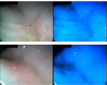
Fig. A3. Case 1. Descending colon, showing endoscopically an irregular and hyperemic mucosa, in some tracts even nodular, and presenting interspersed area of erosion.
Figg. A4 and A5. Case 1. Close up view of a mucosal dise-ased area. With BG endoscopy lesions of the mucosa were visible in dark blue and appeared to be more clearly defined from the remaining mucosa compared to TE.
Fig. A6. Case 1. This imagine shows as with BG endoscopy, fluid present in the colon could hinder the BG visualization, because it could appear in a blue similar to the one of an hyperemic area.
and almost uniformly infiltrated by lymphoid cells. These cells, belonging to the expansion of some follicles constituting the GALT follicular system, were enlarged, with a large nucleus containing two or more medium to large size nucleoli (B phenotype). Mitotic figures were frequently observed, and sometimes atypical. The immunophenotyping of these infil-trating lymphoid cells, performed by immunohistochemistry, showed a large percentages of CD 79a positives cells, with only a small fraction of these cells positives also for CD 21. On the basis of these evidences, the diagnosis of B – large cell GALT-derived lymphoma was performed.
CASE 2
A ten-year-old female Leonberger presenting with vomiting for about a week and a worsening of its general condition for some weeks, was examined at the University Veterinary Teaching Hospital, Camerino University. Five months previously, the dog had been diagnosed with a hypothyroidism, which had been treated accordingly. Laboratory tests revealed a slight hepatopathy and RBC (3.94 1012/L) below reference ranges.
An abdominal ultrasonography (Esaote MyLabTM Class C,
Genoa, Italy; 3-9 MHz micro-convex multifrequency trans-ducer) revealing a thickened portion (area of the gastric body) of the gastric wall and a loss of wall layering was then performed (Fig. B1). Considering the severely compromised clinical conditions of the dog, it was decided to perform a gastroscopy [(Flexible video endoscope, 160 cm length and 9.8 cm Ø, Mercury Produzione®, Foligno, Italy) provided with
13
A I V P A J O U R N A L - I T A L I A N J O U R N A L O F C O M P A N I O N A N I M A L P R A C T I C E - 3 / 2 0 1 5DIAGNOSTICA STRUMENTALE
Fig. B1. Case 2. Ultrasonographic appearance of the thicke-ned gastric body wall associated to the wall layering loss.
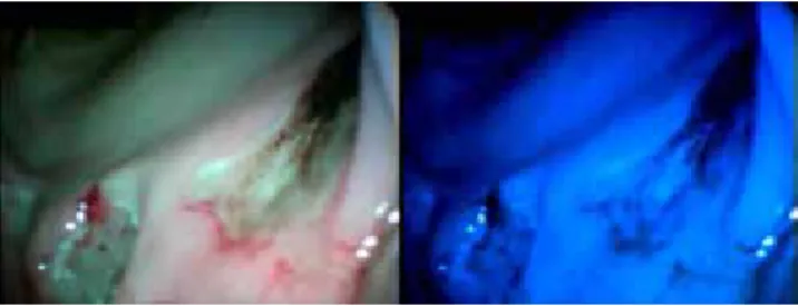
Fig. B2. Case 2. Close up view of mucosal ulcers and dise-ased areas.
Fig. B3. Case 2. Close up view of mucosal ulcers and dise-ased areas.
Fig. B4. Case 2. Close up view of mucosal ulcers and dise-ased areas. Especially in this figure, lesions appeared to be more clearly defined from the remaining mucosa compared to white light endoscopy.
to the white light endoscopy (Figg. B2, B3, and especially B5). Biopsies, performed on diseased areas, revealed at histopathology a severe inflammation of the gastric mucosa that surrounded a deep ulcerative crater. The bottom of the ulcer consisted in a neoplastic tissue classified as gastric adenocarcinoma, intestinal type. Tumor cells described ir-regular tubular structures, with stratification, multiple lumens surrounded by a reduced stroma (“back to back” aspect). The tumor invaded the gastric wall, infiltrating the muscularis mucosae, the submucosa and thence the muscularis propria. Often it showed associated intestinal metaplasia in adjacent mucosa. On the basis of glandular architecture, cellular pleo-morphism and mucosecretion, adenocarcinoma was graded as moderate differentiate.
DISCUSSION
Narrow Band Imaging (NBI, Olympus) is an endoscopic di-agnostic technique, under certain aspects similar to the one used in this study, which allows a better mucosal architec-ture and superficial vascular pattern definition(12),particularly
if associated with high-definition.(18) This technique is based
on the principle that shorter wavelengths penetrate only the superficial portion of the mucosa(12), accordingly, blue light
(narrow bandwidth, 415 nm of central wavelength) and green light (narrow bandwidth, 540 nm of central wavelength) could better define such superficial structures.(5,18) Moreover, these
wavelengths are those corresponding to hemoglobin peaks of absorption.(13,18)
Over the last years, many studies performed in human medicine have investigated the potential of NBI in colorectal diseases(10,11,15,17), evaluating particularly the neoplasm miss rate
compared to white light endoscopy.(7) This technique is
con-sidered useful in differentiating hyperplastic from adenomatous polyps, especially with high-definition instruments(3),while the
capability of improving adenoma detection is not univocal.(11-13,17)
Nevertheless, especially if associated with high-definition, it appears to increase the visualization of flat adenomas(6),while
magnifying NBI could be valuable in determining the invasion depth of early colorectal carcinoma.(4) NBI with magnification
is also considered useful in predicting histopathology and in selecting therapeutic strategies in colorectal tumors.(5,8) Finally,
NBI associated to specific classifications (e.g. NICE classi-fication) could allow, for example, a real-time differentiation between superficial lesions and deeply invasive carcinoma with related therapeutic implications.(1)
The potential of NBI in gastric diseases has been studied
(2,9,14),suggesting that magnifying NBI could be useful in
diagnos-ing early gastric carcinoma and in differentiatdiagnos-ing between small depressed carcinoma and small depressed non-neoplastic lesions;(4) it could also be worthwhile in defining early gastric
cancer margins, with the exception of those cases in which there is a subepithelial spread.(16)
CONCLUSION
To the author’s knowledge, this is the first endoscopic report that uses a BG endoscopy in dogs presenting digestive neoplastic conditions. Information regarding this technique in veterinary medicine literature is absent, and even if, in our cases, the lesions appeared to be more clearly defined with the BG endoscopy, the clinical, endoscopic and pathological significance of this technique in canine colonic and gastric neoplastic infiltrates, is yet to be determined. Furthermore, as in human medicine using the NBI technique, in the pre-sent case, the presence of fecal material also reprepre-sented a limitation for the technique.(11,12)
a single blue + green (BG) filter, restraining wavelengths from 400 to 550 nm].
Endoscopically, the stomach appeared partially filled with food, but at the level of the passage between the gastric body and the antrum many ulcers were found, some of which were bleeding (Figg. B2, B3, B4). A BG endoscopy showed up bleeding areas in dark blue and the lesions appeared to be more clearly defined from the remaining mucosa compared
A I V P A J O U R N A L - I t a l i a n j o u r n a l o f c o m p a n i o n a n i m a l p r a c t i c e - 3 / 2 0 1 5
14
DIAGNOSTICA STRUMENTALE
1. Hayashi N., Tanaka S., Hewett D.G., et al.: Endoscopic prediction of deep submucosal invasive carcinoma: valida-tion of the Narrow-Band Imaging Internavalida-tional Colorectal Endoscopic (NICE) classification. Gastrointestinal Endos-copy, 2013, 78, 625-632.
2. Hayee B., Inoue H., Sato H., et al: Magnification narrow-band imaging for the diagnosis of early gastric cancer: a review of the Japanese literature for the Western endosco-pist. Gastrointestinal Endoscopy, 2013, 78, 452-461. 3. Hewett D.G., Kaltenbach T., Sano Y., et al.: Validation of
a Simple Classification System for Endoscopic Diagnosis of Small Colorectal Polyps Using Narrow-Band Imaging. Gastroenterology, 2012, 143, 599-607.
4. Hirata I., Nakagawa Y., Ohkubo M., et al.: Usefulness of Magnifying Narrow-Band Imaging Endoscopy for the Di-agnosis of Gastric and Colorectal Lesions. Digestion, 2012, 85, 74-79.
5. Hirata M., Tanaka S., Oka S., et al.: Evaluation of microves-sels in colorectal tumors by narrow band imaging magni-fication. Gastrointestinal Endoscopy, 2007, 66, 945-952. 6. Jin X.-F., Chai T.-H., Shi J.-W., et al.: Meta-analysis for
evaluating the accuracy of endoscopy with narrow band imaging in detecting colorectal adenomas. Journal of Gas-troenterology and Hepatology, 2012, 27, 882-887.
7. Kaltenbach T., Friedland S., Soetikno R.: A randomised tandem colonoscopy trial of narrow band imaging versus white light examination to compare neoplasia miss rates. Gut, 2008, 57, 1406-1412.
8. Kanao H., Tanaka S., Oka S., et al.: Narrow-band imaging magnification predicts the histology and invasion depth of colorectal tumors. Gastrointestinal Endoscopy, 2009, 69, 631-636.
9. Kikuste I., Marques-Pereira R., Monteiro-Soares M., et al.: Systematic review of the diagnosis of gastric premalignant conditions and neoplasia with high-resolution endoscopic technologies. Scandinavian Journal of Gastroenterology, 2013, 48, 1108-1117.
10. Kim B.J., Park M.I., Park S.J., et al.: Differential Diagnosis of Colorectal Polyps with Narrow Band Imaging Colonos-copy without Magnification. The Korean Journal of Gastro-enterology, 2014, 63, 276-282.
11. Ng S.C., Lau J.Y.W.: Narrow-band imaging in the colon: Limitations and potentials. Journal of Gastroenterology and Hepatology, 2011, 26, 1589-1596.
12. Rastogi A., Bansal A., Wani S., et al.: Narrow-band imag-ing colonoscopy a pilot feasibility study for the detection of polyps and correlation of surface patterns with polyp histologic diagnosis. Gastrointestinal Endoscopy, 2008, 67, 280-286.
13. Sharma P., Gupta N., Kuipers E.J., et al.: Advanced imag-ing in colonoscopy and its impact on quality. Gastrointes-tinal Endoscopy, 2014, 79, 28-36.
14. Singh R., Hussain A., Loong C.K.: Narrow band imaging with magnification for the diagnosis of lesions in the up-per gastrointestinal tract. World Journal of Gastrointestinal Endoscopy, 2013, 5, 584-589.
15. Tanaka S., Sano Y.: Aim to unify the Narrow Band Imaging (NBI) magnifying classification for colorectal tumors: cur-rent status in Japan from a summary of the Consensus Symposium in ihe 79th Annual Meeting of the Japan Gas-troenterological Endoscopy Society. Digestive Endoscopy, 2011, 23, 131-139.
16. Uedo N., Fujishiro M., Goda K., et al.: Role of narrow band imaging for diagnosis of early-stage esophagogastric can-cer: current consensus of experienced endoscopists in Asia-Pacific region. Digestive Endoscopy, 2011, 23, 58-71. 17. Uraoka T., Higashi R., Saito Y., et al.: Impact of narrow-band imaging in screening colonoscopy. Digestive Endos-copy, 2010, 22, S54-S56.
18. Yao K., Anagnostopoulos G.K., Jawhari A.U., et al.: Opti-cal Microangiography: High-Definition Magnification Colo-noscopy with Narrow Band Imaging (NBI) for Visualizing Mucosal Capillaries and Red Blood Cells in the Large In-testine. Gut and Liver, 2008, 2, 14-18.