In
flammatory biomarker profiling in classical orthostatic hypotension:
Insights from the SYSTEMA cohort
☆
Madeleine Johansson
a, Fabrizio Ricci
b, Nay Aung
c, Richard Sutton
d, Olle Melander
a, Artur Fedorowski
a,e,⁎
a
Department of Clinical Sciences, Malmö, Faculty of Medicine, Lund University, Clinical Research Center, 214 28 Malmö, Sweden
bInstitute for Advanced Biomedical Technologies, Department of Neuroscience, Imaging and Clinical Sciences,“G. d'Annunzio” University, 66100 Chieti, Italy c
William Harvey Research Institute, NIHR Cardiovascular Biomedical Research Unit at Barts, Queen Mary University of London, London, UK d
National Heart and Lung Institute, Imperial College, Hammersmith Hospital Campus, Ducane Road, London W12 0NN, UK e
Department of Cardiology, Skåne University Hospital, 214 28 Malmö, Sweden
a b s t r a c t
a r t i c l e i n f o
Article history:
Received 4 November 2017
Received in revised form 29 November 2017 Accepted 6 December 2017
Available online xxxx
Objective: To investigate the inflammatory biomarker signature associated with classical orthostatic hypotension (OH).
Methods: A cross-sectional study including 778 patients with unexplained syncope and/or orthostatic intolerance undergoing head-up tilt test (HUT) and supine blood sampling. Of these, 98 met diagnostic criteria of classical OH and 181 demonstrated normal haemodynamic response during HUT. Blood plasma samples were analysed by antibody-based Proximity Extension Assay technique simultaneously measuring 57 inflammatory and cancer-related human protein biomarkers. The discovery algorithm was a sequential two-step process of biomarker signature identification by multivariate principal component analysis (PCA), and verification by univariate ANOVA with Bonferroni correction.
Results: Patients with classical OH were older (68 vs. 60 years; pb 0.001) and more likely to be men (58 vs. 41%; pb 0.001). PCA and Bonferroni-adjusted ANOVA identified midkine (MK), immunoglobulin-like transcript 3 (ILT-3), regenerating islet-derived protein 4 (REG-4), and tartrate-resistant acid phosphatase type 5 (TR-AP) as the most robust targeted biomarker signature for OH. In multivariate regression analysis adjusting for age, sex, cardiovascular disease and risk factors, the results remained significant for ILT-3 (p = 0.036), MK (p = 0.008) and REG-4 (p = 0.024), but not for TR-AP.
Conclusions: Targeted protein profiling in classical orthostatic hypotension reveals a biomarker signature associated with immunoregulatory functions and vascular inflammation. Circulating levels of midkine, immunoglobulin-like transcript-3, regenerating islet-derived protein-4 are elevated in orthostatic hypotension, suggesting a complex interplay among inflammation, autonomic dysfunction and atherothrombosis.
© 2017 Elsevier B.V. All rights reserved. Keywords:
Dysautonomia Inflammation Autonomic function Biomarker
Cardiovascular system pathology
1. Introduction
Orthostatic hypotension (OH) is a hallmark sign of autonomic failure frequently observed in patients with neurodegenerative diseases and comorbidities, such as diabetes and hypertension[1–3]. Presence of OH may cause debilitating symptoms and indicates higher risk of car-diovascular disease (CVD) and premature death[2,4,5]. Nevertheless, OH is frequently overlooked in cardiovascular screening programmes, epidemiological studies, and diagnostic work-up of patients with symptoms potentially related to this condition[2].
Traditionally, OH is divided into two main categories: neurogenic and non-neurogenic[2]. Neurogenic OH is a primary manifestation of chronic autonomic failure in neurodegenerative disorders, such as pure autonomic failure, multiple system atrophy and Parkinson disease
[6]. Orthostatic hypotension can also be secondary to various in flamma-tory and non-inflammatory conditions such as multiple myeloma, paraneoplastic syndrome, autoimmune diseases or amyloidosis[4], with a presumable affection of autonomic nervous system, although in many cases the aetiology remains unknown[1]. On the other hand, non-neurogenic OH can be caused by conditions that impair the compensatory mechanisms governed by the autonomic nervous system, such as diabetes and chronic cardiovascular disorders[2], but overlap between neurogenic and non-neurogenic factors in secondary OH may exist[2].
The most severe form of OH, often referred to as classical[7], implies a significant blood pressure reduction within the first 3 min of upright standing[8]. The majority of cases related to neurogenic OH belong
International Journal of Cardiology xxx (2017) xxx–xxx
☆ The authors take responsibility for all aspects of the reliability and freedom from bias of the data presented and their discussed interpretation.
⁎ Corresponding author at: Department of Cardiology, Inga Marie Nilssons gata 46, Skåne University Hospital, 214 28 Malmö, Sweden.
E-mail address:[email protected](A. Fedorowski).
https://doi.org/10.1016/j.ijcard.2017.12.020
0167-5273/© 2017 Elsevier B.V. All rights reserved.
Contents lists available atScienceDirect
International Journal of Cardiology
j o u r n a l h o m e p a g e :w w w . e l s e v i e r . c o m / l o c a t e / i j c a r dto this category[2,7]. However, in at least one third of cases, the aetiology of OH remains elusive, even after an extensive diagnostic work-up[1].
Both neurogenic and non-neurogenic forms of OH may potentially involve activation of inflammatory pathways, as components of the un-derlying pathological process eventually leading to autonomic failure
[9]. Notably, a cholinergic anti-inflammatory pathway that reflexively adjusts macrophage activation via parasympathetic outflow has recent-ly been described[10]. Further, the immune system has been shown to modulate autonomic activity, hence completing the wiring of the so called“inflammatory reflex”[11]. Thus, it is important to explore the expression of inflammatory mediators in OH, as a potential diagnostic tool and therapeutic target in this understudied and difficult-to-treat condition.
In this study, we sought to discover inflammatory biomarkers associated with OH in order to identify a signature which could be potentially useful to understand the pathophysiological pathway underlying the link between classical OH and CVD, as observed in epidemiological studies.
2. Methods 2.1. Study population
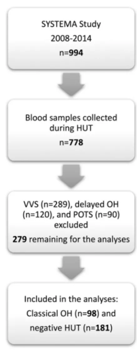
The study was carried out from September 2008 to May 2014 as a part of the Syncope Study of Unselected Population in Malmö (SYSTEMA)[12]. Patients with unexplained syncope and/or symptoms of orthostatic intolerance were referred to the tertiary syncope unit at Skåne University Hospital in Malmö from outpatient care and hospitals in southern Sweden. Additional tests were performed, if indicated, to eliminate any cardiac and neuro-logical causes of the symptoms, e.g. exercise and ambulatory prolonged electrocardiogram (Holter ECG), 2D transthoracic echocardiography, coronary and pulmonary angiography, brain imaging and encephalography. During the study period, 994 patients were exam-ined by head-up tilt test (HUT) according to current European syncope guidelines[7]; of these, 778 patients had blood samples collected during HUT examination (Fig. 1). All patients gave written informed consent. The study protocol conforms to the ethical guide-lines of the 1975 Declaration of Helsinki and has been approved by The Regional Ethical Review Board of Lund University (No. 82/2008).
The PICO model was as follows: patients with unexplained syncope or orthostat-ic intolerance (Population), blood samples and HUT (Intervention), classorthostat-ical OH ver-sus controls (Comparison), targeted protein biomarker discovery and hemodynamic response (Outcome).
2.2. Examination protocol
Patients were taking their regular medications, fasted for 2 h prior to examina-tion but were allowed to drink water at will. They were asked tofill out a question-naire about past medical history. The patients were placed on a tilt table and rested for at least 10 min before blood samples were collected through a venous cannula inserted in the forearm. Subsequently, patients rested for another 10 min to obtain haemodynamically stable parameters; thereafter the standardized 70°HUT was carried out for 20 min followed by nitroglycerine provocation according to the Italian pro-tocol if passive HUT was negative, or until syncope/pre-syncope or pronounced symptoms of orthostatic intolerance occurred[13]. Beat-to-beat blood pressure and ECG was monitored continuously by a validated non-invasive photoplethysmographic method (Nexfin monitor; BMEYE, Amsterdam, Netherlands) with a wrist unit and finger cuff of appropriate size[14].
2.3. Multiplex protein analysis
Plasma biomarkers were measured from supine blood samples (total volume: 30 ml) that had beenfirst centrifuged, then stored as 16 × 250 μl aliquots of EDTA plasma in plastic thermotubes, and frozen at−80 °C. For biomarker analysis, the samples were thawed and examined by the Proximity Extension Assay technique using the Olink Proteomics Proseek Multiplex Oncology I v1 96 × 96 reagents kit, which simultaneously measures 57 inflam-matory and cancer-related human protein biomarkers in plasma (Table S1). In short, a pair of oligonucleotide-labelled antibodies, Proseek probes, binds to the target protein in the plasma sample. When the two Proseek probes are in close proximity, a new polymerase-chain reaction (PCR) target sequence is formed by a proximity-dependent DNA polymerization event. This complex is subsequently detected and quantified using standard real-time PCR. The generated Normalized Protein Expression (NPX) unit is on a log2 scale, which means that a larger number represents a higher protein level in the sample. Additional information about limit of detection, reproducibility and validation is available at the Olink Proteomics website ( http://www.olink.com/products/document-download-center).
2.4. Data analysis
Supine and 3-min HUT BP were calculated over an averaged 30-s period. The supine BP was calculated during a stable period between 1 and 5 min prior to HUT. The 3-min HUT value was calculated after 3 min of HUT.
We defined classical orthostatic hypotension as a sustained drop in systolic BP ≥20 mm Hg and/or drop in diastolic BP (DBP) ≥10 mm Hg after 3 min of passive HUT[8]. A significant drop in BP occurring after 3 min of HUT was defined as delayed OH[15], and these patients were excluded from the analyses. Vasovagal syncope (VVS) was defined as a reproduction of syncope associated with a characteristic pattern of pronounced hypoten-sion, bradycardia or asystole[7], while postural orthostatic tachycardia syndrome (POTS) as a reproduction of symptoms of orthostatic intolerance (lightheadedness, dizziness or dis-comfort) with heart rate increaseN30/min or tachycardia N120/min during HUT[7,8]. Pa-tients with VVS and POTS were excluded from the analyses.
The baroreflex sensitivity (BRS; ms/mm Hg) index was calculated according to the formula: (60 / highest HR during HUT– 60 / supine HR) × 1000 ms / (lowest SBP during HUT− supine SBP), and compared between OH-positive and OH-negative patients. Valsalva maneuver was also performed to further assess nonpostural hemodynamic responses and BRS. Adrenergic BRS failure was featured by clear V-shaped SBP response, as previously reported[16].
We used the Modification of Diet in Renal Disease (MDRD) study equation to calculate the glomerularfiltration rate.
2.5. Statistical analysis
The main characteristics of study population are presented as mean and standard deviation for continuous variables and as percentages for categorical variables.
The discovery algorithm for the identification of potentially relevant biomarkers associated with the presence of OH was a sequential two-step process of i) biomarker signature identification by supervised, multivariate, principal component analysis, and ii) verification by univariate ANOVA with Bonferroni correction.
Fig. 1. Flow-chart summarising the selection process of study population. HUT, head-up tilt; OH, orthostatic hypotension; POTS, postural orthostatic tachycardia syndrome; SYSTEMA, Syncope Study of Unselected Population in Malmö; VVS, vasovagal syncope.
After defining a minimal call rate b 75%, we screened the biomarker panel through supervised principal component analysis, according to the algorithmfirst described by Hastie and Tibsirani[17], which includes the following steps:
1) For each biomarker, compute the standardized univariate logistic regression coeffi-cient which represents the effect size for the outcome (presence or absence of OH); 2) Using an arbitrary effect size thresholdθ from the list 0 ≤ θ1b θ2b ⋯ b θK:
a. Form a reduced data matrix consisting of only those biomarkers whose univariate coefficient exceeds θ in absolute value, and compute the principal components of this matrix;
b. Use these principal components in a multivariate logistic regression model to predict OH status;
3) Select the thresholdθ which gives the best predictive accuracy by 10-fold cross-validation.
Thereafter, for the verification of the selected biomarkers we applied a conservative univariate ANOVA approach, using a Bonferroni-adjusted significance level of p = 0.05/4. Thus, the inter-group (OH + vs. OH−) difference was considered to be statistically significant with a p-value b0.0125. Box plots were generated to display the distribution of biomarker levels between groups.
Furthermore, we performed univariate ordinary least square linear regression models for bivariate correlation between orthostatic SBP change (ΔSBP) and plasma level of selected biomarkers, and multivariate regression models adjusted for age, sex, supine systolic blood pressure, diabetes mellitus, hypertension, antihypertensive treatment, glomerularfiltration rate, prevalent cardiovascular disease and smoking. Finally, we performed a quantile-regression analysis in order to identify differing relationships at different quartiles of SBP changes during HUT.
Statistical analyses were carried out using IBM SPSS Statistics version 23 (SPSS Inc., Chicago, IL, USA) and R Statistical Software (version 2.14.0; R Foundation for Statistical Computing, Vienna, Austria).
3. Results
Of 778 patients with available plasma samples (Fig. 1), we found 98 patients who met classical OH criteria, and 181 patients with normal haemodynamic response during HUT. Descriptive characteristics of the study population are shown inTable 1. Four biomarkers were excluded from the analysis because their call rate was below 75%: erythropoietin (18%), interleukin-2 (7.9%), interferon-gamma (65%) and tumour necrosis factor (6.5%).
3.1. Biomarker signature discovery
The dataset consisted of 279 patients (98 OH and 181 controls). Since the principal component analysis requires pairwise complete data, we did not include markers with high missingness (N5%). This fil-ter resulted in removal of 4 biomarkers (vascular endothelial statin, lipopolysaccharide-induced tumour necrosis factor-alpha factor, MHC class I polypeptide-related sequence A and carcinoembryonic antigen). After removal of all missing data, 262 patients remained. Univariate logistic regression was performed for each of the 49 biomarkers. The re-gression coefficients were then standardized by dividing the coefficient with its standard error. All possible thresholds (Standardized coefficient (θ) ranging from minimum to maximum with 0.05 increments) were used to select groups of biomarkers and construct principal components (PCs). The outcome variable (OH status) was then regressed onto the first two PCs from each group of biomarkers using the binomial link function. This step identified the group of biomarkers which gave the best classification accuracy. The threshold that gave the best classi-fication accuracy (OH+ vs OH−) was selected by ten-fold cross-validation. The following 4 biomarkers reached this threshold: midkine (MK), immunoglobulin-like transcript 3 (ILT-3), regenerating islet-derived protein 4 (REG-4), and tartrate-resistant acid phosphatase type 5 (TR-AP).
3.2. Biomarker verification
As shown inTable 2, all PCA selected biomarkers differed signi fi-cantly in pairwise comparison, even after Bonferroni correction. In multivariate regression analysis adjusting for age, sex, cardiovascular disease and risk factors,ΔSBP was still significantly associated with
ILT-3 (p = 0.036), MK (p = 0.008) and REG-4 (p = 0.024), but TR-AP did not reach statistical significance (Table 3).
Quantile regression analyses investigating the relationships be-tween ILT-3, MK, REG-4 and TR-AP and the quartiles ofΔSBP did not reveal any obvious threshold effect or step function (Fig. S1). Finally, overall results remained unchanged after removing patients with Parkinson disease.
4. Discussion
This study demonstrates that patients with neurogenic and non-neurogenic orthostatic hypotension have elevated plasma levels of several inflammatory biomarkers, particularly immunoglobulin-like transcript 3 (ILT-3), midkine (MK) and regenerating islet-derived protein 4 (REG-4), independently of age, sex, prevalent cardiovascular disease and risk factors.
Thanks to recent technological advances it is possible to measure multiple plasma protein simultaneously. In this study, we applied a novel high throughput multiplex strategy for targeted protein biomark-er discovbiomark-ery to investigate circulating inflammation and cancer-related proteins and their association with OH. Lately, the proteomics technol-ogy has been implemented in a number of studies[18], and this study adds further insights to this emergingfield. Additional understanding of the molecular basis of OH may be of clinical importance in order to improve and personalize therapy in this understudied and difficult to treat condition.
Traditionally, OH has been linked to neurodegenerative diseases and chronic inflammatory conditions, more recently it has been found to be a commonfinding among patients with hypertension and diabetes[2]. However, the relationship between OH and inflammatory responses has not been sufficiently explored. In this study, we provide evidence
Table 1
Patient characteristics according to orthostatic hypotension status (n = 279). Characteristic OH positive n = 98 OH negative n = 181 P value Age (years) 68.1 ± 13.54 59.7 ± 20.5 b0.001 Sex (%, male) 58.2 41.4 0.008 Body-mass index (kg/m2 ) 25.2 ± 4.28 25.8 ± 4.70 0.29 Systolic BP supine (mm Hg) 137.5 ± 25.6 135.5 ± 20.3 0.47 Diastolic BP supine (mm Hg) 72.7 ± 10.2 71.9 ± 9.50 0.54 Heart rate (bpm) supine 69.2 ± 11.9 69.6 ± 11.7 0.78 Systolic BP (mm Hg) HUT min 85.1 ± 23.1 124.8 ± 19.2 b0.001 Diastolic BP (mm Hg) HUT min 55.2 ± 13.6 72.5 ± 10.8 b0.001 Heart rate (bpm) HUT max 80.5 ± 14.7 78.1 ± 14.3 0.20 V-pattern at VM (%) 27.3 1.7 b0.001 BRS index (ms/mm Hg) 2.9 ± 1 12.3 ± 4.8 0.004 Hypertension (n, %) 50.5 39.8 0.09 Ischemic heart disease (%) 10.2 10.5 0.94
Heart failure (%) 4.1 7.2 0.30 Atrialfibrillation (%) 7.1 8.3 0.73 Diabetes mellitus (%) 5.1 8.9 0.25 Parkinson disease (%) 4 0 0.007 Cancer (%) 14.3 9.9 0.28 Smoking (%) 8.2 19.9 0.04 GFR (ml/min) 70 ± 20 81 ± 24 b0.001 LVEF (%) 54 ± 3 54 ± 3 0.92 Beta-blocker (%) 26.3 31.5 0.36 Diuretic (%) 12.1 11.2 0.82 CCB (%) 15.2 12.4 0.51 ACE-I (%) 19.2 8.4 0.009 ARB (%) 16.2 14.6 0.73 Alpha-blocker (%) 4 1.1 0.11 Long-acting nitrate (%) 1 7.3 0.02 OH, orthostatic hypotension; P values for differences between the groups shown as mean and standard deviation for continuous variables and as percentages for categorical variables. ACE-I, angiotensin converting enzyme inhibitor; ARB, angiotensin receptor blocker; BP, blood pressure; BRS, baroreflex slope index; CCB, calcium channel blockers; GFR, glomerularfiltration rate (MDRD formula); HUT min/max, lowest/highest value during passive head-up tilt test; bpm, beats per minute; LVEF, left ventricular ejection fraction; VM, Valsalva maneuver.
supporting the view that autonomic dysfunction underlying OH is not merely a symptom-generating condition, but also a disorder that has complex interplay with important inflammatory and immunological processes.
The elevated levels of MK suggest an acute cytoprotective effect in ischaemia/reperfusion injury related to its anti-apoptotic effect promot-ing angiogenesis and inhibition of cardiac tissue remodelpromot-ing[19]. Moreover, MK facilitates endothelial cell proliferation, and also recruits inflammatory cells to the walls of the vessels promoting neointima formation, vascular stenosis and inflammation, inducing features of plaque vulnerability in atherosclerosis. Upregulation of ILT-3 seems to play a significant role in graft adaptation and protection against the recipient's immune response[20]. Expression of REG-4 is considerably upregulated during inflammation and tissue injury associated with autoimmune diseases, such as active Crohn's disease and ulcerative colitis, and in colorectal cancer[21]. Thesefindings have not been reported previously in OH patients.
4.1. Midkine
MK is a heparin-binding growth factor of low molecular weight involved in the aetiology of inflammatory diseases, e.g. multiple sclero-sis[22,23]. It is activated during oncogenesis, inflammation and tissue repair, and enhances cell proliferation, cell migration, angiogenesis andfibrinolysis. Elevated levels of MK are observed in several malignant tumours and it is also linked to tumour resistance to chemotherapeu-tics. Additionally, deposits of MK are seen in patients with neurodegen-erative diseases, e.g. Alzheimer's disease and multiple system atrophy
[24].
Moreover, results published by Horiba et al.[25]suggest that MK may play a protective role against ischaemia/reperfusion injury and constitutes a new potentially important molecular target for treatment of ischaemic heart disease. Interestingly, MK is induced in cancer tissues where it also promotes angiogenesis and tumour formation by angio-genic and anti-apoptotic activity[19]. Muramatsu et al.[22]found that MK may be useful as a cancer marker, whereas MK itself may be used in treatment of brain and heart diseases. On the other hand, MK-inhibitors can be used in the treatment of malignant tumours, multiple sclerosis, restenosis, renal diseases, and hypertension.
In the heart, preclinical data support a potential role of MK in the pathophysiology of CVD, where it promotes endothelial cell prolifera-tion and enhances plaque infiltration of inflammatory cells. Notably, MK-deficient mice exhibited significantly lower neointimal formation
[26], while systemic administration of MK in apolipoprotein-E knockout mice increased atherosclerosis[27]. Recently, it has also been demon-strated that MK could be used in humans to predict the presence of significant coronary artery disease and higher incidence of acute coronary events[28]. Ultimately, elevated levels of MK are associated with accelerated atherosclerotic plaque formation and progression. Taken together, thesefindings suggest that a sustained and enhanced pro-inflammatory activity might be one of the background mechanisms
underlying the longitudinal association between OH and cardiovascular morbidity.
4.2. Immunoglobulin-like transcript 3
Immunoglobulin-like transcripts (ILTs) are immuno-regulatory proteins that either activate or inhibit immune responses[29]. ILT-3 is an important mediator of the induction of immune tolerance and expressed on monocytes and antigen-presenting cells such as macro-phages and dendritic cells. Although the mechanisms by which ILT3 modulates immune responses is largely unknown, Chang et al. found that down-regulation of ILT3 may result in autoimmune diseases due to excess inflammation and infiltration of T cells in locally affected lesions[30]. Furthermore, studies of human heart transplant recipients demonstrated that rejection-free patients have circulating T-suppressor cells, which cause up-regulation of ILT3 in donor antigen-presenting cells. These results indicate a possibly important mechanism of immune regulation[20].
4.3. Regenerating islet-derived protein 4
REG-4 is associated with inflammatory and metaplastic responses of the gastrointestinal epithelium. It is a critical protein involved in the development of colorectal cancer and overexpression of REG-4 with or without overexpression of matrix metalloproteinase 7 (MMP-7) is a predictive factor of poor prognosis in colorectal cancer[31]. It has been found that REG-4 promotes the proliferation and invasiveness of cancer cells by upregulating the expression of MMP-7, which is involved in matrix degradation within the atherosclerotic lesion, and is associat-ed with severe atherosclerosis, plaque destabilization, and higher incidence of coronary and cerebrovascular events.
4.4. General remarks
Our study confirms that OH and, consequently, cardiovascular autonomic dysfunction are associated with multifactorial mechanisms facilitating cardiovascular disease, including inflammation and autoim-mune mechanisms. Elevated levels of midkine, immunoglobulin-like transcript 3, and regenerating islet-derived protein 4 are in accordance with previous studies that have demonstrated association of OH with neurodegenerative and autoimmune diseases[2,4,32]. Thus, our findings expand the evidence that OH is not merely a haemodynamic phenomenon, but in fact includes a range of dysregulated molecular events heralding malfunction of the immune and circulatory system. 4.5. Strengths and limitations
The present study is based on a large sample of symptomatic individuals and a novel state-of-the-art proteomics chip was used.
Table 2
High throughput multiplex analysis of 4 of 49 oncological biomarkers selected by supervised multivariate principal component analysis, in 89 patients with classical orthostatic hypotension. Plasma concentrations of the assessed proteins are expressed on a log2-scale. Inter-group differences were assessed using analysis of variance method. Bonferroni-corrected significant values (p b 0.0124) are marked in bold.
Biomarker OH positive (n = 98) OH negative (n = 181) P-value Immunoglobulin-like transcript 3 (ILT-3) 2.46 ± 0.64 2.18 ± 0.61 b0.001 Midkine (MK) 7.30 ± 0.57 7.00 ± 0.59 b0.001 Regenerating islet-derived protein 4 (REG-4) 3.68 ± 0.58 3.41 ± 0.57 b0.001 Tartrate-resistant acid
phosphatase type 5 (TR-AP)
5.36 ± 0.50 5.19 ± 0.52 0.007
Table 3
Association between changes in systolic blood pressure during HUT and selected biomarkers in univariate and multivariate regression.
Biomarker Univariate Multivariatea
β 95% CI P-value β 95% CI P-value Immunoglobulin-like transcript 3 (ILT-3) 10.7 5.9–15.5 b0.001 7.8 2.4–13.3 0.021 Midkine (MK) 13.3 8.3–18.4 b0.001 10.4 4.9–15.9 0.001 Regenerating islet-derived protein 4 (REG-4) 12.2 7.1–17.41 b0.001 9.6 4.1–15.0 0.003 Tartrate-resistant acid phosphatase type 5 (TR-AP) 12.2 6.3–18.1 b0.001 5.3 −0.6 -11.2 0.315 a
Adjusted for age, sex, supine systolic blood pressure, diabetes mellitus, hypertension, antihypertensive treatment, glomerularfiltration rate, presence of cardiovascular disease and smoking. Significant associations in multivariate model (p b 0.05) are marked in bold.
All patients were examined according to a standardized protocol with beat-to-beat haemodynamic monitoring, thus minimizing the risk of in-accurate or missed diagnosis of OH. Moreover, we performed a sequen-tial two-step discovery and verification analysis, the former based on a supervised, multivariate, dimensionality reduction technique, achieving the best compromise between best predictive ability and exhaustivity, and the latter using a more conservative approach through univariate ANOVA with Bonferroni adjustment. Nevertheless, this may have resulted in omission of significant information, therefore further studies on independent patient samples are necessary.
Some limitations should be also addressed. Firstly, our study was performed on symptomatic individuals who were unaware of the nature of underlying disorder prior to investigation. Consequently, our study may not be entirely representative of OH detected in the general population through screening programmes or in asymptomatic outpa-tients. Secondly, in order to rule-out possible false positive signals, our findings should be verified and validated with alternative technologies enabling as much sensitive and robust detection and quantification of biomarkers. However, the use of a proximity assay with the require-ment for a dual binding event which ensures minimal background signal, and the robust discovery algorithm, based on a sequential two-step process including principal component analysis and a very strict Bonferroni correction, would make a false positive result less likely.
5. Conclusions
Our study confirms and extends the concept that high throughput multiplex analysis for protein profiling can considerably improve the understanding of autonomic failure. We report here that presence of orthostatic hypotension in patients with a history of unexplained synco-pe and orthostatic intolerance is associated with elevated plasma levels of midkine, immunoglobulin-like transcript 3, and regenerating islet-derived protein 4. These observations support the hypothesis that autonomic dysfunction may be evoked by inflammatory processes, but that it may also maintain a systemic inflammatory milieu with possible detrimental effects on the cardiovascular system.
Contributors
AF, FR, NA, MJ had full access to all the data in the study and take responsibility of the data and accuracy of the data analysis. MJ, OM, RS, AF contributed to the study concept and design. AF, OM, MJ contrib-uted to the acquisition of data. All authors analysed and interpreted the data. AF was the study supervisor. NA, AF, FR did the statistical analysis. MJ, FR, RS, AF drafted the manuscript with critical revision for important intellectual content from all authors.
Transparency
The lead authors (the manuscript's guarantors) affirm that the manuscript is an honest, accurate, and transparent account of the study being reported; that no important aspects of the study have been omitted; and that any discrepancies from the study as planned (and, if relevant, registered) have been explained.
Funding
This work was supported by grants from the Swedish Medical Research Council, the Swedish Heart and Lung Foundation, the Medical Faculty of Lund University, Malmö University Hospital, the Crafoord Foundation, the Ernhold Lundströms Research Foundation, the Region Skåne, the Hulda and Conrad Mossfelt Foundation, the King Gustaf V and Queen Victoria Foundation, and The Wallenberg Foundation.
Competing interests
All authors have completed the ICMJE uniform disclosure atwww. icmje.org/coi_disclosure.pdfand declare: AF reports personal fees from Cardiome Corp. and a patent from ThermoFisher pending outside the submitted work; RS reports personal fees and others from Medtronic Inc. and St. Jude Medical Inc. outside the submitted work; RS performs consultancy for Medtronic Inc.; RS is a member of the speaker's Bureau St. Jude Medical/Abbott Inc.; RS is a shareholder in Boston Scientific Inc., Edwards Lifesciences Inc., Shire PLC, Roche SA and Astrazeneca PLC. There are no other relationships or activities that could appear to have influenced the submitted work.
Licence for publication statement
The Corresponding Author has the right to grant on behalf of all authors and does grant on behalf of all authors, a worldwide licence to the Publishers and its licensees in perpetuity, in all forms, formats and media (whether known now or created in the future), to i) publish, reproduce, distribute, display and store the Contribution, ii) translate the Contribution into other languages, create adaptations, reprints, include within collections and create summaries, extracts and/or, abstracts of the Contribution, iii) create any other derivative work(s) based on the Contribution, iv) to exploit all subsidiary rights in the Contribution, v) the inclusion of electronic links from the Contribution to third party material where-ever it may be located; and, vi) licence any third party to do any or all of the above.
References
[1] D.S. Goldstein, Y. Sharabi, Neurogenic orthostatic hypotension: a pathophysiological approach, Circulation 119 (2009) 139–146.
[2] F. Ricci, R. De Caterina, A. Fedorowski, Orthostatic hypotension: epidemiology, prognosis, and treatment, J. Am. Coll. Cardiol. 66 (2015) 848–860.
[3] A. Fedorowski, P. Burri, O. Melander, Orthostatic hypotension in genetically related hypertensive and normotensive individuals, J. Hypertens. 27 (2009) 976–982.
[4] R. Freeman, Clinical practice. Neurogenic orthostatic hypotension, N. Engl. J. Med. 358 (2008) 615–624.
[5] F. Ricci, A. Fedorowski, F. Radico, M. Romanello, A. Tatasciore, M. Di Nicola, et al., Cardiovascular morbidity and mortality related to orthostatic hypotension: a meta-analysis of prospective observational studies, Eur. Heart J. 36 (2015) 1609–1617.
[6] D. Robertson, The pathophysiology and diagnosis of orthostatic hypotension, Clin. Auton. Res. 18 (Suppl. 1) (2008) 2–7.
[7] A. Moya, R. Sutton, F. Ammirati, J.J. Blanc, M. Brignole, J.B. Dahm, et al., Guidelines for the diagnosis and management of syncope (version 2009): the Task Force for the Diagnosis and Management of Syncope of the European Society of Cardiology (ESC), Eur. Heart J. 30 (2009) 2631–2671.
[8] R. Freeman, W. Wieling, F.B. Axelrod, D.G. Benditt, E. Benarroch, I. Biaggioni, et al., Consensus statement on the definition of orthostatic hypotension, neurally mediat-ed syncope and the postural tachycardia syndrome, Clin. Auton. Res. 21 (2011) 69–72.
[9] T. Pecanha, A.H. Lima, Inflammation and cardiovascular autonomic dysfunction in rheumatoid arthritis: a bidirectional pathway leading to cardiovascular disease, J. Physiol. 595 (2017) 1025–1026.
[10]L.V. Borovikova, S. Ivanova, M. Zhang, H. Yang, G.I. Botchkina, L.R. Watkins, et al., Vagus nerve stimulation attenuates the systemic inflammatory response to endotoxin, Nature 405 (2000) 458–462.
[11] K.J. Tracey, The inflammatory reflex, Nature 420 (2002) 853–859.
[12] N. Isma, R. Sutton, A. Hillarp, K. Strandberg, O. Melander, A. Fedorowski, Higher levels of von Willebrand factor in patients with syncope due to orthostatic hypoten-sion, J. Hypertens. 33 (2015) 1594–1601.
[13]A. Bartoletti, P. Alboni, F. Ammirati, M. Brignole, A. Del Rosso, G. Foglia Manzillo, et al.,‘The Italian Protocol’: a simplified head-up tilt testing potentiated with oral nitroglycerin to assess patients with unexplained syncope, Europace 2 (2000) 339–342.
[14] A. Fedorowski, V. Hamrefors, R. Sutton, J.G. van Dijk, R. Freeman, J.W. Lenders, et al., Do we need to evaluate diastolic blood pressure in patients with suspected orthostatic hypotension? Clin. Auton. Res. 27 (2017) 167–173.
[15] C.H. Gibbons, R. Freeman, Delayed orthostatic hypotension: a frequent cause of orthostatic intolerance, Neurology 67 (2006) 28–32.
[16] I.S. Palamarchuk, J. Baker, K. Kimpinski, The utility of Valsalva maneuver in the diagnoses of orthostatic disorders, Am. J. Phys. Regul. Integr. Comp. Phys. 310 (2016) R243–52.
[17] T. Hastie, R. Tibshirani, J. Friedman, The Elements of Statistical Learning: Data Mining, Inference, and Prediction, Springer, New York, 2013.
[18] C. Hage, E. Michaelsson, C. Linde, E. Donal, J.C. Daubert, L.M. Gan, et al., Inflammatory biomarkers predict heart failure severity and prognosis in patients with heart failure with preserved ejection fraction: a holistic proteomic approach, Circ. Cardiovasc. Genet. 10 (2017).
[19] K. Kadomatsu, P. Bencsik, A. Gorbe, C. Csonka, K. Sakamoto, S. Kishida, et al., Therapeutic potential of midkine in cardiovascular disease, Br. J. Pharmacol. 171 (2014) 936–944.
[20] C.C. Chang, R. Ciubotariu, J.S. Manavalan, J. Yuan, A.I. Colovai, F. Piazza, et al., Tolerization of dendritic cells by T(S) cells: the crucial role of inhibitory receptors ILT3 and ILT4, Nat. Immunol. 3 (2002) 237–243.
[21]A. Granlund, V. Beisvag, S.H. Torp, A. Flatberg, P.M. Kleveland, A.E. Ostvik, et al., Activation of REG family proteins in colitis, Scand. J. Gastroenterol. 46 (2011) 1316–1323.
[22] T. Muramatsu, K. Kadomatsu, Midkine: an emerging target of drug development for treatment of multiple diseases, Br. J. Pharmacol. 171 (2014) 811–813.
[23] H. Takeuchi, Midkine and multiple sclerosis, Br. J. Pharmacol. 171 (2014) 931–935.
[24] K. Kadomatsu, T. Muramatsu, Midkine and pleiotrophin in neural development and cancer, Cancer Lett. 204 (2004) 127–143.
[25]M. Horiba, K. Kadomatsu, K. Yasui, J.K. Lee, H. Takenaka, A. Sumida, et al., Midkine plays a protective role against cardiac ischemia/reperfusion injury through a reduction of apoptotic reaction, Circulation 114 (2006) 1713–1720.
[26] M. Horiba, K. Kadomatsu, E. Nakamura, H. Muramatsu, S. Ikematsu, S. Sakuma, et al., Neointima formation in a restenosis model is suppressed in midkine-deficient mice, J. Clin. Invest. 105 (2000) 489–495.
[27] Y. Takemoto, M. Horiba, M. Harada, K. Sakamoto, K. Takeshita, T. Murohara, et al., Midkine promotes atherosclerotic plaque formation through its pro-inflammatory, angiogenic and anti-apoptotic functions in apolipoprotein E-knockout mice, Circ J. (2017)https://doi.org/10.1253/circj.CJ-17-0043.
[28] N.E. Ibrahim, J.L. Januzzi Jr., C.A. Magaret, H.K. Gaggin, R.F. Rhyne, P.U. Gandhi, et al., A clinical and biomarker scoring system to predict the presence of obstructive coronary artery disease, J. Am. Coll. Cardiol. 69 (2017) 1147–1156.
[29] M.A. Jensen, R.N. Yanowitch, A.T. Reder, D.M. White, B.G. Arnason, Immunoglobulin-like transcript 3, an inhibitor of T cell activation, is reduced on blood monocytes during multiple sclerosis relapses and is induced by interferon beta-1b, Mult. Scler. 16 (2010) 30–38.
[30]C.C. Chang, Z. Liu, G. Vlad, H. Qin, X. Qiao, D.M. Mancini, et al., Ig-like transcript 3 regulates expression of proinflammatory cytokines and migration of activated T cells, J. Immunol. 182 (2009) 5208–5216.
[31] X. Zhu, Y. Han, C. Yuan, W. Tu, G. Qiu, S. Lu, et al., Overexpression of Reg4, alone or combined with MMP-7 overexpression, is predictive of poor prognosis in colorectal cancer, Oncol. Rep. 33 (2015) 320–328.
[32] M. Ruzieh, L. Batizy, O. Dasa, C. Oostra, B. Grubb, The role of autoantibodies in the syndromes of orthostatic intolerance: a systematic review, Scand. Cardiovasc. J. 51 (2017) 243–247.