Open Peer Review
Any reports and responses or comments on the article can be found at the end of the article.
CLINICAL PRACTICE ARTICLE
Early hyaluronidase use in preventing skin necrosis after
treatment with dermal fillers: Report of two cases [version 2;
peer review: 2 approved, 1 not approved]
Francesco Ciancio
,
Maria Stella Tarico , Giuseppe Giudice ,
Rosario Emanuele Perrotta
2 Department of Plastic and Reconstructive Surgery, University of Bari, Bari, 70124, Italy Department of Plastic and Reconstructive Surgery, University of Catania, Catania, 95100, Italy Abstract Injection of dermal fillers, like hyaluronic acid (HA), is a safe procedure, with few and transient side effects such as erythema, bruising and swelling etc. The aim of this report is to provide our protocol for the early treatment of necrotic complications after facial treatment with dermal fillers. We present two cases of skin suffering of the face after dermal infiltration of HA, treated successfully with our early protocol. Our protocol includes the early infiltration of hyaluronidase in the treated areas. We start with infiltration of hyaluronidase distributed over the area to be treated through micro-injections with dosage 40 IU per cm . Our protocol includes the use of systemic corticosteroids for 4 days, anti-aggregation therapy, oral antibiotic, topical cream with nitric oxide and compresses with gauze and warm water. In the skin complications after dermal filler treatment, marked pain and characteristic reticulated erythema in the skin distribution of the affected vessels is often developed. Due to the implementation of our protocol in these patients, we managed to avoid an irreversible necrotic complication of the face in both cases. In this report, our protocol was compared with results published in the literature and allowed us to avoid complications such as skin necrosis with permanent damage. Keywords dermal filler, skin necrosis, filler complications, vascular complications 1 2 1 2 1 2 Referee Status: Invited Referees version 2 published 03 Apr 2019 version 1 published 03 Sep 2018 1 2 3 report report report report report , University of Parma, Nicolò Bertozzi Italy 1 , San Diego Gottfried Lemperle School of Medicine, University of California, USA 2 , Beth Israel Deaconess Robert J. Besaw Medical Center, USA 3 03 Sep 2018, :1388 ( First published: 7 ) https://doi.org/10.12688/f1000research.15568.1 03 Apr 2019, :1388 ( Latest published: 7 ) https://doi.org/10.12688/f1000research.15568.2v2
2 Page 1 of 11
Francesco Ciancio ( )
Corresponding author: [email protected]
: Writing – Original Draft Preparation; : Conceptualization; : Project Administration; :
Author roles: Ciancio F Tarico MS Giudice G Perrotta RE
Data Curation, Validation, Visualization
No competing interests were disclosed.
Competing interests:
The author(s) declared that no grants were involved in supporting this work.
Grant information:
© 2019 Ciancio F . This is an open access article distributed under the terms of the , which
Copyright: et al Creative Commons Attribution Licence
permits unrestricted use, distribution, and reproduction in any medium, provided the original work is properly cited. Ciancio F, Tarico MS, Giudice G and Perrotta RE.
How to cite this article: Early hyaluronidase use in preventing skin necrosis after
F1000Research 2019, :1388 (
treatment with dermal fillers: Report of two cases [version 2; peer review: 2 approved, 1 not approved] 7
)
https://doi.org/10.12688/f1000research.15568.2
03 Sep 2018, :1388 ( )
First published: 7 https://doi.org/10.12688/f1000research.15568.1
Introduction
The use of dermal fillers is increasingly required in cosmetic surgery1. Injection of dermal fillers is a safe procedure, with few
and transient side effects. However, cases of skin necrosis have been reported, including with involvement of vision and ocular globe2. The cause is an impediment of the blood supply by
compression and/or obstruction of the vessel(s) with filler material, and/ or direct injury to the vessel3,4. Several therapeutic
approaches have been described5–7.
The aim of this report is to present our protocol for the early treatment of vascular complications after facial rejuvenation with dermal fillers in order to avoid skin necrosis. We present two cases of vessel damage and skin suffering of the face after dermal infiltration of hyaluronic acid (HA), which occurred in 2017 and was treated successfully with our protocol.
Protocol
Infiltration of hyaluronidase is performed firstly, at a deep dermal level, and is distributed over the area to be treated through micro-injections at a dosage of 40 IU per cm2. The distribution
is homogeneous except for nodular areas in which a double dose is infiltrated (in any case, at least 150 IU of hyaluronidase in 1th
infiltration is used). The lithic enzyme must be infiltrated at the dermal level, homogeneously distributed, in order that from the point of infiltration it is distributed to the vessels by diffusion. The rational use of hyaluronidases is to breakdown HA particles and allow reabsorption. It is well known that hyaluronidases are inactivated by the immune system8, so the treatment must
be repeated to obtain an adequate concentration. In our clinical practice, we use the maximum dosage (40IU per cm2) for three
consecutive days. The areas to be treated are established on the basis of clinical signs of damage/ischemia distribution for which, in selected cases, maintenance doses (40 UI/cm2) can be
applied in the areas most affected. The treatment is repeated after a few hours if the ischemic area shows no improvement with double daily doses but, in all cases, never extended beyond 72h. Systemic corticosteroids for four days (prednisone 25 mg/24h per os) to reduce edema and increase microcirculation perfusion, oral salicylic acid 100 mg per os like anti-platelet, antibiotic prophi-laxis (levofloxacin 500mg / 24h for 4 days) for any infections, topical cream with nitric oxide (3 times a day) to improve blood perfusion, antibiotic therapy, topical cream with nitric oxide, and compresses with gauze and warm water are also recommended in this protocol. In the following days, the evolution of clinical signs should be monitored and therapy continued if needed.
The protocol must be implemented as soon as possible, spe-cifically the cortisone drugs are used in the first 24 hours and continue until the fourth day, the treatment with acetylsalicylic acid 100 mg starts in the first 24 hours and lasts for 10 days, the antibiotic prophylaxis is established. in the first 24 h and continues for 4 days.
Case 1
In June 2017 a 36-year-old female patient was admitted for treatment of infiltration of HA-based dermal fillers. The patient had received treatments with dermal fillers in the past without adverse reactions. Immediately after the dermal filler procedure, the treated areas appeared in good condition without signs of skin suffering. Three days later, at a follow-up examination, the left treated area appeared cyanotic and swollen despite the patient not complaining of discomfort. The skin appeared erythematous with distribution along the left nasolabial folds up to the lateral nasal wall, and the capillary refill time appeared slow or absent (Figure 1). Consequently, treatment with the protocol, as stated above, was performed immediately. We used 40UI of hyaluronidase per cm2, two times a day for 3 days. The patient
received acetylsalicylic acid 100 mg / 24h for 10 days, prednisone 25mg / 24h for 4 days, levofloxacin 500mg / 24h for 4 days, topical cream with nitric oxide 2 times a day and compresses with gauze and warm 3 times a day.
Necrotic complications of the face were avoided in this patient (Figure 2).
Case 2
In August 2017 a 45-year-old woman was treated with HA to fill the region of nasolabial folds. In the past the patient had received similar treatments without adverse reactions. At the clinical check after three days, the patient shows signs of skin suffering. Compared to Case 1 the erythematous area was
Figure 1. A 36-year-old female patient with skin suffering in the left naso-labial fold region after hyaluronic acid infiltration from dermal fillers. Three days after dermal filler treatment, erythematous and blister formation was observed along the nasolabial vessels. Part of the erythema extends to the middle of the nose.
Amendments from Version 1
As recommended by the Review we added details of the dosage on the presented Protocol, we have included the rationale of use for each treatment. We have added a paragraph in the Protocol section indicating the times of use of the drugs and when to begin treatment with each category of drugs.
See referee reports
REVISED
Figure 4. A 45-year-old female patient with hyaluronic acid infiltration from dermal fillers treated with the early protocol.
Left panel: the erythematous lesion has decreased in intensity after 7 days from treatment; middle panel: after 12 days; right panel: after 45 days. The green dots in the picture are the result of a damaged camera, we have not modified the image.
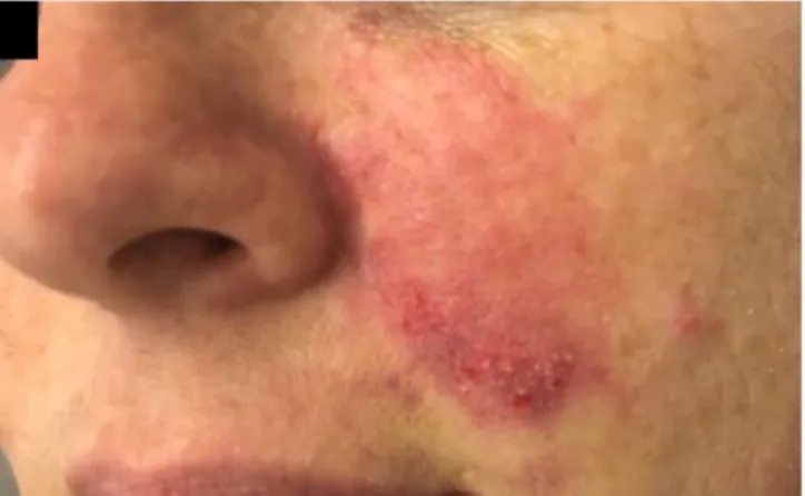
Figure 3. A 45-year-old woman with skin suffering in left naso-labial fold region after hyaluronic acid infiltration from dermal fillers. There is an erythematous halo, blisters and livedo reticularis in the middle third of the left cheek.
Figure 2. A 36-year-old female patient with hyaluronic acid infiltration from dermal fillers treated with the early protocol. Left panel: 7 days after first treatment; middle panel: after 15 days; right panel: clinical check after 45 days. It should be noted that there is no scarring in the final image.
smaller with distribution retained to the medial region of the cheek (Figure 3). Treatment with the protocol, as stated above, was performed immediately and 40UI/cm2 of hyaluronidase was
injected every 12 h per 2 days, after only 1 dose for the third day. Systemic corticosteroids, antiplatelet therapy, antibiotic therapy and local topics were used according to protocol, as expressed in Case 1.
Necrotic complications of the face were avoided in this patient (Figure 4).
Discussion
Damage after dermal fillers can lead to severe consequences, such as skin necrosis, and the involvement of ocular, nerve and muscle structures2. Skin necrosis is the most significant complications
after dermal filler. The incidence of vascular damage after use of fillers has been estimated at 3–9 per 10,000 for HA products, but the true incidence of this complication is unknown9,10. The skin
necrosis after dermal fillers injection is an emergency, the best treatment is often the quickest. The gold standard is prompt injection of hyaluronidase with a dose of 40 IU per cm2 of
affected area. Some authors have described oral antiplatelet drugs, like cardioaspirin while some have cited the use of vasodilator drugs. Today again there is no international standard protocol for the treatment of these complications. In our cases, the patients manifested skin problems probably from filler emboli or direct destruction of the vessels during needle manoeuvres. Interestingly our patients did not experience any pain or discomfort during and after the HA dermal filler procedure. In the literature, some manoeuvres have been reported to reduce the risk of vascular damage, such as aspiration during infiltration11,
low pressure injection, continuously moving the needle or cannula while injecting, injection of small quantities (maximum 0.1 mL of filler per pass)12, observing skin changes during the immediate
post-injection phase, and excellent knowledge of anatomy13–15.
Typically in areas with terminal vascular circulation, the cutaneous vessels suffering may be more likely; in our cases it is one of the most vascularized areas of the face, the nasolabial folds15.
In this kind of complication, an early treatment is the best choice. The gold standard is prompt injection of hyaluronidase16.
De Lorenzi presents a protocol with high doses of hyaluronidase8, where the dosage of hyaluronidases is quantified
on the basis of the regions of the face affected (for example: for
the glabellar region 500 IU of hyaluronidase). We agree with the De Lorenzi that the use of elevated quantities of hyaluronides is required for the treatment of adverse events of vascular filler; however, we believe that a distribution per cm2 is more precise.
More treatments has been described but there is no international standard protocol for the treatment of these complications. As expressed by various Societies of Plastic and Aesthetic Surgery, to minimize the incidence of this type of damage it is essential to contact qualified and trained medical personnel, who follow modern international protocols17–19.
The strengths of our protocol is certainly the result with no residual mark of skin suffering that has been seen in our cases, while the major limitation is the necessary execution of the protocol within 72h from the damage.
Conclusion
Today, the best solution for vascular damage is to categorically avoid dermal fillers treatments with non-medical and untrained personnel. This report aims to offer present a protocol for the
References
1. American Society of Plastic Surgeons: 2012 plastic surgery procedural statistics.
Reference Source
2. Salval A, Ciancio F, Margara A, et al.: Impending Facial Skin Necrosis and Ocular Involvement After Dermal Filler Injection: A Case Report. Aesthetic Plast Surg. 2017; 41(5): 1198–1201.
PubMed Abstract | Publisher Full Text
3. Kim DY, Eom JS, Kim JY: Temporary Blindness After an Anterior Chamber Cosmetic Filler Injection. Aesthetic Plast Surg. 2015; 39(3): 428–430.
PubMed Abstract | Publisher Full Text
4. Schanz S, Schippert W, Ulmer A, et al.: Arterial embolization caused by injection of hyaluronic acid (Restylane). Br J Dermatol. 2002; 146(5): 928–9.
PubMed Abstract | Publisher Full Text
5. Urdiales-Gálvez F, Delgado NE, Figueiredo V, et al.: Treatment of Soft Tissue Filler Complications: Expert Consensus Recommendations. Aesthetic Plast Surg. 2018; 42(2): 498–510.
PubMed Abstract | Publisher Full Text | Free Full Text
6. Vanaman M, Fabi SG, Carruthers J: Complications in the Cosmetic Dermatology Patient: A Review and Our Experience (Part 1). Dermatol Surg. 2016; 42(1): 1–11.
PubMed Abstract | Publisher Full Text
7. Fitzgerald R, Bertucci V, Sykes JM, et al.: Adverse Reactions to Injectable Fillers. Facial Plast Surg. 2016; 32(5): 532–555.
PubMed Abstract | Publisher Full Text
8. DeLorenzi C: New High Dose Pulsed Hyaluronidase Protocol for Hyaluronic Acid Filler Vascular Adverse Events. Aesthet Surg J. 2017; 37(7): 814–825.
PubMed Abstract | Publisher Full Text
9. Beleznay K, Humphrey S, Carruthers JD, et al.: Vascular compromise from soft tissue augmentation: experience with 12 cases and recommendations for optimal outcomes. J Clin Aesthet Dermatol. 2014; 7(9): 37–43.
PubMed Abstract | Free Full Text
10. Rzany B, DeLorenzi C: Understanding, Avoiding, and Managing Severe Filler Complications. Plast Reconstr Surg. 2015; 136(5 Suppl): 196S–203S.
PubMed Abstract | Publisher Full Text
11. Casabona G: Blood Aspiration Test for Cosmetic Fillers to Prevent Accidental Intravascular Injection in the Face. Dermatol Surg. 2015; 41(7): 841–847.
PubMed Abstract | Publisher Full Text
12. Coleman SR: Avoidance of arterial occlusion from injection of soft tissue fillers. Aesthet Surg J. 2002; 22(6): 555–557.
PubMed Abstract | Publisher Full Text
13. Goodman GJ, Roberts S, Callan P: Experience and Management of Intravascular Injection with Facial Fillers: Results of a Multinational Survey of Experienced Injectors. Aesthetic Plast Surg. 2016; 40(4): 549–555.
PubMed Abstract | Publisher Full Text
14. Lazzeri D, Agostini T, Figus M, et al.: Blindness following cosmetic injections of the face. Plast Reconstr Surg. 2012; 129(4): 995–1012.
PubMed Abstract | Publisher Full Text
15. de Maio M, DeBoulle K, Braz A, et al.: Facial Assessment and Injection Guide for Botulinum Toxin and Injectable Hyaluronic Acid Fillers: Focus on the Midface. Plast Reconstr Surg. 2017; 140(4): 540e–550e.
PubMed Abstract | Publisher Full Text
16. Cavallini M, Antonioli B, Gazzola R, et al.: Hyaluronidases for treating complications by hyaluronic acid dermal fillers: Evaluation of the effects on cell cultures and human skin. Eur J Plast Surg. 2013; 36(8): 477–484.
Publisher Full Text
17. The British Association of Aesthetic Plastic Surgeons: BAAPS Consumer Safety Guidelines. Accessed February 2, 2016.
Reference Source
18. The American Society for Aesthetic Plastic Surgery: Patient Safety Guidelines by ASAPS.
Reference Source
19. Colombo G, Caregnato P, Stifanese R, et al.: A vast intranasal filler-induced granulomatous reaction: a case report. Aesthetic Plast Surg. 2010; 34(5):
660–663.
PubMed Abstract | Publisher Full Text
20. Feller-Heppt G, Haneke E, Heppt MV: Diagnosis and management of filler adverse effects: an algorithm. Facial Plast Surg. 2014; 30(6): 647–55.
PubMed Abstract | Publisher Full Text
21. Lo Russo G, Spolveri F, Ciancio F, et al.: Mendeley: an easy way to manage, share, and synchronize papers and citations. Plast Reconstr Surg. 2013; 131(6):
946e–7e.
PubMed Abstract
early treatment of vascular damage after dermal fillers. Our early-implementation protocol has been compared with results presented in the literature and allowed us to avoid complications such as skin necrosis with permanent damage20,21.
Consent
Written informed consent was obtained from the patients for the publication of this case report and any associated images.
Data availability
All data underlying the results are available as part of the article and no additional source data are required.
Grant information
The author(s) declared that no grants were involved in supporting this work.
Open Peer Review
Current Referee Status:
Version 2 10 April 2019 Referee Report https://doi.org/10.5256/f1000research.20633.r46715 Gottfried Lemperle Division of Plastic Surgery, San Diego School of Medicine, University of California, San Diego, CA, USA My impression is still that both cases with superficial necroses would have healed easily by nature, only-and that Dilorenzi´s experiments on hyaluronidase penetration through dead arteries are not sufficient proof to recommend intralesional hyaluronidase injections: there is no hyaluronic acid outside of the arteries. No competing interests were disclosed. Competing Interests:I have read this submission. I believe that I have an appropriate level of expertise to state that I do not consider it to be of an acceptable scientific standard, for reasons outlined above.
03 April 2019 Referee Report https://doi.org/10.5256/f1000research.20633.r46716 Robert J. Besaw Beth Israel Deaconess Medical Center, Boston, MA, USA Ciancio and colleagues have provided a revised version of their article which includes additional information on rationale, dosing, and administration of the components of their newly proposed protocol. With this information, the protocol should be much more reproducible for others in the community. It will be up to the community to further determine clinical benefit for this regimen in larger studies. No competing interests were disclosed. Competing Interests: Reviewer Expertise: Clinical trials, outcomes research
I have read this submission. I believe that I have an appropriate level of expertise to confirm that it is of an acceptable scientific standard.
Version 1
01 April 2019 Referee Report
https://doi.org/10.5256/f1000research.16981.r45979 Robert J. Besaw Beth Israel Deaconess Medical Center, Boston, MA, USA Ciancio and colleagues present a new protocol for preventing skin necrosis for patients undergoing treatment of dermal fillers. As the use of dermal fillers become increasingly common, this could offer a new approach to mitigating potential serious adverse events. This study offers an interesting new proof of concept with two cases that underwent the protocol. Ideally, the authors will next present a larger observational study to further support their concept and demonstrate the benefits of this regimen. To make sure that others would be able to duplicate such a procedure, it's important to be specific. The authors should take the time to explain in more detail why each of the components is included in the regimen, i.e. what each piece is adding. Also, would recommend detailing when systemic corticosteroids should begin (prior to rest of the regimen? after?). If doses/timing is all done on a case-by-case basis, then this must be stated. Overall, nice outcomes in two cases from an interesting protocol. Will require a larger study to demonstrate if this protocol warrants more widespread uptake.
Is the background of the cases’ history and progression described in sufficient detail? Yes
Are enough details provided of any physical examination and diagnostic tests, treatment given and outcomes?
Partly
Is sufficient discussion included of the importance of the findings and their relevance to future understanding of disease processes, diagnosis or treatment?
Yes
Is the conclusion balanced and justified on the basis of the findings? Partly
No competing interests were disclosed.
Competing Interests:
Reviewer Expertise: Clinical trials, outcomes research
I have read this submission. I believe that I have an appropriate level of expertise to confirm that it is of an acceptable scientific standard, however I have significant reservations, as outlined above. Author Response 01 Apr 2019 , University of Bari, Italy Francesco Ciancio Revision: Page 7 of 11
As recommended by the Review we added (in red) details of the dosage on the presented protocol, we have included the rationale of use for each treatment. We have added a paragraph in the section of the Protocol indicating the times of use of the drugs and when to begin treatment with each category of drugs. The authors have no conflicts of interest Competing Interests: 04 December 2018 Referee Report https://doi.org/10.5256/f1000research.16981.r41307 Gottfried Lemperle Division of Plastic Surgery, San Diego School of Medicine, University of California, San Diego, CA, USA Sorry to say: but these 2 cases are not representative for an outcome of any therapy; they would have been solved by themselves, i.e. by Nature within one week. Check your own publication (below) or google images: much worse superficial necroses and infections healed without obvious scarring. - My PC contains at least 10 serious cases from Brazil and elsewhere, where cosmeticians and even hairdressers are allowed to inject dermal fillers. The reason for skin necrosis in the face is always intra-arterial injection: therefore, its prevention is using a cannula or better: moving the needle or a cannula back and forth during injection. The facial artery can easily be palpated from intraorally just above the mucosa and way beneath the nasolabial fold. To inject HA soo deeply shows the lack of knowledge of the anatomical layers of the face. Hyaluronidase is a logical therapy (and the present Gold Standard, I know) - if it could be injected into the embolized artery. DeLorenzi´s experiments on blocked dead arteries in hyaluronidase are not convincing that this enzyme dissipates through the arterial wall from the outside. I just reviewed a manuscript from Beijing where similar experiments in a living rabbit ear showed slight recovery of the necrosis only when hyaluronidase was injected intra-arterially within the first 4 hours. It is difficult to demonstrate an effect of any therapy in emergencies. To make it short, as long as there is no proof that hyaluronidase enters blocked arteries from the outside (why should it?) we should stick to an immediate physical procedure to try to spread the intra-arterial HA by retrograde scratching with a fingernail through the oral mucosa and emptying it thereby - or massive massaging of the blocked area in all directions hoping to distribute the HA to other branches. I have not done it - but it sounds logical to me. References
1. DeLorenzi C: Transarterial degradation of hyaluronic acid filler by hyaluronidase.Dermatol Surg. 2014;
(8): 832-41 |
40 PubMed Abstract Publisher Full Text
2. DeLorenzi C: New High Dose Pulsed Hyaluronidase Protocol for Hyaluronic Acid Filler Vascular Adverse Events.Aesthet Surg J. 2017; 37 (7): 814-825 PubMed Abstract Publisher Full Text | 3. Zhu GZ, Sun ZS, Liao WX, Cai B, Chen CL, Zheng HH, Zeng L, Luo SK: Efficacy of Retrobulbar Hyaluronidase Injection for Vision Loss Resulting from Hyaluronic Acid Filler Embolization.Aesthet Surg J. 2017; 38 (1): 12-22 PubMed Abstract Publisher Full Text |
4. Chen YC, Wu HM, Chen SJ, Lee HJ, Lirng JF, Lin CJ, Chang FC, Luo CB, Guo WY: Intra-Arterial Thrombolytic Therapy Is Not a Therapeutic Option for Filler-Related Central Retinal Artery Occlusion.
Thrombolytic Therapy Is Not a Therapeutic Option for Filler-Related Central Retinal Artery Occlusion.
. 2018; (3): 325-329 |
Facial Plast Surg 34 PubMed Abstract Publisher Full Text
5. Kim HJ, Kwon SB, Whang KU, Lee JS, Park YL, Lee SY: The duration of hyaluronidase and optimal timing of hyaluronic acid (HA) filler reinjection after hyaluronidase injection.J Cosmet Laser Ther. 2018; 20 (1): 52-57 PubMed Abstract Publisher Full Text |
Is the background of the cases’ history and progression described in sufficient detail? No
Are enough details provided of any physical examination and diagnostic tests, treatment given and outcomes?
Yes
Is sufficient discussion included of the importance of the findings and their relevance to future understanding of disease processes, diagnosis or treatment?
No
Is the conclusion balanced and justified on the basis of the findings? No
No competing interests were disclosed.
Competing Interests:
I have read this submission. I believe that I have an appropriate level of expertise to state that I do not consider it to be of an acceptable scientific standard, for reasons outlined above.
Author Response 15 Dec 2018 , University of Bari, Italy Francesco Ciancio It is necessary to clarify about the review of Gottfried Lemperle. We agree on the use of blunt cannulas and on infiltration techniques with continuous moviments, small quantities of filler and anything else procedure to reduce the risks of ischemic damage from filler infiltration. The reviewer affirms that “these 2 cases are not representative for an outcome of any therapy; they would have been solved by themselves, i.e. by Nature within one week” we do not agree with him. The signs of vascular complications after dermal fillers are described in the literature . In fact, the early signs are blisters (3 days), erithema, livedo reticularis (up to a few days) and then crusting, necrosis (after days 6), slough, and finally healing by secondary intention − a process that may take six weeks or more. Therefore, in our clinical opinion, the photos shown in the paper are indicative of early signs of vascular compromise from dermal fillers and require integrated early treatment. The presumption that the scar results would be the same even without treatment must be scientifically proven. We do not agree when he says "Delorenzi's Experiments on Online Blonde Dead Arteries in Hyaluronidase Are Are not Convincing This Enzyme Dissipates Through The Arterial Wall From The Outside ... to Make It Short, As Long As there is not Proof That Hyaluronidase Enters Blocked Arteries from the Outside (Why Whyu Shted it?)" . In literature there are many papers that state the effectiveness of the use od hyaluronidases in the subcutaneous tissue, near the affected area. The skin damage may result from extrinsic obstruction of the vessels so the necrosis may also occur secondary to local edema or to occlusion of adjacent vessels, secondary to the hydrophilic properties of the product, motivated the 1 2 Page 9 of 11
1. 2. 3. 4. 5. extravascular use of hyaluronidases. Use of this enzyme around the vessels is finalized to reduce the extrinsic vessel’s obstruction by hyaluronic acid. Several studies showed that the injection of hyaluronidase, throughout the area around the vascular occlusion point, promote its intravascular penetration and facilitate removal of the HA that is obstructing the vessel . De Lorenzi's experiment on sections of arteries and facial veins appears interesting and the concept of "flooding" the affected area deserves respect. Until there is no contrary evidence, it is a logical and appropriate rational. So we are in disagree with the reviewer. Urdiales-Gálvez F, Delgado NE, Figueiredo V, et al.: Treatment of Soft Tissue Filler Complications: Expert Consensus Recommendations. Aesthetic Plast Surg. 2018; 42(2): 498–510. DeLorenzi C: Transarterial degradation of hyaluronic acid filler by hyaluronidase.Dermatol Surg. 2014; 40 (8): 832-41 DeLorenzi C: New High Dose Pulsed Hyaluronidase Protocol for Hyaluronic Acid Filler Vascular Adverse Events.Aesthet Surg J. 2017; 37 (7): 814-825 Hirsch RJ, Cohen JL, Carruthers JD (2007) Successful manage- ment of an unusual presentation of impending necrosis following a hyaluronic acid injection embolus and a proposed algorithm for management with hyaluronidase. Dermatol Surg 33(3):357–360 Bachmann F, Erdmann R, Hartmann V, Wiest L, Rzany B (2009) The spectrum of adverse reactions after treatment with injectable fillers in the glabellar region: results from the Injectable Filler Safety Study. Dermatol Surg 35(Suppl 2):1629–1634 No competing interests were disclosed. Competing Interests: 03 October 2018 Referee Report https://doi.org/10.5256/f1000research.16981.r38457 Nicolò Bertozzi Department of Medicine and Surgery, Division of Plastic Surgery, Cutaneous, Mininvasive, Regenerative and Plastic Surgery Unit, University Hospital of Parma, University of Parma, Parma, Italy Very good written article about the author's experience with the use of hyaluronidase in preventing skin necrosis after dermal filler infiltration. Their protocol is clearly stated and the pictures are very representative of the clinical course of the two patients treated. Discussion could be more complete by citing further authors. Thanks for your effort.
Is the background of the cases’ history and progression described in sufficient detail? Yes
Are enough details provided of any physical examination and diagnostic tests, treatment given and outcomes?
Yes
Is sufficient discussion included of the importance of the findings and their relevance to future understanding of disease processes, diagnosis or treatment?
Yes
3-4-5
Yes
Is the conclusion balanced and justified on the basis of the findings? Yes
No competing interests were disclosed.
Competing Interests:
I have read this submission. I believe that I have an appropriate level of expertise to confirm that it is of an acceptable scientific standard.
The benefits of publishing with F1000Research: Your article is published within days, with no editorial bias You can publish traditional articles, null/negative results, case reports, data notes and more The peer review process is transparent and collaborative Your article is indexed in PubMed after passing peer review Dedicated customer support at every stage For pre-submission enquiries, contact [email protected] Page 11 of 11