Abstract. – OBJECTIVE: To evaluate the prognostic value of 18F-FDG PET/CT in terms of
survival in patients with locally advanced rec-tal cancer (LARC) who had undergone surgery preceded by neoadjuvant chemoradiotherapy (nCRT). Moreover, the existence of correlation between Overall Survival (OS) and Disease Free Survival (DFS) with pathological staging ((y)pT-NM and TRG) was evaluated.
PATIENTS AND METHODS: A total of 58 pa-tients with biopsy-proven of LARC were includ-ed. All patients underwent conventional diag-nostic/staging procedures to characterize the rectal lesion. The first whole-body 18F-FDG PET/
CT was performed 1 week before the beginning of nCRT (baseline scan). The second 18F-FDG
PET/CT was scheduled at 5-6 weeks from nCRT completion (post-nCRT scan). Survival was eval-uated in 3 different restaging classification sys-tems, based on focusing only on primary lesion (TRG), loco-regional evaluation (ypTNM) and whole-body 18F-FDG PET/CT evaluation (VRA).
RESULTS: Among the 58 patients at the end of the observation, 46/58 patients (79.3%) were alive and 12/58 (20.7%) were dead. This work demonstrated a higher percentage of patients with TRG complete response (39.7%) compared to literature (24.6%), with longer Overall Surviv-al (OS) and Disease Free SurvivSurviv-al (DFS) in re-sponders even if without statistically significant differences.
CONCLUSIONS: The present study highlights the predictive and prognostic potential role of
18F-FDG PET/CT in assisting physicians on
per-sonalized decision in the selective risk-adapted treatment strategy, and to schedule the correct follow-up approach.
Key Words:
18F-FDG PET/CT, Locally advanced rectal cancer (LARC), Neoadjuvant chemoradiotherapy (nCRT), Total mesorectal excision (TME).
Introduction
The management of locally advanced rectal cancer (LARC) includes surgery preceded by ne-oadjuvant chemoradiotherapy (nCRT). The treat-ment strategy has been impletreat-mented by less inva-sive procedures such as total mesorectal excision (TME) that allows sphincter preservation.
This strategy is possible thanks to a reliable evaluation of tumor response assessment. The evaluation’s methods of the response to nCRT reported in literature are very diversified, wi-thout any agreement of its benefits or efficacy; in fact, only in few studies1,2 complete
remis-sion is reached. Nevertheless, 5-year survival of LARC patients undergoing nCRT can vary from 83 to 90%, but it is not defined whether post-therapy restaging has predictive survi-val role2. Consequently, it is not established
whether whole body clinical restaging with imaging technique, or pathological assessment by (y)pTNM or local response Tumor Regres-sion Grade (TRG) is more useful for survival prediction. Molecular imaging with positron emission tomography/computed tomography
(PET/CT) using [18F]
2-fluoro-2-deoxy-D-glu-A. NICCOLI ASABELLA
1, M. SIMONE
2, A. BALLINI
3, C. ALTINI
1, C. FERRARI
1,
V. LAVELLI
1, R. DE LUCA
2, F. INCHINGOLO
4, G. RUBINI
11Department of Interdisciplinary Medicine, Nuclear Medicine Unit, University of Bari “Aldo Moro”,
Bari, Italy
2Department of Surgery Oncology, National Cancer Research Centre, Giovanni Paolo II Tumor
Institute, Bari, Italy
3Department of Basic Medical Sciences, Neuroscience and Sense Organs, University of Bari Aldo
Moro, Bari, Italy
4Department of Interdisciplinary Medicine, School of Medicine, University of Bari Aldo Moro, Bari, Italy
All authors contributed equally to this work
Predictive value of
18F-FDG PET/CT on survival
in locally advanced rectal cancer after
neoadjuvant chemoradiation
cose (18F-FDG) is a hybrid imaging modality that allows assessing molecular and
morpho-logic information at the same time3. 18F-FDG
PET/CT has gained wide acceptance in onco-logy with many clinical applications because it represents an efficient tool for whole body staging and re-staging and there is increasing evidence that can significantly contribute to therapy response assessment, influencing the-rapeutic management and treatment planning, and to prediction of prognosis in oncologic
pa-tients3. 18F-FDG PET/CT has proven efficacy
with special emphasis for the staging of rectal carcinomas and some studies have also shown its validity in the evaluation of the response to
therapies 1,3. The aim of our study was to
eva-luate the prognostic value of 18F-FDG PET/CT
in terms of survival in patients with LARC who had undergone nCRT. We also evaluated the existence of correlation between Overall Survi-val (OS) and Disease Free SurviSurvi-val (DFS) with pathological staging ((y)pTNM and TRG).
Patients and Methods
Patients
58 patients with biopsy-proven of LARC were included. All patients underwent conventional diagnostic/staging procedures to characterize the rectal lesion. The following exclusion criteria were applied: pregnancy, aged younger than 18 years, previous rectal treatment (chemotherapy, radiotherapy or surgery), presence of distant me-tastases at the time of diagnosis, neoadjuvant the-rapy contraindications. Written informed consent was obtained from all patients. Characteristics of patients and disease at the initial staging are reported in Table I. Patients were followed for at least 5 years.
Treatments
Neoadjuvant chemotherapy, consisting of
5-fluorouracil (435 mg/m2/d) and leucovorin (20
mg/m2/d) for 32-34 days, was intravenously
ad-ministered. The whole pelvic field received 25 fractions of 180 cGy/d over over 5 weeks, for a total of 5040 cGy, using a 4-field box technique. Neoadjuvant chemotherapy was started con-currently on the first day of radiotherapy. All patients were scheduled to TME 8 weeks after completion of the nCRT. The same surgical team with improved experience (M.S. and R. D. L.) operated all patients.
18F-FDG PET/CT
The first whole-body 18F-FDG PET/CT was
performed 1 week before the beginning of nCRT
(baseline scan). The second 18F-FDG PET/CT was
scheduled at 5-6 weeks from nCRT completion (post-nCRT scan). Images were acquired with a discovery LSA PET/CT device (GE Healthcare, Waukesha, WI, USA) that integrates a PET (ad-vance nxI) with 16-slice CT scanner (light speed plus). All patients, before 18F-FDG (Sparkle srl,
Casarano (LE), Italy) administration fasted for at least 8 h and had a capillary blood glucose <160 mg/mL. The image acquisition was obtained 50 min after the intravenous injection of 3.7 MBq/
kg of 18F-FDG. The CT scan was carried out from
the external acoustic meatus to the root of the thi-gh with patients lying on their back with hands above their head. The CT acquisition parameters were: 340 mA (auto), 120 kV, slice thickness 3.75 mm, tube rotation time 0.8 ms, collimation field of view (FOV) of 50 cm. The CT data were used for attenuation correction of PET scanning, whi-ch was performed immediately after the acquisi-tion of CT images. The CT scans were performed without administration of contrast medium. The PET acquisition was obtained in caudal-cranial direction; PET was reconstructed with a matrix of 128x128, ordered subset expectation maximum iterative reconstruction algorithm (two iterations, 28 subsets), 8 mm Gaussian filter and 50 cm field of view.
Image Analysis
Two nuclear medicine physicians with at le-ast 8 years of experience (C.A. and C.F.) blindly and independently analyzed data using a
dedi-cated Advantage™ Workstation (version 3.2; GE
Healthcare, Waukesha, WI, USA). Baseline and
post-nCRT 18F-FDG PET/CT scans were analyzed
by “MultiVol CONF PETCT” program that al-lows the simultaneous observation of both scans. Qualitative analysis was performed by visual re-sponse assessment (VRA) and patients were then classified into the following categories: Complete Response (CR) if there was complete absence of
pathological 18F-FDG uptake sites, Partial
Re-sponse (PR) if there was a remarkable reduction
of 18F-FDG uptake, Stable Disease (SD) if there
were no changes from the baseline 18F-FDG PET/
CT and Progressive Disease (PD) in case of
in-creased uptake of 18F-FDG or onset of new 18
F-F-DG uptake areas from the baseline 18F-FDG PET/
CT. CR and PR were considered “VRA respon-ders” while SD and PD were considered “VRA
non responders”. Volumes of interest (VOIs) were drawn ssemiautomatically on the rectal area of
the abnormal 18F-FDG uptake corresponding to
the tumor in the baseline scan. Semiquantitative analysis was performed calculating Standardized
Uptake Values (SUVmax and SUVmean), using the
maximum and mean activity values within each VOI with the highest radioactivity concentration, normalized to the injected dose and patient’s body weight. Metabolic Tumor Value (MTV) and Total Lesion Glycolysis (TLG) were evaluated using a fixed threshold of 40% of SUVmax, both for ba-seline (MTVbaseline, TLGbaseline) and for post-nCRT scan (MTVpost-nCRT, TLGpost-nCRT)4-7. The SUV
max
and SUVmean values of the baseline scan (SUV
ba-seline) and the post-nCRT scan (SUVpost-nCRT) were
used to calculate response index (RI%) with the formula [(SUVbaseline−SUVpost-nCRT)/SUVbaseline] x100. ΔMTV and ΔTLG were also calculated as the difference between baseline and post-nCRT scan values.
Response Evaluation and Follow Up
The assessment of the tumor response to nCRT was performed according to Mandard’s Tumor
Regression grade (TRG score)8 and by the
evalua-tion of the (y)pTNM categories according to the
International Union against Cancer4. According
to the TRG, the patients were divided into two groups: “TRG responders” (TRG I) and “TRG non-responders” (TRG II to V). According to (y) pTNM patients CR, PR, SD and PD were assigned comparing with the TNM initial staging; patien-ts were then divided into “(y)pTNM responders” and “(y)pTNM non-responders”. The clinical fol-low up was performed by expert gastrointestinal surgeons (M.S. and R.D.) who collected all the data about the instrumental procedures from the third month after surgery to the end of the study.
Statistical Analysis
All semiquantitative data were expressed as me-dians and compared by the Mann-Whitney U test. Overall survival (OS) was defined as the time from surgery until death for any cause or to the last follow-up. Disease-free survival (DFS) was defined as the time from surgery to the documen-ted local or distant recurrence (whichever occur-red first) or last follow-up. OS and DFS rates were estimated with their 95% CI using the Kaplan-Meier method and compared with the log-rank test. Hazard ratios (HR) were derived from Cox regression analysis. A univariate analysis asses-sed the correlation of pre- and post-surgical
cha-racteristics (considered as dichotomous variables) with DFS and OS. A p-value ≤0.05 was conside-red statistically significant. Statistical evaluations were carried out using SPSS 20.0 for Mac (IBM Corp., IBM SPSS Statistics for Windows, Ar-monk, NY, USA).
Results
Metabolic 18F-FDG PET/CT Response
Evaluation
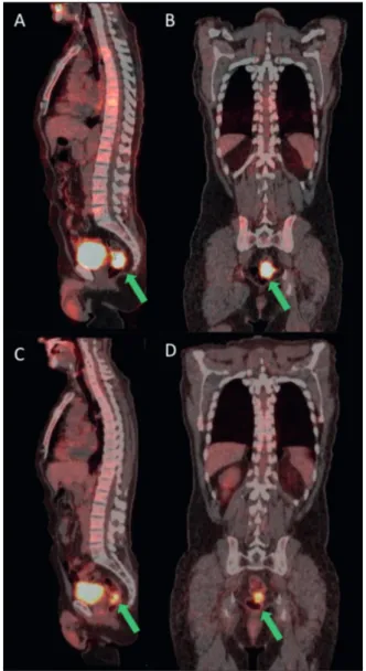
According the VRA, 12/58 patients (20.7%) were considered CR, 36/58 patients (62.1%) PR, 4/58 patients (6.9%) PD and 6/58 patients (10.3%) SD. Then, 48/58 patients (17.2%) were considered “VRA Responders” and 10/58 patients (82.8%) “VRA Non-responders”. Description of the se-miquantitative parameters in the whole popula-tion and in subgroups are reported on Table II. There were no differences statistically significant for semiquantitative parameters among CR, PR, SD and PD and between “VRA Responders” and “VRA Non-responders” (p> 0.05). An exemplar case of a PR patient is showed on Figure 1.
Pathological Response Evaluation
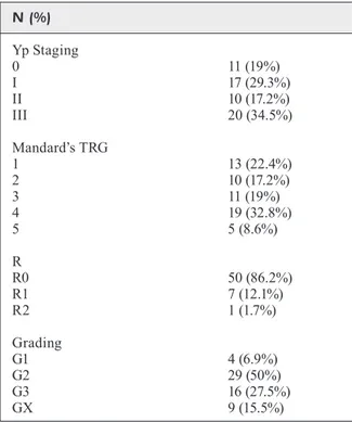
According to the TRG criterion, the surgi-cal specimen showed 23/58 “TRG responders” (39.7%) and 35/58 “TRG non-responders” (60.3%). According the (y)pTNM 10/58 patients (17.2%) were considered CR, 17/58 patients (29.3%) PR, 10/58 patients (17.2%) PD and 21/58 patients (36.3%) SD. Next, 27/58 patients (46.6%) resulted “(y)pTNM responders”, while 31/58 (53.4%) re-sulted “(y)pTNM non-responders”. All the patho-logical characteristics are described in Table III.
Survival Characteristics
Among the 58 patients at the end of the obser-vation, 46/58 patients (79.3%) were alive and 12/58 (20.7%) were death. Furthermore 33/58 patients (56.9%) had relapse: 22/33 patients had relapse at the 3 month follow up (at rectal anastomosis in 7 patients, liver in 5, lungs in 7, lymph nodes, bones and peritoneum in 1 patient respectively); 2/33 at the 6 month follow up (rectal anastomosis); 5/33 at the 12 month follow up (rectal anastomosis in 3, liver and lungs in 1 patient respectively); 4/33 at the 24 month follow up (rectal anastomosis, liver, lungs and lymph nodes in 1 patient respectively).
Exemplar cases of patients with rectal relapses and distant metastases onset in follow up are re-ported in Figure 2 and 3. Median follow up was
63 months (range 3-96). Kaplan Meier curves showed a global OS of 83.51 months (SD 3.30) and DSF of 45.72 months (SD 5.79). About the semiquantive parameters, only age is significant-ly related to the OS (OR=1.123 p=0.007). All the remnant parameters are not related neither to the OS and DSF. Cox test showed neither of variables and semiquantitative parameters have significant
correlation. Regarding metabolic 18F-FDG PET/
CT response evaluation, log rank test did not show statistical difference for OS and DFS neither for
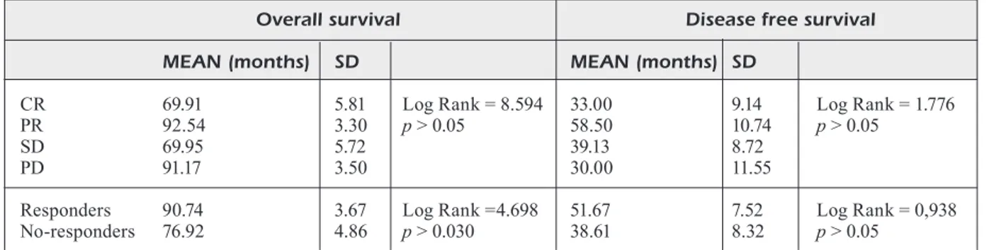
the distinction in “VRA responders” and “VRA non-responders”, neither for the distinction in CR, PD, PR and SD. Results are reported in Table IV. In relation to pathological Response Evalua-tion by TRG, log rank test did not show statistical difference for OS and DFS for the distinction in “TRG responders” and “TRG non-responders”. Results are reported in Table V.
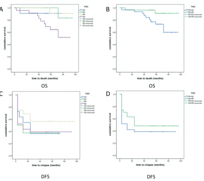
About pathological response evaluation by (y) pTNM, log rank test showed statistical difference for OS both for the distinction in “(y)pTNM re-sponders” and “(y)pTNM non-rere-sponders” and for the distinction in CR, PD, PR and SD (p=0.030). For DFS no statistical significance was found in difference between “(y)pTNM responders” and “(y)pTNM non-responders” and among CR, PD, PR and SD. The survival curves are showed in Figure 4 and results are reported in Table VI.
Discussion
The nCRT treatment led to a better outcome of patients with LARC with increased 5-year survi-val rate; the most recent and wide study with 336 LARC patients showed 73.5% survivors followed
for a mean of 60.4 months9. Survival seems to
be related to the response to therapy, in particu-lar it is greater if the therapy has led to complete tumor eradication then the lack of any patholo-gical change or even tumor progression despite
the nCRT10. The tumor response to nCRT varies
considerably among patients, the complete disap-pearance of the tumor is reached in about 20% of
cases11. In our work, 17.2% of patients achieved
a complete pathological response (ypT0N0M0). Assessment of disease parameters as potential predictors of the long-term outcome of patients with LARC undergoing nCRT can help in iden-tifying patients to whom conservative surgery might be offered11. Despite various attempts at
Table I. Baseline characteristics of study population. No. of patients 58
Sex
Male 39 (67.2%)
Female 19 (32.8%)
Age (years old) Mean 66 (range: 38-89) Clinical staging
II 33 (56.9%)
III 25 (43.1%)
Figure 1. 70-year-old male with a vegetans lesion at 7 cm from the anal verge. Baseline ¹8F-FDG PET/CT sagittal (A)
and coronal (B) images show the rectal lesion, SUVmax: 17.2 (green arrows). The post nCRT ¹8F-FDG PET/CT
sa-gittal (C) and coronal (D) images show reduction of rectal lesion size and pathological uptake, SUVmax: 10.6 (green arrows). Patient was classified as PR and his OS was 80 months and a liver lesion onset at the 24-month follow up.
predicting response and long-term outcome
ba-sed on molecular profiling of tumors, there are no markers adequately validated about such as probiotics and bioactive molecules for this pur-Table II. 18F-FDG PET/CT semi-quantitative parameters.
Mean (±SD) All VRA VRA
patients responders non-responders CR PR SD PD SUV maxbaseline 18.37 (±8.91) 20.17 (±8.57) 9.72 (±4.27) 22.37 (±9.53) 19.44(±8.24) 9.65(±3.32) 9.82 (±6.04)
SUV meanbaseline 9.35 (±4.3) 10.30 (±4.05) 4.76 (±2.10) 11.64(±4.67) 9.86(±3.79) 4.73(±1.79) 4.80 (±2.80)
MTVbaseline 24.81 (±13.7) 26.12 (±13.80) 18.50 (±11.83) 26.93 (±16.39) 25.85(±13.08) 19.48(±6.61) 17.04 (±18.49)
TLGbaseline 247.23 (±221.20) 20.17 (±8.57) 100.80 (±110.41) 356.64 (±306.30) 251.43(±191.88) 90.89(±48.73) 115.67 (±179.24)
SUV maxpost-nCRT 6.75 (±3.1) 6.28(±2.88) 8.99(±3.47) 3.30 (±1.75) 7.28(±2.48) 7.58(±3.90) 11.10 (±0.90)
SUV meanpost-nCRT 3.11 (±1.53) 2.88 (±1.39) 4.24(±1.75) 1.51 (±0.81) 3.33(±1.24) 3.48(±1.89) 5.37 (±0.61)
MTVpost-nCRT 23.17 (±15.20) 23.43 (±16.44) 21.92 (±6.99) 34.19 (±23.31) 19.84(±11.80) 21.88(±6.68) 21.98 (±8.49) TLGpost-nCRT 70.54 (±57.42) 70.84 (±61.05) 69.08 (±37.62) 80.55 (±63.73) 67.61(±60.71) 76.05(±43.23) 58.63 (±29.80) RI max% 58.02 (±22.07) 63.55 (±17.71) 31.45 (±22.36) 73.80 (±11.67) 60.13(±18.17) 26.76(±23.67) 38.48 (±21.36) RI mean% 61.05 (±24.17) 66.96 (±19.42) 32.65 (±25.44) 78.33 (±9.63) 63.17(±20.45) 29.45(±26.70) 37.44 (±26.51) ΔMTV 26.23 (±31.48) 65.58 (±31.95) 12.21 (±26.09) 24.13 (±34.72) 30.83(±31.32) 8.67(±21.24) 17.52 (±35.05) ΔTLG 53.18 (±34.61) 59.99 (±13.80) 20.50 (±29.28) 57.16 (±36.48) 60.94(±30.65) 18.36(±23.49) 23.70 (±40.36) Figure 2. 65-year-old female VRA classified as PR; TRG was II and ypTNM was I. ¹8F-FDG PET/CT MIP (A), coronal (B) and
axial (C) images performed after nCRT showed a small rectal lesion with SUVmax of 5.4. Follow up at 3 months ¹8F-FDG PET/
tes death and progression is associated with an
increased 18F-FDG uptake. A recent systematic
review23 including five studies demonstrated that
a CR on 18F-FDG PET/CT after nRCT is
predi-pose12,13. Purely morphological diagnostic
techni-ques are not able to predict treatment response because anatomical changes onset lately than
functional tissues modifications14-16. It is well
known the role of 18F-FDG PET/CT in the
mana-gement of oncological patients and there is also a growing consensus in its usefulness in evaluating the response to therapies in several oncological
diseases17. Furthermore, there is a growing
num-ber of studies18-20 that have investigated the
pro-gnostic value of 18F-FDG PET/CT. About LARC,
since there is a certain degree of heterogeneity in the applied methodology, literature reports different results, not directly comparable1. This
difference is also observed because the
qualitati-ve assessment of 18F-FDG PET/CT images can be
associated with the analysis of semiquantitative parameters. To date these parameters include not only SUVs, that are related to increased tumor aggressiveness and poor long-term prognosis, but also MTV and TLG, which are more
representa-tive of the entire tumor burden7,21. Their role in
predicting outcome is still under discussion and we did not relate them to OS and DFS because we did not find significant statistical differences
among the different groups. de Geus-Oei et al22
showed that the chemotherapy-induced changes in glucose metabolism of the lesion are highly predictive for patient outcome: an increase in
ra-Table III. Pathological characteristics of the study population. N (%) Yp Staging 0 11 (19%) I 17 (29.3%) II 10 (17.2%) III 20 (34.5%) Mandard’s TRG 1 13 (22.4%) 2 10 (17.2%) 3 11 (19%) 4 19 (32.8%) 5 5 (8.6%) R R0 50 (86.2%) R1 7 (12.1%) R2 1 (1.7%) Grading G1 4 (6.9%) G2 29 (50%) G3 16 (27.5%) GX 9 (15.5%)
Figure 3. 70-year-old male, VRA classified as CR. Follow up ¹8F-FDG PET/CT at 3 months: MIP (A), axial (B, C, E) and
ctive of OS, but not of DFS. In our analysis, CR patients had a better OS and DFS only compa-red with PD patients, but this difference was not statistically significant, even when we compare OS and DFS in “VRA Responders” and “VRA Non-responders”. However, we must keep in mind the limited number of events (cancer dea-ths) observed in this small patient population that likely restrict the statistical power of these data. Literature reports that the pathologic stage ((y) pTNM) and the extent of residual cancer (TRG) at completion of nCRT better correlate with pro-gnosis than the clinical stage11,24. For this reason,
we chose to evaluate survival in the 3 different restaging classification systems, based on focu-sing only on primary lesion (TRG), loco-regional
evaluation (ypTNM) and whole-body 18F-FDG
PET/CT evaluation (VRA). Our work
demon-strated a higher percentage of patients with TRG complete response (39.7%) compared to literature (24.6%), with longer OS and DFS in responders even if without statistically significant differen-ces. The rate of (y)pTNM CR of our study was lower than literature (17.2% vs. 22.6%), and OS and DFS in CR patients was shorter than the other groups; otherwise, OS and DFS in PR pa-tients were the longest. These results show that (y)pTNM, not providing total body information, is not a complete evaluation system for predicting outcome. The statistically significant difference in OS between “(y)pTNM responders” and “(y)
pTNM non-responders” is a further confirm 24.
The difference in OS and DFS between respon-ders and non-responder groups was higher accor-ding to TRG and (y)pTNM compared to VRA, this is probably due to the inserting of PR pa-Table VI. Survival according VRA evaluation.
Overall survival Disease free survival
MEAN (months) SD MEAN (months) SD
CR 69.91 5.81 Log Rank = 8.594 33.00 9.14 Log Rank = 1.776
PR 92.54 3.30 p > 0.05 58.50 10.74 p > 0.05
SD 69.95 5.72 39.13 8.72
PD 91.17 3.50 30.00 11.55
Responders 90.74 3.67 Log Rank =4.698 51.67 7.52 Log Rank = 0,938 No-responders 76.92 4.86 p > 0.030 38.61 8.32 p > 0.05
Table IV. Survival according VRA evaluation.
Overall survival Disease free survival
MEAN (months) SD MEAN (months) SD
CR 66.56 6.63 Log Rank = 2.872 39 10.45 Log Rank = 3.802
PR 86.30 3.75 p > 0.05 46.5 7.4 p > 0.05
SD 72.96 10.74 61.66 16.93
PD 59.50 6.02 10.5 4.97
Responders 83.44 3.64 Log Rank =0.00 46.25 6.38 Log Rank = 0,037 No-responders 79.35 7.38 p > 0.05 41.2 13.002 p > 0.05
Table V. Survival according TRG evaluation.
Overall survival Disease free survival
MEAN (months) SD MEAN (months) SD
Responders 72.51 4.08 Log Rank = 0.049 47.35 7.44 Log Rank = 1.727 No-responders 83.36 4.10 p > 0.05 39.5 7.39 p > 0.05
tients in “VRA responders”; this choice is related to the possibility of fibrotic tissue keeping weak
18F-FDG uptake for long time. The small number
of our sample is a limit of the study, but the ex-tended patient follow-up (median 91 months) is a feature rarely found in other reports and strength of our study.
Conclusions
We highlight the potential role of 18F-FDG in
assisting physicians on personalized decision.
The predictive and prognostic value of 18F-FDG
PET/CT may be pivotal in the selective risk-adap-ted treatment strategy and to schedule the correct follow-up approach.
Conflict of Interest
The Authors declare that they have no conflict of interest.
Acknowledgment
Dino Rubini for imaging technical support.
Author Contributions
A.N.A. conceived the study and contributed to data analysis and interpretation, and manuscript revision; C.F., R.D.L. and V.L. gave a scientific contribution for data interpretation; A.C wrote the first draft of the manuscript and supervised diagnos-tic imaging procedure; A.B. and F.I. contributed to the manu-script revision and bibliographic research; M.S. and G.R. made substantial contributions to the conception and design of the study, diagnosis and coordination, supervised the manuscript and gave final approval of the version to be published. All the authors have read and approved the final manuscript.
Figure 4. Survival curves for (y)pTNM evaluation. (A) OS in CR, PR, SD and PD; (B) OS in “(y)pTNM responders” and “(y) pTNM non-responders”; (C) DFS in CR, PR, SD and PD; (D) DFS in “(y)pTNM responders” and “(y)pTNM non-responders”.
References
1) AvAllone A, Aloj l, CArACò C, Delrio P, PeCori B, TA -TAngelo F, SCoTT n, CASAreTTi r, Di gennAro F, Mon -TAno M, SilveSTro l, BuDillon A, lASToriA S. Early
FDG PET response assessment of preoperative radiochemotherapy in locally advanced rectal cancer: correlation with long-term outcome. Eur J Nucl Med Mol Imaging 2012; 39: 1848-1857. 2) roMBouTS AjM, Hugen n, elFerink MAg, nAgTegAAl
iD, De WilT jHW. Treatment interval between
neo-adjuvant chemoradiotherapy and surgery in rectal cancer patients: a population-based study. Ann Surg Oncol 2016; 23: 3593-3601.
3) ruBini g, AlTini C, noTAriSTeFAno A, MerenDA n, ru -Bini D, iAnorA AA, ASABellA An. Role of 18F-FDG
PET/CT in diagnosing peritoneal carcinomatosis in the restaging of patient with ovarian cancer as compared to contrast enhanced CT and tumor marker Ca-125. Rev Esp Med Nucl Imagen Mol 2014; 33: 22-27.
4) lee Sj, kiM jg, lee SW, CHAe YS, kAng BW, lee Yj,
PArk jS, CHoi gS. Clinical implications of initial
FDG-PET/CT in locally advanced rectal cancer treated with neoadjuvant chemoradiotherapy. Cancer Chemother Pharmacol 2013; 71: 1201-1207.
5) lArSon SM, erDi Y, AkHurST T, MAzuMDAr M, MACAPin -lAC HA, Finn rD, CASillA C, FAzzAri M, SrivASTAvA n,
Yeung HW, HuMM jl, guilleM j, DoWneY r, kArPeH
M, CoHen Ae, ginSBerg r. Tumor treatment
respon-se barespon-sed on visual and quantitative changes in global tumor glycolysis using PET-FDG imaging. The visual response score and the change in total lesion glycolysis. Clin Positron Imaging 1999; 3: 159-171.
6) niCColi-ASABellA A, AlTini C, De luCA r, FAnelli M,
ruBini D, CAliAnDro C, MonTeMurro S, ruBini g.
Pro-spective analysis of 18F-FDG PET/CT predictive value in patients with low rectal cancer treated with neoadjuvant chemoradiotherapy and con-servative surgery. Biomed Res Int 2014; 2014: 952843.
7) AlTini C, niCColi ASABellA A, De luCA r, FAnelli M,
CAliAnDro C, QuArTuCCio n, ruBini D, CiSTAro A,
MonTeMurro S, ruBini g. Comparison of (18)F-FDG
PET/CT methods of analysis for predicting re-sponse to neoadjuvant chemoradiation therapy in patients with locally advanced low rectal cancer. Abdom Imaging 2015; 40: 1190-1202.
8) MAnDArD AM, DAliBArD F, MAnDArD jC, MArnAY j,
HenrY-AMAr M, PeTioT jF, rouSSel A, jACoB jH, Segol
P, SAMAMA g, eTAl. Pathologic assessment of tumor
regression after preoperative chemoradiotherapy of esophageal carcinoma. Clinicopathologic cor-relations. Cancer 1994; 73: 2680-2686.
9) Akgun e, ozkok S, Tekin M, YolDAS T, CAliSkAn C, koSe
T, kArABuluT B, SezAk M, elMAS n, ozuTeMiz o. The
effects of chemoradiotherapy on recurrence and survival in locally advanced rectal cancers with curative total mesorectal excision: a prospective, nonrandomized study. World J Surg Oncol 2017; 15: 205.
10) AvAllone A, Delrio P, PeCori B, TATAngelo F, PeTril -lo A, SCoTT n, MArone P, Aloi l, SAnDoMeniCo C,
lASToriA S, iAFFAioli vr, SCAlA D, ioDiCe g, BuDillon
A, CoMellA P. Oxaliplatin plus dual inhibition of
thy-midilate synthase during preoperative pelvic ra-diotherapy for locally advanced rectal carcinoma: long-term outcome. Int J Radiat Oncol Biol Phys 2011; 79: 670-676.
11) CAPirCi C, ruBello D, CHieriCHeTTi F, CrePAlDi g, FAnTi
S, MAnDoliTi g, SAlviATo S, Boni g, rAMPin l, Poli -Co C, MAriAni g. Long-term prognostic value of
18F-FDG PET in patients with locally advanced rectal cancer previously treated with neoadjuvant radiochemotherapy. AJR Am J Roentgenol 2006; 187: W202-W208.
12) TATullo M, SiMone gM, TArullo F, irlAnDeSe g, viTo
D, MArrelli M, SAnTACroCe l, CoCCo T, BAllini A,
SCACCo S. Antioxidant and antitumor activity of a
bioactive polyphenolic fraction isolated from the brewing process. Sci Rep 2016 27; 6: 36042. 13) BAllini A, SAnTACroCe l, CAnTore S, BoTTAliCo l, Di
-PAlMA g, De viTo D, gArgiulo C, SAini r, inCHingolo
F. Probiotics efficacy on oxidative stress values in inflammatory bowel disease: a randomized dou-ble-blinded placebo-controlled pilot study. Endo-cr Metab Immune Disord Drug Targets 2018 (in press). PMID: 29692270.
14) STABile iAnorA AA, loruSSo F, niCColi ASABellA A, Di
MAggio P, Fonio P, loSCo M, ruBini g. [Multidetector
CT for the assessment of the groin region]. Re-centi Prog Med 2012; 103: 483-488.
15) niCColi-ASABellA A, noTAriSTeFAno A, ruBini D, Al -Tini C, FerrAri C, MerenDA n, FAnelli M, ruBini g.
18F-FDG PET/CT in suspected recurrences of epithelial malignant pleural mesothelioma in asbestos-fibers-exposed patients (comparison to standard diagnostic follow-up). Clin Imaging 2013; 37: 1098-1103.
16) ABAkAY A, koMek H, ABAkAY o, PAlAnCi Y, ekiCi F,
TekBAS g, TAnrikulu AC. Relationship between
18F-FDG PET/CT findings and survival of 177 pa-tients with malignant pleural mesothelioma. Eur Rev Med Pharmacol Sci 2013; 17: 1233-1241. 17) FerrAri C, niCColi ASABellA A, MerenDA n, AlTini C,
FAnelli M, Muggeo P, De leonArDiS F, Perillo T, SAn -Toro n, ruBini g. Pediatric hodgkin lymphoma:
predictive value of interim 18F-FDG PET/CT in therapy response assessment. Medicine (Balti-more) 2017; 96: e5973.
18) CiSTAro A, niCColi ASABell A A, CoPPolino P, QuAr -TuCCio n, AlTini C, CuCinoTTA M, Alongi P, BAlMA
M, SAnFiliPPo S, BuSCHiAzzo A, PiCCArDo A, FAnelli
M, SAMBuCeTi g, BoMAnji j, BAlDAri S, BiSi g, FAnTi
S, ruBini g. Diagnostic and prognostic value of
18F-FDG PET/CT in comparison with morpho-logical imaging in primary adrenal gland mali-gnancies - A multicenter experience. Hellenic Journal of Nuclear Medicine. Hell J Nucl Med 2015; 18: 97-102.
19) grASSeTTo g, CAPirCi C, MArzolA MC, rAMPin l,
CHonDrogiAnniS S, MuSTo A, CrePAlDi g, MiniCozzi
pro-gnostic role of 18F-FDG-PET/CT. Abdom Imaging 2012; 37: 575-579.
20) nuvoli S, Fiore v, BABuDieri S, gAlASSi S, BAgellA P,
SolinAS P, SPAnu A, MADeDDu g. The additional role
of 18F-FDG PET/CT in prosthetic valve endocar-ditis. Eur Rev Med Pharmacol Sci 2018; 22: 1744-1751.
21) urSino S, FioriCA F, ColoSiMo C, MiCuCCi M, STeFAnelli
A, BerreTTA M, PAnAreo S, De BiASi v, Feggi lM, zini g,
CArTei F. Metabolic responses in non-small cell lung
cancer after hypofractionated stereotactic radiothe-rapy PET and hypofractionated radiotheradiothe-rapy. Eur Rev Med Pharmacol Sci 2012; 16: 755-762.
22) De geuS-oei lF, vAn lAArHoven HWM, viSSer eP, Her -MSen r, vAn Hoorn BA, kAMM Yj, krABBe PF, CorSTenS
FH, PunT Cj, oYen Wj. Chemotherapy response
evaluation with FDG PET in patients with colo-rectal cancer. Ann Oncol 2007; 19: 348-352. 23) krug B, CroTT r, De CAnniere l, D’HonDT l, vAnDer
BorgHT T. A systematic review of the predictive
va-lue of 18F-fluoro-2-deoxyglucose positron emis-sion tomography on survival in locally advanced rectal cancer after neoadjuvant chemoradiation. Colorectal Dis 2013; 15: e627-633.
24) leCCiSoTTi l, gAMBACorTA MA, De WAure C, STeFAnelli
A, BArBAro B, veCCHio FM, CoCo C, PerSiAni r, Cru -CiTTi A, TorTorelli AP, giorDAno A, vAlenTini v. The
predictive value of 18F-FDG PET/CT for asses-sing pathological response and survival in locally advanced rectal cancer after neoadjuvant radio-chemotherapy. Eur J Nucl Med Mol Imaging 2015; 42: 657-666.