Introduction
Partum-related hemorrhage is the leading cause of morbidity and mortality among pregnant women resulting in 140,000 deaths each year.1Although the majority of these deaths occur in low income countries, excessive bleeding at delivery is also frequent in high resource countries. In Australia, Canada and the US, 3-7% of deliveries in 2005 were complicated by pri-mary postpartum hemorrhage.2
The bleeding risk is expected to be higher in women with inherited thrombocytopenias (ITs) because of low platelet counts and associated defects of platelet function in some dis-orders. Nevertheless, few studies have investigated this topic
and there is little evidence to guide management of pregnancy and delivery.3Fetal and neonatal outcomes are also not well-described.
One of the forms of IT most investigated with respect to pregnancy is biallelic Bernard-Soulier Syndrome (bBSS), proba-bly because of its early identification in the middle of the last century. A systematic review of the literature published in 2010 identified 16 relevant articles, all case reports, describing 30 deliveries in 18 bBSS women.4Excessive bleeding occurred in 18 cases of which blood transfusion was required in 15. All women survived, but 2 required emergency hysterectomy. Concerning the fetus, there was one intra-uterine death caused by gastrointestinal bleeding and one neonatal death due to
©2014 Ferrata Storti Foundation. This is an open-access paper. doi:10.3324/haematol.2014.105924 Manuscript received on February 17, 2014. Manuscript accepted on April 22, 2014.
Correspondence: [email protected]
Pregnancy in women with inherited thrombocytopenias is a major matter of concern as both the mothers and the newborns are potentially at risk of bleeding. However, medical management of this condition cannot be based on evidence because of the lack of consistent information in the literature. To advance knowledge on this matter, we performed a multicentric, retrospective study evaluating 339 pregnancies in 181 women with 13 different forms of inherited thrombocytopenia. Neither the degree of thrombocytopenia nor the severity of bleeding tendency wors-ened during pregnancy and the course of pregnancy did not differ from that of healthy subjects in terms of miscar-riages, fetal bleeding and pre-term births. The degree of thrombocytopenia in the babies was similar to that in the mother. Only 7 of 156 affected newborns had delivery-related bleeding, but 2 of them died of cerebral hemorrhage. The frequency of delivery-related maternal bleeding ranged from 6.8% to 14.2% depending on the definition of abnormal blood loss, suggesting that the risk of abnormal blood loss was increased with respect to the general pop-ulation. However, no mother died or had to undergo hysterectomy to arrest bleeding. The search for parameters predicting delivery-related bleeding in the mother suggested that hemorrhages requiring blood transfusion were more frequent in women with history of severe bleedings before pregnancy and with platelet count at delivery below 50 x 109/L.
Analysis of 339 pregnancies in 181 women with 13 different
forms of inherited thrombocytopenia
Patrizia Noris,1Nicole Schlegel,2Catherine Klersy,3Paula G. Heller,4Elisa Civaschi,1Nuria Pujol-Moix,5Fabrizio Fabris,6
Remi Favier,7,8Paolo Gresele,9Véronique Latger-Cannard,10,11Adam Cuker,12Paquita Nurden,13Andreas Greinacher,14
Marco Cattaneo,15Erica De Candia,16Alessandro Pecci,1Marie-Françoise Hurtaud-Roux,2Ana C. Glembotsky,4
Eduardo Muñiz-Diaz,17Maria Luigia Randi,6Nathalie Trillot,18Loredana Bury,9Thomas Lecompte,19,20
Caterina Marconi,21Anna Savoia,22,23and Carlo L. Balduini1on behalf of the European Hematology Association –
Scientific Working Group on Thrombocytopenias and Platelet Function Disorders
1Department of Internal Medicine, University of Pavia-IRCCS Policlinico San Matteo Foundation, Italy; 2National Reference Centre on
Inherited Platelet Disorders and Service d’Hématologie Biologique, CHU Robert Debré and Paris 7 Denis Diderot University, Paris, France; 3Service of Biometry and Statistics, IRCCS Policlinico San Matteo Foundation, Pavia, Italy; 4Institute of Medical Research Alfredo
Lanari, University of Buenos Aires, Argentina; 5Universitat Autònoma de Barcelona, Institut de Recerca Biomèdica Sant Pau, Spain; 6Department of Medicine-DIMED, University of Padova Medical School, Italy; 7AP-HP, Armand Trousseau Children’s Hospital,
Haematological Laboratory, French Reference Center for Inherited Platelet disorders, Paris, France; 8Inserm UMR1009, Villejuif, France; 9Department of Internal Medicine, University of Perugia, Italy; 10Centre de Compétence Nord-Est des Pathologies Plaquettaires from the
frame of the Reference French Centre, France; 11Service d’Hématologie Biologique, Centre Hospitalo-Universitaire, Nancy, France; 12Department of Medicine and Department of Pathology & Laboratory Medicine, Perelman School of Medicine, University of
Pennsylvania, Philadelphia, PA, USA; 13Plateforme Technologique et d’Innovation Biomédicale, Hôpital Xavier Arnozan, Pessac, France; 14Institut für Immunologie und Transfusionsmedizin, Greifswald, Germany; 15Medicina III, Ospedale San Paolo, Dipartimento di Scienze
della Salute, Università degli Studi di Milano, Italy; 16Servizio Malattie Emorragiche e Trombotiche, Istituto di Medicina Interna e
Geriatria, Policlinico Agostino Gemelli, Università Cattolica del Sacro Cuore, Roma, Italy; 17Immunohematology Department, Banc de
Sang i Teixits de Catalunya, Barcelona, Spain; 18Institut d’Hématologie-Transfusion, Pôle Biologie Pathologie Génétique, CHRU, Lille,
France; 19Département des Spécialités de Médecine, Service d’Hématologie, Hôpitaux Universitaires de Genève, Suisse; 20Université de
Genève, Faculté de Médecine, Suisse; 21Genetica Medica, Dipartimento di Scienze Mediche Chirurgiche, Policlinico Sant'Orsola-Malpighi,
University of Bologna, Italy; 22Department of Medical Sciences, University of Trieste, Italy; and 23Institute for Maternal and Child Health –
IRCCS Burlo Garofolo, Trieste, Italy
antepartum intracranial hemorrhage. On this basis, it has been concluded that bBSS is associated with a very high risk of serious bleeding in the mother and the neonate.
Some information on pregnancy outcome is available also for MYH9-related disease (MYH9-RD), one of the most frequent forms of IT. A recent review of the literature exam-ined 25 case reports and one case series describing a total of 75 pregnancies in 40 women.5Postpartum hemorrhage in the mother occurred in 4 cases, while no obvious bleeding complications were reported among the newborns. Based on these data, MYH9-RD does not seem to increase bleed-ing risk either in mothers or neonates.
Limited data on pregnancy outcomes have been provided for patients with mild to moderate thrombocytopenia due to monoallelic BSS (mBSS), in all cases induced by the p.Ala156Val substitution in GPIb alpha (Bolzano mutation).6 Overall, 20 women delivered 34 children with no excessive maternal or neonatal bleeding. Although the authors did not provide information on management of pregnancies and childbirths, this study suggests that women with mBSS have gestational outcomes similar to healthy subjects.
A moderate risk of bleeding during delivery has been reported in a series of subjects with thrombocytopenia induced by ANKRD26 mutations (ANKRD26-related thrombocytopenia, ANKRD26-RT).7Thirteen patients gave birth, either vaginally or by caesarean section, with bleed-ing complications in 3 women. No information was provid-ed on prophylactic treatment or neonatal outcomes.
The paucity of evidence presently available, therefore, indicates that maternal and neonatal bleeding risk during pregnancy and delivery may vary with different forms of IT, ranging from very severe to very mild. However, the evi-dence concerns only four ITs and largely derives from case reports or series of patients not specifically investigated with respect to pregnancy.
To remedy this scarcity of information, we performed a retrospective, multicentric study aimed at systematically collecting and analyzing pregnancy outcomes in a large series of patients with well-defined forms of IT.
Methods
Patients
This study was promoted by the Scientific Working Group on Thrombocytopenias and Platelet Function Disorders of the European Hematology Association (EHA) and was announced at the 2013 EHA Congress in Stockholm. Clinical centers that, based on scientific publications and/or personal knowledge, represent points of reference for ITs were invited to participate. Forty-five institutions took part in the study. They were asked to analyze their databases and extract data on pregnancies in women with ITs. A series of questions had to be answered for each enrolled case related to the base-line characteristics of the mother, the course of pregnancy, and the management and outcome of delivery, with particular attention to the bleeding events in the mother and the newborn.
Only women with diagnoses confirmed by genetic analysis were eligible for the study. As an exception, positivity of the immunofluorescence test for MYH9-related disease (MYH9-RD) was considered sufficient for making the diagnosis in patients where mutational screening was not available because of its very high sensitivity and specificity.8
Neonates for whom genetic screening was not available were considered affected from the same illness as the mother whenever
they were thrombocytopenic and the mother had a dominant dis-order. In case of recessive disorders, documentation that the par-ents were carriers of the disease was required.
The Institutional Review Board of the IRCCS Policlinico San Matteo Foundation of Pavia, Italy, approved the study protocol. The study was performed in accordance with the Declaration of Helsinki, and each center complied with local ethical rules.
Classification of bleeding
Because of the retrospective nature of the study, it was not pos-sible to use the most recent bleeding scores that are most suited to patients with primary hemostasis defects. Spontaneous bleeding tendency in the mother before pregnancy was, therefore measured by the World Health Organization bleeding scale: grade 0 indicates no bleeding; grade 1, petechiae; grade 2, mild blood loss; grade 3, gross blood loss; grade 4, debilitating blood loss.9
The amount of blood loss has been used by most authors to identify post-partum hemorrhages.10However, this parameter was
available for only a few patients, and we could not use it. Therefore, we identified two definitions for excessive bleeding at delivery: “excessive bleedings requiring blood transfusion” (EBBT), based on transfusion of platelets and/or red blood cells during or after delivery to treat bleeding (prophylactic platelet transfusions prior to delivery were not considered for definition of EBBT), and “all excessive bleedings” (AEB), which includes not only patients receiving platelets and/or red blood cells, but also subjects who did not receive blood products and were judged by the treating physi-cian as presenting larger than normal blood loss. Thus, EBBT is a narrow definition that is expected to identify the most serious blood loss, while AEB is a broader definition that also encompasses less severe bleedings.
The same criteria were used to define increased bleedings with previous surgery. Any type of bleeding in newborns was consid-ered abnormal and recorded. Miscarriage was defined as sponta-neous expulsion from the uterus of the products of conception before viability. Pre-term birth was defined as live birth of a neonate of less than 37 weeks gestational age. Platelet count was measured by cell counters available in each center.
Statistical analysis
Data were described as median and 25th-75th percentiles if
contin-uous and as counts if categorical. EBBT and AEB incidence and 95% confidence intervals (95%CI) were computed. Generalized linear regression models were used to compare platelet counts between and within groups; Huber-White robust standard errors were calculated to account for intra-patient correlation. Logistic regression was used to assess the association of a series of patients’ characteristics with bleeding; Odds Ratios (OR) and 95%CI were computed. ROC curve analysis was used to identify the optimal cut offs for the association of platelets counts and bleedings. Stata 13 (StataCorp, College Station, TX, USA) was used for computa-tion. Two-sided P<0.05 was considered statistically significant.
Results
Not all data were available for all investigated patients, and results described below report the number of investi-gated cases for which data were available. In case the num-ber of investigated patients is not reported, it is intended that all patients were studied.
Womens’ base-line characteristics
One hundred and eighty-one women with 13 different forms of IT who had a total of 339 pregnancies were inves-tigated retrospectively by 45 centers in 20 countries. In 169
Table 1. Diagnosis and base-line features of investigated women with different forms of inherited thrombocytopenia who had one or more pregnancies.
Disorder N. of Median N. of patients with a Median platelet WHO bleeding N. of N. of N. of N. of
(abb., women age in years previous diagnosis/ count before pregnancy scale*, N. patients patients patients patients
phenotype at diagnosis, medical treatment/ x109/L, 25th-75th of patients with a receiving receiving with EBBT with AEB
MIM number) 25th-75thpercentile splenectomy for percentile history of spontaneous surgery prophylactic at surgery at surgery
immune bleeding of grades platelet N. of evaluated N. of
thrombocytopenia 0-1-2-3-4 transfusions patients evaluated
N. of evaluated evaluated patients MYH9-related 98 31, 19-42 41/32/11 40, 23-64 39-21-30-5-3 67 26 (67) 9 (67) 20 (67) disease (MYH9-RD, 600208) ANKRD26-related 23 21, 11-40 8/6/3 54, 30-84 6-4-7-3-3 12 2 (11) 0 (11) 1 (11) thrombocytopenia (ANKRD26-RT, 188000) Biallelic 13 33, 26-36 3/3/1 50, 23-92 5-0-5-2-1 8 1 (7) 1 (7) 2 (7) Bernard-Soulier syndrome (bBSS, 231200) Monoallelic 24 31, 19-41 1/1/0 86, 77-111 12-4-3-5-0 13 1 (13) 0 (13) 3 (13) Bernard-Soulier syndrome (mBSS, 231200) ACTN1-related 9 21, 17-40 0/0/0 77, 65-94 2-3-2-2-0 4 0 (4) 0 (4) 2 (4) thrombocytopenia (ACTN1-RT, 615193) Familial platelet 4 37, 26-38 1/1/0 116, 93-138 0-0-3-0-1 2 0 (2) 0 (2) 1 (2) disorder and predisposition
to acute myelogenous leukemia (FPD/AML, 601399)
ITGB3-related 3 16, 11-37 0/0/0 78, 65-114 1-0-0-2-0 3 3 (3) 0 (3) 0 (3)
thrombocytopenia (ITGB3-RT, 187800)
Platelet-type 2 27, 27-28 1/0/0 80, 30-130 0-1-0-0-1 1 0 (1) 0 (1) 1 (1)
von Willebrand disease (VWDP, 177820) Gray platelet 1 13 1/1/0 65 0-0-0-1-0 1 1 (1) 0 (1) 0 (1) syndrome (GPS, 139090) FLNA-related 1 23 0/0/0 43 1-0-0-0-0 1 0 (1) 0 (1) 0 (1) thrombocytopenia (FLNA-RT, nd) TUBB1-related 1 4 1/0/0 100 0-1-0-0-0 1 0 (1) 0 (1) thrombocytopenia (TUBB1-RT, 613112) 0 (1) Velocardiofacial 1 34 0/0/0 75 0-1-0-0-0 1 0 (1) 0 (1) 0 (1) syndrome (VCFS, 192430) CYCS-related 1 25 0/0/0 35 0-1-0-0-0 1 0 (1) 0 (1) 0 (1) thrombocytopenia (CYCS-RT, 612004) Total 181 29 (19-39) 57/44/15 57 (30-82) 66-36-50-20-9 114 34 (112) 10 (112) 30 (112)
*World Health Organization bleeding scale: grade 0, no bleeding; grade 1, petechiae; grade 2, mild blood loss; grade 3, gross blood loss; grade 4, debilitating blood loss; EBBT: excessive bleeding requiring blood transfusion; AEB: all excessive bleedings; 25th-75th: 25th-75thpercentiles.
women, diagnoses were confirmed by the identification of mutations in the candidate genes, while in 12 subjects a diagnosis of MYH9-RD was made on the basis of a positive immunofluorescence test for the distribution of MYH9 pro-tein in neutrophils.8Of note, 18 of 24 women with the diag-nosis of mBSS had the p.Ala156Val substitution in GPIb alpha (Bolzano mutation).6
Patients’ features before pregnancy are reported in Table 1. A feature common to many investigated patients was the difficulty of making a correct diagnosis of IT, as shown by the observation that 57 subjects were initially misdiagnosed
with immune thrombocytopenia. Forty-four of these women received unnecessary treatment, including splenec-tomy in 15 cases. Difficulties in diagnosis is also demon-strated by the finding that the inherited origin of thrombo-cytopenia was recognized in adulthood in most cases, with a mean age at diagnosis of 30 years. These findings under-score the need to maintain a high index of suspicion for ITs in the evaluation of patients with thrombocytopenia.11
Thrombocytopenia was on average moderate, with a mean platelet count of 60 x 109/L in the whole case series and a mean platelet count higher than 45 x 109/L in all
dis-orders for which more than one patient was examined. However, severely reduced platelet counts were observed in a few cases. In this regard, it is important to note that platelet counts were measured by electronic cell counters and that these instruments are known not to recognize very large platelets.12It is, therefore, expected that the degree of thrombocytopenia was overestimated in patients with inherited macrothrombocytopenias, especially those with
MYH9-RD and bBSS who typically have giant platelets.
Spontaneous bleeding tendency before pregnancy was on average mild. WHO grade 3 and 4 bleeding were reported in only 20 and 9 of 181 subjects, respectively.
Perioperative AEB and EBBT were reported in 30 and 10 of 112 women receiving surgery, respectively.
Gestations
No variation in severity of the pre-existing bleeding ten-dency was reported during pregnancy (data not shown).
The course of gestation was uneventful in 304 cases, while miscarriage was reported in 34 pregnancies (10.1%, 95%CI: 7.2-14.0). One therapeutic interruption of pregnan-cy because of fetal malformation was reported. The
inci-dence of miscarriage is superimposable to that observed in a large population of white women in the US (10.2%).13 Pregnancy loss occurred in the first trimester in 29 cases, in the second in 5 cases, and in the third in one case.
Pre-term birth was recorded for 30 pregnancies (9.9%, 95%CI: 6.8-13.7), and thus the frequency of this event was not significantly different from that observed in a general population of Western countries (7.4%).14 All pre-term deliveries occurred at a gestational age of 36 or 37 weeks.
Newborns
Diagnostic definition was possible in 278 newborns: 156 were affected by ITs, while 122 were unaffected.
Comparison of platelet counts in the 126 affected new-borns for which this information was available with base-line platelet counts in their mothers revealed that the degree of thrombocytopenia was similar, although the small observed difference was statistically significant (mean platelet count in the newborns 69 x 109platelets/L, in the mothers 58 x 109/L; P=0.020).
Bleeding signs consisting of petechiae were observed in 5 affected neonates, while fatal cerebral hemorrhages were
Table 2. Characteristics of pregnancy, delivery and newborns in women with inherited thrombocytopenias. The number of investigated patients is reported (in brackets) in case of missing data.
Disorders N. of Median Median N. of term/ N. of vaginal N. of N. of N. of N. of N. of N. of
pregnancies/ age in platelet preterm deliveries/ deliveries with deliveries withdeliveries with deliveries with healthy affected
miscarriages years at count at deliveries cesarean general/ prophylactic EBBT, % AEB, % newborns newborns
delivery, delivery (N. of sections spinal or platelet incidence, CI incidence, CI affected by with
25th-75th x109/L, evaluated (N. of epidural/no transfusion (N. of (N. of the mother bleedings
percentile 25th-75th deliveries) evaluated anesthesia (N. of evaluated evaluated disorder (N. of
percentile deliveries) (N. of evaluated deliveries) deliveries) (N. of evaluated
(N. of evaluated deliveries) evaluated babies)
evaluated deliveries) babies)
deliveries) MYH9-RD 185/21 28, 24-32 60, 34-80 142/20 94/69 54/34/73 31 13, 8.3, 4.5- 24, 15.3, 10.1-22 58/94 6 (94) (86) (162) (163) (163) (161) 13.8 (156) (156) (152) ANKRD26-RT 48/6 29, 25-30 34, 28-76 41/1 27/15 5/2/27 4 3, 7.1, 1.4- 5, 11.9, 17/23 0 (23) (26) (42) (42) (34) (42) 19.4 (42) 3.9-25.6 (42) (40) bBSS 22/1 32, 28-35 40, 26-65 16/5 8/13 9/4/7 6 1, 4.7, 0.1-23.8 3, 14.2, 3-36.3 12/3 (15) 0 (3) (15) (21) (21) (20) (21) (21) (21) mBSS 42/3 29, 25-34 86, 66-105 37/2 29/10 6/4/28 0 1, 2.5, 0.1-13.4 5, 12.8, 4.2- 19/18 1(18) (33) (39) (39) (38) (39) (39) 27.4 (39) (37) ACTN1-RT 18/1 24, 22-26 72, 55-88 17/0 17/0 0/0/17 0 0, 0, 0-19.5 0, 0, 0-19.5 6/11 0 (11) (11) (17) (17) (17) (17) (17) (17) FPD/AML 9/2 27, 21-29 92, 80-105 7/0 4/3 0/1/4 1 1, 14.2, 0.3- 1, 14.2, 0.3- 3/3 0(3) (4) (7) (7) (5) (7) 57.8 (7) 57.8 (7) (6) ITGB3-RT 3/0 28, 24-29 82, 58-110 (3) 3/0 (3) 0/3 (3) 1/0/0 (1) 2 (3) 0, 0, 0-70.7 (3) 0, 0, 0-70.7 (3) 1/2 (3) 0 (2) VWDP 5/0 30, 29-32 110, 58-113 (5) 4/1 (5) 3/2 (5) 2/0/3 (5) 0 (5) 1, 20, 0.5-71.6 (5)3, 60, 14.6-94.7 (5) 3/0 (3) 0 (0) GPS 3/1 25, 25-28 47, 40-55 (2) 2/0 (2) 2/0 (2) 2/0/0 (2) 2 (2) 0, 0, 0-84.1 (2) 1, 50, 1.2-98.7 (2) 2/0 (2) 0 (0) FLNA-RT 1/0 28 82 (1) 1/0 (1) 1/0 (1) 0/0/1 (1) 0 (1) 0, 0, 0-97.5 (1) 0, 0, 0-97.5 0/1 (1) 0 (1) (1) (1) TUBB1-RT 1/0 39 48 (1) 0/1 (1) 0/1 (1) 1/0/0 (1) 0 (1) 0, 0, 0-97.5 (1) 0, 0, 0-97.5 (1) 0/0 (0) 0 (0) VCFS 1/0 33 (0) 1/0 (1) 1/0 (1) 0/1/0 (1) 0 (1) 0, 0, 0-97.5 (1) 0, 0, 0-97.5 (1) 0/1 (1) 0 (1) CYSC-RT 1/0 28 50 (1) 1/0 (1) 1/0 (1) 0/0/1 (1) 0 (1) 0, 0, 0-97.5 (1) 0, 0, 0-97.5 (1) 1/0 (1) 0 (0) TOTAL 339/35 28, 25-32 65, 37-85 272/30 187/116 80/46/161 46 (301) 20, 6.7, 4.1- 42, 14.1, 10.4- 122/156 () 7 (156) (188) (302) (303) (289) 10.2 (296) 18.6 (296) 278
reported in 2 infants born by vaginal delivery to 2 MYH9-RD mothers with greatly reduced platelet counts (12 and 16 x 109/L, respectively). Platelet count was severely reduced in one newborn (7 x 10⁹/L) with the same MYH9 mutation as the mother, while neither testing for MYH9-RD nor a platelet count were carried out in the other neonate because he died shortly after birth.
Deliveries
Table 2 describes maternal characteristics at delivery as well as management of childbirth and bleeding events. Platelet count at delivery was available in 188 cases, and comparison with base-line non-pregnant platelet count revealed mild differences (mean platelet count before preg-nancy 56 x 109/L, at delivery 65 x 109/L; P=0.071). Thus, only limited changes in platelet count are expected to occur during pregnancy in patients with ITs.
Prophylactic platelet transfusions were given in prepara-tion for delivery in 46 of 301 evaluable cases, while other prophylactic treatments, consisting of steroids, desmo-pressin, tranexamic acid or recombinant activated factor VII, were given in 9, 5, 2 and one case, respectively.
Mean platelet count at delivery was lower in women given prophylactic platelet transfusions than in women not receiving this treatment (40 vs. 69 x 109/L; P<0.001). Similar results were obtained when baseline non-pregnant platelets counts were considered (38 vs. 65 x 109/L; P<0.001). This indicates that platelet count may have been a parameter used by physicians to determine the need for prophylactic platelet transfusion.
One hundred and sixteen of 303 births were by caesarean section. In the 289 deliveries for which information on pain control was available, general anesthesia was performed in 80 cases (27.7%), and spinal or epidural anesthesia in 46 (15.9%). No bleeding complications related to these proce-dures were reported.
AEB occurred in 42 cases (14.2% of deliveries, 95%CI: 10.4-18.7) and EBBT in 20 cases (6.8% of deliveries, 95%CI: 4.17-10.2). In case of EBBT, platelet transfusions were given in 6 deliveries, red cell transfusions in 9 and both red blood cell and platelet concentrates in 5. Since the incidence of abnormal bleeding at delivery in the normal population, as evaluated by the amount of blood loss, is 3-7% in high resource countries,2 the frequency of AEB appears to be higher in ITs than in the general population. Comparison of the need for red blood cell transfusion in our case series is possible with that observed in the general population: fif-teen deliveries in women with ITs required erythrocyte transfusions (5.06%, 95%CI: 2.8-8.0), while the need for this treatment ranged from 0.5 to 1.2% in non-thrombocy-topenic women.2,15,16 Altogether, these results indicate that bleeding risk at delivery is increased in ITs.
No women died from complications of childbirth or required hysterectomy to stop bleeding.
Correlations between mothers’ features and bleedings
at delivery
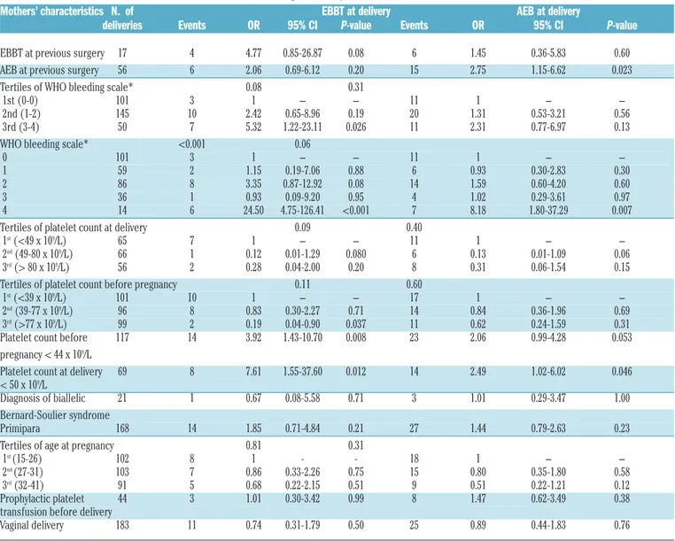
Some predictors of AEB and EBBT at delivery were iden-tified (Table 3). Considering EBBT at delivery, a trend towards statistically significant association was found with EBBT at previous surgery (OR 4.7, 95%CI: 0.8-26.9), while a significant correlation was found with a history of grade 3 or 4 (OR 5.32, 95%CI: 1.22-23.11) and grade 4 (OR 24.50, 95%CI: 4.75-126.41) of WHO bleeding scale.
EBBT was also significantly associated with a base-line
platelet count in the lowest tertile, and a trend towards sig-nificance was found between EBBT and platelet count in the lowest tertile at delivery (platelets >49 x 109/L). ROC analysis identified platelet counts of 44 and 50 x 109/L as the optimal cut offs of platelet counts before pregnancy and at delivery, respectively, for the identification of patients with a higher risk for EBBT. The incidence of EBBT in mothers with platelet counts below these cut-off values before preg-nancy and at delivery was 3.92 (95%CI: 1.43-10.70) and 7.61 (95%CI: 1.55-37.60) times higher, respectively, than in patients with higher platelet counts. Both differences were statistically significant.
EBBT was not less frequent in vaginal deliveries than in caesarean sections and was not reduced in women who received prophylactic platelet transfusions prior to delivery. Table 2 shows that the incidence of bleeding requiring blood transfusion was not significantly different in different forms of IT. However, this finding is reliable for MYH9-RD,
ANKRD6-RT, bBSS, mBSS and ACTN1-related
thrombocy-topenia (ACTN1-RT) because of the high number of inves-tigated deliveries, while it is not dependable for all other disorders because of the low number of observed child-births.
A history of surgical bleeding and lower platelet counts also correlated with AEB, though to a lesser extent than with EBBT. As with EBBT, other examined maternal char-acteristics did not predict AEB at delivery.
Discussion
Inherited thrombocytopenias have long been considered exceedingly rare, but recent advances have facilitated diag-nosis and greatly increased the number of reported patients.17Although population studies have not yet been performed, it has recently been calculated that the preva-lence of ITs in Italy is at least 2.7 in 100,000.18The improved knowledge of ITs has also changed our view of their clinical picture. It has been shown that bleeding tendency is mild in the majority of patients and spontaneous life-threatening hemorrhage is uncommon/rare.11 Nevertheless, hemostatic challenges always need careful attention. Pregnancy and delivery are especially critical because both mothers and affected newborns are at risk of hemorrhage. However, published data are insufficient to provide an evidence-based approach to management during pregnancy and delivery. The present study on 339 pregnancies in 181 women with 13 different ITs provides, for the first time, the opportunity to systematically examine this topic in detail.
Results of our study on the one hand suggest that the course of pregnancy in ITs does not differ from that of healthy women; on the other hand, the risk of bleeding with childbirth appears to be increased in both mothers and neonates.
Neither thrombocytopenia nor bleeding tendency wors-ened during pregnancy. The incidence of pregnancy loss was superimposable to that observed in healthy women, and the incidence of pre-term birth was not increased in women with ITs. No intrauterine bleeding was reported in the 156 fetuses who inherited IT. However, antepartum gastrointestinal19 and intracranial bleedings20 with fatal con-sequences have been previously described in the fetuses of 2 bBSS mothers. A risk of major bleeding in utero must, therefore, be borne in mind, though such events were not observed in our study cohort. Regarding delivery-related
neonatal bleedings, our data indicate that the risk is low, although fatal hemorrhages may occur. Indeed, only 7 of 156 affected newborns had bleeding. In 5 cases, bleeding consisted of petechiae, but 2 neonates, both born by vaginal delivery to mothers with severe thrombocytopenia due to
MYH9-RD, died of cerebral hemorrhage. To our
knowl-edge, delivery-related fatal intracranial hemorrhages have not been previously reported in neonates with ITs. The small number of neonates affected by ITs other than
MYH9-RD does not allow us to exclude a risk of severe
neonatal hemorrhage with these conditions, though none were observed in our series. Similarly, we cannot conclude that vaginal delivery is associated with a greater risk of neonatal hemorrhage, because this type of birth was chosen in the vast majority of MYH9-RD women, as well as in other forms of ITs. Thus, infants delivered vaginally by severely thrombocytopenic women with MYH9-RD must be considered at risk for intracranial bleeding; we cannot exclude the possibility that other forms of IT or caesarean section may expose newborns to similar risk. The incidence
of delivery-related maternal bleeding in our case series ranged from 6.8% to 14.2% depending on the criterion used for defining abnormal bleeding. Comparison with healthy women is difficult, because most commonly used definitions of partum-related hemorrhages in the general population rely on the amount of blood loss (>500 mL for vaginal deliveries, and >750-1500 mL for caesarean births),2 but this information was not available for the majority of our patients. Instead, we adopted two different criteria for increased bleeding: one based on the need to give blood products (EBBT), the other taking into account not only blood transfusions, but also the judgment of the obstetri-cian that bleeding was excessive (AEB). The former defini-tion may underestimate the occurrence of hemorrhages as defined by the entity of blood loss, because it is conceivable that not all women losing more than 500 mL of blood after vaginal delivery or more than 750 mL after caesarean sec-tion received blood transfusions. Also the latter definisec-tion risks underestimating the frequency of bleedings because it includes a subjective assessment and it has been shown that
Table 3. Correlations between mother characteristics and bleeding at delivery.
Mothers’ characteristics N. of EBBT at delivery AEB at delivery
deliveries Events OR 95% CI P-value Events OR 95% CI P-value
EBBT at previous surgery 17 4 4.77 0.85-26.87 0.08 6 1.45 0.36-5.83 0.60
AEB at previous surgery 56 6 2.06 0.69-6.12 0.20 15 2.75 1.15-6.62 0.023
Tertiles of WHO bleeding scale* 0.08 0.31
1st (0-0) 101 3 1 - - 11 1 -
-2nd (1-2) 145 10 2.42 0.65-8.96 0.19 20 1.31 0.53-3.21 0.56
3rd (3-4) 50 7 5.32 1.22-23.11 0.026 11 2.31 0.77-6.97 0.13
WHO bleeding scale* <0.001 0.06
0 101 3 1 - - 11 1 -
-1 59 2 1.15 0.19-7.06 0.88 6 0.93 0.30-2.83 0.30
2 86 8 3.35 0.87-12.92 0.08 14 1.59 0.60-4.20 0.60
3 36 1 0.93 0.09-9.20 0.95 4 1.02 0.29-3.61 0.97
4 14 6 24.50 4.75-126.41 <0.001 7 8.18 1.80-37.29 0.007
Tertiles of platelet count at delivery 0.09 0.40
1st(<49 x 109/L) 65 7 1 - - 11 1 -
-2nd(49-80 x 109/L) 66 1 0.12 0.01-1.29 0.080 6 0.13 0.01-1.09 0.06
3rd(> 80 x 109/L) 56 2 0.28 0.04-2.00 0.20 8 0.31 0.06-1.54 0.15
Tertiles of platelet count before pregnancy 0.11 0.60
1st(<39 x 109/L) 101 10 1 - - 17 1 -
-2nd(39-77 x 109/L) 96 8 0.83 0.30-2.27 0.71 14 0.84 0.36-1.96 0.69
3rd(>77 x 109/L) 99 2 0.19 0.04-0.90 0.037 11 0.62 0.24-1.59 0.31
Platelet count before 117 14 3.92 1.43-10.70 0.008 23 2.06 0.99-4.28 0.053
pregnancy < 44 x 109/L
Platelet count at delivery 69 8 7.61 1.55-37.60 0.012 14 2.49 1.02-6.02 0.046
< 50 x 109/L
Diagnosis of biallelic 21 1 0.67 0.08-5.58 0.71 3 1.01 0.29-3.47 1.00
Bernard-Soulier syndrome
Primipara 168 14 1.85 0.71-4.84 0.21 27 1.44 0.79-2.63 0.23
Tertiles of age at pregnancy 0.81 0.31
1st (15-26) 102 8 1 - - 18 1 -
-2nd (27-31) 103 7 0.86 0.33-2.26 0.75 15 0.80 0.35-1.80 0.58
3rd(32-41) 91 5 0.68 0.22-2.15 0.51 9 0.51 0.22-1.21 0.12
Prophylactic platelet 44 3 1.01 0.30-3.42 0.99 8 1.47 0.62-3.49 0.38
transfusion before delivery
Vaginal delivery 183 11 0.74 0.31-1.79 0.50 25 0.89 0.44-1.83 0.76
*World Health Organization bleeding scale: grade 0, no bleeding; grade 1, petechiae; grade 2, mild blood loss; grade 3, gross blood loss; grade 4, debilitating blood loss; EBBT: excessive bleed-ing requirbleed-ing blood transfusion; AEB: all excessive bleedbleed-ings; OR: Odds Ratio; CI: confidence intervals.
visual estimation tends to under-evaluate blood loss at delivery with respect to more accurate measurements.21 Since the incidence of increased bleeding at delivery in the general population ranges from 3% to 7%,2AEB in our case series was higher than normal, while EBBT was at the upper limit of normal range. However, the observation that red blood cell transfusions at delivery were given much more frequently in our patients (5% of deliveries) than in the general population (0.5-1.2%) strongly suggests that bleeding risk is increased in ITs.
Because of this, we searched for mothers’ parameters able to predict delivery-related bleedings. EBBT at childbirth was more frequent in women with higher grades of the WHO bleeding scale before pregnancy, and a trend toward statistical significance was found between EBBT at delivery and EBBT at previous surgeries. Also the degree of throm-bocytopenia before pregnancy and at delivery was related to EBBT, although with different degrees of statistical signif-icance. Based on this finding, we searched for the cut-off value of platelet count able to predict EBBT by ROC curve analysis, and found that platelet count less than 50 x 109/L at delivery and less than 44 x 109/L before pregnancy were significantly associated with higher frequency of hemor-rhage requiring blood products. Thus, the bleeding tenden-cy before pregnantenden-cy and the degree of thrombotenden-cytopenia are both important to predict severe bleeding at childbirth. Of note, experts’ recommendations for management of pregnancy in patients with immune thrombocytopenia identified platelet count 50 x 109/L as the minimum value required for safe delivery.22,23
Broadly similar results were obtained in the search for correlations between AEB at delivery in the mothers and their previous bleeding tendency as well as their platelet counts. However, the risk deriving from unfavorable fea-tures was lower and had lower statistical significance than for EBBT, probably because the definition of AEB includes a subjective judgment of milder bleeding events.
No other investigated maternal characteristics predicted increased bleeding at delivery. Based on the common notion that severe hemorrhages are frequent on the occa-sion of hemostatic challenges in patients with bBSS and the previously published data on delivery in these patients,4it is surprising that bleeding events at childbirth in our case series were no more frequent in bBSS than in other forms of IT. This unexpected finding could have several explana-tions. First, the base-line bleeding tendency in our bBSS women was milder than that observed in other series of unselected bBSS patients, as shown by the observation that WHO grade 3 and 4 bleeding were reported only in 3 of our 13 women, while they were described in 7 of 10 patients of a previous case series.24It may be that our group of bBSS patients was enriched in forms with mild bleeding tenden-cy because some women with severe bBSS have undergone hysterectomy for heavy menstruation or have been discour-aged from having a pregnancy by the fear of bleeding at delivery. Moreover, the frequency of prophylactic platelet transfusions in bBSS women was twice that of women with other ITs, and this may have masked a higher risk of bleeding. The observation that only one AEB (not requiring blood products) was reported in 15 deliveries of bBSS patients not preceded by prophylactic platelet transfusions supports the hypothesis that giving platelets in preparation for delivery may be unnecessary in women with mild forms of bBSS. Finally, the previous systematic review, which was based on case reports and, therefore, was highly
susceptible to reporting bias, could have overestimated bleeding risk at delivery.
Another surprising finding was the similar incidence of increased bleeding at delivery in mothers receiving or not receiving prophylactic platelet transfusions, in that this result might suggest that platelet transfusions were ineffec-tive. However, the observation that platelet count was lower in women given transfusions provides a credible alternative explanation and suggests that prophylactic platelet infusions were effective in reducing the incidence of hemorrhage.
Our study has some limitations. First, only a small num-ber of eligible patients with some forms of IT were identi-fied by the participating centers (Table 1), likely reflecting the rarity of these disorders. Creation of larger international registries is required to further improve knowledge of preg-nancy outcomes in these rare disorders. Another limitation derives from the failure of automated cell counters to iden-tify very large platelets and the resulting underestimation of platelet count in inherited macrothrombocytopenias, espe-cially bBSS and MYH9-RD.12It is thus likely that the degree of thrombocytopenia in these conditions was overestimat-ed in our study. Moreover, data were missing in a relevant number of patients enrolled in the study, a limitation inher-ent to retrospective investigations. Finally, we compared the outcome of pregnancy in women with ITs with that reported in general population. Comparison with non-thrombocytopenic women from these same institutions from the same time periods would be more appropriate, but obtaining the required information from 45 centers in 20 countries seemed to us a hopeless undertaking.
In conclusion, our study showed that delivery-related bleeding risk is higher in ITs than in the general population for both mothers, who may have blood loss requiring blood transfusions, and affected newborns, who may rarely pres-ent with fatal intracranial hemorrhage. Nevertheless, deliv-ery occurs without bleeding complications in the vast majority of mothers and neonates. Our study also identified the degree of thrombocytopenia and a history of severe bleeding tendency in the mother as potentially useful parameters to predict the risk of delivery-related bleedings.
Collaborators
In alphabetical order Sophie Bayart,1 Anne Bauters,2
Schéhérazade Benabdallah-Guedira,3 Françoise Boehlen,4-5
Jeanne-Yvonne Borg,6 Roberta Bottega,7 James Bussel,8
Daniela De Rocco,9Emmanuel de Maistre,10Michela Faleschini,9
Emanuela Falcinelli,11 Silvia Ferrari,12 Alina Ferster,13 Tiziana
Fierro,11 Dominique Fleury,14 Pierre Fontana,4-5 Chloé James,15
Francois Lanza,16 Véronique Le Cam Duchez,6 Giuseppe
Loffredo,17 Pamela Magini,18 Dominique Martin-Coignard,19
Fanny Menard,20 Sandra Mercier,21 Annamaria Mezzasoma,11
Pietro Minuz,22Ilaria Nichele,23Lucia D. Notarangelo,24Tommaso
Pippucci,18 Gian Marco Podda,25 Catherine Pouymayou,26 Agnes
Rigouzzo,27 Bruno Royer,28 Pierre Sie,29 Virginie Siguret,30Catherine
Trichet,31 Alessandra Tucci,32 Béatrice Saposnik,33 Dino Veneri22. 1Service d’Hémostase Bio-Clinique, Centre Régional de traitement
des maladies hémorragiques de Rennes-Bretagne, CHU de Rennes, Rennes, France; 2Institut d’Hématologie-Transfusion, Pôle
Biologie Pathologie Génétique, CHRU Lille, France; 3Université
Mohamed V Rabat, Faculté de Médecine et de Pharmacie, Hôpital
Avicenne, Rabat, Morocco; 4Division of Angiology and
Haemostasis, Department of Medical Specialisations, Faculty of Medicine and University Hospitals of Geneva, Geneva, Switzerland; 5Geneva Platelet Group, Faculty of Medicine,
University of Geneva, Geneva, Switzerland; 6CHU Charles
Nicolle, Unité Hémostase-Hématologie, Rouen, France; 7Institute
for Maternal and Child Health – IRCCS Burlo Garofolo, Trieste, Italy; 8Weill Medical College of Cornell University, New York,
NY, USA; 9Department of Medical Sciences, University of Trieste,
Trieste, Italy; 10Service d’hématologie Biologie, Centre
Hospitalo-Universitaire Dijon, France; 11Department of Internal Medicine,
University of Perugia, Perugia, Italy; 12Department of
Medicine-DIMED; University of Padova Medical School, Padova, Italy;
13Unité d’Hémato-Oncologie pédiatrique, Hôpital Universitaire
des Enfants Reine Fabiola, Bruxelles, Belgique; 14Service de
Néphrologie, CH Valenciennes, Valenciennes, France;
15Laboratoire d'Hématologie and National Reference Centre on
Inherited Platelet Disorders, CHU Haut Lévêque, Pessac, France;
16INSERM UMR-S949/EFS-Alsace, Strasbourg, France;
17Department of Oncology, Azienda Santobono-Pausilipon,
Pausilipon Hospital, Napoli, Italy; 18Genetica Medica,
Dipartimento di Scienze Mediche Chirurgiche, Policlinico Sant'Orsola-Malpighi - University of Bologna, Bologna, Italy;
19Laboratoire de Génétique, CH Le Mans, France; 20Centre
Hospitalier de la côte basque, Bayonne, France;21Service de
Génétique Clinique, Centre de Référence Anomalies du Développement du Grand Ouest, CHU Rennes-Hôpital Sud, Rennes, France; 22Department of Medicine and Haematology,
University Hospital of Verona, Verona, Italy; 23Department of Cell
Therapy and Hematology, San Bortolo Hospital, Vicenza, Italy;
24Department of Pediatrics, University of Brescia, Brescia, Italy;
25Medicina III, Ospedale San Paolo, Dipartimento di Scienze della
Salute, Università degli Studi di Milano, Italy; 26Laboratoire
d’Hématologie and National Reference Centre on Inherited Platelet Disorders, CHU La Timone, Marseille, France; 27AP-HP, Armand
Trousseau children Hospital, Department of Anesthesiology, Paris, France; 28Hématologie clinique et thérapie cellulaire, CHU Amiens,
France; 29Laboratoire d'Hématologie and National Reference
Centre of Inherited Platelet Disorders, CHU Rangueil, Toulouse, France; 30Service d' Hématologie Biologique, CHU Hôpital
Européen Georges Pompidou, Paris, France; 20Service de Biologie
Clinique Secteur Hématologie, CH Victor Dupouy, Argenteuil, France; 32Hematology Unit, Spedali Civili Hospital and University
of Brescia, Brescia, Italy; 33National Reference Centre on Inherited
Platelet Disorders and Service d’Hématologie Biologique, CHU Robert Debré and Paris 7 Denis Diderot University, Paris, France.
Contribution: Collaborators contributed to characterization or collection of patients, had the opportunity to revise the manuscript and approved it.
Funding
This work was supported by a grant from Telethon Foundation, Italy (no. GGP10089).
Authorship and Disclosures
Information on authorship, contributions, and financial & other disclosures was provided by the authors and is available with the online version of this article at www.haematologica.org.
References
1. Edhi MM, Aslam HM, Naqvi Z, Hashmi H. Post partum hemorrhage: causes and man-agement. BMC Res Notes. 2013;6(1):236. 2. Knight M, Callaghan WM, Berg C,
Alexander S, Bouvier-Colle MH, Ford JB, et al. Trends in postpartum hemorrhage in high resource countries: a review and recommen-dations from the International Postpartum Hemorrhage Collaborative Group. BMC Pregnancy Childbirth. 2009 Nov 27;9:55. 3. Bolton-Maggs PH, Chalmers EA, Collins
PW, Harrison P, Kitchen S, Liesner RJ, et al. A review of inherited platelet disorders with guidelines for their management on behalf of the UKHCDO. Br J Haematol. 2006;135 (5):603-33.
4. Peitsidis P, Datta T, Pafilis I, Otomewo O, Tuddenham EG, Kadir RA. Bernard Soulier syndrome in pregnancy: a systematic review. Haemophilia. 2010;16(4):584-91. 5. Hussein BA, Gomez K, Kadir RA.
May-Hegglin anomaly and pregnancy: a system-atic review. Blood Coagul Fibrinolysis. 2013;4(5):554-61
6. Noris P, Perrotta S, Bottega R, Pecci A, Melazzini F, Civaschi E, et al. Clinical and laboratory features of 103 patients from 42 Italian families with inherited thrombocy-topenia derived from the monoallelic Ala156Val mutation of GPIb (Bolzano mutation). Haematologica. 2012;97(1):82-8. 7. Pippucci T, Savoia A, Perrotta S, Pujol-Moix
N, Noris P, Castegnaro G, et al. Mutations in ANKRD26 are responsible for a frequent form of inherited thrombocytopenia: analy-sis of 78 patients from 21 families. Blood. 2011;117(24):6673-80.
8. Savoia A, De Rocco D, Panza E, Bozzi V,
Scandellari R, Loffredo G, et al. Heavy chain myosin 9-related disease (MYH9 -RD): neu-trophil inclusions of myosin-9 as a pathog-nomonic sign of the disorder. Thromb Haemost. 2010;103(4):826-32.
9. Miller AB, Hoogstraten B, Staquet M, Winkler A. Reporting results of cancer treat-ment. Cancer. 1981;47(1):207-14.
10. American College of Obstetrics and Gynecology practice bulletin: Clinical Management Guidelines for Obstetricians-Gynecologists number 76, October 2006: Postpartum Hemorrhage. Obstet Gynecol. 2006;108(4):1039-47.
11. Balduini CL, Savoia A, Seri M. Inherited thrombocytopenias frequently diagnosed in adults. J Thromb Haemost. 2013;11(6): 1006-19.
12. Noris P, Klersy C, Gresele P, Giona F, Giordano P, Minuz P, et al. Platelet size for distinguishing between inherited thrombo-cytopenias and immune thrombocytopenia: a multicentric, real life study. Br J Haematol. 2013;162(1):112-9.
13. Mukherjee S, Velez Edwards DR, Baird DD, Savitz DA, Hartmann KE. Risk of miscar-riage among black women and white women in a U.S. Prospective Cohort Study. Am J Epidemiol. 2013;177(11):1271-8. 14. Beck S, Wojdyla D, Say L, Betran AP,
Merialdi M, Requejo JH, et al. The world-wide incidence of preterm birth: a system-atic review of maternal mortality and mor-bidity. Bull World Health Organ. 2010; 88(1):31-8.
15. Roberts CL, Ford JB, Algert CS, Bell JC, Simpson JM, Morris JM. Trends in adverse maternal outcomes during childbirth: a population-based study of severe maternal morbidity. BMC Pregnancy Childbirth. 2009;25 (9):7.
16. Kuklina EV, Meikle SF, Jamieson DJ, Whiteman MK, Barfield WD, Hillis SD, et al. Severe obstetric morbidity in the United States: 1998-2005. Obstet Gynecol. 2009; 113(2 Pt 1):293-9.
17. Balduini CL, Savoia A. Genetics of familial forms of thrombocytopenia. Hum Genet. 2012;131(12):1821-32.
18. Balduini CL, Pecci A, Noris P. Inherited thrombocytopenias: the evolving spectrum. Hamostaseologie. 2012;32(4):259-70. 19. Peng TC, Kickler TS, Bell WR, Haller E.
Obstetric complications in a patient with Bernard–Soulier syndrome. Am J Obstet Gynecol. 1991;165(2):425-6.
20. Fujimori K, Ohto H, Honda S, Sato A. Antepartum diagnosis of fetal intracranial hemorrhage due to maternal Bernard– Soulier syndrome. Obstet Gynecol. 1999; 94 (5 Pt 2):817-9.
21. Stafford I, Dildy GA, Belfort MA. Visually estimated and calculated blood loss in vagi-nal and cesarean delivery. Am J Obstet Gynecol. 2008;199(5):519.e1-7.
22. Letsky EA, Greaves M. Guidelines on the investigation and management of thrombo-cytopenia in pregnancy and neonatal alloim-mune thrombocytopenia. Maternal and Neonatal Haemostasis Working Party of the Haemostasis and Thrombosis Task Force of the British Society for Haematology. Br J Haematol. 1996; 95(1):21-6
23. Gill KK, Kelton JG. Management of idio-pathic thrombocytopenic purpura in preg-nancy. Semin Hematol. 2000;37(3):275-89. 24. Savoia A, Pastore A, De Rocco D, Civaschi
E, Di Stazio M, Bottega R, et al. Clinical and genetic aspects of Bernard-Soulier syn-drome: searching for genotype/phenotype correlations. Haematologica. 2011;96(3): 417-23.