Retrospective Study
Improving Visual Activity Using Gabor Patch Technology in Stargardt Disease:
A Retrospective Study
Mincarelli Chiara
1, Gallenga Carla Enrica
2*, Colangelo Luigi
3, Lobefalo Lucio
4, Di Carlo Pier Luigi
4, Nuri Mohamed
5,
Parmeggiani Francesco
2and Perri Paolo
21CNO Pescara, Orthoptic Service, Pescara, Italy
2Department of Biomedical Surgical Specialistic Sciences, Section Ophthalmology, University of Ferrara, Ferrara, Italy 3Salus Health Center, section Ophthalmology, Battipaglia, Italy
4Department of Experimental Surgical Sciences, Section Ophthalmology, G D’Annunzio University, Chieti-Pescara, Italy 5Tripoli Eye Hospital, Tripoli, Libya
*Corresponding Author: Gallenga Carla Enrica, Department of Biomedical Surgical Specialistic Sciences, Section Ophthalmology, University of Ferrara, Ferrara, Italy.
Received: December 31, 2017; Published: February 19, 2018
Abstract
Gabor patch technology is a non-invasive, patient-specific treatment based on visual stimulation and facilitation of neural connec-tions responsible for vision. The treatment improved BCVA in Stargardt patients. Mean BCVA, for the 10 eyes, has raised from 0.81 (+- 0.17) logMar to 0.49 (+-0.07) logMar. Mean ETDRS’ chart lines improvement has been 3.18 +-2.05. This improvement appears to be retained for at least 12 months after treatment, consistent with the disease progression.
Keywords: Stargardt Disease; Gabor Patch Technology; Brain Plasticity; Contrast Sensitivity; ABCA4 Gene (1q); Lipofuscin Pisciform-Like Fleks Hyper-Autofluorescent; Retinal Pigmented Epithelium Hypo-Autofluorescent Spot; Dyschromatopsia; Macular Atrophy; Auto-somal Recessive Transmission
Introduction
The Stargardt Disease, bilateral symmetric maculopathy, represents the 7% of all retinal hereditary dystrophies with prevalence of 1/10.000; the ABCA4 gene (1q), part of the 7 families ABCA - ABCG of the ATP binding box, is involved in the recessive form, coding for a photoreceptor membrane protein transferring the disc all-trans-retinal to the cytoplasm, where anomalous stable dimers A2PEHE are stored up and via photoreceptor external article phagocytosis transferred to the Pigmented Epithelium (PE) cells where the phagolyso-somes can only make a partial degradation to A2E (lipofuscin constituent), detergent molecule toxic for the PE. Photoreceptor degenera-tion expressing the genetic anomaly is the consequence of PE cell loss; the involvement of macular area is ought to be correlated to the highest cone and rod density in peri- and foveolar zones [1]. Diagnosis is based on the juvenile onset of rapid visual loss stabilizing in a long lasting low vision condition, never ending in blindness, macular granular atrophic pattern with peripapillary and peripheral sparing, pisciform-like flecks of lipofuscin hyper-autofluorescent which comes to a standstill with time but disappearing later on the atrophic mac-ular area and small hypo-autofluorescent spots due to the PE changes, FA choroidal silence, OCT imaging with clivus loss, retinal thinning, loss of photoreceptor line outside the atrophy, multifocal ERG with reduced foveal peak and reduced amplitude of peripheral rings, with a red-green dyschromatopsy as a general rule and a pedigree compatible with autosomal recessive transmission. Waiting for gene therapy, no medical treatment (i.e. isotretinoin, fenretinide, etc.) has been shown to increase the Best Corrected Visual Acuity (BCVA) [2]. Gabor patch technology RevitalVision™ (RV) is a non-invasive, patient-specific treatment based on visual stimulation and facilitation of neural
connections responsible for vision. The technology involves the use of an internet-based computer generated visual training exercise re-gime using sets of patient specific stimuli based on Gabor patches, to sharpen contrast sensitivity and, therefore, to improve visual acuity.
Cortical neurons in the visual cortex function as highly specialized image analyzers or filters, responding only to specific parameters of a visual image, such as orientation and spatial frequency, and visual processing involves the integrated activity of many neurons, with interneural interactions effecting both excitation and inhibition [3]. Visual contrast activates neurons involved in vision processing, and neural interactions determine the sensitivity for visual contrast at each spatial frequency, and the combination of neural activities set Con-trast Sensitivity Function (CSF) [3,4]. The relationship between neuronal responses and perception are mainly determined by the signal-to-noise ratio (S/N ratio) of neuronal activity, and the brain pools responses across many neurons to average out noisy activity of single cells, thus improving S/N ratio, leading to improved visual performance and acuity [5]. Low vision acts as a major cause of QoL loss [6].
Studies have shown that the noise of individual neurons can be lowered under experimental control by appropriate choice of stimu-lus conditions and the ability to detect low contrasts can be increased dramatically through control of stimustimu-lus parameters [7-10]. This precise control of stimulus conditions leading to increased neuronal efficiency, is fundamental in initiating the neural modifications that are the basis for brain plasticity [11,12]. Brain plasticity (the ability to adapt to changed conditions in acquiring new skills) has been dem-onstrated in many basic tasks [13-15], with evidence pointing to physical modifications in the adult cortex during repetitive performance [16-18].
The Gabor patch technology has already shown in the last 20 years to specifically promote neuronal interactions, using a set of patient-specific stimuli that improve neuronal efficiency [19,20] and induce improvement of CSF due to a reduction of noise and increase in signal strength [21-24]. As visual perception quality depends both on the input received through the eye and the processing in the visual cortex, this technology compensates for blurred inputs coming from the retina, activating a subjective PRL and by enhancing the neural process-ing in the visual cortex.
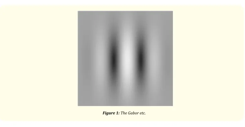
Technology foundations: The building block of these visual stimulations is the Gabor patch (Figure 1), which efficiently activates and matches the shape of receptive field in the Visual Cortex.
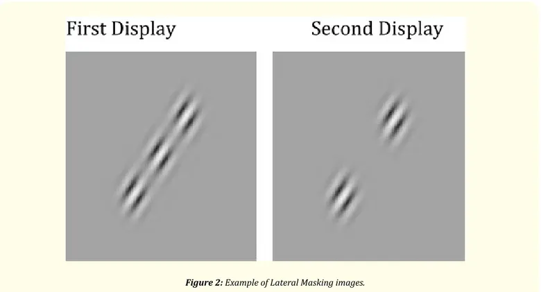
The fundamental stimulation-control technique is called “Lateral Masking”, where collinearly oriented flanking Gabors are displayed in addition to the target, central, Gabor patch. The patient is exposed to two short displays in succession, in a random order. The patient’s task is to identify which display contains the target Gabor image (Figure 2). The system provides the patient with audio feedback for an incorrect response. The task is repeated and a staircase is applied until the patient reaches the visual threshold level.
Figure 2: Example of Lateral Masking images.
Home Rehabilitation Protocol: The Gabor patch RV Treatment System is a software-based, interactive system tailored and continu-ously adaptive to the individual visual abilities. In the first stage, the subject is exposed to a set of visual perception tasks, aimed to analyze and identify each subject’s neural inefficiencies or deficiencies. Based on this analysis, a treatment plan is initialized, and subject specific-ity is achieved by administering patient-specific stimuli in a controlled environment (Scheme 1).
Scheme 1: The patient at home is connected to and fro via the Cloud to the software-based, interactive system tailored and
Each session is designed to train, directly and selectively, those functions in the visual cortex, V1 area, via GLG, which were diagnosed to be further enhanced. At each session an algorithm analyzes the patient’s responses and accordingly adjusts the level of visual difficulty to the range most effective for further improvement. Between sessions, the progress of the patient is taken into account by the algorithm for the next session generation. Thus, for each subject an individual training schedule is designed based on the initial state of visual performance, severity of dysfunction and progress in course of treatment. The treatment is applied in successive 30-minute sessions, administered 2 - 3 times a week, a total of approximately 30 - 50 sessions, according to the capacity of improvement perceived at the check-points: every 5 sessions, subject’s visual acuity is tested in order to continuously monitor subject’s progress, stopping at the end of the increasing trend. The average entire treatment duration is around 3 - 4 months.
Results
RV Treatment Group. To evaluate the efficacy of RV Gabor patch Technology in enhancing BCVA in patients with Stargardt disease, 5 Stargardt patients (10 eyes) verified at Ophthalmoscopy/ERG/ FA/ICG/OCT/Ishihara tables, 3 male, 2 female, mean aged 35 (+- 11.11 y.c), caucasians, were selected and informed according to the Helsinki declaration. Investigations included manifest and cycloplegic re-fraction, LogMAR BCVA.
As for CS Function, results are not included in the analysis in this paper, since no standard detection method has been used. Exclusion criteria
- Age (<18 > 50 y. o.)
- Other ocular disease impairing the visual axis, neurological disease or mental impairment. Inclusion criteria
- BCVA stable in the last 6 month in both eyes.
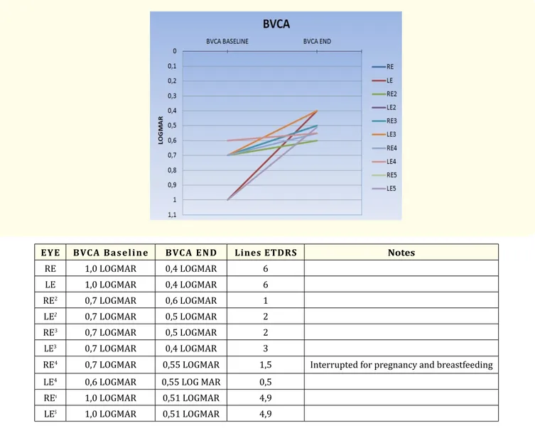
Patients have completed over 30 treatment session. Mean BCVA, for the 10 eyes, has raised from 0.81 (+- 0.17) logMar to 0.49 (+-0.07) logMar. Mean ETDRS’ chart lines improvement has been 3.18 +-2.05 (Table 1, a and b).
EYE BVCA Baseline BVCA END Lines ETDRS Notes
RE 1,0 LOGMAR 0,4 LOGMAR 6 LE 1,0 LOGMAR 0,4 LOGMAR 6 RE2 0,7 LOGMAR 0,6 LOGMAR 1 LE2 0,7 LOGMAR 0,5 LOGMAR 2 RE3 0,7 LOGMAR 0,5 LOGMAR 2 LE3 0,7 LOGMAR 0,4 LOGMAR 3
RE4 0,7 LOGMAR 0,55 LOGMAR 1,5 Interrupted for pregnancy and breastfeeding
LE4 0,6 LOGMAR 0,55 LOG MAR 0,5
REs 1,0 LOGMAR 0,51 LOGMAR 4,9
LES 1,0 LOGMAR 0,51 LOGMAR 4,9
One subject, included in the analysis, has quit the treatment in advance for pregnancy. Results suggest that Gabor patch treatment improves BCVA in Stargardt patients. This improvement appears to be retained for at least 12 months after treatment, consistent with the disease progression. More research is needed with prospective randomized studies with a larger population and for a longer follow up to confirm also the tenure of the positive trend pointed out.
Disclosure
Authors have not financial interest.
Bibliography
1. Meunier I and Puech B. “Dystrophies maculaires avec dépots fluorescents”. In Cohen SY and Gaudric A. Rétine. Lavoiser, Paris 2 (2012): 26-43.
2. Frezzotti R and Guerra R. “Oftalmologia essenziale”. CEA, Milano (2008).
3. Hubel DH and Wiesel TN. “Receptive fields, binocular interaction and functional architecture in the cat’s visual cortex”. Journal of
Physiology 160.1 (1962): 106-154.
4. Polat U. “Functional architecture of long-range perceptual interactions”. Spatial Vision 12.2 (1999): 143-162.
5. Geisler WS and Albrecht DG. “Visual cortex neurons in monkeys and cats: detection, discrimination, and identification”. Visual
Neu-roscience 14.5 (1997): 897-919.
6. Gallenga PE., et al. “Low Vision and blindness in Abruzzo”. New Trends in Ophthalmology 2 (1989): 137-142.
7. Polat U and Sagi D. “Lateral interactions between spatial channels: suppression and facilitation revealed by lateral masking experi-ments”. Vision Research 33.7 (1993): 993-999.
8. Polat U and Sagi D. “Spatial interactions in human vision: from near to far via experience- dependent cascades of connections”.
Pro-ceedings of the National Academy of Sciences of the United States of America 91.4 (1994): 1206-1209.
9. Polat U., et al. “Collinear stimuli regulate visual responses depending on cell’s contrast threshold”. Nature 391 (1998): 580-584. 10. Kasamatsu T., et al. “Colinear facilitation promotes reliability of single-cell responses in cat striate cortex”. Experimental Brain
Re-search 138.2 (2001): 163-172.
11. Dosher BA and Lu ZL. “Mechanisms of perceptual learning”. Vision Research 39.19 (1999): 3197-221.
12. Sale A., et al. “Noninvasive strategies to optimize brain plasticity: from basic research to clinical perspective”. Neural Plasticity (2013): 863970.
13. Morrone MC., et al. “Developmental changes in optokinetic mechanisms in the absence of unilateral cortical control”. Neuroreport 10.13 (1999): 2723-2729.
Aknowledgement
Mrs Elena Caravaggi, Talshir MT, for technical support.
Note: Partially presented as poster at IV International Course of Orbitoplastic Surgery in Amalfi Coast, 2016. L. Colangelo ed. ISBN 9791220019774, Salerno, Italy (2017): P04, 10-11.
14. Lobefalo L., et al. “Recupero funzionale nei pazienti ambliopi dopo trattamento con biofeedback”. Annali Di Ottalmologia E Clinica
Oculistica 128 (2002): 209-213.
15. Verboschi F., et al. “Riabilitazione visiva con biofeedback microperimetrico in pazienti con malattia di Stargardt”. XIII° Low Vision Accademy – Roma (2012).
16. Sagi D and Tanne D. “Perceptual learning: learning to see”. Current Opinion in Neurobiology 4.2 (1994): 195-199. 17. Gilbert CD. “Adult Cortical Dynamics”. Physiological Reviews 78.2 (1998): 467-485.
18. Verdina T., et al. “Biofeedback rehabilitation of eccentric fixation in patients with Stargardt disease using the MP1 microperimeter”. XIII° Low Vision Accademy – Roma (2012).
19. Polat U and Sagi D. “In Maturational Windows and Adult Cortical Plasticity”. Eds. Julesz, B. and Kovâcs (Addison-Wesley), I (1995) 1-15.
20. Solari R., et al. “RevitalVision vs Fotostimolazione customizzata in pazienti con ipovisione moderata: analisi preliminary”. XIII° Low Vision Accademy – Roma (2012).
21. Breitmeyer BG and Ogmen H. “Recent models and findings in visual backward masking: A comparison, review, and update”.
Percep-tion and Psychophysics 62.8 (2000): 1572- 1595.
22. Carrasco M., et al. “Spatial covert attention increases contrast sensitivity across the CSF: Support for signal enhancement”. Vision
Research 40.10-12 (2000): 1203-1215.
23. Chung ST., et al. “Spatial-frequency characteristics of letter identification in central and peripheral vision”. Vision Research 42.18 (2002): 2137-2152.
24. Cass JR and Spehar B. “Dynamics of collinear contrast facilitation are consistent with long-range horizontal striate transmission”.
Vi-sion Research 45.21 (2005): 2728-2739.