https://doi.org/10.1007/s00592-020-01504-w
ORIGINAL ARTICLE
Baseline SD‑OCT characteristics of diabetic macular oedema patterns
can predict morphological features and timing of recurrence
in patients treated with dexamethasone intravitreal implants
Chiara M. Eandi1,2,3 · Daniele De Geronimo4 · Daniela Giannini4 · Maria Sole Polito1 · Gian Marco Tosi5 ·
Giovanni Neri5 · Yannick Le Mer3 · Monica Varano4 · Mariacristina Parravano4
Received: 16 January 2020 / Accepted: 11 February 2020 / Published online: 29 February 2020 © The Author(s) 2020
Abstract
Aims To evaluate the timing and spectral-domain optical coherence tomography (SD-OCT) features of diabetic macular oedema (DME) recurrence according to baseline OCT patterns in patients treated with dexamethasone implant (DEX-I).
Methods This is a retrospective observational study (72 eyes/65 patients). Best-corrected visual acuity, timing of DME recurrence, and SD-OCT pattern [intraretinal cysts (IRC), IRC plus subretinal fluid (mixed), external limiting membrane (ELM), ellipsoid (IS/OS) layer integrity] were assessed at baseline and monthly until first DME recurrence.
Results Forty-two (58.3%) and 30 (41.6%) DME eyes had an IRC and mixed DME pattern at baseline, respectively. Twenty-four out of thirty mixed eyes (80%) relapsed without subretinal fluid. At baseline, mixed eyes showed similar changes in ELM and IS/OS (60 and 76.6% of eyes, respectively) versus IRC eyes (42.8 and 80.9% of eyes). After DME recurrence, more mixed eyes at baseline showed ELM and IS/OS changes (63.3 and 86.6%) than IRC eyes (50 and 76.2%). 33.3% of mixed eyes had DME recurrence at ≥ 6 months from first DEX-I implant versus 19% of IRC eyes.
Conclusions Mixed DME eyes were treated with DEX-I relapse later and more frequently without subretinal fluid than IRC eyes. SD-OCT characteristics of different DME patterns at baseline can predict morphological features and timing of DME recurrence.
Keywords Diabetic macular oedema · Dexamethasone implant (DEX-I) · Intravitreal treatment · Baseline characteristics · Recurrence · Spectral-domain optical coherence tomography
Introduction
Diabetic macular oedema (DME), a macular thickening secondary to diabetic retinopathy (DR), results from a blood–retinal barrier defect that leads to vascular leakage and fluid accumulation [1]. In patients with diabetes, DME is a leading cause of visual impairment and loss [2] and has been reported in almost 30% of patients with a duration of disease > 20 years [3].
DME has been related to the expression of several inflam-matory factors, including vascular endothelial growth factor (VEGF), intercellular adhesion molecule-1 (ICAM-1), inter-leukin-6 (IL-6), monocyte chemotactic protein-1 (MCP-1), and leukostasis [4, 5]. Moreover, the expression of these factors has been related to both vascular permeability of the retina along with the severity of disease, thus confirming their important pathogenetic role [4]. While achieving con-trol of glycemia is essential to limit the progression of DME,
This article belongs to the topical collection Eye Complications of Diabetes, managed by Giuseppe Querques.
Chiara M. Eandi and Daniele De Geronimo have contributed equally to this work.
* Chiara M. Eandi [email protected]
1 Department of Surgical Sciences, University of Torino, C.
Dogliotti 14, 10126 Turin, Italy
2 Department of Ophthalmology, Jules-Gonin Eye Hospital,
University of Lausanne, Fondation Asile des aveugles, Lausanne, Switzerland
3 Department of Ophthalmology, Fondation Ophtalmologique
A. De Rothschild, Paris, France
4 IRCCS - Fondazione Bietti, Rome, Italy
5 Ophthalmology Unit, Department of Medicine, Surgery
several treatment options for patients with DME are also in widespread use [2, 6]. Indeed, in the past decade, advances in the understanding of the pathogenesis of DME have led to the development of new therapies with anti-inflamma-tory action, especially steroids and VEGF inhibitors, which have resulted in several novel therapeutic applications [2, 7]. While intravitreal anti-VEGF agents have been shown to be effective in improving best-corrected visual acuity (BCVA) and decreasing central retinal thickness (CRT), it has been suggested that they should be used with caution due to pos-sible systemic adverse events [2]. Moreover, they are not appropriate for all patients, and not all patients respond to anti-VEGF treatment; compliance to therapy also remains suboptimal due to the numerous injections required [8, 9].
In addition to anti-VEGF agents, in diabetic animal mod-els, intravitreal corticosteroids have been shown to block the production of several inflammatory mediators, such as VEGF and ICAM-1, and inhibit leukostasis [10, 11]. In a clinical context, dexamethasone has been shown to have the highest relative efficacy among all corticosteroids that are routinely used to treat DME [6].
Dexamethasone intravitreal implant (DEX-I) is a matrix based on micronized dexamethasone embedded in a bio-degradable copolymer of polylactic-co-glycolic acid that slowly releases the steroid into the vitreous over a period of months [12, 13]. DEX-I has been studied extensively in patients with DME. Based on the MEAD study, the Food and Drug Administration (FDA) and European Medicines Agency (EMA) approved DEX-I for the treatment of DME [14].
Several subsequent studies further demonstrated that DEX-I could improve BCVA and CRT in patients with DME and thus represents a viable treatment option [15, 16]. Simi-lar results were obtained from the analysis of real-life data [17, 18]. A meta-analysis of four randomized clinical trials involving 521 eyes with DME reported that DEX-I is asso-ciated with improvements in BCVA that are non-inferior to anti-VEGF therapy, with superior anatomic outcomes at 6 months [6]. Moreover, compared to anti-VEGF agents, DEX-I requires fewer injections with no significant differ-ences in the rates of adverse events, although there was some concern over raised intraocular pressure and cataract com-pared to anti-VEGF therapy. Given these favourable charac-teristics, DEX-I may be considered as first-choice therapy in selected cases, such as for pseudophakic eyes, failure of an anti-VEGF-agent, or in patients who are unwilling or unable to undergo frequent intravitreal injections [6].
To date, there is still limited evidence on the impact that individual characteristics of DME may have on the recur-rence of DME following the implant of DEX-I. Some evi-dence has been presented that the DME morphologic sub-types, as defined by optical coherence tomography (OCT), may be associated with greater reductions in CRT in patients
with DME. In particular, the serous retinal detachment (SRD) subtype has been associated with a greater reduction in the CRT than the diffuse retinal thickening (DRT) sub-type [19], and in another study, the cystoid macular oedema (CME) and SRD subtypes showed greater reduction in CRT than the DRT subtype [20]. To shed further light on this aspect, we evaluated the spectral-domain (SD)-OCT mor-phological features of DME recurrence according to baseline OCT patterns in patients treated with DEX-I.
Materials and methods
Study design and patient population
This was a retrospective observational study. Informed con-sent was obtained from all subjects. All research procedures described in this study adhered to the tenets of the 1964 Declaration of Helsinki and its later amendments.
Clinical charts were retrieved from the four participating centres, and the pooled data were analysed. We included patients with a diagnosis of DME who had been treated with DEX-I during the period from 1 January 2017 to 30 June 2018 and followed at least until the first recurrence of DME at four referral centres (University of Torino, IRCCS-Fon-dazione Bietti in Rome, University of Siena, and Rothschild Foundation in Paris).
All patients had data relating to BCVA and SD-OCT fea-tures available at baseline and at each follow-up examina-tion. BCVA was measured using the Early Treatment Dia-betic Retinopathy Study (ETDRS) charts and reported as LogMar. Follow-up examinations included maximal answer (defined as macula dry or with the minimum amount of intra- or subretinal fluid) and information on DME recur-rence (mean recurrecur-rence timing, characteristics of DME on SD-OCT). Baseline data also included the following: demo-graphic data, diabetes duration, per cent glycated haemoglo-bin (HbA1c) level, and information on previous intravitreal treatment. Exclusion criteria were: macular oedema second-ary to causes other than diabetes; previous treatment with intraocular corticosteroids; previous anti-VEGF intravitreal injections within the 6 months before treatment with the DEX implant; previous macular laser; and previous pars plana vitrectomy.
SD-OCT images were acquired with a Spectralis HRA + OCT instrument (Heidelberg Engineering, Heidel-berg, Germany, version 6.4.7.0). The scanning protocol included a high-resolution 20° × 20° volume scan centred in the central macula. CRT was measured using the retina map pattern and the provided ETDRS grid in the central millime-tre. Each section was obtained using ART (automatic real-time) eye tracking, and 16 scans were averaged to improve the signal-to-noise ratio. The following SD-OCT features
were considered: CRT, DME pattern classified according to the presence of intraretinal cysts (IRC) or IRC plus subreti-nal fluid (mixed) pattern, the integrity of the extersubreti-nal limit-ing membrane (ELM), and the ellipsoid junction (IS/OS).
The BCVA and SD-OCT characteristics were evaluated by two expert observers (CME and MP) at baseline and then monthly after DEX-I treatment until the first recurrence of DME. Patients were retreated according to a pro re nata (PRN) regimen if there was a recurrence of DME, defined as the presence of intra- or subretinal fluid on SD-OCT, also in the absence of visual impairment.
Statistical analysis
The normal data distribution was tested using the one-sam-ple Kolmogorov–Smirnov test. All continuous variables were expressed as mean ± standard deviation, while cat-egorical variables as frequency and percentage. T test and Mann–Whitney test were performed as appropriate. Contin-gency tables (chi-square test) were used to investigate the relationship between pre- and post-recurrence DME pat-tern and between baseline DME patpat-tern and pre- and post-recurrence morphological parameters (ELM and IS/OS). A one-way repeated measures analysis of variance (ANOVA) for each group was conducted to evaluate the null hypoth-esis that there is no change in functional and morphologi-cal parameter values when measured at baseline, maximal answer, and after recurrence in study groups. Post hoc tests were performed using the Bonferroni correction. Statistical evaluation was performed using SPSS (IBM SPSS Statistic 25). A p value < 0.05 was considered statistically significant.
Results
Baseline demographic characteristics
Considering pooled data from the four centres, baseline information and follow-up data were available on a total of 72 eyes from 65 patients with DME and receiving a DEX-I. The cohort included 39 males (60%) and 26 females (40%), with a mean age of 57.2 ± 8.2 years, mean duration of type 2 diabetes of 17.2 ± 8.8 years, and mean per cent HbA1C
at baseline of 8.2 ± 1.8 (66.0 ± 17.3 mmol/mol). Forty-three of the 72 eyes (59.7%) were phakic, and 29 (40.3%) were pseudophakic. Thirty-nine of the 72 eyes (54.2%) were naïve to treatment, and 33 of 72 eyes (45.8%) were switched from previous anti-VEGF treatment (Table 1).
SD‑OCT features
Forty-two (58.3%) and 30 (41.6%) eyes presented with an IRC and mixed DME pattern at baseline, respectively.
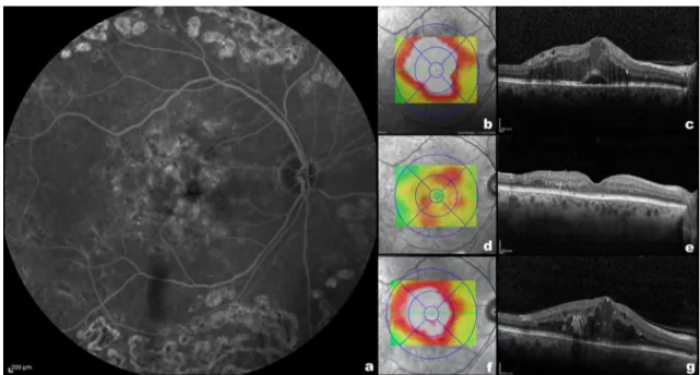
Maximal answer (macula dry or with the minimum amount of intra- or subretinal fluid) timing was 1.83 ± 0.80 months for all patients, 1.83 ± 0.82 months for IRC group, and 1.83 ± 0.79 for the mixed group. Twenty-four of 30 mixed eyes (80%) treated with DEX-I relapsed without subreti-nal fluid (Table 2). A significant relationship was found between basal and post-recurrence DME pattern, X2 (1, n = 72) = 6.19, p = 0.013. A Mann–Whitney test showed that no differences were present in mean recurrence timing of baseline IRC and mixed groups (IRC group 5.74 ± 1.98 months; mixed group 6.27 ± 2.69 months, p = 0.168). However, a higher percentage of mixed eyes (33.3%) had a recurrence of DME after 6 months or longer after the first DEX-I implant in comparison with IRC eyes (19%) (Table 2). Figure 1 shows the SD-OCT features of a representative patient with mixed pattern DME before, 2 months after, and at the time of recurrence 7 months after treatment with DEX-I.
At baseline, mixed eyes showed similar changes in ELM and IS/OS (60 and 76.6% of eyes, respectively) compared with IRC eyes (42.8 and 80.9% of eyes, respectively). After
Table 1 Demographic and baseline characteristics of patients
n number, SD standard deviation, HbA1c glycated haemoglobin
a 66.0 ± 17.3 mmol/mol
Characteristic
Patients/eyes (n) 65/72
Male/female (n) 39/26
Age, mean ± SD (years) 57.2 ± 8.2
Duration of diabetes (years) mean ± SD 17.2 ± 8.8 (range 5–45)
HbA1c % (mean ± SD) 8.2 ± 1.8a
Phakic/pseudophakic (n eyes) 43/29
Naïve/switched (n eyes) 39/33
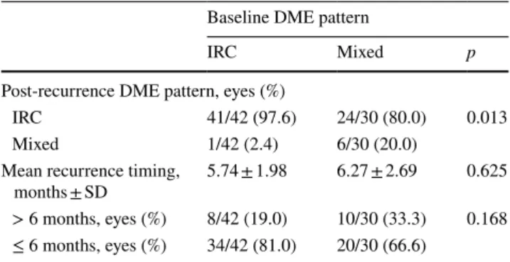
Table 2 Baseline DME pattern versus post-recurrence DME pattern and timing of recurrence
DME diabetic macular oedema, IRC intraretinal cysts, SD standard
deviation
Baseline DME pattern
IRC Mixed p
Post-recurrence DME pattern, eyes (%)
IRC 41/42 (97.6) 24/30 (80.0) 0.013
Mixed 1/42 (2.4) 6/30 (20.0)
Mean recurrence timing,
months ± SD 5.74 ± 1.98 6.27 ± 2.69 0.625
> 6 months, eyes (%) 8/42 (19.0) 10/30 (33.3) 0.168 ≤ 6 months, eyes (%) 34/42 (81.0) 20/30 (66.6)
a recurrence of DME, baseline mixed eyes showed greater changes in ELM and IS/OS (63.3% and 86.6% of eyes, respectively) compared with IRC eyes (50 and 76.2% of eyes, respectively) (Table 3).
By T test, a significant difference was found between basal CRT of the IRC and mixed groups at baseline (IRC 516 ± 136 μm; mixed 600 ± 116 μm; p = 0.008). Moreover, significant differences between groups were found in CRT at post-recurrence (IRC at baseline 462 ± 131 μm; mixed at baseline group 614 ± 150 μm; p = 0.006).
In both the IRC and mixed groups, ANOVA indicated a significant effect of time on CRT (p < 0.01). Post hoc tests indicated that pairwise differences in the IRC group were significant between baseline (516 ± 136 μm) and maximal answer (307 ± 76 µm) time (p = 0.009) and between maximal
answer and post-recurrence (460 ± 135 μm) time (p < 0.01). In the mixed group, there was a significant decrease in CRT values between baseline (600 ± 116 µm) and maximal answer (336 ± 93 μm) time, as well as an increase between maximal answer and post-recurrence (499 ± 145 μm) time. The maximal answer for both groups was at month 2.
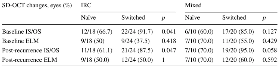
At baseline, treatment-naïve eyes showed fewer IS/OS changes than switched eyes in both subgroups of DME pat-terns (66.7% vs. 91.7% in IRC and 60% vs. 85% in the mixed group) (Table 4). After a recurrence of DME, a similar trend was observed: Naïve eyes showed fewer IS/OS changes than switched eyes in both subgroups of DME patterns (61.1% vs. 87.5% in IRC and 70% vs. 95% in the mixed group).
At baseline and after recurrence of DME, a non-statis-tically significant difference in terms of ELM changes was
Fig. 1 Left: Fluorescein angiography a of a patient with diabetic macular oedema before the treatment with dexamethasone intra-vitreal implants (DEX-I). Top right: Spectralis thickness map (b) and B-scan c of the same patient before the treatment with DEX-I, showing intraretinal cysts and subretinal fluid (mixed pattern). Mid-dle right: Spectralis thickness map (d) and B-scan e of the same
patient 2 months after the treatment with DEX-I showing a reduc-tion in the macular thickness and no intra- or subretinal fluid. Bottom right: Spectralis thickness map (f) and B-scan g of the same patient 7 months after the treatment with DEX-I showing new diabetic macu-lar oedema with intraretinal cysts without subretinal fluid
Table 3 Spectral-domain optical coherence tomography (SD-OCT) characteristics by group
CRT central retinal thickness, ELM external limiting membrane, IRC intraretinal cysts, IS/OS ellipsoid
junction, SD standard deviation
SD-OCT characteristics IRC Mixed p
Baseline IS/OS changes, eyes (%) 34/42 (80.95) 23/30 (76.67) 0.659
Baseline ELM changes, eyes (%) 18/42 (42.86) 18/30 (60.00) 0.151
Post-recurrence IS/OS changes, eyes (%) 32/42 (76.20) 26/30 (86.70) 0.268 Post-recurrence ELM changes, eyes (%) 21/42 (50.00) 19/30 (63.30) 0.262
Mean baseline CRT (μm ± SD) 516 ± 136 600 ± 116 0.008
found between treatment-naïve and switched eyes in both subgroups of DME patterns (baseline: 50% vs. 37.5% in IRC and 70% vs. 55% in the mixed group; after recurrence: 50% vs. 50% in IRC and 70% vs. 60% in the mixed group) (Table 4).
BCVA characteristics
A T test showed no differences between baseline BCVA (IRC 0.394 ± 0.287 LogMar; mixed 0.491 ± 0.334 LogMar) in the IRC and mixed groups (p = 0.195) and no differences between groups for post-recurrence BCVA (IRC at baseline 0.437 ± 0.296 LogMar; mixed at baseline 0.290 ± 0.191 Log-Mar; p = 0.202). In both the IRC and mixed groups, ANOVA indicated a significant time effect (p < 0.01) on BCVA. Post hoc tests indicated that pairwise differences in the IRC group were significant between baseline (0.394 ± 0.28 LogMar) and maximal answer (0.301 ± 0.21 LogMar) time (p = 0.009) and between maximal answer and post-recurrence time (0.377 ± 0.21 LogMar; p < 0.01). In the mixed group, a sig-nificant difference between maximal answer (0.420 ± 0.36 LogMar) and post-recurrence time (0.488 ± 0.037 LogMar; p = 0.015) was found.
Of note, no relevant safety issues were encountered dur-ing follow-up in this patient cohort, confirmdur-ing the previ-ously reported favourable safety profile with DEX-I.
Discussion
Overall, the present analysis found that mixed DME eyes treated with DEX-I relapsed with only intraretinal fluid, without subretinal fluid, and at a later time compared to IRC eyes. Thus, the present data would seem to indicate that the SD-OCT characteristics that define different DME patterns at baseline may be useful in predicting the morphological features and timing of recurrence of DME. These findings further confirm the utility of SD-OCT in identifying param-eters that can be predictive of a better and longer response to DEX-I.
The possibility of identifying morphological biomarkers in DME that can predict a better response to DEX-I is of substantial clinical interest. In this regard, OCT is a fast
and noninvasive examination routinely used in daily clini-cal practice, and its application in this setting has important implications for the management of patients with diabetic maculopathy. Moreover, the present analysis suggests that patients with an IRC pattern may relapse more frequently and sooner. This provides the clinician with additional infor-mation that can help to individualize treatment with DEX-I and help in selecting patients who may be the most appro-priate candidates for this therapy. For example, in patients who are predicted to relapse earlier, more frequent follow-up may be warranted in order to modulate treatment accord-ingly or to allow for earlier switching to another treatment, as with other therapies such as anti-VEGF [21]. One addi-tional advantage of better prediction of relapse and time to relapse is that early implantation of DEX-I has been associ-ated with better visual outcomes. Indeed, significantly more eyes showing a robust early response demonstrated ≥ 10-let-ter long-10-let-term gain in BCVA compared to eyes with poor early response [22].
Considering specific OCT features, in the present analy-sis, we found that mixed eyes treated with DEX-I relapsed later than IRC eyes. We could speculate that a higher per-centage of mixed eyes had a recurrence of DME later than IRC eyes because the presence of SRF in mixed eyes could denote an inflammatory nature of macular oedema that could be better opposed by the action of DEX-I. These results are consistent with the study by Zur et al., which found that eyes with DME and subretinal fluid (SRF), no hyperreflec-tive foci (HRF), and a continuous IS-OS layer responded better to DEX-I than those without these features [23]. The recurrence of DME without SRF observed in 80% of mixed eyes treated with DEX-I could also confirm the inflamma-tory nature of SRF, which do not reappear in the recurrence of DME after DEX-I treatment. In support of this hypoth-esis, previous studies have reported that higher concentra-tions of inflammatory cytokines in the vitreous and aqueous humour are present in eyes with SRF, thus suggesting the presence of a significant inflammatory component [4, 24]. Moreover, in our study, eyes with SRF at baseline showed greater changes in ELM than eyes without SRF (60% vs. 42.8%, respectively). This finding is of interest, consider-ing that previous authors have reported that the integrity of the ELM seems to be a key factor in preventing fluid
Table 4 SD-OCT changes of ELM and IS/OS layers by subgroup of patients (naïve, switched)
ELM external limiting membrane, IRC intraretinal cysts, IS/OS ellipsoid junction
SD-OCT changes, eyes (%) IRC Mixed
Naïve Switched p Naïve Switched p
Baseline IS/OS 12/18 (66.7) 22/24 (91.7) 0.041 6/10 (60.0) 17/20 (85.0) 0.127 Baseline ELM 9/18 (50) 9/24 (37.5) 0.418 7/10 (70.0) 11/20 (55.0) 0.429 Post-recurrence IS/OS 11/18 (61.1) 21/24 (87.5) 0.047 7/10 (70.0) 19/20 (95.0) 0.058 Post-recurrence ELM 9/18 (50.0) 12/24 (50.0) 1 7/10 (70.0) 12/20 (60.0) 0.592
from passing from the outer retina into the subretinal space [25]. The post-recurrence OCT features of eyes with DME showed that eyes with SRF had more changes in both the IS/OS and ELM layers compared to eyes without SRF (86.7 and 63.3%, respectively, for mixed eyes vs. 76.2 and 50% for IRC eyes). In this regard, we speculate that SRF at baseline could be associated with damage to the IS/OS in the long term. Moreover, since there are no changes at the level of the ELM between the baseline and the post-recurrence the low rate of recurrence of SRD might depend on the timing of retreatment that was performed as soon as intraretinal fluid appeared and before the development of subretinal fluid.
DEX-I is significantly associated with improved anatomi-cal outcomes (although not necessarily BCVA) and has been recommended as first-choice therapy for pseudophakic eyes, those resistant to anti-VEGF agents, or for patients who are reluctant to receive frequent intravitreal injections [6]. In fact, several studies have reported that DEX-I is effective for the treatment of DME, even in refractory cases that have failed to respond to other therapies, substantiating its utility in these patients [19, 26].
We also examined the difference between eyes naïve to treatment and eyes switched from previous intravitreal anti-VEGF in all cases. Interestingly, at baseline and at the post-recurrence time, treatment-naïve eyes showed fewer IS/OS changes than switched eyes in both groups, with a significant difference in IRC eyes and a statistical trend in mixed eyes. Zur et al. [23] reported that eyes with a continuous IS/OS layer respond better to DEX-I, and thus we can speculate that eyes naïve to treatment have better preservation of the IS/ OS layer compared to switched eyes and thus have a better response to DEX-I. Notwithstanding, the efficacy of DEX-I in DME has been confirmed in both naïve and refractory patients [26, 27], as well as in real-world analyses [28].
Studies on prognostic indicators with the use of DEX-I in patients with DME have reported that visual and ana-tomical outcomes of treatment with DEX-I may be predicted by baseline visual acuity and intraretinal fluid morphology [29]. Recent data have further indicated that elevated MCP-1 aqueous humour levels and DRT pattern at baseline are bio-markers that predict future favourable anatomic response to DEX-I [30]. Thus, the present data add to the growing list of clinical markers that can help predict response to DEX-I. This is important as DEX-I is recommended in current guidelines as first- or second-line therapy in subjects with DME [31, 32] and represents a valid therapeutic alternative to other medical treatments, as also demonstrated in direct comparisons with anti-VEGF agents at 12 and 24 months [33, 34].
The present study has some limitations, such as its retro-spective design and relatively low number of patients, and larger studies with additional ophthalmologic parameters are warranted to confirm our findings. Moreover, we decided
not to include in the current analysis some anatomical prog-nostic parameters, such as hyperreflective foci and disor-ganization of retinal inner layers (DRIL), because of their lower application on clinical routine examination. How-ever, it could be interesting in a further study to evaluate the association between these parameters, already described as important clinical and prognostic factors for DME [35, 36], and the pattern of DME.
We can nonetheless confirm the utility of SD-OCT in identifying parameters that can be predictive of better and longer response to DEX-I, thus reinforcing the need to fur-ther study combinations of SD-OCT and metabolic biomark-ers. One such candidate for study is intracellular adhesion molecule 1, which has been shown to correlate with subreti-nal fluid height in DME [37].
In conclusion, most mixed DME eyes treated with DEX-I relapse without subretinal fluid and at a later time than IRC eyes. The SD-OCT characteristics of different DME patterns at baseline can help to predict the morphological features and timing of DME recurrence.
Acknowledgements Preliminary results of this study were presented at the EASDec Meeting, Amsterdam, 17 May 2019, and the Euretina Meeting, Paris, 5–8 September 2019. Writing and editorial assistance was provided to the authors by Patrick Moore and Ray Hill, independ-ent medical writers, and funded by Allergan plc, Dublin, Ireland, at the request of the investigator. All authors met the ICMJE authorship criteria. Neither honoraria nor payments were made for authorship. Funding Neither honoraria nor payments were made for authorship. The research for this paper was, in part, financially supported by the Italian Ministry of Health and Fondazione Roma. The funders had no role in study design, data collection and analysis, decision to publish, or preparation of the manuscript.
Compliance with ethical standards
Conflict of interest MP reports personal fees from Allergan, Bayer, Novartis, outside of the submitted work. YLM reports personal fees from Allergan and Novartis, outside of the submitted work. The other authors have nothing to disclose.
Human and animal rights disclosure Institutional review board approval for a retrospective chart review was obtained from the indi-vidual centres (University of Torino, IRCCS-Fondazione Bietti in Rome, University of Siena, and Rothschild Foundation in Paris), as required by local guidelines.
Informed consent disclosure Informed consent was obtained from all individual participants included in the study.
Open Access This article is licensed under a Creative Commons Attri-bution 4.0 International License, which permits use, sharing, adapta-tion, distribution and reproduction in any medium or format, as long as you give appropriate credit to the original author(s) and the source, provide a link to the Creative Commons licence, and indicate if changes were made. The images or other third party material in this article are included in the article’s Creative Commons licence, unless indicated otherwise in a credit line to the material. If material is not included in
the article’s Creative Commons licence and your intended use is not permitted by statutory regulation or exceeds the permitted use, you will need to obtain permission directly from the copyright holder. To view a copy of this licence, visit http://creat iveco mmons .org/licen ses/by/4.0/.
References
1. Bhagat N, Grigorian RA, Tutela A, Zarbin MA (2009) Diabetic macular edema: pathogenesis and treatment. Surv Ophthalmol 54:1–32
2. Zhang L, Wang W, Gao Y, Lan J, Xie L (2016) The efficacy and safety of current treatments in diabetic macular edema: a system-atic review and network meta-analysis. PLoS ONE 11:e0159553 3. Klein R, Klein BE, Moss SE, Davis MD, DeMets DL (1984) The
Wisconsin epidemiologic study of diabetic retinopathy. IV. Dia-betic macular edema. Ophthalmology 91:1464–1474
4. Funatsu H, Noma H, Mimura T, Eguchi S, Hori S (2009) Asso-ciation of vitreous inflammatory factors with diabetic macular edema. Ophthalmology 116:73–79
5. Miyamoto K, Khosrof S, Bursell SE, Rohan R, Murata T, Cler-mont AC, Aiello LP, Ogura Y, Adamis AP (1999) Prevention of leukostasis and vascular leakage in streptozotocin-induced dia-betic retinopathy via intercellular adhesion molecule-1 inhibition. Proc Natl Acad Sci USA 96:10836–10841
6. He Y, Ren XJ, Hu BJ, Lam WC, Li XR (2018) A meta-analysis of the effect of a dexamethasone intravitreal implant versus intravit-real anti-vascular endothelial growth factor treatment for diabetic macular edema. BMC Ophthalmol 18:121
7. Ford JA, Lois N, Royle P, Clar C, Shyangdan D, Waugh N (2013) Current treatments in diabetic macular oedema: systematic review and meta-analysis. BMJ Open 3:e002269
8. Brown DM, Nguyen QD, Marcus DM, Boyer DS, Patel S, Feiner L, Schlottmann PG, Rundle AC, Zhang J, Rubio RG, Adamis AP, Ehrlich JS, Hopkins JJ, Ride, Rise Research Group (2013) Long-term outcomes of ranibizumab therapy for diabetic macular edema: the 36-month results from two phase III trials: RISE and RIDE. Ophthalmology 120:2013–2022
9. Nguyen-Khoa BA, Goehring EL, Werther W, Fung AE, Do DV, Apte RS, Jones JK (2012) Hospitalized cardiovascular events in patients with diabetic macular edema. BMC Ophthalmol 12:11 10. Tamura H, Miyamoto K, Kiryu J, Miyahara S, Katsuta H, Hirose
F, Musashi K, Yoshimura N (2005) Intravitreal injection of cor-ticosteroid attenuates leukostasis and vascular leakage in experi-mental diabetic retina. Invest Ophthalmol Vis Sci 46:1440–1444 11. Wang K, Wang Y, Gao L, Li X, Li M, Guo J (2008) Dexametha-sone inhibits leukocyte accumulation and vascular permeability in retina of streptozotocin-induced diabetic rats via reducing vascular endothelial growth factor and intercellular adhesion molecule-1 expression. Biol Pharm Bull 31:1541–1546
12. Chang-Lin JE, Burke JA, Peng Q, Lin T, Orilla WC, Ghosn CR, Zhang KM, Kuppermann BD, Robinson MR, Whitcup SM, Welty DF (2011) Pharmacokinetics of a sustained-release dexametha-sone intravitreal implant in vitrectomized and nonvitrectomized eyes. Invest Ophthalmol Vis Sci 52:4605–4609
13. Haghjou N, Soheilian M, Abdekhodaie MJ (2011) Sustained release intraocular drug delivery devices for treatment of uveitis. J Ophthalmic Vis Res 6:317–329
14. Boyer DS, Yoon YH, Belfort R Jr, Bandello F, Maturi RK, Augus-tin AJ, Li XY, Cui H, Hashad Y, Whitcup SM, Ozurdex Mead Study Group (2014) Three-year, randomized, sham-controlled trial of dexamethasone intravitreal implant in patients with dia-betic macular edema. Ophthalmology 121:1904–1914
15. Cebeci Z, Kir N (2015) Role of implants in the treatment of dia-betic macular edema: focus on the dexamethasone intravitreal implant. Diabetes Metab Syndr Obes 8:555–566
16. Dugel PU, Bandello F, Loewenstein A (2015) Dexamethasone intravitreal implant in the treatment of diabetic macular edema. Clin Ophthalmol 9:1321–1335
17. Chhablani J, Bansal P, Veritti D, Sambhana S, Sarao V, Pichi F, Carrai P, Massaro D, Lembo A, Mansour AM, Banker A, Gupta SR, Hamam R, Lanzetta P (2016) Dexamethasone implant in diabetic macular edema in real-life situations. Eye (Lond) 30:426–430
18. Lam WC, Albiani DA, Yoganathan P, Chen JC, Kherani A, Maberley DA, Oliver A, Rabinovitch T, Sheidow TG, Tourville E, Wittenberg LA, Sigouin C, Baptiste DC (2015) Real-world assessment of intravitreal dexamethasone implant (0.7 mg) in patients with macular edema: the CHROME study. Clin Ophthal-mol 9:1255–1268
19. Castro-Navarro V, Cervera-Taulet E, Navarro-Palop C, Monferrer-Adsuara C, Hernandez-Bel L, Montero-Hernandez J (2019) Intra-vitreal dexamethasone implant Ozurdex(R) in naive and refractory patients with different subtypes of diabetic macular edema. BMC Ophthalmol 19:15
20. Koytak A, Altinisik M, Sogutlu Sari E, Artunay O, Umurhan Akkan JC, Tuncer K (2013) Effect of a single intravitreal bevaci-zumab injection on different optical coherence tomographic pat-terns of diabetic macular oedema. Eye (Lond) 27:716–721 21. Herbaut A, Fajnkuchen F, Qu-Knafo L, Nghiem-Buffet S, Bodaghi
B, Giocanti-Auregan A (2017) Switching to aflibercept in diabetic macular edema not responding to ranibizumab and/or intravitreal dexamethasone implant. J Ophthalmol 2017:8035013
22. Al-Khersan H, Hariprasad SM, Chhablani J, Dex Implant Study Group (2017) Early response to intravitreal dexamethasone implant therapy in diabetic macular edema may predict visual outcome. Am J Ophthalmol 184:121–128
23. Zur D, Iglicki M, Busch C, Mariussi A, Mariussi M, Loewenstein A, International Retina Group (2018) OCT biomarkers as func-tional outcome predictors in diabetic macular edema treated with dexamethasone implant. Ophthalmology 125:267–275
24. Sonoda S, Sakamoto T, Yamashita T, Shirasawa M, Otsuka H, Sonoda Y (2014) Retinal morphologic changes and concentra-tions of cytokines in eyes with diabetic macular edema. Retina 34:741–748
25. Gaucher D, Sebah C, Erginay A, Haouchine B, Tadayoni R, Gaud-ric A, Massin P (2008) Optical coherence tomography features during the evolution of serous retinal detachment in patients with diabetic macular edema. Am J Ophthalmol 145:289–296 26. Iglicki M, Busch C, Zur D, Okada M, Mariussi M, Chhablani JK,
Cebeci Z, Fraser-Bell S, Chaikitmongkol V, Couturier A, Gian-cipoli E, Lupidi M, Rodriguez-Valdes PJ, Rehak M, Fung AT, Goldstein M, Loewenstein A (2019) Dexamethasone implant for diabetic macular edema in naive compared with refractory eyes: the International Retina Group Real-Life 24-Month Multicenter Study. The IRGREL-DEX Study. Retina 39:44–51
27. Escobar-Barranco JJ, Pina-Marin B, Fernandez-Bonet M (2015) Dexamethasone implants in patients with naive or refractory dif-fuse diabetic macular edema. Ophthalmologica 233:176–185 28. Mello Filho P, Andrade G, Maia A, Maia M, Biccas Neto L,
Muralha Neto A, Moura Brasil O, Minelli E, Dalloul C, Iglicki M (2019) Effectiveness and safety of intravitreal dexamethasone implant (Ozurdex) in patients with diabetic macular edema: a real-world experience. Ophthalmologica 241:9–16
29. Lee H, Kang KE, Chung H, Kim HC (2018) Prognostic factors for functional and anatomic outcomes in patients with diabetic macular edema treated with dexamethasone implant. Korean J Ophthalmol 32:116–125
30. Figueras-Roca M, Sala-Puigdollers A, Zarranz-Ventura J, Alba-Linero C, Alforja S, Esquinas C, Molins B, Adan A (2019) Ana-tomic response to intravitreal dexamethasone implant and baseline aqueous humor cytokine levels in diabetic macular edema. Invest Ophthalmol Vis Sci 60:1336–1343
31. Schmidt-Erfurth U, Garcia-Arumi J, Bandello F, Berg K, Chakravarthy U, Gerendas BS, Jonas J, Larsen M, Tadayoni R, Loewenstein A (2017) Guidelines for the management of diabetic macular edema by the European Society of Retina Specialists (EURETINA). Ophthalmologica 237:185–222
32. Guidelines for Diabetic Eye Care. Updated (2017). http://www. icoph .org/downl oads/ICOGu ideli nesfo rDiab eticE yeCar e.pdf. Accessed 8 Apr 2019
33. Fraser-Bell S, Lim LL, Campain A, Mehta H, Aroney C, Bryant J, Li J, Quin GJ, McAllister IL, Gillies MC (2016) Bevacizumab or dexamethasone implants for DME: 2-year results (The BEV-ORDEX Study). Ophthalmology 123:1399–1401
34. Gillies MC, Lim LL, Campain A, Quin GJ, Salem W, Li J, Good-win S, Aroney C, McAllister IL, Fraser-Bell S (2014) A rand-omized clinical trial of intravitreal bevacizumab versus intravitreal
dexamethasone for diabetic macular edema: the BEVORDEX study. Ophthalmology 121:2473–2481
35. Vujosevic S, Bini S, Midena G, Berton M, Pilotto E, Midena E (2013) Hyperreflective intraretinal spots in diabetics without and with nonproliferative diabetic retinopathy: an in vivo study using spectral domain OCT. J Diabetes Res 2013:491835
36. Sun JK, Lin MM, Lammer J, Prager S, Sarangi R, Silva PS, Aiello LP (2014) Disorganization of the retinal inner layers as a predic-tor of visual acuity in eyes with center-involved diabetic macular edema. JAMA Ophthalmol 132(11):1309–1316
37. Zhu D, Zhu H, Wang C, Yang D (2014) Intraocular soluble intracellular adhesion molecule-1 correlates with subretinal fluid height of diabetic macular edema. Indian J Ophthalmol 62:295–298
Publisher’s Note Springer Nature remains neutral with regard to jurisdictional claims in published maps and institutional affiliations.