Maria Antonietta Mazzei, Susanna Guerrini, Nevada Cioffi Squitieri, Giusi Imbriaco, Raffaele Chieca, Serenella Civitelli, Vinno Savelli, Francesco Giuseppe Mazzei, Luca Volterrani
Magnetic resonance imaging: Is there a role in clinical
management for acute ischemic colitis?
Maria Antonietta Mazzei, Susanna Guerrini, Nevada Cioffi Squitieri, Giusi Imbriaco, Luca Volterrani, Department of Hu-man Pathology and Oncology, Section of Radiological Sciences, University of Siena, 53100 Siena, Italy
Raffaele Chieca, Oncologic Department, UOC Gastroenterol-ogy and Digestive Endoscopy, Azienda Ospedaliera Universitaria Senese, 53100 Siena, Italy
Serenella Civitelli, Division of Surgery, Azienda Ospedaliera Universitaria Senese, University of Siena, 53100 Siena, Italy Vinno Savelli, Department of Surgery and Bioengineering, Sec-tion of Surgery, University of Siena, 53100 Siena, Italy
Francesco Giuseppe Mazzei, Department of Diagnostic Imag-ing, Azienda Ospedaliera Universitaria Senese, 53100 Siena, Italy Author contributions: Mazzei MA, Guerrini S, Cioffi Squitieri N, Imbriaco G, Chieca R, Civitelli S, Savelli V, and Mazzei FG substantial contributions to conception and design, acquisition of data, analysis and interpretation of data; Mazzei MA and Volter-rani L drafting the article and revising it critically for important intellectual content; Mazzei MA and Volterrani L final approval of the version to be published.
Correspondence to: Maria Antonietta Mazzei, MD, Assistant Professor of Radiology, Department of Human Pathology and Oncology, Section of Radiological Sciences, University of Siena, Viale Bracci 10, 53100 Siena,
Italy. [email protected]
Telephone: +39-577-585700 Fax: +39-577-44496
Received: August 9, 2012 Revised: November 26, 2012 Accepted: December 15, 2012
Published online: February 28, 2013
Abstract
AIM: To validate the utility of magnetic resonance im-aging (MRI) for the clinical management of acute isch-emic colitis (IC).
METHODS: This is a magnetic resonance (MR) pro-spective evaluation of 7 patients who were proved to have acute IC on the basis of clinical, endoscopic and computed tomography (CT) findings and who were imaged in our institution between February 2011 and
July 2012. The mean age of the patients was 72.28 years. Abdominal CTs were obtained using a 64-detec-tor row configuration for all patients with un-enhanced and contrast-enhanced scans, in the late arterial phase (start delay 45-50 s) and in the portal venous phase (start delay 70-80 s). The MR examinations were per-formed using a 1.5T superconducting magnet, using Fast Imaging Employing Steady State Acquisition and T2-weighted fast-recovery fast-spin echo sequences in axial and coronal plane. CT and MRI examinations were analysed for the presence of colonic abnormalities and associated findings.
RESULTS: Segmental involvement was seen in 6 pa-tients (85.71%), with a mean length of involvement of 412 mm (range 145.5-1000 mm). Wall thickness varied between 6 mm and 17.5 mm (mean 10.52 mm) upon CT examinations and from 5 to 15 mm (mean 8.8 mm) upon MR examinations. The MRI appearance of the colonic wall varied over the time: Type Ⅰ appearance with a 3 layer sandwich sign was seen in 5 out of 12 examinations (41.66%), patients underwent MR within a mean of 36 h (ranging from 1 to 54 h) after the CT examination. Type Ⅱ and Ⅲ appearance with a 2 layer sign, was seen in 4 examinations (33.33%), patients underwent MR within a mean of 420.5 h (ranging from 121 to 720 h) after the CT examination. In the remain-ing three MRI examinations, performed within a mean of 410 h (ranging from 99.5 to 720 h) the colonic wall appeared normal.
CONCLUSION: MRI, only using precontrast images, may be used as a substitute for invasive procedures in diagnosis and follow-up of acute IC.
© 2013 Baishideng. All rights reserved.
Key words: Ischemic colitis; Magnetic resonance ima-ging; Medical management; Colon; Computed tomog-raphy
BRIEF ARTICLE doi:10.3748/wjg.v19.i8.1256 © 2013 Baishideng. All rights reserved.
Mazzei MA, Guerrini S, Cioffi Squitieri N, Imbriaco G, Chieca R, Civitelli S, Savelli V, Mazzei FG, Volterrani L. Magnetic reso-nance imaging: Is there a role in clinical management for acute ischemic colitis? World J Gastroenterol 2013; 19(8): 1256-1263 Available from: URL: http://www.wjgnet.com/1007-9327/full/ v19/i8/1256.htm DOI: http://dx.doi.org/10.3748/wjg.v19.i8.1256
INTRODUCTION
Ischemic colitis (IC) is a relatively common disease[1] and
it is considered the most frequent form of intestinal isch-emia and the second most frequent cause of lower gas-trointestinal bleeding[2]. It represents the consequence of
an acute or, more commonly, chronic decrease or block-age in the colonic blood supply, which may be either oc-clusive or non- ococ-clusive in origin[3]. The original insult
causing the ischemic event can rarely be established, but frequently occurs in elderly patients with diffuse disease in small segmental vessels and various co-morbidities. To-day, with the introduction of new therapies, pharmaco-logical causes could also be considered[4,5]. The anatomic
damage results in ischemic necrosis of variable severity that can range from superficial mucosal involvement to full-thickness transmural necrosis[6]. The treatment
de-pends on the acuteness and severity of the presentation[3].
Most cases of IC are transient and resolve spontaneously and such patients do not require specific therapy, instead very mild cases can be managed on an outpatient basis with a liquid diet, close observation and antibiotics[7]. IC
rarely presents itself in a gangrenous form (acute fulmi-nant IC).
The incidence of IC is underestimated because it often has a mild transient nature, clinical presentation can be nonspecific and highly variable, therefore, the diagno-sis largely depends on clinical suspicion. In this context the role of imaging techniques remains controversial[8].
Standard radiology yields non-specific and late findings, while computed tomography (CT), the main technique for the noninvasive diagnosis of mesenteric ischemia, is well suited to confirm the clinical suspicion of IC, to suggest IC when it is unsuspected and to diagnose complications, however it requires the use of radiation and an iodinates contrast agent, limiting the possibility to use this technique in a short term follow-up[9-14].
Re-cently Iacobellis et al[15] has proposed magnetic resonance
imaging (MRI) as a substitute for invasive procedures in diagnosing and grading acute IC, allowing for the early identification of pathological findings and by defining the evolution of ischemic lesions with 7T magnetic reso-nance imaging (7T-MRI) on an animal model with acute IC. The purpose of this study was to validate the utility of MRI in the clinical management of acute IC. In par-ticular to show our experience in daily practice, focusing the attention both on the diagnosis and follow-up of this pathological condition.
MATERIALS AND METHODS
Patients populationAll human procedures were approved by our Institutional Care Committee.
This is a magnetic resonance (MR) prospective evalu-ation of 11 patients who were proved to have acute in-testinal ischemia on the basis of clinical, endoscopic and CT findings and who were imaged in our institution be-tween February 2011 and July 2012. Among the initial 11 patients, 7 were included in the study because they were identified as having IC. Two patients with extensive small intestinal infarction because of the occlusion of superior mesenteric artery and 2 patients with the occlusion of superior mesenteric vein were not included.
Of the 7 patients, 2 were men and 5 women, with a mean age of 72.28 years (range, 54-97 years). Clinical charts, CT and MR examinations were reviewed. Imaging evaluation was requested because of the following clini-cal findings: sudden-onset of abdominal pain in all the patients (n = 7) (100%), bloody diarrhoea or bright red
rectal blood in 4 patients (57.14%), nausea and vomiting in 1 patient (14.29%), elevated white blood cells (WBC) in 5 patients (71.42%), elevated Lactate Dehjdrogenase (LDH) levels in 6 patients (85.71%).
Five (71.42%) of these patients had arteriosclerotic cardiovascular disease on the basis of the findings from a clinical evaluation or cardiac work-up. Their mean age was 77 years. One patient (14.29%, 54 years old), during treatment of Lenalidomide for a relapsed multiple my-eloma and one patient (14.29%, 80 years old) had hypo-tensive episodes associated with a recent exacerbation of chronic pancreatitis.
The diagnosis of IC was suspected on the basis of the clinical presentation and CT findings and was con-firmed in all the patients: in 5 patients, an endoscopic procedure alone (sigmoidoscopy or colonoscopy) was performed, alternatively an endoscopic procedure and biopsy was performed on 1 patient; and 1 patient, under-went endoscopy and surgery with histological specimen evaluation.
Multidetector-row computed tomography imaging protocol
Abdominal CTs were obtained using a 64-detector row configuration for all patients (Discovery CT 750HD, General Electric Healthcare, Milwaukee, WI, United States). In all patients the examination was performed with a spiral technique in a cranio-caudal direction (from the base of the lungs to the pelvic brim) and supine posi-tion. All patients underwent un-enhanced and contrast-enhanced CT, in the late arterial phase (start delay 45-50 s) and in the portal venous phase (start delay 70-80 s) with an intravenous injection of 2 mL/kg of non-ionic contrast material (Iopamiro 370; Bracco Diagnostics, Milan, Italy), followed by 40 mL of saline solution using a peristaltic semiautomated power injector (4 mL/s flow
rate, SIAS 757, Bologna Italy) with an 18-gauge needle in the antecubital vein. Oral medium contrast was not given to any of the patients; rectal air or rectal contrast material was not administered. The following technical parameters were used: effective slice thickness of 3.75 mm for plain acquisition, 1.25 mm in the late arterial phase and 2.5 mm in the portal venous phase; beam pitch of 0.938, re-construction interval of 0.8 mm, tube voltage of 120-140 KVp and reference mAs of 250/700 mA. Automatic tube current modulation was used to minimise radiation exposure. Standard reconstruction algorithm was used. Patients were instructed not to breath during helical im-aging to avoid motion artefacts.
MRI techniques
The MR examination was performed using a 1.5T super-conducting magnet (SIGNA HD 1.5, General Electric Healthcare, Milwaukee, WI, United States), using the fol-lowing sequences: Fast Imaging Employing Steady State Acquisition (FIESTA, TR/TE, 3.8/1.6; matrix size, 192 × 320; section thickness, 6 mm; intersection gap, 1 mm; field of view, 480 mm × 480 mm; NEX, 1; breath-hold) on coronal plane and T2-weighted fast-recovery fast-spin echo sequence (FRFSE: TR/TE, 6000/70, matrix size, 320 × 256; section thickness, 4mm; intersection gap, 0.4 mm; field of view, 480 mm × 480 mm; NEX, 4; respira-tory triggering) in axial and coronal plane. An eight chan-nel body phase-array surface coil was employed. Oral and intravenous medium contrast were not given to any of the patients.
Image analysis and comparison
CT scans and MR images were evaluated by two Radiolo-gists (Mazzei MA and Mazzei FG) experienced in gastro-intestinal imaging, reaching a consensus agreement. The following were assessed: the location and length of the colonic segment involved; the appearance and degree of wall thickening; the presence of a double-halo or target configuration (two or three concentric rings); pericolic streakiness, peritoneal fluid or blood, presence of mural, mesenteric, or portal venous gas, and free intra-peritoneal air or other relevant abdominal findings were also recorded.
The bowel wall was considered thickened if it mea-sured more than 3 mm in diameter. Right side colonic involvement was defined as abnormalities affecting a seg-ment or the entire ascending colon including the hepatic flexure. Left-side involvement was defined as abnormali-ties starting at or distal to the splenic flexure. Finally, the gross appearance of the affected colonic wall at CT was divided into three morphologic groups according to Balthazar et al[14]: (1) Type Ⅰ CT (acute IC), wall
thicken-ing with heterogeneous enhancement and zones of low attenuation compatible with severe colonic edema; there was enhancement of the mucosa consistent with an acute process, a shaggy contour, a loss of colonic haustra, with varying degrees of pericolic streakiness; (2) Type Ⅱ CT (subacute IC), the CT appearance showed concentric and
symmetric mild mural thickening and homogeneous at-tenuation of the wall of the colon with a sharply defined contour and with or without minimal pericolic streaki-ness; and (3) Type Ⅲ CT (gangrenous IC): there was cir-cumferential intramural air consistent with pneumatosis coli.
A similar classification was realised by the analysis of MR images and a correlation between the gross appear-ance of the affected colonic wall on both CT and MRI and the time from the onset.
RESULTS
Patient demographic information (age and sex), pre-exist-ing disease, number of CT and MR examinations durpre-exist-ing hospitalisation, diagnostic proof (S = surgery, B = biopsy and E = endoscopy) and status at the time of discharge (D = died; L = living) for the 7 patient are summarised in Table 1. The diagnostic CT examinations were performed within a mean of 45.5 h (range, 2-168 h) after the clini-cal onset of symptoms, while the first MR examinations were performed in 4 patients within 48 h (mean of 14.8 h; range, 1-40 h) after the date of the CT examination, in 5 patients between 48 h and 15 d (mean of 137.5 h, range 54-240 h), and in 3 patients after 15 d from the CT examinations (mean of 524 h, range 384-720 h). Three patients underwent only one MRI examination, 3 patients were studied twice and 1 patient was studied three times, for a total of 12 MRI examinations. The duration of the MRI follow-up of the 7 patients considered varied from 1 to 30 d from the date of the CT examination.
Among the 7 patients, 6 (85.71%) exhibited a seg-mental involvement of the colon and 1 patient (14.29%) had the entire colon involved. The length of involvement in the patients with a segmental distribution, obtained us-ing 2D reformat reconstructions on CT images, ranged from 145.5 to 1000 mm, with a mean length of 412 mm. The thickness of the wall of the colon in the affected segments varied from 6 to 17.5 mm, with a mean bowel wall thickness of 10.52 mm on CT images, and from 5 to 15 mm, with a mean bowel wall thickness of 8.8 mm, on MR images. Two out of 6 patients (33.33%) with a segmental involvement of the colon exhibited a left-sided and splenic flexure colitis, 1 (16.66%) a left-left-sided colitis only, 1 (16.66%) a left-sided colitis and IC of the transverse colon and splenic flexure, 1 (16.66%) an IC of the transverse colon only, and 1 (16.66%) an IC of the sigmoid colon. In the 1 patient with ischemic pancolitis, the diagnosis was confirmed at colonoscopy and total colectomy.
The gross appearance of the affected colonic wall at CT, divided into three morphologic groups, according to Balthazar et al[14], showed that Type Ⅰ CT was present
in 5 out of 7 patients (71.4%) and Type Ⅱ CT in 2 out of 7 patients (28.6%). The diagnostic CT examinations in 5 patients with Type Ⅰ CT appearance was performed within a mean of 36 h (ranging from 2 and 48 h) after the clinical onset of symptoms, showing an acute form of
Type Ⅰ appearance were performed within a mean of 36 h (ranging from 1 to 54 h) after the CT examination and the same patients with Type Ⅰ MRI showed the Type Ⅰ appearance at CT, according to an acute form of IC (Figures 1 and 2); (2) Type Ⅱ MRI, in the sec-ond group, which consisted of 2 patients, there was a wall thickening with a 2 layer sign (high signal intensity of the inner layer and low signal intensity of the outer layer. The MR examinations with Type Ⅱ appearance were performed within a mean of 420.5 h (ranging from IC.
As the morphologic CT groups, the gross appearance of the affected colonic wall upon MRI could be divided into 3 groups: (1) Type Ⅰ MRI, in the first group, wall thickening with a 3 layer sandwich sign showing high signal intensity of the intermediate layer and the low sig-nal intensity of the inner and outer layers, were present in 5 MR examinations (4 patients, 1 patient had 2 MR examinations with an interval of 48 h due to worsen-ing of clinical symptoms). The MR examinations with
Table 1 Patients demographic characteristic
Patient Sex Age (yr) Pre-existing disease Number of CT/MR Diagnostic proof Status of discharge
1 F 54 Multiple myeloma relapsed treated with Lenalinomide 1/2 E L 2 F 89 Ischemic heart disease, hypertension, rheumatoid arthritis 1/1 E L 3 F 80 Hypertension, thrombotic disease 1/2 E + B L 4 F 97 Diverticulitis, pancreatitis, hypertension in treatment 1/1 E L
5 M 67 Crohn’s disease 1/1 B D
6 F 57 Hypertension in treatment 1/2 E L
7 M 62 Heart failure 1/3 E L
CT: Computed tomography; MR: Magnetic resonance; F: Female; M: Male; B: Biopsy; E: Endoscopy; D: Died; L: Living.
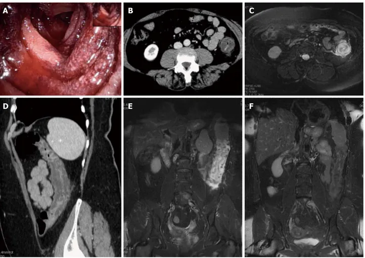
D
C B
A
E F
Figure 1 Magnetic resonance imaging follow-up of a patients with ischemic colitis resolved promptly. Ischemic colitis (IC) of left side colon in a 57 yr old
woman with a recent history of acute hypertensive crisis, who presented with left lower quadrant pain and massive rectal bleeding. A: Endoscopic procedure showed multiple necrotic area; B and C: Contrast-enhanced computed tomography (CT) and axial T2 fast-recovery fast-spin echo sequence (FRFSE) magnetic resonance imaging (MRI), after 32 h from CT, showed acute IC (Type Ⅰ CT and MRI) with wall thickening, three layer sandwich sign and a mild amount of free fluid in the paraco-lic gutter; D and E: 2D coronal reformat CT and coronal T2 FRFSE MRI, at the same time, showed the entire involved tract; F: Ischemia risolved without compparaco-lications with conservative therapy as shown in the follow-up MRI.
121 to 720 h) after the CT examination, according to a subacute form of IC; and (3) Type Ⅲ MRI, in the third group, which consisted of 2 patients, the MR appearance showed a wall thickening with a inversed 2 layer sign (high signal intensity of the outer layer and low signal intensity of the inner layer ). The MR examinations with Type Ⅲ
appearance were performed within a mean of 420.5 h (ranging from 384 to 457 h) after the CT examination, according to a subacute form of IC.
In the remaining 3 MRI examinations, performed within a mean of 410 h (ranging from 99.5 to 720 h) the colonic wall appeared normal.
No patients had circumferential intramural air consis-tent with pneumatosis coli.
Mild to moderate amounts of free abdomen fluid were present in all patients (100%) on the CT examina-tion, and in 4 out of 4 of the first MR examinations (performed within 48 h from the date of CT examina-tion) and in 1 out of 5 in the second MRI examinations (performed between 48 h and 15 d from the date of CT examination).
The free fluid was mainly located in the paracolic gut-ters and in the Douglas. No high-attenuation fluid consis-tent with blood was present. No reduction in caliber of mesenteric vessels was found.
On the basis of the result of the initial CT examina-tion, endoscopic findings and clinical evaluaexamina-tion, no sur-gery was performed on any of the patients. Sursur-gery was
performed 3 wk after the initial episode because of the development of a sealed-off perforation of the trans-verse colon in one of the patients.
The site of the affected tract and it’s length (mm), gross appearance on CT (type according Balthazar et al[14],
thickness of the affected wall and presence or absence of peritoneal fluid) and on MRI (type, thickness of the af-fected colonic wall and presence or absence of peritoneal fluid), in relation to the date of each examination were reported in Table 2.
DISCUSSION
The diagnosis of IC largely depends on clinical suspicion, especially since many other conditions (e.g., infectious
colitis, inflammatory bowel disease, diverticulitis, colon cancer) are presented with abdominal pain, diarrhoea and hematochezia[3]. Endoscopy has become the
diag-nostic test of choice in establishing the diagnosis of IC, although, it can be limited because it could be performed without bowel preparation to prevent hypoperfusion caused by dehydrating cathartics; in addition a minimal air insufflation should be used to prevent perforation[7,8].
From a radiological point of view, a CT examination is actually considered the main technique for the nonin-vasive diagnosis of mesenteric ischemia and also in cases of acute abdomen from different and various origins, be-cause it can suggest IC when it is unsuspected, can diag-D
C B
A
E F
Figure 2 Magnetic resonance imaging follow-up of a patient with ischemic colitis and worsening of clinical symptoms. Ischemic colitis (IC) of sigmoid colon
in a 62-year-old man with left lower quadrant pain and elevate lactate dehjdrogenase levels, who presented with melena and a recent history of stenting procedures for ischemic cardiopathy. A: Endoscopic procedure showed multiple necrotic area; B: 2D (two dimensional) coronal reformat contrast-enhanced computed tomogra-phy (CT) showed acute IC (Type Ⅰ CT); C-E: The patient had 2 magnetic resonance examinations (C and D-E) with an interval of 48 h due to worsening of clinical symptoms, with an increase of the length and thickness of the involved tract (D-E); F: The ischemic process resolved without complication after parenteral nutrition, as showed in the follow-up magnetic resonance imaging, performed after 384 h from the date of CT examination.
nose complications and exclude other illnesses. However it requires the use of ionizing radiation and an iodinates contrast agent, limiting the possibility to use this tech-nique in a short term follow-up[9-14].
Recently Iacobellis et al[15] has reported that MRI can
play a relevant role in the diagnostic management of acute IC and may be substituted for other invasive en-doscopic procedures in the diagnosis and grading of IC when an ischemic injury is suggested. Prior publications have described the feasibility of using MRI to evaluate a full range of colonic disease processes, including only one case of IC but without pathological confirmation and using precontrast and postcontrast imaging[16]. Our
study is based on the assumption that a parallel between the experimental colonic ischemic damage in the animal model and humans is reasonable[15]. Then our aim has
been to validate the utility of MRI in the clinical diagno-sis and follow-up of IC, using combined Fast Imaging Employing Steady State Acquisition on a coronal plane and T2-weighted fast-recovery fast-spin echo sequences, both on an axial and coronal plane. About 71.4% of patients that underwent a CT examination within 48 h showed Type Ⅰ of gross appearance of an involved co-lonic wall (wall thickening with heterogeneous enhance-ment and zones of low attenuation compatible with severe colonic edema and enhancement of the mucosa consistent with an acute process, a shaggy contour, a loss of colonic haustra, with varying degrees of pericolic streakiness). The same patients with Type Ⅰ CT showed Type Ⅰ appearance at MRI, according to an acute form of IC. The reason why the gross appearance of the in-volved colonic wall had a thickened and edematous ap-pearance is related to the fact that usually IC is a form of non-occlusive ischemic disease and in most cases, however, there is no evidence of obstruction of a major artery or vein. Then a decrease in blood flow to 20% of the normal flow, associated with small-vessel disease (hypoxia), and reperfusion injury when the blood flow is reestablished are the responsible factors[17,18].
Conse-quently, any part of the colon can be involved, with no
correlation established between the length and site of the involvement and distribution of the superior mesenteric or inferior mesenteric artery or vein[17,19,20]. Segments
commonly affected by IC are the splenic flexure (Griffith point) because the marginal artery of Drummond (a system of arcades connecting the major arteries) is oc-casionally tenuous here and is absent in upto 5% of pa-tients, and the anastomotic plexus between the inferior mesenteric artery distribution and the hypogastric vascu-lar supply (point of Sudeck) at the rectosigmoid junction, because it is distal to the last collateral connection with proximal arteries[7,21]. In our case population a segmental
distribution was apparent in 85.71% (6 patients).
The striking differences in the gross morphology of ischemic segment as detected at MRI is probably related to the timing of the examination and to the pathophysi-ology of the developing anoxic process. In the initial phases of anoxia, mucosal damage occurs first; with more severe and prolonged forms of anoxia, submucosa hemorrhage, edema, and pericolic congestive and edema-tous changes developing later due to the reperfusion event. Indeed, in the subacute phase (MRI examination performed between 3 and 30 d) the gross morphology has been changed (MRI Type Ⅱ or Ⅲ) with a reduction of the thickness of the involved wall (mean 7.2 mm, range 5-8 mm) and a double ring appearance (high signal intensity of the inner layer and low signal intensity of the outer layer for Ⅱ type MRI and high signal intensity of the outer layer and low signal intensity of the inner layer for Type Ⅲ MRI), probably for the reduction of edematous phenomena like the CT Type Ⅱ according to Balthazar.
Although up to 85% of cases of IC managed conser-vatively improve within 1 or 2 d and resolve completely within 1 or 2 wk, close to one-fifth of patients develop peritonitis or deteriorate clinically and require surgery. Surgical resection is required when an irreversible isch-emic injury and chronic colitis develop as both can lead to bacteremia and sepsis, colonic stricture, persistent ab-dominal pain and bloody diarrhoea, and protein-loosing
Table 2 Gross appearance on computed tomography and magnetic resonance imaging
Patient Involved tract/length (mm) CT Ⅰ range 2-168 h1 MRI Ⅰ whitin 48 h2 MRI Ⅱ > 48 h <15 d2 MRI Ⅲ > 15 d2
1 Left side colon and splenic flexure/314 Type Ⅰ/12 mm; free fluid
Type Ⅰ/10 mm; free fluid
Type Ⅲ/6 mm; no free fluid 2 Left side colon, splenic flexure and
transverse colon/609
Type Ⅰ/12 mm; free fluid
Type Ⅱ/7 mm; no free fluid 3 Left side colon and splenic flexure/380 Type Ⅱ/9.5 mm;
free fluid
Type Ⅱ/7 mm; no free fluid
normal appearance; no free fluid 4 Transverse colon/219 Type Ⅱ/6 mm;
free fluid
normal appearance; no free fluid 5 Entire colon/1000 Type Ⅰ/8.5 mm;
free fluid
Type Ⅰ/9.5 mm; free fluid 6 Left side colon/218 Type Ⅰ/17.5 mm;
free fluid
Type Ⅰ/15 mm; free fluid
Normal appearance; no free fluid 7 Sigmoid colon/145.5 Type Ⅰ/8.2 mm;
free fluid Type Ⅰ/7 mm; free fluid Type Ⅰ/8 mm; free fluid Type Ⅲ/5 mm; no free fluid MRI: Magnetic resonance imaging; CT: Computed tomography. 1 After the clinical onset; 2 After the date of the CT examination.
enteropathy[8]. The advantage of the use of MRI for
clinical management of IC, is the possibility to perform a short term follow-up without the employment of ion-izing radiation or intravenous contrast material. As dem-onstrated in our study MRI could be used, instead of CT to suggest the diagnosis of the IC in the proper clinical setting, particular when a segmental distribution is evi-dent, in the depiction of other abnormal conditions that may be seen in patients suspected of having IC, and in a short term follow-up, when a clinical worsening occurs, to adequately manage the patient.
Some limitations of the present study should be out-lined. Firstly, our patient selection process does not allow for an evaluation of the sensitivity or specificity of MRI for the diagnosis or detection of IC. The other major limitation of both CT and MR imaging in the diagnosis of colonic ischemia is the lack of specificity. The gross morphologic features overlap with those of inflamma-tory colitis, although the segmental distribution is more often seen in ischemia[22]. In spite of the limited number
of subjects who were examined, we are convinced that MRI can provide a valid imaging for the identification of pathological findings of acute IC. Moreover MRI in combination with clinical suspicion, endoscopic and his-tological findings, can play a key role in the diagnosis and management of IC. In particular MRI can discriminate patients with urgent operative intervention from patients in which a follow-up can be proposed as an alternative to surgery. All these allow for an earlier detection and ef-fective follow-up of IC with a possible earlier adequate treatment.
ACKNOWLEDGMENTS
We thank Miss Julia Hassall for reviewing the manuscript and Miss Francesca Seri and Mr. Duccio Guerrieri for performing the majority of MR examinations.
COMMENTS
BackgroundThe incidence of ischemic colitis (IC) is underestimated because it often has a mild transient nature, clinical presentation can be nonspecific and highly variable, therefore, the diagnosis largely depends on clinical suspicion. In this context the role of imaging techniques remains controversial. Standard radiol-ogy yields non-specific and late findings, while computed tomography (CT), the main technique for the noninvasive diagnosis of mesenteric ischemia, is well suited to confirm the clinical suspicion of IC, to suggest IC when it is unsus-pected and to diagnose complications, however it requires the use of radiation and an iodinates contrast agent, limiting the possibility to use this technique in a short term follow-up.
Research frontiers
The role of magnetic resonance imaging (MRI) in the diagnostic management of acute IC is still controversial, and nothing is known about the in vivo magnetic resonance findings of IC or about the relationship between MR findings and the onset of clinical symptoms.
Innovations and breakthroughs
To be known, this is the first study using MRI for the evaluation of IC and for the comparison between MRI and CT findings in this pathological condition. Applications
MRI in combination with clinical suspicion, endoscopic and histological findings,
can play a key role in the diagnosis and management of IC. In particular MRI can discriminate patients with urgent operative intervention from patients in which a follow-up can be proposed as an alternative to surgery. In particular the advantage of the use of MRI for clinical management of IC, is the possibility to perform a short term follow-up without the employment of ionizing radiation or intravenous contrast material.
Terminology
Fast imaging employing steady state acquisition sequence (FIESTA): provides images of fluid filled structures with very short acquisition times. The FIESTA sequence uses the T2 steady state contrast mechanism to provide high signal noise ratio images with strong signal from fluid tissues while suppressing back-ground tissue for contrast and anatomic detail of small structures; IC: inflamma-tion of the colon due to colonic ischemia resulting from alterainflamma-tions in systemic circulation or local vasculature.
Peer review
This paper is well written, is original and has good information. It merits to be admitted to publish without susbtantial changes.
REFERENCES
1 Boley SJ, Schwartz S, Lash J, Sternhill V. Reversible vascular
occlusion of the colon. Surg Gynecol Obstet 1963; 116: 53-60 [PMID: 13968597]
2 Paterno F, Longo WE. The etiology and pathogenesis of
vas-cular disorders of the intestine. Radiol Clin North Am 2008;
46: 877-885, v [PMID: 19103137]
3 Theodoropoulou A, Koutroubakis IE. Ischemic colitis:
clinical practice in diagnosis and treatment. World J
Gastro-enterol 2008; 14: 7302-7308 [PMID: 19109863 DOI: 10.3748/
wjg.14.7302]
4 Rha SE, Ha HK, Lee SH, Kim JH, Kim JK, Kim JH, Kim PN,
Lee MG, Auh YH. CT and MR imaging findings of bowel ischemia from various primary causes. Radiographics 2000;
20: 29-42 [PMID: 10682769]
5 Westgeest HM, Akol H, Schreuder TC. Pure
naratriptan-induced ischemic colitis: a case report. Turk J Gastroenterol 2010; 21: 42-44 [PMID: 20533112 DOI: 10.4318/tjg.2010.0047] 6 Stamatakos M, Douzinas E, Stefanaki C, Petropoulou C,
Arampatzi H, Safioleas C, Giannopoulos G, Chatziconstan-tinou C, Xiromeritis C, Safioleas M. Ischemic colitis: surging waves of update. Tohoku J Exp Med 2009; 218: 83-92 [PMID: 19478463]
7 Baixauli J, Kiran RP, Delaney CP. Investigation and
man-agement of ischemic colitis. Cleve Clin J Med 2003; 70: 920-91, 920-91, 920-91, passim [PMID: 14650467]
8 Elder K, Lashner BA, Al Solaiman F. Clinical approach to
colonic ischemia. Cleve Clin J Med 2009; 76: 401-409 [PMID: 19570972 DOI: 10.3949/ccjm.76a.08089]
9 Angelelli G, Scardapane A, Memeo M, Stabile Ianora
AA, Rotondo A. Acute bowel ischemia: CT findings. Eur
J Radiol 2004; 50: 37-47 [PMID: 15093234 DOI: 10.1016/
j.ejrad.2003.11.013]
10 Romano S, Romano L, Grassi R. Multidetector row comput-ed tomography findings from ischemia to infarction of the large bowel. Eur J Radiol 2007; 61: 433-441 [PMID: 17157468 DOI: 10.1016/j.ejrad.2006.11.002]
11 Mazzei MA, Mazzei FG, Marrelli D, Imbriaco G, Guerrini S, Vindigni C, Civitelli S, Roviello F, Grassi R, Volterrani L. Computed tomographic evaluation of mesentery: diagnostic value in acute mesenteric ischemia. J Comput Assist Tomogr 2012; 36: 1-7 [PMID: 22261763]
12 Mazzei MA, Guerrini S, Cioffi Squitieri N, Imbriaco G, Mazzei FG, Volterrani L. Non-obstructive Mesenteric Isch-emia after Cardiovascular Surgery: not so uncommon. Ann
Thorac Cardiovas 2013; In press
13 Mazzei MA, Guerrini S, Cioffi Squitieri N, Genovese EA, Mazzei FG, Volterrani L. [Diagnosis of acute mesenteric ischemia/in-farction in the era of multislice CT]. Recenti Prog Med 2012; 103: 435-437 [PMID: 23096727 DOI: 10.1701/1166.12884]
14 Balthazar EJ, Yen BC, Gordon RB. Ischemic colitis: CT evalua-tion of 54 cases. Radiology 1999; 211: 381-388 [PMID: 10228517] 15 Iacobellis F, Berritto D, Somma F, Cavaliere C, Corona M,
Cozzolino S, Fulciniti F, Cappabianca S, Rotondo A, Grassi R. Magnetic resonance imaging: a new tool for diagnosis of acute ischemic colitis? World J Gastroenterol 2012; 18: 1496-1501 [PMID: 22509081 DOI: 10.3748/wjg.v18.i13.1496] 16 Chung JJ, Semelka RC, Martin DR, Marcos HB. Colon
dis-eases: MR evaluation using combined T2-weighted single-shot echo train spin-echo and gadolinium-enhanced spoiled gradient-echo sequences. J Magn Reson Imaging 2000; 12: 297-305 [PMID: 10931593]
17 Brandt LJ, Boley SJ. Ischemic and vascular lesions of the bowel. In: Sleisenger MH, Fordtran JS, editors. Gastrointes-tinal disease: pathophysiology, diagnosis, management. 5th ed. Philadelphia: Saunders, 1993: 1940-1945
18 Zimmerman BJ, Granger DN. Reperfusion injury. Surg Clin
North Am 1992; 72: 65-83 [PMID: 1731390]
19 Wittenberg J, Athanasoulis CA, Williams LF, Paredes S, O’ Sullivan P, Brown B. Ischemic colitis. Radiology and patho-physiology. Am J Roentgenol Radium Ther Nucl Med 1975; 123: 287-300 [PMID: 1115306]
20 Bharucha AE, Tremaine WJ, Johnson CD, Batts KP. Ischemic proctosigmoiditis. Am J Gastroenterol 1996; 91: 2305-2309 [PMID: 8931407]
21 Roger AI, David S. Intestinal blood flow and disease of vascular impairment. In: Haubrich WS, Schaffner F, Berk JE, editors. Gastroenterology. 5th ed. Philadelphia: Saunders, 1995: 1212-1234
22 Philpotts LE, Heiken JP, Westcott MA, Gore RM. Colitis: use of CT findings in differential diagnosis. Radiology 1994; 190: 445-449 [PMID: 8284397]
P- Reviewer Rodrigo L S- Editor Wen LL L- Editor A E- Editor Zhang DN