DIPARTIMENTO DI INGEGNERIA CHIMICA, MATERIALI, AMBIENTE
Corso di Dottorato di ricerca in Ingegneria chimica XXX ciclo
Buccal and Topical drug delivery
Candidato:
Gabriele Varani
Relatore: Chiar.ma Prof.ssaAlessandra Adrover
Anno Accademico 2016–2017
Ph.D Thesis. Sapienza-University of Rome Author: Gabriele Varani, Roma, 2017 email: [email protected]
This thesis has been typeset by LATEXand the Book class.
A thesis submitted in partial fulfillment of the requirements for the degree of Doctor of Philosophy in Ingegneria chimica.
Ottobre 2017 Thesis not yet defended.
Dedicated to Riccardo...
...perchè vorrei portarti in tutti i
posti che sogni.
Abstract
The aim of this work is to investigate new and classical techniques, methods and formu-lations for topical and buccal release.
All the formulations proposed are based on natural and biocompatible polymer ma-trices such as gellan gum, scleroglucan and hydroxypropylmethylcellulose.
The proposed formulations are tested by in vitro release tests. In fact they represents a valid support and a useful starting point for the realization of a potentially usable in-vivo pharmaceutical formulation that may have commercial utility.
The research work is both experimental and theoretical. Each topic presents a more chemical-pharmaceutical part, based on the formulation preparation and release exper-iments, and a more theoretical-numerical approach that allows a correct interpretation and description of the experimental data obtained.
Release from hydrogels and thin films require different modelling approaches. Also the physico-mathematical description of different release experiments (different release devices such as Franz cell, millifluidic device and USP II) requires different theoretical and numerical techniques.
The outcome of an accurate model development is of fundamental importance for future design of pharmaceutical formulations with prescribed release properties.
In addition the formulations are investigated through rheological, mechanical, thermo-analytic and mucoadhesive tests in order to have a more comprehensive picture of their practical utilization.
Contents
1 General Introduction 1
1.1 Thin films for buccal drug delivery . . . 1
1.1.1 Anatomy of the oral mucosa . . . 2
1.1.2 Oral thin films: a general presentation . . . 6
1.1.3 Polymers for the preparation of thin films . . . 8
1.1.4 Mucoadhesive properties of buccal films . . . 9
1.1.5 Films evaluation . . . 10
1.1.6 Manufacturing techniques . . . 13
1.1.7 In vitro dissolution testing of buccal thin films . . . 15
1.2 Hydrogels for topical drug delivery . . . 19
1.2.1 Anatomy of skin . . . 20
1.2.2 Formulation design of Hydrogels . . . 23
1.2.3 Hydrogels evaluation . . . 27
1.2.4 In vitro release from Hydrogels . . . 29
1.3 Oral thin films and Hydrogels in this work . . . 33
1.3.1 Gellan gum, HPMC, Scleroglucan . . . 33
1.3.2 Cyclodextrins(CD) . . . 36
2 Aim of the work 41 3 Scleroglucan based hydrogel for topical drug delivery 43 3.1 Introduction . . . 43
3.2 Materials and methods . . . 44
3.2.1 Chemicals . . . 44
3.2.2 Synthesis of carboxymethyl scleroglucan (Scl − CM300) . . . 44
3.2.3 Hydrogel preparation . . . 44
3.2.4 Rheological characterization . . . 45
3.2.5 Release studies with Franz diffusion cell . . . 45
3.2.6 Mathematical models of permeation experiments in a vertical Franz cell . . . 46
3.2.7 Primary skin irritation experiments . . . 48
3.3 Results . . . 49
3.3.1 Physical hydrogel preparation and characterization . . . 49
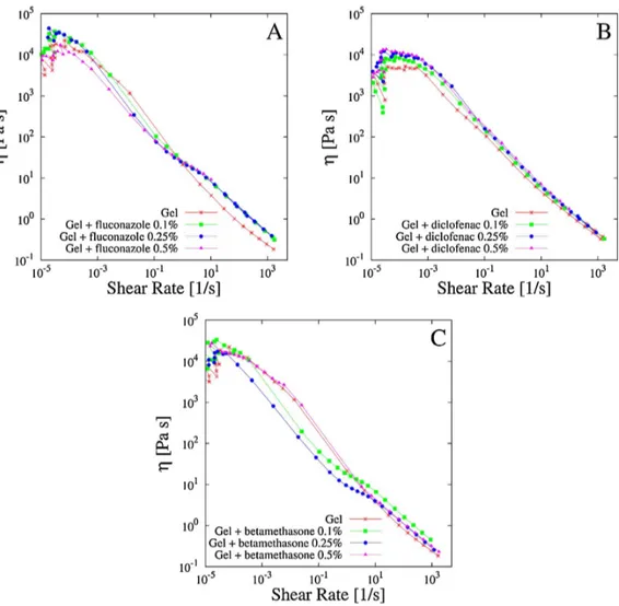
3.3.2 Rheological characterization of drug loaded Scl − CM300 physical hydrogels . . . 49
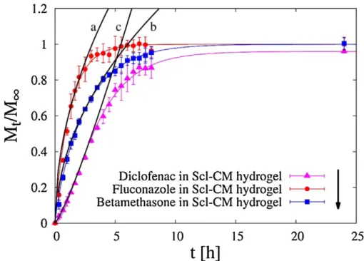
3.3.3 Release studies . . . 53
3.3.4 Primary skin irritation experiments . . . 57
3.4 Conclusion . . . 58
4 Drug delivery from Oral thin films 59 4.1 Introduction . . . 59
4.2 Materials and method . . . 59
4.2.1 Chemicals . . . 59
4.2.2 Film forming technology . . . 60
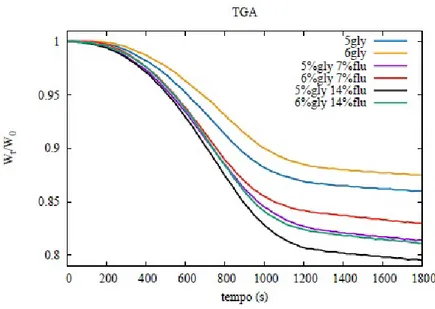
4.2.3 Thermogravimetric analysis . . . 60
4.2.4 Swelling . . . 61
4.2.5 USP II, Paddle apparatus . . . 62
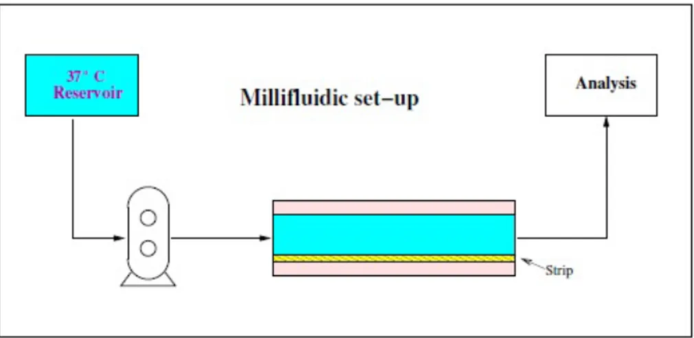
4.2.6 Novel millifluidic device . . . 62
4.3 Results . . . 65
4.3.1 Swelling model for OTF . . . 65
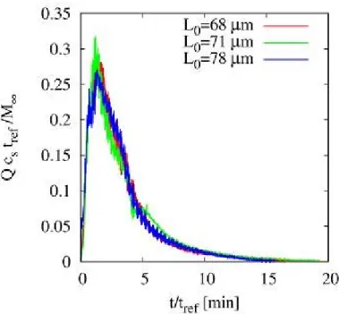
4.3.2 Drug release time scales . . . 69
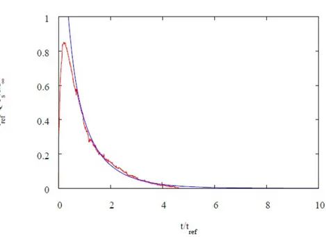
4.3.3 Model of release . . . 71
4.3.4 2D-Model . . . 75
4.4 Conclusion . . . 79
5 Release analysis from HPMC based erodible thin films 81 5.1 Introduction . . . 81
5.1.1 Rapid disintegrating film (RDF) . . . 81
5.1.2 Furosemide features . . . 82
5.2 Materials and method . . . 83
5.3 Results and discussion . . . 84
5.3.1 Preliminary analysis . . . 84
5.3.2 Release from Franz Cell . . . 85
5.3.3 Swelling-Erosion tests . . . 95
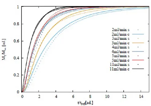
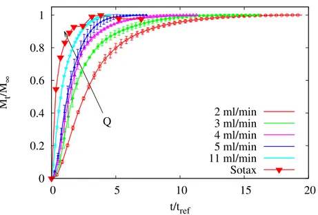
5.3.4 Sotax Vs Millifluidic device . . . 96
5.4 Conclusion . . . 98
6 Preventing drug crystallization in glycerol-plasticized gellan gum thin films 99 6.1 Introduction . . . 99
6.2 Materials and method . . . 100
6.2.1 Materials . . . 100
6.2.2 Preparation of polymeric films . . . 100
6.2.3 Preparation of inclusion complex cyclodextrins-drug . . . 101
6.2.4 Rheological studies . . . 101
6.2.5 Thickness measurements . . . 101
6.2.6 Thermogravimetric analysis . . . 101
6.2.7 Differential scanning calorimetry . . . 101
6.2.8 Tensile tests . . . 102
6.2.9 Swelling studies . . . 102
6.2.10 Mucoadhesion tests . . . 102
6.2.11 Uniformity of drug content tests . . . 102
6.2.12 In vitro release studies . . . 103
6.3 Results and discussion . . . 103
6.3.1 Preparation of GG:Gly thin films . . . 103
6.3.2 Mechanical properties of GG:Gly thin films . . . 106
6.3.3 Swelling studies . . . 108
6.3.4 Evaluation of the mucoadhesive properties of GG:Gly thin films . . 108
6.3.5 Drug loaded films . . . 109
6.3.6 Release studies . . . 113
6.4 Conclusion . . . 115
7 General conclusions 117
Chapter 1
General Introduction
1.1
Thin films for buccal drug delivery
The oral route is the most important method of administration for systemic effect, due to low cost, ease of administration and high level of patient compliance. Among the pharmaceutical dosage form for oral delivery, the conventional tablet seems to be most popular, because of its ease of transportability and comparatively low manufacturing cost [1]. Nonetheless, two main types of disadvantages can be ascribed to the oral route: first pass drug metabolism in the liver and pre-systemic elimination of the drug in the gastrointestinal tract (GI first pass) could lessen the effective biodisponibility to an un-acceptable level, or even destroy the drug, as it is the case of biological active ingredients as proteins and peptides; moreover, a fast administration of emergency drugs and agents with a rapid onset action could not be possible [2]. In these cases, the traditional choice is parenteral drug administration, which is, on the patient side, uncomfortable, uneasy and undesired. Difficulties associated with parenteral delivery and poor drug availability, together with substantial efforts focused on placing a drug or drug delivery system in a particular region of the body for extended periods of time, provided the impetus for exploring alternative routes, such as the mucosal layer lining a number of regions of the gastrointestinal tract, the airways, mouth, the ear, nose, and the eye.
Mucosal tissues can interact with the hydrophilic macromolecules of drug matrices, bringing about an adhesive attachment called mucoadhesion. This interaction retains a formulation in intimate contact with the adsorption site. The buccal region of oral cavity is the most attractive site for the delivery of drugs. Buccal drug delivery involves the administration of desired drug through the buccal mucosal membrane lining of the oral cavity. This route is useful for mucosal (local effect) and transmucosal (systemic effect) drug administration. In the first case, the aim is to achieve a site-specific release of the drug on the mucosa, whereas the second case involves drug absorption through the mucosal barrier to reach the systemic circulation [3–7].
The buccal mucosa is permeable, with a rich blood supply, more robust and have more tolerance to potential sensitizers in comparison to the other mucosal tissues. No activation of the drug absorption is required. Local modification of tissue permeability, inhibition of protease activity and reduction in immunogenic response are allowed.
On the other hand, the environment of the oral cavity presents some significant chal-lenges for systemic drug delivery, given that the mucosa has barrier properties. The principle physiological environment of the oral cavity, in terms of pH, fluid volume and composition, is shaped by the secretion of saliva.Saliva covers the surface area of the mouth (around 217 cm2)with a layer of average thickness 70-100 µm for adults and 60-90 µm for children. It is continuously secreted at average flow rate of 0.3-1 ml/min, but can be elicitated up to 7.07 ml/min by stimulating agents; flow rates < 0.1 ml/min must be considered pathological. The volume of saliva in the mouth ranges from 0.8 ml, after swallowing, to 1.1 ml just before swallowing [8–11]. The continuous secretion of saliva
Figure 1.1: Structure of oral cavity.
leads to subsequent dilution of the drug; saliva swallowing can provoke the partial or total removal of the dosage form. The volume of saliva constantly present in the mouth, around 1 ml, provides a relatively low fluid volume available for drug release from delivery systems compared to the GI tract [12, 13].
The different types of buccal dosage forms are buccal tablets, ointments, gels, patches and thin films [14]. Over and above the mentioned demands, oral delivery systems alternative to tablets and syrups have been of interest since the latest 1970’s, as a possible improvement for a particular, and not so small, class of patients who have difficulties in swallowing. This disorder affects more frequently for old people, children and mentally illness, but the oral administration without swallowing could be helpful also for travelling or military patients who may not have ready access to water [15, 16].
1.1.1
Anatomy of the oral mucosa
Buccal region is that part of the mouth bounded anteriorly and laterally by the lips and the cheeks, posteriorly and medianly by the teeth and/or gums, and above and below by the reflections of the mucosa from the lips and cheeks to the gums.
Numerous racemose, mucous, or serous glands are present in the sub mucous tissue of the cheeks. The buccal glands are placed between the mucous membrane and buccinator muscle: they are similar in structure to the labial glands, but smaller ( figure 1.1).
About five, of a larger size than the rest, are placed between the masseter and buc-cinator muscles around the distal extremity of the parotid duct; their ducts open in the mouth opposite the last molar tooth. They are called molar glands. Maxillary artery sup-plies blood to buccal mucosa and blood flow is faster and richer (2.4ml/min/cm2) than that in the sub lingual, gingival and palatal regions, thus facilitates passive diffusion of drug molecules across the mucosa.
The thickness of the buccal mucosa is measured to be 500–800 µm and is rough textured, hence suitable for retentive delivery systems. The turnover time for the buccal
epithelium has been estimated at 5–6 days. Buccal mucosa composed of several layers of different cells as shown in figure 1.2. The epithelium is similar to stratified squamous epithelia found in rest of the body and is about 40–50 cell layers thick.
Lining epithelium of buccal mucosa is the nonkeratinized stratified squamous epithe-lium that has thickness of approximately 500–600 µm and surface area of 50.2 cm2.
Basement membrane, lamina propria followed by the sub mucosa is present below the epithelial layer. Lamina propria is rich with blood vessels and capillaries that open to the internal jugular vein [17].
The barriers such as saliva,mucus,membrane coating granules, basement membrane etc retard the rate and extent of drug absorption through the buccal mucosa. The main penetration barrier exists in the outermost quarter to one third of the epithelium.
Oral mucosa, a barrier to permeability
The effective permeability coefficient (Peff) values reported in the literature across the buccal mucosa for different molecules range from a lower limit of 2.2 · 10−9 cm/s for
dextran 4000 across rabbit buccal membrane to an upper limit of1.5 · 10−−5 cm/s for
both benzylamine and amphetamine across rabbit and dog buccal mucosa, respectively. This range clearly demonstrates the presence of a permeability barrier in the oral mucosa, which is mostly imposed by the oral epithelium acting as a protective layer for the tissues beneath, and as a barrier to the entry of foreign material and microorganisms. However, this range is estimated to be 4–4000 times more permeable than that of skin. The permeability barrier property of the oral mucosa is predominantly due to inter cellular materials derived from the so-called "membrane coating granules" (MCGs). MCGs are spherical or oval organelles that are 100–300 nm in diameter and found in both keratinized and non-keratinized epithelia.
These organelles have also been referred to as "small spherically shaped granules", "corpusula", "small dense granules", "small lamellated bodies", "lamellated dense bod-ies", "keratinosomes", "transitor dense bodbod-ies", and "cementsomes" . However, most of these descriptive names have not fully defined the functions of this cellular species. MCGs were first named as such because it was believed that they were subject to ex-ocytosis from the cytoplasm of the stratum spinosum of keratinized epithelia following thickening of these cells. Nonetheless, it is actually the contents of MCGs that are subject to exocytosis prior to the onset of membrane thickening.
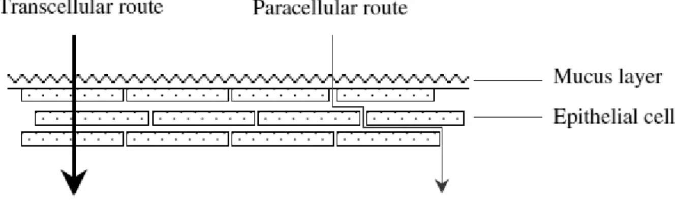
The main mechanisms responsible for the penetration of various substances include simple diffusion (paracellular, transcellular), carrier-mediated diffusion, active transport, and pinocytosis or endocytosis. Recent evidence has shown that passive diffusion is the primary mechanism for the transport of drugs across the buccal mucosa, although carrier-mediated transport has been reported to have a small role.
Two routes of passive transport are available in the buccal epithelium(figure 1.3); one involves the transport of compounds through the inter cellular spaces between the cells (paracellular), and the other involves passage into and across the cells (transcellular). De-pending on the nature of the permeant, the molecular geometry, lipophilicity, and charge, either of the transport pathways across buccal epithelium can be selected. While consid-erable evidence has been presented to document that most compounds diffuse through the buccal mucosa by passive diffusion or simple Fickian diffusion, some are transported by a carrier mediated process across the buccal mucosa. Glucose, monocarboxylic acids and salicylic acid, and nicotinic acid, are examples of substances which utilize a carrier-mediated diffusion mechanism for permeation across buccal epithelium [18].
Mucus
The epithelial cells of buccal mucosa are surrounded by the intercellular ground substance called mucus with the thickness varies from 40 µm to 300 µm. Though the sublingual glands and minor salivary glands contribute only about 10% of all saliva, together they produce the majority of mucus and are critical in maintaining the mucin layer over the
Figure 1.2: A sketch of the buccal mucosa.
oral mucosa. It serves as an effective delivery vehicle by acting as a lubricant allowing cells to move relative to one another and is believed to play a major role in adhesion of mucoadhesive drug delivery systems.
At buccal pH, mucus can form a strongly cohesive gel structure that binds to the epithelial cell surface as a gelatinous layer. Mucus molecules are able to join together to make polymers or an extended three-dimensional network.Different types of mucus are produced, for example G, L, S, P and F mucus, which form different network of gels. Other substances such as ions, protein chains, and enzymes are also able to modify the interaction of the mucus molecules and, as a consequence, their bio-physical properties.
Mucus is composed chiefly of mucins and inorganic salts suspended in water. Mucins are a family of large, heavily glycosylated proteins composed of oligosaccharide chains attached to a protein core. Three quarters of the protein core are heavily glycosylated and impart a gel like characteristic to mucus. Mucins contain approximately 70–80% carbohydrate, 12–25% protein and up to 5% ester sulphate.
The dense sugar coating of mucins gives them considerable water-holding capacity and also makes them resistant to proteolysis, which may be important in maintaining mucosal barriers. Mucins are secreted as massive aggregates by prostaglandins with molecular masses of roughly 1 to 10 million Da. Within these aggregates, monomers are linked to one another mostly by non covalent interactions, although intermolecular disulphide bonds also play a role in this process.
Oligosaccharide side chains contain an average of about 8–10 monosaccharide residues of five different types namely L-fucose, D-galactose, D-glucosamine, N-acetyl-D-galactosamine and sialic acid. Amino acids present are serine, threonine and proline. Because of the presence of sialic acids and ester sulfates, mucus is negatively charged at physiological salivary pH of 5.8–7.4 [19–21].
Saliva
The mucosal surface has a salivary coating estimated to be 70 µm thick, which act as unstirred layer. Within the saliva there is a high molecular weight mucin named MG1 that can bind to the surface of the oral mucosa so as to maintain hydration, provide lubrication, concentrate protective molecules such as secretory immunoglobulins, and limit the attachment of microorganisms. Several independent lines of evidence suggest that saliva and salivarymucin contribute to the barrier properties of oral mucosa.
The major salivary glands consist of lobules of cells that secrete saliva; parotids through salivary ducts near the upper teeth, submandibular under the tongue, and the sublingual through many ducts in the floor of the mouth. Besides these glands, there are 600–1000 tiny glands called minor salivary glands located in the lips, inner cheek area (buccal mucosa), and extensively in other linings of the mouth and throat.
Total output from the major and minor salivary glands is termed as whole saliva, which at normal conditions has flow rate of 1–2 ml/min. Greater salivary output avoids potential harm to acid-sensitive tooth enamel by bathing the mouth in copious neutral-izing fluid.
With stimulation of salivary secretion, oxygen is consumed and vasodilator sub-stances are produced; and the glandular blood flow increases, due to increased glandular metabolism.
Saliva is composed of 99.5% water in addition to proteins, glycoproteins and elec-trolytes. It is high in potassium (7·plasma), bicarbonate (3·plasma), calcium, phospho-rous, chloride, thiocyanate and urea and low in sodium(1/10·plasma).
The normal pH of saliva is 5.6-7. Saliva contains enzymes namely α-amylase (breaks 1–4 glycosidic bonds), lysozyme (protective, digests bacterial cell walls) and lingual lipase (break down the fats). Saliva serves multiple important functions. It moistens the mouth, initiates digestion and protects the teeth from decay. It also controls bacterial flora of the oral cavity. Because saliva is high in calcium and phosphate, it plays a role in mineralization of new teeth repair and precarious enamel lesions. It protects the teeth
by forming “protective pellicle”. This signifies a saliva protein coat on the teeth, which contains antibacterial compounds.
However, salivary flow rate may play role in oral hygiene. Intraoral complications of salivary hypofunction may cause candidiasis, oral lichen planus, burning mouth syn-drome, recurrent aphthous ulcers and dental caries.
A constant flowing down of saliva within the oral cavity makes it very difficult for drugs to be retained for a significant amount of time in order to facilitate absorption in this site.
In general, intercellular spaces pose as the major barrier to permeation of lipophilic compounds, and the cell membrane which is lipophilic in nature acts as the major trans-port barrier for hydrophilic compounds because it is difficult to permeate through the cell membrane due to a low partition coefficient. Permeabilities between different regions of the oral cavity vary greatly because of the diverse structures and functions. In gen-eral, the permeability is based on the relative thickness and degree of keratinization of these tissues in the order of sublingual> buccal> palatal. The permeability of the buccal mucosa was estimated to be 4–4000 times greater than that of the skin [13, 17, 18, 22, 23].
1.1.2
Oral thin films: a general presentation
Similar in size, shape and thickness to a postage stamp, pharmaceutical thin films (figure 1.4) were developed from skin patches technology. They firstly appeared in the 1970’s as breath mint in the confectionery industry and gradually spread in cosmetics, nutraceuti-cal and over-the-counter products. After the approval of Zuplenz and Ondasetron from the Food and Drug Administration (USA) in 2010, they can be considered now as the most advanced oral solid dosage form [24,25]. Pharmaceutical thin films are equivalently called in literature "strips", "dissolving films" and "orodispersible films" by the Euro-pean Medicines Agency [26, 27]. Also the term "wafer" is improperly used, although this word refers to a similar kind of dosage form. Pharmaceutical films are typically designed to be placed on or under the tongue for oral administration, but they can be used in principle for any other mucosal tissue [28].
Films for oral administration are equivalently called "buccal" or "oral" films. De-pending on thickness and disintegration time, three categories can be qualitatively iden-tified. In order of size (increasing): flash release films, mucoadhesive melt-away wafers and mucoadhesive sustained release wafers [29]. Disintegration time is usually scaled to thickness. In the present default of any standardized definition or method, this classi-fication should be considered merely suggestive. Focus of this work is mainly on films with thickness < 100µm.
In conctat with biological fluids, films rapidly disintegrate and dissolve to release the medication, in the mouth (buccally or sublingually) and/or via the small intestines (en-terically), without the need of water and improving the efficacy of the active ingredients. In the mouth, a thin film gets instantly wet by saliva, rapidly hydrates and adheres onto the site of application. [30]. Unlike other rapid dissolving dosage forms, films can be produced with a manufacturing process that is competitive with the manufacturing costs of conventional tablets [31]. Just few drugs have been already commercialized in this form because of the complexity associated mainly with its novelty. Among marketed products, there are anti-emethic, antihistaminic, analgesic. The first marketed product under medical prescription has been Zuplenz/Ondasetron.
An extensive pioneer medical work exploring the administration of this drug in oral strips is [32]. Strips have been recognized as an effective and highly patient compliant drug delivery system [33]. Compliance is significative in particular when considering cases of mental-illness, emesis, or children, geriatric, disphagic patients. In addition, pharmaceutical industries show an increasing interest in oral films, as they can be used to elongate patent-life of existing active principles by a new administration form [34, 35].
In summary, the principal advantages of oral strips are [36–38, 55]:
• unobstructivity, high compliance for patients with swallowing problems, no special training is required for the administration of dosage form;
• no need of water, drug is wet by saliva;
• quick dissolution and release, with a relatively large surface area and a very high ratio area/thickness, for rapid wetting;
• precision in the administered dose is ensured from each of the strips; • systemic and local action;
• they can be mucoadhesive; • overcoming of first pass effect;
• to be not so brittle as oral dissolving tablets, and so there are less transport and storage difficulties;
• no special set up for industries are required and production costs are competitive; • new business opportunities, like patent extension, product promotion and product
differentiation.
On the other hand, most significative disadvantages are:
• expensive packaging, since the dosage form is moisture sensitive;
• taste masking of drug should be done and there are limited taste masking options; • drugs which irritate the mucosa are forbidden;
• high doses of drug cannot be incorporated. OTF are typically made of [39]:
• a filming hydrophilic polymer (around 45% by weight); • an active pharmaceutical ingredient (API, up to 30 % ); • plasticizer (0-20 %);
• other additives, such as fillers, surfactants, saliva stimulating agents and sweeting agents (up to % 40).
Drug and polymer are the essential components of OS. Knowledge and techniques about including drugs in thin films have been continuously increasing in the last ten years [40–54]. Desiderable characteristics are a low required dose ( < 40 mg), low molecular weight, possibly good taste.
The drug should also be stable and soluble both in saliva and water and partially unionized in water. The active component must be able to pass the mucosa barrier, not provoking irritations. Eligible API are anti emetic, neuroleptics, cardiovascular agents, analgesics, anti allergic, anti epileptics, anxiolytics, sedatives, hypnotics, diuretics, tiparkinsonism agents, anti-bacterial agents and drugs used for erectile dysfunction, an-tialzheimers,expectorants, antitussive.For mucosal and transmucosal administration, con-ventional dosage forms are not able to assure therapeutic drug levels in the mucosa and circulation because of the physiological removal of the oral cavity (washing effect of saliva and mechanical stress), which take the formulation away from the mucosa, resulting in a very short exposure time and unpredictable distribution of the drug on the site of action/absorption. To obtain the therapeutic action, it is therefore necessary to prolong and improve the contact between the active substance and the mucosa [20].
1.1.3
Polymers for the preparation of thin films
Polymers are the backbone of film formulations and various polymers are available for the preparation of thin films [55]. The polymers can be used alone or in combination with other polymers to achieve the desired film properties. The polymers employed should be non-toxic, non-irritant, and absence of leachable impurities is required.Water-soluble polymers are used as film formers to produce a thin film with rapid disintegration, good mechanical strength, and good mouth feel effects. Both natural and synthetic polymers are used for film preparation [56, 57]. Availability of diverse polymers allows imparting specific properties in the thin films. For instance, gelatine are available in different molecular weights, and thus the appealing and glossy films could be obtained with the gelatin having a high molecular weight. Pullulan is frequently used for producing a thin film with great solubility,high mechanical strength and they are stable over a wide range of temperatures. The blending of chitosan and high methoxy pectin (HMP) or low methoxy pectin (LMP) resulted in a thin film exhibiting an excellent mechanical strength. The film forming polymers such as hydroxypropyl cellulose (HPC), methyl cellulose, and carboxymethyl cellulose (CMC) produce a thin film with less water vapor barrier due to hydrophilic nature which aids in water retention [58, 59].
The basic idea of pharmaceutical thin strips is exactly that a dissolved component could be immobilized in a solid film, and be released from a gel as soon as the dosage form comes in conctat with biological fluids.
Natural and semi-natural polymers have also been reported in the literature as mu-coadhesive. Chitosan was first introduced in 1994 by Guo for its use in mucoadhesive film formulations [68]. Following Carbopol and HPMC as polymeric matrices for mu-coadhesive films, chitosan exhibited better adhesion than acacia in a peeling test using an Instron 4201.
Mucoadhesive films are thin and flexible retentive dosage forms, and release drug di-rectly into a biological substrate. They facilitate in extending residence time at the appli-cation site leading to prolonged therapeutic effects [65]. Majority of the thin film having mucoadhesive properties are hydrophilic in nature and undergoes swelling and form a chain interaction with the mucin.Among the several studied polymers, the most com-pelling mucoadhesion properties are exhibited by chitosan, hyaluronan, cellulose deriva-tives, polyacrylates, alginate, gelatin and pectin [66]. Compared with non-ionic polymers, the cationic and anionic polymers facilitate strong interaction with mucus [67].
Plasticizers can significatively help OTF formulation. They improve mechanical prop-erties such as flexibility and brittleness, by reducing the glass transition temperature of the filming polymer. Plasticizers should be compatible with the drug, as well as other additives used in the preparation of OTF. Most attractive plasticizers for OTF are glyc-erol, propylene glycol, low molecular weight polyethylene glycols, phthalate derivatives
like dibutyl phthalate, citrate derivatives such as tributyl, triethyl, acetyl citrate, cas-tor oil [60, 61]. Improving palatability is probably the first reason that why additives are added in OTF. A simple obscuration technique, which means mixing and blend-ing bitter tastblend-ing API with pleasurable taste substances, can be used. Also, barrier techniques, which includes complexation, polymeric coating and coated particle, have recently appeared. Sweetening and flavoring agents are typical additives for palatabil-ity improvement. Sweetening agents are the most major part of the food product or in pharmaceutical dosage forms proposed to be disintegrated or dissolved in the oral cavity. Natural as well as artificial sweetening agents are used to improve the palatability of the formulation. Common agents are sugar, dextrose, lactose, mannitol, sucrose, xyli-tol, malixyli-tol, acesulfame potassium, talin, glycyrrhizin, sucralose, aspartame, saccharin, essential oils or water soluble extracts of menthol, wintergreen, peppermint, sweet mint, spearmint, vanillin, cherry, chocolate, cinnamon, clove, lemon, orange. Sweetening agent are generally used either alone as in combination between the concentrations of 3 to 6 % by weight of the film. Selection of flavoring agents is depending on which type of drug is to be incorporated in the formulation. The recognition of the oral disintegrating / dissolving formulation by an individual, depends on the initial flavor quality, which is observed in the first few seconds after the product has been consumed, and on the after taste of the formulation, which lasts for at least about 10 min.
Other functional additives are surfactants and saliva stimulating agents. Surfactants are used as a solubilizing or wetting dispersing agent so that the film gets dissolved within seconds and releases active agent immediately. Some of the commonly used are sodium lauryl sulfate, benzalkonium chloride, tweens, poloxamer 407. Saliva stimulating agents, as citric acid, malic acid, lactic acid, ascorbic acid and tartaric acid, are used to increase the rate of production of saliva. This would aid in the more rapidly disintegration of fast dissolving film formation. Saliva stimulating agents are used alone as well as in combination between 2 to 6 % w/w by weight. Finally, fillers and colorants can be added to improve film’s aspect and handling. Typical colorants are natural coloring agents, and natural juice concentrates, pigments such as titanium oxide, silicon dioxide and zinc oxide. Maximum colorants’s concentration is 1 % by weight [62–64].
In common terms, polymers are understood as excipients, but it has become an es-sential component while designing and formulating thin films.Therefore, understanding the properties of polymers such as chemistry, rheology, and physicochemical proper-ties of polymer seems to be imminent for maximizing their uses to develop a thin film. The selection of appropriate polymer during the development of polymeric thin films may be critical; thereby, several points should be considered according to the require-ments.Therefore, it is imperative to consider the appropriate polymer for producing a thin film with a better performance that assures high therapeutic success.
1.1.4
Mucoadhesive properties of buccal films
Bioadhesion is the general term describing adhesion between any biological and synthetic surface. Mucoadhesion is a specific term describing the particular interaction of a mucosal membrane with a synthetic surface [69]. The phenomenon of mucoadhesion has been explained by applying any of the five theories of adhesion into the interaction of the dosage form and the biological substrate [70, 71]: electronic [72], adsorption [73, 74], wetting [75], diffusion [76], and fracture theory [77]; here, we briefly summarize theories related to mucoadhesion theory. Since mucoadhesive buccal films include the interaction of a dry polymeric matrix that undergoes hydration, drug release, and sometimes erosion, the phenomenon is very complex. Smart has defined four possible scenarios for the analysis of the mucoadhesion process based on the hydration state of the dosage form and on the amount of mucus layer available for mucoadhesion [78].
Mucoadhesive buccal films can be classified as a "case 3" scenario since they are solid dry substrates that come in contact with a mucosa having thin or discontinuous mucus layers. Relevant to the analysis of the mucoadhesion of polymeric films on the buccal mucosa are the adhesion theories of adsorption and diffusion. The adsorption
theory states that the main contributors to the adhesive bond are the inter-polymer interactions, such as hydrogen bonds and van der Waals forces [79]. The diffusion theory assumes that polymeric chains from the solid substrate, i.e. the mucoadhesive film, and the biological substrate, i.e. mucin in the mucosa layer, interdiffuse across the adhesive interface. Important variables in this process are the diffusion coefficient of the polymer into the mucin layer and vice versa, the contact time, and the molecular chain length and their mobility [79, 80].
Most of the mucoadhesive phenomena have two main stages that control the perfor-mance of the dosage form: the contact stage and the consolidation stage (figure 1.5) [81]. Since mucoadhesive films are dosage forms that are brought in contact with the biological membrane by the patient, the contact stage is initiated by the patient. During the con-tact process, the film will start dehydrating the mucus gel layer and will itself hydrate, initiating the interpenetration of the polymeric chains into the mucus and vice versa.
Figure 1.5: Contact and consolidation stage of mucoadhesion.
For mucoadhesive films, which usually are designed to remain for prolonged times in contact with the buccal mucosa, a second stage, the consolidation stage, needs to take place in order to maintain this bond. In the consolidation stage, the mucoadhe-sive strength will be determined by the polymer in the formulation, and how readily the dosage form hydrates upon contact with the mucus gel layer. This process is explained by the dehydration theory, which explains that when a material capable of gelation, such as a mucoadhesive polymer in a buccal film, is brought into contact with an aqueous viscous colloid, water will move until equilibrium is reached between the two layers [77]. The strength of the mucoadhesive bond will then be determined by the extent of in-termixing that occurs after water migrates and reaches equilibrium. Mucoadhesive films have been designed to remain in contact with the buccal mucosa for therapeutic purposes for prolonged periods of time. The measurement of the mucoadhesive strength and time of mucoadhesion [82] are very important parameters.
1.1.5
Films evaluation
Many different characteristics play a significative role in production and handling of pharmaceutical strips, such as mechanical strength, structure, dissolution or storage be-haviour. Tests on films assess basic properties which are meaningful in research and quality control. Here we go into details of the main type of analysis.
Tensile/mechanical properties. Mechanical properties indicate at what extent the film can withstand force or stress during processing, packaging, transport and handling. Tensile testing is usually conducted using a texture analyzer device (figure 1.6,1.7), con-ceptually similar to a classical stress-strain test. The film is inserted and elongated until it breaks. Tensile strength, percent elongation at break, elastic modulus (Young’s modu-lus), tensile energy to break are measured.Some general behaviors of films observed from stress—strain curves are shown in figure 1.8. Part of the literature suggests to follow
ASTM International Test Method for Thin Plastic Sheeting D882. Moderate tensile strength and low elastic modulus are desidered features. The mechanical properties of pharmaceutical strips are main determined by the hydrophilic polymer and plasticizer used in formulation. Typical values are tensile strength 1-30 MPa, Young’s module 10-2000 MPa, elongation at break 1-150 % [83–86, 107].
Film forming capacity and appearance. Film forming capacity and appearance
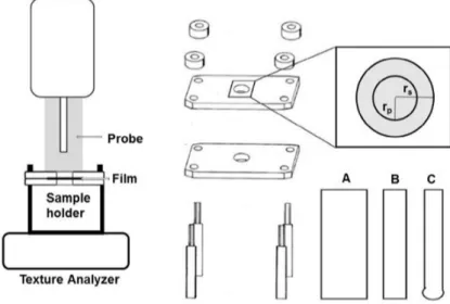
Figure 1.6: Experimental setup (left) and sample holder for the film preparation (right), where rs indicates radius of samples, and rp indicates radius of probe. Geometry of
cylindrical probes A and B and spherical probe C is shown on the right bottom [105].
Figure 1.7: Determination of percent elongation of thin films using a texture analyzer, where a = initial length of the film in the sample holder opening, a0 = initial length -radius of probe, b = displacement of the probe, c0 + r = length after strain, c0 = length of a0 after strain, r = radius of the probe [105].
of films are two qualitative concepts. Film forming capacity is the ability of film formers to form desired films. It is categorized according to strip forming capacity such as very poor, poor, average, good, very good, excellent. Appearance of strip is evaluated by visual observation such as transparent, semi transparent,opaque [87, 88].
Thickness. Thickness is measured at different points (normally 5) by a micrometric screw gauge.
Folding endurance. Folding endurance is determined by folding a film up to it break at the folding point. The number of attempt required to break the film is the folding endurance value [89, 90].
Solid state characterization. Solid state properties of films can be analyzed by means of differential scanning calorimetry, Fourier transform infrared spectroscopy, X-Ray diffraction [91].
Figure 1.8: Examples of stress–strain curves obtained from polymeric thin films.
Figure 1.9: Schematic illustration of the apparatus used for dissolution studies of films.
expansion of the matrix in aqueous media. This phenomenon is simultaneous to the set-up of a gel phase. These polymers form physical-linked gels and are bio erodible. So as, a true swelling equilibrium, as in the sense of cross-linked gel such as Poly-methyl-metacrilate, does not exist. Nonetheless, a pseudo-swelling equilibrium can be individ-uated [92]. Swell is measured by immerging a sample in a swelling medium (generally saliva or water at fixed pH and temperature), then weighing the sample, time by time, until an equilibrium weight is reached. The film can be put in a Petri dish, in a net wire or a beaker. The swelling degree S is usually calculated as (equation (4.10)):
S= M − M0 M0
(1.1)
where M is the measured weight and M0 is the initial weight. Common polymers for
OTF can swell from 0.2 to 50 times, depending on the formulation [93, 94].
In-vitro dissolution testing. Dissolution testing evaluates the duration of drug re-lease from the dosage form and its pace. Dissolution tests for OTF have been performed in diffusion cells [95], USP paddle II(figure 1.9) or basket (I) apparatus, USP IV flow-through apparatus [96]. In the literature, many authors have done some improvisation on the dissolution apparatus, while others have employed Franz diffusion cells (FDC) for testing the drug release from the polymeric films.A major barrier with respect to film in dissolution testing is the placing of the samples. Several methods have been practiced, where the film is attached on the inner side of the glass vessels or the stirring element using an adhesive tape [106].
Uniformity of drug content. A prerequisite for therapeutic efficacy, safety, and regulatory approval of a medicine is drug content uniformity. Failure to achieve a high degree of accuracy with respect to the amount of drug in individual unit doses of the film
can result in therapeutic failure, non reproducible effects, and, importantly, toxic effects to the patient. Drug content is measured by dissolving a known weight of the film for analysis. An assay of film area rather than weight would be more appropriate for assess-ing drug content uniformity. Drug uniformity in OTF can normally have a variation of ± 15 %. Drug content uniformity is a crucial aspect in film manufacturing [97].
In vitro Bioadhesion measurement. In vitro bioadhesion measurement method was first reported [98] in evaluation of the adhesive properties of patches using a micropro-cessor based on advanced force gauze equipment with porcine buccal membrane as a model tissue under simulated buccal conditions. Data collection and calculations were performed using the Data Plot software package of the instrument. Two parameters, namely the work of adhesion and peak detachment force were used to study the buc-cal adhesiveness of patches. The work of adhesion was determined from the area under force-distance curve while the peak detachment force was the maximum force required to detach the film from the tissue [107].
1.1.6
Manufacturing techniques
Manufacturing processes for OTF preparation have thrived in the last two decades. Sol-vent casting, semi solid casting, hot-melt extrusion, solid-dispersion extrusion, and rolling are the main technological families. An overview is presented here [99–104].
Casting solution. This technique require raw materials with few trivial prerequisites: the polymer must be soluble in a volatile solvent or water, a stable solution with a reason-able minimum solid content and medium viscosity is sought, formation of a homogeneous film and release from the casting support must be possible. Water soluble polymers-hydrocolloids good to prepare OTF include: hydroxypropylmethyl cellulose (HPMC), hydroxypropyl cellulose (HPC), Pullulan, sodium alginate, pectin, carboxymethyl cellu-lose (CMC), Poly-vinyl alcohol (PVA). Water-soluble ingredients are dissolved to form a clear, viscous solution. The API and other agents are dissolved apart in a suitable solvent. This second mixture is later vigorously mixed with the water solution. The entrapped air is removed by vacuum. Deaeration is necessary to obtain uniform film property and thickness. The resulting solution is cast as a film, allowed to dry, and cut into pieces to the desired size (figure 1.10). Specific types of equipment, such as rollers, are required for pouring the solution on an inert base. The clearance between the roller and the substrate determines the required thickness of the film. Drying the film, removes the solvent and helps to obtain the finished product. Usually, glass, plastic, inox or teflon plates are used as an inert base for film casting.
This manufacturing technology is the most frequent in laboratory. When transferred to production scale, several problems can be encountered, that include how casting the film, obtaining uniform thickness, and proper drying [108, 109] . Air entrapment may tend to produce non- uniform films. Deaeration step is imperative to get a uniform film which may be achieved by vacuum assisted machines.
Hot-melt extrusion. In the hot-melt extrusion (HME) process, appropriate amounts of polymer, drug, plasticizer and additives are blended into an uniform powdered mix-ture, prior to feeding through the hopper of the preheated extruder and be transferred into the heated barrel by a rotating extruder screw (figure 1.11). The API and other excipients are mixed in a dry state. An advantage of this process is the complete elimi-nation of the solvent. Films cool and are cut to the desired size. The high temperature makes the process suitable only for thermostable drugs. Homogeneous films are obtained, with thickness < 1 mm [110, 111].
Semi-solid casting. Firstly, a water solution of film forming polymer is prepared. This mixture is added to a solution of an acid insoluble polymer (e.g. cellulose acetate phthalate, cellulose acetate butyrate), which was prepared in ammonium or sodium hy-droxide. The appropriate amount of plasticizer is added to obtain a gel mass. Finally, the gel mass is casted in films or ribbons, using heat controlled drums. The thickness of the film is about 0.1-1.1 mm. The ratio of the acid insoluble polymer to film forming polymer should be 1:4 [112].
Figure 1.10: Commercial manufacturing of film based on solvent-casting [55].
Figure 1.11: Hot-melt extrusion system for the preparation of films [55].
Solid dispersion extrusion. Immiscible components are extrude with drug, preparing solid dispersions. The latter are shaped in to films by means of dies [112].
Rolling-method. The film is prepared by pre-mixing of an active ingredient and excipi-ents, followed by subsequent addition of the solvent. The pre-mix or master batch, which includes the film- forming polymer, polar solvent, and any other additives except drug is added to the master batch feed tank. A pre-determined amount of the master batch is controllably fed via a metering pump and control valve to first and second mixers. The required amount of the drug is added through an opening in each of the mixers. After the drug has been blended with the master batch pre-mix for a sufficient time to provide a uniform matrix, a specific amount of the uniform matrix is then fed to the pan through the second metering pump. The film is finally formed on the inert substrate and carried away via the support roller ( figure 1.12). Thus the wet film is then dried using controlled bottom drying, desirably in the absence of external air currents or heat on the top (exposed) surface of the film [113].
Figure 1.12: Schematic overview of Rolling-print technology for the preparation of films [55].
1.1.7
In vitro dissolution testing of buccal thin films
Absorption of drugs in living organisms schematically depends on three steps [114]:
• release from the dosage form;
• dissolution or solubilization of the drug under physiological conditions; • permeability across targeted tissues.
The purpose of in-vitro dissolution testing is to offer substantial information about steps 1 and 2, without resorting to test animals. Because of the critical nature of the first two of these steps, vitro dissolution testing may be relevant to the prediction of in-vivo performance. It concerns all solid pharmaceutical dosage forms. Most common applications of dissolution testing are:
• assess the lot-to-lot quality of a drug product; • guide development of new formulations;
• ensure continuing product quality and performance after certain changes,such as changes in the formulation, the manufacturing process, the site of manufacture, and the scale-up of the manufacturing process.
New drug applications (NDAs) submitted to the Food and Drug Administration (FDA) contain bio availability data and in-vitro dissolution data, that, together with chemistry, manufacturing, and controls (CMC) data, characterize the quality and performance of the drug product. In-vitro specifications for generic products should be established based on a dissolution profile. For new drug applications, as well as generic drug applications, dissolution specifications should be based on acceptable clinical, bio availability, and/or bio equivalence batches. Dissolution specifications are set depending on the class of the active substance, with the aim of predicting the likelihood of achieving a successful in vivo-in vitro correlation (IVIVC). FDA Bio pharmaceutics Classification System (BCS) divides active substances in four classes high permeability-high solubility (1), low solu-bility - high permeasolu-bility (2), high solusolu-bility - low permeasolu-bility (3), low solusolu-bility - low permeability (4). This classification is focused on oral administration and it is derived from a classical mathematical model of drug absorption through the bowels [115].
Considering the release time, drugs are classified as "immediate release" if 85 % of the substance is released from the dosage form in 30-45 min. Slower dissolution corresponds to modified release dosage forms, such as extended and delayed release [116]. The agencies responsible of dissolution testing standardization are the national Pharmacopeias.
In the last thirty years, European Pharmacopeia, Japanese Pharmacopeia and US Pharmacopeia have been working for a common harmonization of dissolution testing procedures. Thanks to this, regulatory requirements have progressively reached a good grade of agreement [117, 118].
Buccal films have not been included yet in any standardization. At present all liter-ature works have assumed traditional oral dissolution as a starting point [119].
Some words in the nomenclature of dissolution testing [120]:
• apparatus: the basic unit for the in-vitro performance testing of dosage units. The apparatus consists of a container (vessel) for the dosage unit and dissolution medium, a device for promoting motion of the dissolution medium (stirring ele-ment), temperature control and support to hold the vessel and stirring element in a fixed orientation. Typically, six to eight apparatuses are grouped in a dissolution test assembly.
• stirring element: a paddle, rotating basket and shaft, or other device for pro-moting the movement of dissolution medium relative to the dosage unit under test.
• assembly: a combination of multiple apparatuses providing temperature control, controlled unified motion of stirring elements, and providing the opportunity for simultaneous or individual start of the apparatuses.
• dissolution system: test assembly connected to sampling and filter unit but with-out instrumentation such as UV/VIS spectrophotometer or HPLC chromatograph.
Four different apparatuses are in use [121]:
• Basket apparatus (USP 1), figure 1.13; • Paddle apparatus (USP 2), figure 1.14;
• Reciprocating Cylinder (USP 3), not accepted in the Japanese Pharmacopeia, figure 1.15;
• Flow through cell (USP 4), figure 1.16.
Figure 1.13: USP1-Basket apparatus.
Immediate-release, modified release and extended release tablets are usually tested in dissolution baths, using USP 2 paddles. Floating capsules and tablets generally prefer USP 1 baskets [122, 123]. Both USP 1 and UPS 2 apparatuses comprehend a stirred vessel with nominal capacity from 1 to 4 liters. They differ only in the stirring element. USP 2 is generally recommended as the first choice in dissolution apparatuses.
Despite of its universal diffusion, USP 2 have been deeply criticized [124]. Its hy-drodynamic conditions have been judged highly unreliable and great part of the uncon-trolled variability typical of the dissolution test have been ascribed to hydrodynamic effects. Tests is conducted in a small agitated vessel operated at Reynolds numbers in the transitional regime (2100< Re < 10000): under such conditions, flow behavior in stirred-tanks is known to be both time-dependent and strongly heterogeneous. Con-sequently, the hydrodynamics in the vicinity of a tablet would likely be both position
Figure 1.14: USP2-Paddle apparatus.
Figure 1.15: USP3-Reciprocating cylinder.
and time-dependent. Fluctuations in the flow introduce variability in the evolution of processes that are affected by hydrodynamics, such as shearing of the tablet surface, de-agglomeration of particles, mass transfer from the solid to the liquid, suspension and mixing of tablet fragments. Changes in the agitation speed can alter the measured dis-solution rates and impact the ability to correlate in vitro disdis-solution tests with in vivo performance [125, 126].
In experimental and computational fluid dynamics studies, a low recirculation zone was observed in the lower part of the hemispherical vessel bottom, where the tablet dissolution process takes place. This region is the most critical, considering that the
Figure 1.16: USP4-Flow through cell.
dissolving tablet will likely be at this location during the dissolution test. The exact tablet location has a significant impact on the dissolution profile [127, 128].
Drug release in USP 3 can be significatively affected by different agitation patterns [129].
The flow-through cell(USP 4), also referred to as the column (-type) method and USP 4 apparatus, was born of the need to overcome some of the limitations set by existing compendial methods, particularly the USP 1 and USP 2 apparatuses. It is now in widespread use. The flow-through cell offers definite advantages in regards to its low volume hold-up, the ability to maintain sink conditions (particularly for sparingly soluble drugs), and the ease of changing dissolution media (by analogy with physiological changes along the gastro-intestinal tract). Flow-through is also called the "infinite sink" condition. The flow-through cell is claimed to possess ideal hydrodynamic conditions for mild agitation, homogeneity and mathematically definable solvent flow pattern. Different types of cells are available for testing tablets, powders, suppositories, hard- and soft-gelatin capsules, implants, semisolids, suppositories, and drug-eluting stents. For orally administered solid dosage forms, two different cells are described: the large cell (22.6-mm internal diameter) and the small cell (12-mm internal diameter) that provide approximate volumes of 19 ml and 8 ml, respectively, for dissolution (cell volumes without glass beads).
Usually the bottom cone of the cell is filled with small glass beads (about 1-mm diameter), and one bead (about 5-mm diameter) is positioned at the apex to prevent material from descending into the inlet tubing. Different amounts of small glass beads can be used according to the experimental setup. The sample can be placed upon a holder, but also on or within the glass-bead bed. For dispersed systems (i.e., suspensions, powders), mixing of the sample within the glass-bead bed has also been reported [130– 133].
1.2
Hydrogels for topical drug delivery
Topical preparations are used for the localized effects at the site of their application by virtue of drug penetration into the underlying layers of skin or mucous membranes. The main advantage of topical delivery system is to bypass first pass metabolism. Avoidance of the risks and inconveniences of intravenous therapy and of the varied conditions of absorption, like pH changes, presence of enzymes, gastric emptying time are other ad-vantage of topical preparations. Semi-solid formulation in all their diversity dominate the system for topical delivery, but foams, spray, medicated powders, solution, and even medicated adhesive systems are in use.
The topical drug delivery system is generally used where the others system of drug administration fails or it is mainly used in pain management, contraception, and urinary incontinence. Over the last decades the treatment of illness has been accomplished by administrating drugs to human body via various routes namely oral, sublingual, rectal, parental, topical, inhalation etc.
Topical drug delivery can be defined as the application of a drug containing formu-lation to the skin to directly treat cutaneous disorders (e.g. acne) or the cutaneous manifestations of a general disease (e.g. psoriasis) with the intent of confining the phar-macological or other effect of the drug to the surface of the skin or within the skin. Topical activities may or may not require intra-cutaneous penetration or deposition [134, 135]. Topical drug delivery systems include a large variety of pharmaceutical dosage form like semisolids, liquid preparation, sprays and solid powders. Most widely used semisolid preparation for topical drug delivery includes gels, creams and ointments [136].
The advantages of topical drug administration are [137]
• Avoids gastrointestinal (GI) drug absorption difficulties caused by GI pH, enzymatic activity and drug interactions with food, drink, and other orally administered drugs.
• A substitute for other routes of administration (e.g. oral administration, intra-venous injection) when that route is unsuitable, as with vomiting, swallowing prob-lems, resistant children and diarrhoea.
• Patient acceptability is better as this drug delivery system is non-invasive, avoiding the inconvenience of parenteral therapy.
• Avoids the first-pass effect, possibly avoiding the deactivation by digestive and liver enzymes.
• Reduction of doses as compare to oral dosage forms.
• Ability to dissolve a wide range of medications with different chemical properties, making combination therapy with one transdermal cream possible.
• Provides extended therapy with a single application, improving compliance. • Drug therapy may be terminated rapidly by removal of the application from the
skin surface.
• Less greasy and can be easily removed from the skin.
On the other hand, the limitations of transdermal drug delivery (TDD) are functions of skin physiology and drug bio-activity [141]. The excellent barrier properties of skin are well known [138, 139], and currently limit TDD to only the most potent drugs.
In terms of dosage, this criterion implies that drugs, the daily dose of which is 10-20 mg or less, are potential candidates for TDD.
There are, in addition, further constraints determined by the physicochemical prop-erties of the drug. For a drug molecule to reach the cutaneous microvasculature, and, hence, the systemic circulation, it must traverse both the lipophilic stratum corneum and the much more hydrophilic viable epidermis [140]. Therefore, drugs with a reasonable
Figure 1.17: Longitudinal section of skin.
partition coefficient and possessing solubility both in oil and in water are most ideal. A highly lipophilic compound, for example, may readily enter and diffuse within the stratum corneum but be unable to penetrate deeper into the skin. The other major disadvantage of TDD is the potential elicitation of either allergic or irritant responses by the drug or the adhesive of the device.
1.2.1
Anatomy of skin
The human body has two systems that protect it from the harmful organisms existing in the environment. The internal defense system destroys microorganisms and bacteria that have already attacked the body. The external defense system prevents microbial microorganisms to enter the body. Skin is biggest external defense system. Skin covers the outside of the body but has other functions beside the defense mechanism. It serves as a mechanical barrier between the inner part of the body and the external world [142]. Temperature of skin varies in a range of30◦C to 40◦C degree depending on the
environmental conditions [143].
Skin is the largest organ in the body. It consists of three layers(figure 1.17). The outer layer is called epidermis, the middle layer is dermis and the inner most layer is hypodermis [147–149].
Epidermis
Consists of epithelial cells. Among these cells, both living cells and dead cells can be found. These new cells at the bottom of epidermis divide fast and push the older cells upward. The epidermis does not have any direct source of blood veins to provide nu-trition. It takes its nutrients from the diffusion of necessary molecules from a rich vas-cular network in the underlying dermis. Epidermal cells are connected very strongly by desmosomes. Desmosomes are in contact with the intracellular keratin filmates. Keratin filmates produce keratin. Keratin cells accumulate and crosslink with the other keratin cells in the cytosol during their maturation. Afterward when the older cells die, this
network of keratin fibroses remains and provides a tough and hard protective layer in epidermis, called protective keratinized layer. This layer is waterproof and airtight. It prevents most substances to enter the body or leave from the body. In diseased skin, particularly burns, epidermis is destroyed causing potential loss of body fluid and an increase in susceptibility to microbial infections, leading to fatal consequences untreated.
Cell types that exist in the epidermis are (figure 1.19):
• Keratinocytes; these are the main cell types in epidermis (95% of cells);
• Melanocytes; these are the pigment producer cells and found in the basal layers of epidermis;
• Langerhans cells; these cells are important immunological cells and can be found in the mid dermis as well Merkel cells; these cells are found in the basal layer of epidermis and are one part of amine precursor and decarboxylation system [144, 145].
Epidermis consists of five layers, namely from inside to outside(figure 1.18):
• stratum germinativum (basal layer); • stratum spinosum;
• stratum granulosum; • stratum lucidum; • stratum corneum.
Stratum corneum is the outer most layer of epidermis and has a thickness of 10-20 µm when it is dry and 40 µm when it is hydrated and becomes swollen [145].
Stratum corneum has a structure of "bricks and mortar" arrangement. In this model the keratin rich corneocytes (bricks) are sitting in the intracellular lipid rich matrix (mortar) [145].
Corneocytes (the bricks) create 85 % of stratum corneum and intracellular lipids (15%) are arranged in 15-20 layers. Stratum corneum consists of 70% proteins, 15% lipids and only 15% water [146].
Molecules can basically permeate through skin by two different pathways(figure 1.20).The first pathway is called the transappendegeal route. In this route the molecules should permeate through skin by permeation through sweat glands and across the hair folli-cles. The number of molecules which can penetrate through this pathway is very limited. The second pathway of penetration through skin is the transepidermal pathway. In this pathway molecules should pass through stratum corneum as multilayered barrier. This pathway has two micro pathways: the intracellular micro pathway and the transcellular micro pathway [145].
Dermis
Dermis is positioned under epidermis and is characterized by lots of elastin fibres that provide the stretching ability as well as lots of collagen that provides the strength to the skin. Blood vessels found in dermis provide nutrients for both dermis and epidermis. Dermis also plays a major role in temperature regulation. Nerves present there are re-sponsible for pressure and pain sensations. Dermis has a thickness of 3-5 mm. In addition to elastin fibres, blood vessels and nerves, an interfibrillar gel of glycosaminoglycan, salt, water, lymphatic cells and sweet glands are parts of dermis [145]. Cell types found in dermis are:
• Fibroblasts: collagen producing cells; • Macrophages: scavenger cells
Figure 1.18: Epidermal and skin layer.
Figure 1.20: Structure of stratum corneum and penetration pathways.
• Mast cells: responsible for immunological reactions and interactions with eosinophils [144]
Dermis plays an important role as connection to other skin layers also. Changes in the metabolism in dermis can influence growth integrity of the epidermis, hair follicles and skin glands [143].
Hypodermis
Hypodermis is the inner layer of skin. It is the contact layer between skin and the underlying tissues in body such as muscles and bone [137]. Sweat glands, sebaceous glands and hair follicles enfold in epidermis but they stem from dermis. Sweat glands release a dilute salt solution into the surface of skin. The evaporation of this solution makes skin cool and this is important for temperature regulation of both body and skin. Sweet glands are present all over the body. The amount of dilutions (sweet) that gets produced depends on environmental temperature, the amount of heat generating skeletal muscle activity and various emotional factors. The sebaceous glands produce sebum. Sebum is an oily liquid released into hair follicles and from there onto the skin surface. Sebum protects both hair and skin from drying out and provides waterproof layer [142].
1.2.2
Formulation design of Hydrogels
To treat skin infection various hydrogels based formulations are prepare and apply top-ically for local action. Hydrogels have been used to deliver active component. Instead of conventional creams, the hydrogels have been formulated for better patient compli-ance [150].
Gels that consist of an aqueous dispersion medium that is gelled with a suitable hydrophilic gelling agent are known as hydrogels. Hydrogels are three-dimensional hy-drophilic polymer networks, which have the ability to absorb large quantities of wa-ter [154]. Hydrophilic polymers such as hydroxypropyl methylcellulose (HPMC), Car-bopol and sodium alginate have been previously investigated as gelling agents [155, 156].
Hydrogels can be formed via chemical or physical crosslinks, which provide a networked structure and physical stability. These physical crosslinks include entanglements, crys-tallites, Van der Waals interactions or hydrogen bonding. Hydrogels formed from phys-ical crosslinks are known as "reversible" or "physphys-ical" hydrogels [153, 157]. In contrast, hydrogels known as "chemical" or "permanent" gels are formed via covalently bonded crosslinked networks [151, 152].
Some of the topically applied preparations are shown in figure 1.21.Topically applied gels are classified by two schemes [158]. The first scheme divides gels in two types of gel systems. These are called as inorganic and organic gel systems. Most inorganic hydrogels
Figure 1.21: General classification of gel.
are two-phase systems, such as aluminum hydroxide gel and bentonite magma. Bentonite has also been used as an ointment base in about 10% to 25% concentrations. Most organic gels are single-phase systems and may include such gelling agents as carbomer and tragacanth and those that contain an organic liquid, such as Plastibase.
The second classification scheme divides gels into hydrogels and organogels with some additional sub categories. Hydrogels include ingredients that are soluble in water; they include organic hydrogels, natural and synthetic gums, and inorganic hydrogels. Ex-amples include hydrophilic colloids such as silica, bentonite, tragacanth, pectin, sodium alginate, methylcellulose carboxymethyl cellulose sodium, and alumina, which in high concentration form semisolid gels. Sodium alginate has been used to produce gels that can be employed as ointment bases. In concentrations greater than 2.5% and in the presence of soluble calcium salts, a firm gel, stable between pH 5 and 10, is formed.
Methylcellulose, hydroxy ethylcellulose, and sodium CMC are among the commercial cellulose products used in ointments. They are available in various viscosity types, usually high, medium, and low. Organogels include the hydrocarbons, animal and vegetable fats, soap base greases, and the hydrophilic organogels.
Topical gel may include the following components:
• Gel forming agent or polymer; • Drug Substance;
• Penetration Enhancers;
Gel forming agent or Polymer [159–170].Different classes of polymeric materials have been used to achieve rate controlled drug delivery. The mechanism of drug release depends upon the physicochemical properties of the drug and polymer. The following criteria should be satisfied for a polymer to be used in a topical system:
• Molecular weight, chemical functionality of polymer must allow diffusion and release of the specific drug.
• The polymer should permit the incorporation of a large amount of drug. • The polymer should not react, physically or chemically with the drug.
• The polymer should be easily manufactured and fabricated into the desired product and inexpensive.
• The polymer must be stable and must not decompose in the presence of drug and other excipients used in the formulation, at high humidity conditions, or at body temperature.
• Polymers and its degradation products must be nontoxic. No single material may have all these attributes, certain excipients may be incorporated to alter some properties for example Co-solvents such as ethanol, propylene glycol, PEG 400 could be added to increase drug solubility.
Gel forming polymers are classified as below:
• Natural Polymers:
– Proteins-E.g. Collagen, Gelatin, Xanthin, Gellum Gum;
– Polysaccharides-E.g. Agar, Alginic acid, Sodium or potassium carrageenan, Tragacanth, Pectin, Guar Gum, Cassia tora;
• Semi synthetic Polymers:
– Cellulose Derivatives-E.g. Carboxymethyl cellulose Methylcellulose, Hydrox-ypropyl cellulose, HydroxHydrox-ypropyl methylcellulose, Hydroxyethyl cellulose;
• Synthetic Polymers:
– Carbomer-E.g. Carbopol-940, Carbopol-934, Carbopol -941; – Poloxamer;
– Polyacrylamide; – Polyvinyl Alcohol;
– Polyethylene and its copolymers;
• Inorganic Substances-E.g. Aluminum Hydroxide bentonite; • Surfactants-E.g. Cetosteryl alcohol, Brij-96;
Drug Substance [159–170].Drug Substance plays a very important role in the suc-cessful development of a topical product. The important drug properties that effect its diffusion through gels as well as through skin are as follows.
• Physicochemical properties:
– Drug should have a molecular weight of less than 500 Daltons.
– Drugs highly acidic or alkaline in solution are not suitable for topical delivery. – Drug must have adequate lipophilicity.
– A saturated aqueous solution of the drug should have a pH value between 5 and 9.
• Biological properties:
– The drug should not be directly irritated to the skin.
– Drugs, which degrade in gastrointestinal tract or are inactivated by hepatic first pass effect, are suitable for topical delivery.