UNIVERSITA’ DI PISA
FACOLTA’ DI MEDICINA E CHIRURGIA
DOTTORATO DI RICERCA IN SCIENZE ENDOCRINE E
METABOLICHE
Direttore: Chiar.mo Prof. Aldo Pinchera
ROLE OF SORTILIN
IN THE POST-ENDOCYTIC
TRAFFICKING OF
THYROGLOBULIN
Relatore: Dott. Michele Marinò
Dottoranda: Dott.ssa Roberta Botta
INDICE
INTRODUCTION
4
THE Vps10p-DOMAIN RECEPTOR FAMILY 5 Background 5
Evolutionary history and gene organization 6
Localization 7
Specific structural features 8 Function 9
Sortilin 10
THYROGLOBULIN 14
Structure, synthesis and secretion 14
Trafficking 15
MICROPINOCYTOSIS AND LYSOSOMAL DEGRADATION 16
ENDOCYTOSIS AND OTHER PATHWAYS OF TG TRAFFICKING 16
Tg endocytic receptors 17
THE THYROID AGR 19
NAGR 20
MEGALIN 20
AIM OF THE STUDY
24
Materials 29
Experimental animals and related procedures 30
Histology and Immunohistochemistry 31
Preparation of tissue extracts 31
Serum assays in mice 32
Cell cultures and related procedures 32
Preparation of cell extracts 34
Immunofluorescence Staining 34
Immunolectronmicroscopy 35
Immunoprecipitation experiments 36
Western blotting 36
Biosensor binding assays 37
Cell binding, uptake, degradation, recycling and transcytosis assays 38
ELISA 40
Data presentation and statistics 40
RESULTS
42
Expression of sortilin in thyroid epithelial cells 43
Expression of sortilin in thyroid epithelial cells is TSH-dependent 46
Tg is a sortilin ligand 49
Sortilin is not a Tg endocytic receptor 52
Sortilin does not bind to Tg in the biosynthetic pathway 57
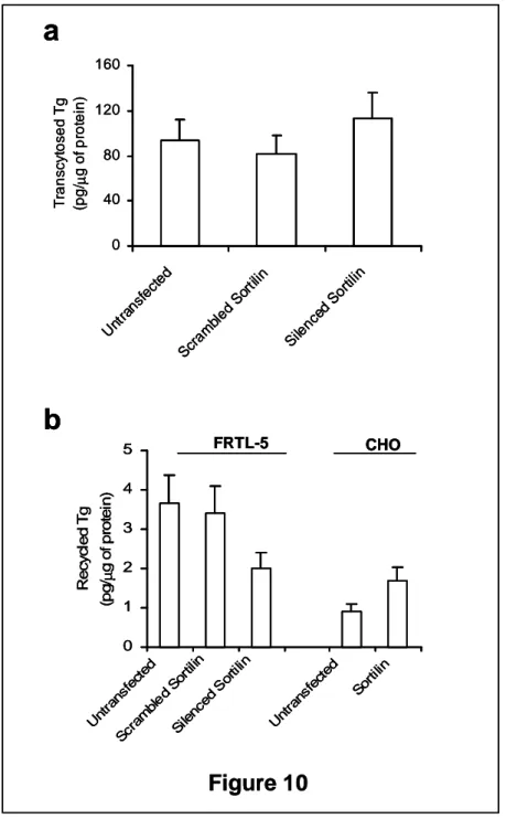
Sortilin-Tg interactions in the endocytic pathway result in Tg recycling 61
DISCUSSION
63
THE Vps10p-DOMAIN RECEPTOR FAMILY
Background
Vps10p-domain (Vps10p-D) receptors are type-I transmembrane proteins sharing as a common feature and hallmark an N-terminal Vps10p-D domain, which was named after the yeast-sorting protein Vps10p (Vps, for vacuolar protein sorting defective). The structure of Vps10p receptor is characterized by a large N-terminal luminal moiety and a short cytoplasmic domain. The luminal part contains two homologous regions, each characterized by a C-terminal motif of 10 cysteine residues (10CC motif), the spacing of which is conserved [1]. The family of mammalian type-I transmembrane receptors containing a Vps10p domain comprises five members, namely sortilin, SorCS1, SorCS2, SorCS3, and SorLA [2-7]. All of them carry a single Vps10p-D situated at the N-terminus of their luminal/extracellular moiety (Fig. 1). In sortilin, also known as neurotensin receptor-3 [8], the Vps10p-D makes up the entire luminal/extracellular part of the receptor, but additional modules are found in the other four receptors. In SorLA, the Vps10p-D is followed by 5 low-density lipoprotein receptor (LDLR) class B repeats flanked by an EGF precursor-type repeat, a cluster of 11 LDLR class A repeats, and 6 fibronectin type-III repeats. Due to the presence of the 11 LDLR class A repeats, SorLA was also named LR11 [9]. The mutually highly homologous SorCS1, SorCS2, and SorCS3 contain a leucine-rich segment between the Vps10p-D and the transmembrane domain. Structure prediction of the leucine-rich segment suggests a beta-sandwich fold and relates the domain to the E-set superfamily [10]. Following the extracellular and transmembrane segment,
each receptor carries a short (40–80 amino acids) cytoplasmic domain comprising typical motifs for interaction with cytosolic adaptor molecules.
Fig.1: Structural organization of the Vps10p-domain receptor family
Evolutionary history and gene organization
Receptors containing a Vps10p-D have been identified in several eukaryotes, but not all protein domain combinations are present in all phyla. Mammalian Vps10p-D receptor genes are large (up to 600 kb) and span multiple, usually small, exons [7, 11]. Sequence similarity between the distinct mRNAs and proteins in an individual organism is very high for SorCS1 and SorCS3, e.g., 88% overall identity between the human mRNAs [12]. Whereas the other Vps10p-D receptor genes are dispersed
throughout the genome, SorCS1 and SorCS3 are located adjacent to each other on one chromosome, e.g., human chromosome 10q23.3. This indicates that one gene originates from the other by gene duplication. Within the receptor family, splice variants are known only for SorCS1. The alternative use of composite internal/terminal and terminal exons results in different receptor isoforms with identical extracellular and transmembrane parts but with different cytoplasmic domains [13-15]. The alternative cytoplasmic domains differ in sequence and length and present different consensus motifs for interaction with cytosolic adaptor proteins.
Localization
Analysis of the expression of the Vps10p-D receptors was performed in mice and men. In the adult, all receptors are prominently expressed in the nervous system but are also found in other tissues and organs [2, 3, 5-7, 9, 12, 16, 17]. During embryonic and postnatal development, all five genes are expressed in a dynamic and sometimes transient fashion [6, 12, 18-20]. Although the developmental expression of the receptors predominates in the nervous system, sortilin is also highly expressed in the embryonic lung and SorLA is expressed in several developing glands, such as the thyroid gland, and shows a strong expression in the embryonic lung and kidney. In addition to the nervous system, in the adult high expression was found for SorLA in liver, testis, and kidney, and sortilin is highly expressed in testis and skeletal muscle. SorCS1 is expressed in adult liver, kidney, and heart, and SorCS2 expression was found in adult lung and testis.
Specific structural features
The aminoacid similarity among all Vps10p-D proteins is relatively limited, but two structural features are conserved from yeast to mammals and characterize the domain: an N-terminus comprising a consensus motif for processing by proprotein convertases (Fig. 1) and a C-terminal segment containing 10 conserved cysteine residues (10CC motif).
Sortilin, SorLA, SorCS1, and SorCS3 are synthesized as precursors and converted in the trans-Golgi network to mature receptors by proprotein convertase-mediated cleavage and subsequent dissociation of their propeptides [7, 8, 14, 21-24]. The Vps10p-D of sortilin and SorLA need the propeptide cleavage to expose their ligand-binding region. Their propeptides bind to the mature receptors with high affinity and inhibit binding of ligands, therefore propeptide cleavage affects sortilin and SorLA for ligand binding [22, 23]. The prevention of premature binding of ligands to the receptors was suggested as one function of the propeptides [23, 25]. In addition, the propeptides may have a chaperone-like function. It has been shown for sortilin that its propeptide promotes the transport of the receptor through Golgi compartments but seems to be required for receptor folding [25]. However, SorCS1 and SorCS3 do not bind their own propeptides and neither SorLA, SorCS1, nor SorCS3 depend on their propeptides for transport in the biosynthetic pathway [14, 24, 25]. Therefore it is likely that the propeptides have different functions or may be redundant for some of the Vps10p-D family members.
The C-terminal segment of the Vps10p-Ds is characterized by 10 cysteine residues. Exchanging the 10CC module of sortilin and SorLA leads to a corresponding change in affinity for receptor-specific ligands [25]. Therefore it was suggested that the 10CC module contributes to the binding of specific ligands [25]. Based on a computational approach, it was proposed that the N-terminal part of the Vps10p-D of sortilin has a beta-propeller fold [26]. Recently, the crystal structure of the sortilin Vps10p-D in complex with its ligand neurotensin revealed that the N-terminal part has indeed a 10-bladed beta propeller structure and is followed by two small domains, designated 10CC-a and 10CC-b, which constitute the 10CC module. Both have a low content of secondary structure and interact extensively with the beta-propeller [27].
Function
It is well established that in mammalian cells many newly synthesized hydrolytic enzymes are targeted to lysosomes by binding to mannose-6-phosphate receptors (MPRs). The yeast vacuole is equivalent to the lysosomes of higher eukaryotes. Vacuolar protein sorting in yeast and lysosomal protein sorting in mammalian cells may share similar mechanisms, but no mannose-6-phosphate-mediated sorting has been found in yeast. The established pathway involves the yeast protein Vps10p as a sorting receptor [1, 28, 29]. The identification of Vps10p-D receptors in mammals caused speculation that these receptors are also capable of mediating sorting of ligands from the trans-Golgi network (TGN) to late endosomes or lysosomes [28, 29].
In their short cytoplasmic domains, the Vps10p-D receptors carry typical consensus motifs for interaction with cytosolic adaptor proteins, internalization, and intracellular sorting. In accordance, sortilin, SorLA, and SorCS1a and c have a prominent intracellular vesicular and perinuclear localization [5, 8, 14, 15, 22, 30-32]. Surprisingly, SorCS3 predominates on the cellular surface and shows only minor intracellular expression [24]. Sortilin, SorLA, SorCS1a, SorCS1c, and SorCS3 can also mediate endocytosis [14, 15, 22, 31-33].
Sortilin
Sortilin is internalized via clathrin-coated pits, and the identified internalization signals are known to facilitate binding to the AP-2 complex [7]. Sortilin delivers internalized ligands to lysosomes and transports cargo, from the TGN to endosomes, presumably mediated by Golgi-localized, c-adaptin-ear-containing, ADP-ribosylation factor-binding proteins (GGAs) [31, 34]. Recent results demonstrate that trafficking of sortilin is also facilitated by AP-1, and that the endosome-to-TGN retrieval is mediated by the retromer complex [35-38]. The sortilin cytoplasmic domain resembles that of MPRs, and sortilin and MPRs colocalize in endosomes and endosome-to-TGN carrier vesicles [37]. Furthermore, sortilin conveys lysosomal targeting of sphingolipid activator proteins (SAPs), acid sphingomyelinase, and cathepsin D and H to lysosomes [36, 39, 40]. Taken together, these data support a model of sortilin being an endocytic and intracellular sorting receptor contributing the targeting of ligands to lysosomes and the sorting between the Golgi apparatus and
endosomes. Therefore, the role of sortilin in Golgi-endosomal trafficking is comparable to the function of MPRs.
Sortilin is expected to serve additional cell- and tissue-specific functions as it is expressed in several specialized cell types. For instance, sortilin represents one of the major components of glucose transporter isoform 4 (Glut4)-containing vesicles of adipocytes and myocytes [41, 42]. In fat and skeletal muscle cells, Glut4 is translocated to the cell surface in response to insulin through a system of specialized vesicles. In adipocytes, sortilin shows colocalization with Glut4. Moreover, sortilin plays an essential role in the formation of Glut4-containing storage vesicles and may be involved in the regulation of the insulin-responsive glucose transport system [43, 44].
Initially, sortilin was purified by LDL-receptor-associated protein (RAP) affinity chromatography [5]. The binding of RAP to the sortilin Vps10p-D is remarkable as it appears to be the only RAP-binding receptor structurally not related to the LDL receptor family. In addition, binding and internalization of several other, unrelated ligands, such as lipoprotein lipase, apolipoprotein A–V, the unprocessed forms of the nerve growth factor (proNGF) and brainderived nerve growth factor (proBDNF), and the neuropeptide neurotensin was demonstrated [8, 23, 33, 34, 45-47], and several findings suggest that sortilin alone or in complex with the G-protein-coupled neurotensin receptor-1 may modulate neurotensin signalling [48].
Sortilin binds the pro-neurotrophins proNGF and proBDNF in concert with the neurotrophin receptor p75NTR [45, 46]. The neurotrophins NGF and BDNF promote
neuronal survival and growth, but by contrast the unprocessed forms induce cell death. Pro-neurotrophins are released by neurons and glia, particularly when cell death prevails, for instance, in the developing hypothyroid cerebral cortex, the prodromal and end-stages of Alzheimer’s disease, or following brain trauma or seizure [49-54]. They form a death receptor complex in which sortilin specifically binds the pro-domain of pro-neurotrophins with high affinity, whereas p75NTR simultaneously interacts with the mature domain. The formation of this ternary complex is crucial for the pro-apoptotic function, as sortilin antagonists rescue cultured super cervical ganglion (SCG) neurons from proNGF- and proBDNF-induced cell death [45, 46]. Sortilin-deficient mice (Sortilin-/- mice) are born viable and show no gross abnormalities [51]. Neurons from Sortilin-/- mice are resistant to pro-neurotrophin-induced apoptosis. In the developing retina, these mice show reduced neuronal apoptosis indistinguishable from p75NTR-deficient mice. Although sortilin deficiency does not affect developmentally regulated apoptosis of sympathetic neurons, it prevents their age-dependent degeneration. Furthermore lesioned motor neurons from sortilin-/- mice are protected from cell death [51]. In agreement, the release of recombinant sortilin ectodomain at injury sites reduces death of axotomized sensory neurons to the same degree as neutralizing antibodies against the BDNF prodomain [55]. Thus, the formation of the ternary complex consisting of pro-neurotrophin, sortilin, and p75NTR triggers apoptotic signalling under pathological conditions, as well as in specific stages of neuronal development and aging. In conclusion, sortilin serves not only as a general intracellular sorting receptor, it also
mediates specialized functions, for instance as a co-receptor determining a specific signalling pathway.
THYROGLOBULIN
Structure, synthesis and secretion
Thyroglobulin (Tg) is a 660-kDa glycoprotein composed of two identical 330-kDa subunits (monomers) [56]. Complete sequences of human, bovine, mouse and rat Tg cDNA have been obtained [57-60]. The 8.5-kb human Tg gene is located on the long arm of chromosome 8 and encodes 2,767 amino acid residues, representing the Tg monomer [56, 57, 61]. Transcription of the Tg gene requires a complex interplay of at least three transcription factors (TTF1, TTF2, and Pax 8), which are also involved in transcription of thyroperoxidase (TPO) and of the thyroid sodium/iodide symporter (NIS) genes [62, 63]. The thyroid-stimulating hormone (TSH) up-regulates Tg synthesis by acting directly on the Tg promoter and indirectly through the thyroid transcription factors [56, 62]. Tg is produced exclusively by thyrocytes and follows the usual biosynthetic pathway. It is synthesized and initially processed in the endoplasmic reticulum with the formation of dimers and with addition of N-linked glycoside residues, and then is further processed in the Golgi apparatus, especially by modification of carbohydrate residues [64, 65]. Tg is transported via vesicles from the trans-Golgi network to the apical surface of thyrocytes, where it is released into the lumen of thyroid follicles and stored as the major protein component of colloid (>95%) [56]. There is evidence that secretion of Tg is a regulated process rather than a constitutive process [56, 66]. This may be useful in achieving a balance between release and uptake of Tg, which, under normal conditions, must be equal. Formation of thyroid hormone residues within the Tg molecule occurs at the cell-colloid
interface by coupling of tyrosyl residues of Tg with iodide, a process that is carried out by TPO and other additional enzymes. Five hormonogenic sites have been identified in human Tg [56], and it has been estimated that, under physiological conditions, a Tg molecule contains on average 2.28 molecules of thyroxine (T4) and 0.29 molecules of triiodothyronine (T3) [67]. However, the degree of hormone content varies among different Tg molecules [56, 67], and Tg molecules that are poor in iodide and hormone content are usually referred to as immature. Tg molecules within the colloid also differ with respect to the extent and type of glycosylation, and there is a correlation between the iodine content of Tg and the nature of its complex carbohydrate residues [56, 68]. Because newly secreted Tg molecules are soluble and adjacent to the apical surface of thyrocytes, they are the first available for uptake by thyrocytes [69, 70]. Newly secreted molecules that escape uptake proceed into the colloid, where they undergo further modifications, characterized in part by the formation of highly concentrated (up to 750 mg/ml), insoluble, covalently cross-linked, multimerized forms [70, 71]. Thus, most of the Tg in the colloid is in storage, to be solubilized and taken up by thyrocytes only under special circumstances, such as iodine deprivation or intense TSH stimulation.
Trafficking
Tg is the precursor of thyroid hormones and its intrathyroidal metabolism is central in thyroid hormone synthesis and secretion. Although some pathways of Tg trafficking in thyroid epithelial cells have been characterized, other probably remain
to be elucidated, and likely their understanding may be useful for providing the molecular basis, and possibly the cure, for several, yet unexplained, forms of dyshormonogenetic hypothyroidism.
MICROPINOCYTOSIS AND LYSOSOMAL DEGRADATION
Hormone release requires degradation of hormone-rich Tg molecules. To some extent, Tg degradation and release of T4, but not of T3, can take place within the
follicle lumen through the action of extracellular proteases located at the apical surface of thyrocytes [70]. However, the main pathway of hormone release is lysosomal degradation, which occurs following micropinocytosis of Tg by thyroid epithelial cells. In rodents, thyroid epithelial cells can internalize Tg by macropinocytosis, but in most species, including humans, uptake of Tg occurs mainly by micropinocytosis [70, 72]. The majority of investigators agree that fluid phase micropinocytosis is the major route of uptake leading to hormone release, because of the very high concentrations of Tg within the colloid [70, 72]. Both forms of micropinocytosis involve the formation of small vesicles at the plasma membrane which invaginate to form intracellular vesicles that fuse with endosomes [70, 72], which in turn fuse with lysosome.
ENDOCYTOSIS AND OTHER PATHWAYS OF TG TRAFFICKING
Interactions between Tg and endocytic receptors on the apical membrane of thyroid epithelial cells result in Tg uptake and delivery to post-endocytic pathways. Several receptors have been postulated or demonstrated to mediate Tg endocytosis and some
are involved in thyroid homeostasis. Because endocytic receptors take up substances that are present in very low concentrations in extracellular fluids, it is unlikely that they mediate uptake of large amounts of Tg [70, 72]. Receptor-mediated endocytosis involves specific binding of certain ligands to cell surface receptors, often with high affinity, with the result that even minor components of the extracellular fluid can be internalized in large amounts [73]. Receptors may contribute to hormone release under special circumstances, for example iodine deficiency, but under physiological conditions they are more likely to sort Tg molecules to post-endocytic pathways that do not lead to hormone release, namely recycling or transcytosis [72]. The contribution of the various means of endocytosis depends on the biochemical features of Tg, especially hormone content and degree of glycosylation (Fig. 2). Ultimately, both fluid phase endocytosis and receptor mediated endocytosis are aimed at the same goal, namely to render hormone release effective, which is achieved not only by targeting of Tg to lysosomes, but also by favoring complete glycosylation of immature Tg molecules through their recycling, or by eliminating hormone-poor Tg molecules from the colloid by transcytosis. The latter process is probably especially important when hormone-poor Tg molecules are present in excess within the colloid.
Tg endocytic receptors
Several molecules expressed on the membrane of thyroid ephitelial cells have been proposed to function as Tg endocytic receptors. Two of them, the asialoglycoprotein receptor (AGR) and megalin, are well characterized receptors. The characteristics of
acetylglucosamine receptor (NAGR)] are unknown. As detailed below, the three identified Tg receptors probably deliver Tg to different, post-endocytic pathways (Fig. 2). It is likely that other receptors for Tg also exist [72], but their nature and roles are not established.
Fig.2: Schematic representation of the known Tg endocytic pathways and of the receptors involved.
Early Endosome Late Endosome Lysosome T4 T4 T4 T4T3 T4 T4 T4 T4T 3 Recycling Endosome Golgi Endoplasmic Reticulum Fluid phase Transcytosis Endosome Apical Basal Colloid Blood ? Hormone-rich Tg Hormone-poor Tg Poorly glycos. Tg AGR NAGR Megalin Early Endosome Late Endosome Lysosome T4 T4 T4 T4T3 T4 T4 T4 T4T 3 T4 T4 T4 T4T 3 Recycling Endosome Golgi Endoplasmic Reticulum Fluid phase Transcytosis Endosome Apical Basal Colloid Blood ? Hormone-rich Tg Hormone-poor Tg Poorly glycos. Tg AGR NAGR Megalin
THE THYROID AGR
The existence of a thyroid AGR similar to the liver receptor was postulated because removal of sialic acid units from Tg increases its binding to thyroid membranes [74]. Indeed, AGR is expressed by thyroid ephitelial cells and expression of AGR in thyroid PC C13 cells is TSH-dependent [75-77], suggesting a thyroid-specific function of the receptor. By immunohistochemistry, AGR can be found on the apical membrane of TEC in rat thyroid sections [76], directly facing the follicular lumen, therefore in the ideal position to mediate Tg endocytosis.
Asialo-Tg binds to AGR in solid phase assays and it also binds to the native receptor in PC C13 cells [75-77]. In addition, Tg uptake by PC C13 cells can be in part reduced by an antibody against AGR [77], suggesting that, at least in cultured cells, AGR is involved in Tg endocytosis.
AGR binds especially to Tg molecules with a low degree of glycosylation, a feature that makes it an ideal candidate to be involved in Tg recycling. In addition, other studies showed that binding of asialo-Tg to thyroid membranes occurs optimally at low pH [74], which should prevent dissociation of Tg from the receptor due to the acidic pH of endosomes, as it sometimes occurs in ligand recycling or transcytosis. Nevertheless, degradation of Tg in PC C13 cells is reduced by an antibody against AGR [77], suggesting that, at least in part, AGR delivers Tg to lysosomes, which presumably should result in hormone release. It is possible that, also in view of its TSH dependence, AGR may facilitate maximal hormone release under special circumstances, for example iodine deficiency. However, a dual role with AGR
mediating to some extent also Tg recycling cannot be completely excluded (Fig. 2). Unfortunately, no in vivo data are available, and therefore the actual impact of AGR on thyroid function remains to be elucidated.
NAGR
Asialo-agalacto-Tg, obtained by digestion of asialo-Tg with galactosidase, bears exposed N-acetylglucosamine residues and it binds to thyroid membranes to a greater extent than undigested Tg [78]. In addition, asialo-agalacto-bovine serum albumin (BSA) binds to thyroid membranes with high affinity (saturation point 13 nM), and binding can be inhibited by unlabeled native Tg and to an even greater extent by asialo-Tg and asialo-agalacto-Tg [79]. These findings suggest the existence of a NAGR capable of interacting with Tg, which may be responsible for recycling of poorly glycosylated Tg molecules (Fig. 2). In support of this, asialo-agalacto-BSA is released undegraded following endocytosis by cultured TEC, and ovomucoid, a glycoprotein with exposed N-acetylglucosamine residues, accumulates in the Golgi following endocytosis by cultured TEC [80]. Although NAGR is very likely to mediate recycling, the exact identity of the receptor is unknown, as previous attempts to identify it failed. In addition, because studies in vivo are not available, the impact of NAGR on thyroid function remains to be established.
MEGALIN
Megalin is a member of the low density lipoptotein (LDL)-receptor family expressed by a restricted group of absorptive cells, including thyroid ephitelial cells, where it
can be found on the apical surface [72], thus in the ideal position to mediate Tg endocytosis. Megalin expression in thyroid ephitelial cells is up-regulated by TSH [72, 81], suggesting a thyroid-specific function.
Tg binds to purified megalin with high affinity (Kd ~9-11 nM), both in solid phase assays and to the native receptor in FRTL-5 cells [81]. In the latter, megalin competitors reduce Tg uptake by ~50% [81], suggesting the receptor is involved in Tg endocytosis. In most instances, megalin-mediated uptake of ligands results in their delivery to lysosomes [81]. However, certain ligands undergo a different intracellular fate, namely transcytosis, which is the case for Tg, representing one of the mechanisms by which Tg enters the bloodstream. Thus, transport of Tg across FRTL-5 cell layers is reduced by megalin competitors [72, 81], and portions of the megalin ectodomain (secretory components) remain complexed with transcytosed Tg [82]. Furthermore, in conditions associated with increased megalin expression, due to TSH (hypothyroid rats) or TSH-like (patients with Graves’ disease) stimulation, a relatively high proportion of serum Tg is complexed with megalin secretory components [82] and serum Tg levels in megalin KO mice are reduced [83].
The molecular mechanisms responsible for targeting of the Tg-megalin complex to transcytosis are known only in part [84]. Although binding of Tg to megalin is optimal at low pH, transcytosis is only minimally affected by increasing intracellular pH, suggesting that pH resistance is not a major factor [85]. The calcium-calmodulin pathway and phosphoinositide 3-kinase (PI3-K) affect Tg transcytosis. Calmodulin antagonists reduce transcytosis and increase T3 release, indicating calmodulin favors
transcytosis prior to Tg sorting [81]. In contrast, a PI3-K inhibitor increases Tg transcytosis but does not affect T3 release, suggesting that PI3-K exerts an inhibitory effect at a post-sorting stage [86].
A major role in determining targeting of Tg to transcytosis is related to the ability of Tg to bind to cell surface heparinoids. As other megalin ligands, Tg is a heparin-binding protein and heparin and megalin heparin-binding sites are functionally related [81]. Occupation of a major heparin-binding sequence of rat Tg (2489-2503) abolishes Tg binding to megalin. In addition, transcytosis of rat Tg in FRTL-5 cells is reduced by enzymatic removal of cell surface heparan sulfate proteoglycans (HSPGs) as well as by an antibody against Tg2489-2503 [83]. In this regard, optimal exposure of Tg2489-2503 is crucial, and a greater exposure of Tg2489-2503 in hormone-poor rat Tg is responsible for its preferential transcytosis compared with hormone-rich Tg [83]. The role of Tg binding to heparinoids appears to be more important in rodents than in humans, because Tg2489-2583 is identical in mice and rats, but differs in humans by 6 residues, resulting in a weaker Tg binding to heparin [87].
As mentioned above, transcytosis is preferential for hormone-poor Tg molecules. Thus, Tg transport across FRTL-5 cells is greater for hormone-poor than for hormone-rich Tg [83]. In addition, megalin-mediated transcytosis in vivo, estimated by the proportion of serum Tg complexed with megalin secretory components, is enhanced by inhibition of hormone formation within Tg due to thionamide treatment [83]. This selective mode of transcytosis renders hormone release more effective, by preventing hormone-poor Tg to enter the lysosomal pathway, thereby avoiding
competition with hormone-rich Tg, as well as wasteful transcytosis of hormone-rich Tg. Thus, megalin KO mice are hypothyroid [88]. Whether megalin deficiency exists in humans and it causes thyroid dysfunction remains to be established.
Sortilin is a 100 kDa member of the recently discovered family of Vps10p domain proteins, which comprises also SorLA ( 250 kDa) and SorCS 1, 2 and 3 ( 130kDa). The hallmarkof the family is a Vps10p domain at the N-terminus, which in sortilin makesup the entire luminal receptor. In addition to the Vps10p ectodomain, the structure of Vps10p proteins is characterized by a transmembrane segment and a shortcytoplasmic tail, where motifs for interaction with cytosolic adaptor molecules are present. Functional sorting sites, namely dileucines, acidic clusters and tyrosine-based motifs, which are known to be involved in endocytosis and intracellular trafficking, have been established in all Vps10p members. The structural heterogeneity of the cytoplasmic tail suggests differential and/or alternative functions of Vps10p proteins, which is being progressively unveiled especially in the case of sortilin. Thus, sortilin is capable of mediating endocytosis of ligands if expressed on the cell membrane, but it can also transport proteins from the Golgi to late endosomes when expressed in the Golgi, its most abundant and usual location.
Sortilin shares at least two ligands with the LDL receptor family member megalin, namely lipoprotein lipase (LPL) and the receptor associated protein (RAP). In the thyroid, megalin serves as a high affinity endocytic receptor for thyroglobulin (Tg), which led us to the hypothesis that, because of similarities between sortilin and megalin, Tg may be a sortilin ligand. The relevance of the issue stems from the fact that although some pathways of Tg trafficking in thyroid epithelial cells (thyrocytes) have been characterized, other probably remain to be elucidated, and likely their understanding may be useful for providing the molecular basis, and possibly the cure,
for several, yet unexplained, forms of dyshormonogenetic hypothyroidism. Thus, Tg is the precursor of thyroid hormones and its intrathyroidal metabolism is central in thyroid hormone synthesis and secretion. Briefly, Tg is synthesized by thyroid epithelial cells and then secreted into the lumen of thyroid follicles, where it undergoes hormone formation by coupling of its tyrosyl residues with iodide. Hormone-rich Tg molecules can either proceed further into the follicle, where they arrange into aggregates that form the colloid, or they can undergo proteolytic cleavage, resulting in hormone release[70]. To some extent, Tg degradation can take place within the follicle lumen through the action of extracellular proteases, but the main route of hormone release is lysosomal degradation, which occurs following micropinocytosis of Tg by thyroid epithelial cells. It is generally believed that fluid phase-mediated uptake is the main route of Tg micropinocytosis leading to lysosomal degradation and hormone release, but endocytic receptors for Tg located at the apical membrane of thyroid epithelial cells also exist and some of them have been characterized. In most instances, Tg internalized by endocytic receptors is thought to undergo intracellular fates other than lysosomal degradation, either recycling or transcytosis. In the recycling pathway, internalized Tg molecules are transported to the Golgi and then re-secreted back into the colloid. The responsible receptor has not been identified, and the process is thought to serve for poorly glycosilated Tg molecules to undergo completion of glycosylation within the Golgi. Transcytosis, a route through which Tg is transported intact from the follicle lumen to the bloodstream, is mediated by megalin and involves preferentially hormone-poor Tg
molecules. By preventing hormone-poor Tg to enter the lysosomal pathway and compete with hormone-rich Tg for degradation, transcytosis renders hormone release more effective. It is unknown how targeting of Tg towards the various intracellular post-endocytic pathways occurs and which molecules are involved.
The aim of the present thesis was:
− study the expression of sortilin in thyroid epithelial cells;
− investigate the interaction between sortilin and the thyroid hormone precursor, thyroglobulin;
− study the role of sortilin-Tg interaction in thyroid homeostasis and the function of sortilin in the thyroid gland.
MATERIALS
AND
Materials
rTg was purified from frozen rat thyroid glands (Pel-Freeze Biologicals, Rogers, ARK); hTg was purified from thyroid glands obtained at surgery from patients with Graves’ disease, upon signed informed consent; 5Tg was purified from FRTL-5 cell supernatants, as described. hTg was labeled with 125I-Na (Perkin Elmer, Waltham, MA) using IODO beads (Pierce, Rockford, IL) (specific activity: 1,500-7,000 cpm/ng). Tg was labeled with biotin using EZ-Link Sulfo-NHS-LC-Biotin (Pierce). hTg was labeled with 20 nm gold particles as described [89]. The content in T3 of the
three Tg preparations was measured using a competitive T3 assay from MP
Biomedicals (Orangeburg, NY). Briefly, 25 µg of Tg were diluted in Tg, T3 and T4
-free human serum, collected from thyroidectomized and radioiodine-ablated patients upon signed informed consent. Samples were added to anti-mouse IgG coated wells together with HRP-conjugated T3 and a mouse anti-T3 antibody. The content of T3 was
estimated based on a T3 standard curve and it was expressed as Mol/Mol of Tg.
The extracellular domain of sortilin (s-sortilin) was purified as described [90]. Two rabbit polyclonal antibodies against sortilin, one against the luminal part (Vps10p-domain) and one against the sortilin tail were described previously [23]. A mouse monoclonal antibody against sortilin was generated by immunization of mice with the purified sortilin-Vps10p domain. Soluble sorCS3 was purified as described [24]. RAP was purified as described [91]. Normal goat and swine sera, neurotensin, unlabeled or biotin-labeled BSA, normal rabbit IgG (rIgG) and p-Nitrophenyl-phosphate were from Sigma (Sigma, St. Louis, MO). A rabbit anti-Tg antibody was
from Axell (Westbury, NY). A mouse anti-Tg antibody, either unlabeled or labeled with biotin, was from Dako Corporation, (Carpinteria, CA). Rabbit anti-GAPDH and FITC-labeled anti-rabbit IgG were from Santa Cruz (Santa Cruz,CA). Biotin-labeled swine anti-rabbit IgG was from Dako Corporation. TRITC-labeled streptavidin was from Pierce. Texas red-conjugated anti-rabbit IgG was from AbCam (Cambridge, UK). HRP-conjugated anti-rabbit and anti-mouse IgG were from BioRad (Hercules, CA). Alkaline phosphatase (ALP) conjugated streptavidin was from Amersham (Freiburg, Germany). HRP-streptavidin was from Dakopatts (Glostrup, Denmark)and from Amersham.Normal human thyroid tissue samples were obtained at surgery from the contralateral normal thyroid lobe of six patients who underwent total thyroidectomy for differentiated thyroid cancer upon signed informed consent. Rat brain and testicles were obtained from Pel-Freeze Biologicals.
Experimental animals and related procedures
Twelve eight week old C57Bl6 female mice were from Jackson Laboratories (Bar Harbor, ME). Six mice received 0.05% methimazole (C4H6N2S, Sigma,) and 0.5%
sodium perchlorate (NaClO4, Sigma) in drinking water for five days, as described
previously [92]. Six mice were untreated. Prior to sacrifice, blood samples were collected. Sera were stored at -20°C until use. Mice were sacrificed and the thyroid glands were harvested and either stored at -80°C for preparation of tissue extracts, or fixed in paraformaldehyde, dehydrated and embedded in paraffin for histological and immunohistochemical studies. Animal care, treatment and sacrifice procedures were
in accordance with institutional guidelines. Animal studies were approved by the local review board.
Histology and Immunohistochemistry
Five µm sections from paraffin embedded specimens were deparaffinized, rehydrated and either subjected to standard hematoxylin and eosin (H & E) staining, or to immunohistochemistry, using a previously described procedure [93]. Briefly, sections were incubated with 3% H2O2, microwaved with antigenic unmasker
(MS-UNMASKER 30PA-0285, Diapath, Bergamo, Italy), and blocked with normal swine serum (Sigma). Sections were incubated overnight at 4°C with rabbit anti-sortilin (130 µg/ml in PBS containing 0.1% BSA) or rabbit anti-Tg (1:100). Sections were washed with PBS, and then incubated with biotin-labeled anti-rabbit IgG, followed by HRP-conjugated streptavidin and 3,3’-diaminobenzidine tetra-hydrochloride. Sections were counterstained with hematoxylin.
Preparation of tissue extracts
Tissue samples were washed, minced and homogenized. Samples were incubated in lysis buffer [50 mM Tris pH 8.6, 0.5 M NaCl, 10% Triton X-100, 0.01% deoxycholate, 20 mM EDTA, 0.2% NaN3 and 10% protease inhibitor cocktail
solution (Roche Diagnostics, Mannehim, Germany)] and spun for 30 minutes at 10,000 x g. Supernatants, were collected and briefly sonicated. Protein concentrations was measured with the Bradford method.
Serum assays in mice
FT4 was measured by equilibrium dialysis immunoassay (Nichols, San Juan
Capistrano, CA). Serum TSH was kindly measured by Dr. Samuel Refetoff, Departments of Medicine and Pediatrics, University of Chicago, with a previously described radioimmunoassay [94].
Cell cultures and related procedures
FRTL-5 cells (American Type Culture Collection. Rockville, MD) were cultured as described [95], in Coon's F12 medium containing 5% newborn calf serum and a mixture of five hormones plus bovine TSH (Sigma) 1 mU/ml. In certain experiments, cells were cultured for 24-72 hours either in medium lacking TSH, or in medium containing TSH at various concentrations. FRTL-5 cells were membrane-labeled with EZ-Link Sulfo-NHS-LC-Biotin according to the manufacturer’s instructions. In certain experiments, sortilin in FRTL-5 cells was silenced by transient transfection
with the following sense mRNA sequence:
5’-CCCGUCUACUUCACCGGGCUGGCUU-3’. The following scrambled sortilin sequence was used as a control: 5’-CCCUCCAUUACCCGGCGGUGUGCUU-3’. Transfections were performed using Lipofectamine 2000 (Invitrogen, Carlsband, CA). Expression of sortilin after transfection was determined by RT-PCR as follows. RNA samples were isolated using the Trizol reagent (Invitrogen). Single-stranded cDNA was synthesized from 1 µg of total RNA using the SuperScript First-Strand Synthesis System (Invitrogen) for RT-PCR. The following PCR primers were designed with
Primer3 software21 [96]: forward: CAGACAGACCTCCCCTTTCA; reverse: AATTCTTGGACACCCACAGG. PCR was performed with SYBR® Green PCR Master Mix (Applied Biosystems, Foster City, CA). The reaction conditions were: two minutes at 50°C (1 cycle), 10 minutes at 95°C (1 cycle), 15 seconds at 95°C and one minute at 60°C (50 cycles). Gene-specific PCR products were continuosly measured with the ABI PRISM 7900 sequence detection system (Applied Biosystems). Samples were normalized using a housekeeping gene (β-actin). Sortilin protein was measured in cell extracts by Western blotting as reported below.
COS-7 cells (American Type Culture Collection) were cultured in Dulbecco’s modified Eagle’s medium containing 10% fetal bovine serum. COS-7 cells were transiently transfected using the diethylaminoethyl-dextran method [97] with either one or more of the following cDNAs: i) a previously described full length sortilin cDNA cloned into a pcDNA3 expression vector [23] and ii) a previously described [98] bovine Tg cDNA, cloned into a pCB6 expression vector, kindly provided by Dr. Peter Arvan, (Division of Metabolism, Endocrinology and Diabetes, University of Michigan, Ann Arbor, MI). For co-transfection experiments we used the following control cDNAs: human MC4, cloned into a pcDNA3 expression vector [99]. In certain experiments in which COS-7 cells had been transfected with Tg cDNA or co-transfected with Tg plus sortilin cDNAs, following transfection, cells were cultured for 48 hours in medium containing Tg-free human serum in place of fetal bovine serum. Then, supernatants were collected and cell extracts were prepared for Tg detection by Western blotting.
CHO cells (American Type Culture Collection) were cultured in Ham’s F12 medium containing 10% fetal bovine serum. CHO cells were stably transfected with full length sortilin cDNA using Lipofectamine 2000.
Preparation of cell extracts
Cells were detached from plates with 0.5 mM EDTA in PBS, washed with PBS, and incubated on ice for one hour in lysis buffer (1% Triton X-100, 1% deoxycholate in H2O). Samples were spun for 10 minutes at 10,000 x g, the pellets were discarded
and the supernatants were collected. Protein concentrations were measured with the Bradford method.
Immunofluorescence Staining
FTRL-5 cells were cultured on glass cover slips and fixed. Before fixation, in certain experiments cells were incubated at 37°C with biotin-labeled hTg(100 µg/ml in serum-free cell medium containing 5 mM CaCl2, 0.5 mM MgCl2 and 0.5% BSA). Cells were washed with PBS, fixed with 4% formaldehyde, permeabilized with 0.2% Triton X-100 in PBS, and blocked with PBS containing either 5% serum (either goat or donkey), 5% BSA and 0.1% Tween-20. Cells were incubated at room temperature for 2-16 hours with 5% goat serum, 5% BSA and 0.1% Tween-20 in PBS, containing either one of the following antibodies or antibody combinations: i) mouse anti-sortilin (20 µg/ml); ii) rabbitanti-sortilin(0.88 or 4.4 mg/ml) and iii) mouse anti-Tg (1:400). Cells were washed three times with PBS containing 0.1% Tween-20, and once with
PBS alone and then incubated for one hour at room temperature with either one or with a combination of the following secondary antibodies, depending on the primary antibodies used: i) FITC-labeled anti-rabbit IgG (diluted 1:500); ii) TRICT-labeled anti-mouse IgG (1:100) and iii) Texas red-labeled anti-rabbit IgG (1:200). In experiments in which biotin-labeled Tg was used, the second incubation was performed with TRITC-labeled streptavidin (1:2000) alone, or, in experiments in which the anti-Tg antibody was added with rabbit anti-sortilin, together with FITC-labeled anti-rabbit IgG. Cells incubated with secondary antibodies or streptavidin alone were used as negative controls. After the second incubation cells were washed as reported above. Nuclei were counterstained with the nuclear marker DAPI (Santa Cruz). After completion of all incubation steps, cells were extensively and rapidly washed with H2O and finally mounted on glass slides. Staining was assessed using an
Eclipse 80i fluorescence microscope (Nikon, Chiyoda-ku, Tokyo, Japan).
Immunolectronmicroscopy
FRTL-5 cells were cultured until confluence on high density large pore (3 µm) filters in cell culture inserts (Becton Dickinson, Mountain View, CA). In certain experiments cells were incubated at 37°C with gold-labeled Tg (50 µg/ml in serum-free medium containing 5 mM CaCl2, 0.5 mM MgCl2 and 0.5% BSA). Cells were fixed with 4% paraformaldehyde, 0.5% glutaraldehyde in 50 mM phosphate buffer pH 7.2, rinsed in the same buffer and dehydrated with ethanol. Then, cells were embedded in LR white methacrylate resin and gradually raised in absolute ethanol
from 30% to 100%. Cells were placed in gelatine and allowed to polymerize in the presence of benzoyl peroxide and ultrathin sections were obtained. For immunogold staining, cells were blocked with nickel grids. For sortilin staining, sections were then incubated with rabbit anti-sortilin (1:1000). Grids were washed and incubated with 10 nm gold-labeled goat anti-rabbit IgG (British BioCell International, UK). Grids were washed and sections were stained with uranyl acetate. Sections were analyzed with a Jeol JEM EX II transmission electron microscope (JEOL Ldt, Tokio, Japan). Sections incubated with the secondary antibody alone were used as a control.
Immunoprecipitation experiments
Samples were incubated overnight at 4°C with protein A beads coupled with rabbit anti-sortilin (650 µg), or, as a control, with normal rabbit IgG. Beads were spun down, washed extensively, and incubated with reducing Laemmli buffer, following which beads were spun down again. Supernatants were collected and subjected to Western blotting for sortilin or Tg (see below). In certain experiments with cell extracts, cells had been pre-incubated with a membrane permeable cross-linker [Dithiobis succinimidyl propionate (DSP), Pierce].
Western blotting
Samples were subjected to SDS-PAGE and then blotted onto nitrocellulose membranes, which were then incubated with either one of the following: i) rabbit anti-sortilin (88 to 440 µg/ml) or ii) rabbit anti-GAPDH(1:200), or iii) rabbit anti-Tg
(1:2500), followed by HRP-conjugated anti-rabbit IgG (1:3000); iv) mouse anti-sortilin (2.5 µg/ml) followed by HRP-conjugated anti-mouse IgG (1:3000), or v) horseradish peroxidase (HRP)-conjugated streptavidin (1:3000).Bands were detected by ECL and their density was measured using ImageJ (National Institute of Health, Bethesda, MD).
Biosensor binding assays
All assays were performed using a BIAcore 3000 instrument (GE Healthcare Europe GmbH, Uppsala, Seweden) with CM5 sensor chips maintainedat 20°C. The sensor surface was under a continuous flow(5 µl/min) of 10 mM HEPES, pH 7.4, 3.0 mM EDTA, 150 mM NaCl, and 0.005% surfactant P20. The carboxylated dextran matrix of flow cells 1–4 was activated by injection of 0.2 M N-ethyl-N-(3-dimethylaminopropyl)carbodiimide and 0.05 M N-hydroxy-succimide in water (240 µl). A 10 mM sodium acetate solution (pH4.0) of 10–30 µg/ml of purified receptor (s-Sortilin, s-SorCS3) was then injected over flow cells 2–4 at 5 µl/min, and the remainingbinding sites in all four flow cells were subsequently blockedby injection of 1 M ethanolamine, pH 8.5. The amount of immobilizedprotein, as determined by the relative response, varied between60 and 80 fMol/mm2. Binding of the three Tg preparations was determined by injecting40-µl aliquots (0.01–5 µM ligand, 5 µl/min) through all flow cells, either alone or in the presence of competitors (RAP, neurotensin). Unless otherwise stated, the sampleswere dissolved in 10 mM HEPES, pH 7.4, 150 mM NaCl, 1.5 mM CaCl2,1 mM EGTA, 0.005% surfactant P20, which
was also used as runningbuffer. The surface plasmon resonance signal was expressed in relative response units; i.e. the difference in response between the immobilized protein flow cell and the corresponding controlflow cell (i.e. flow cell 1, activated and blocked but withoutprotein). Regeneration of the sensor chip after each cycle of analysis was performed with 10 mM glycine/HCl, pH 4.0, 500 mM NaCl, 20 mM EDTA, and 0.005% surfactant P20. The BIAevaluation 3.1 software was used to determine kinetic parameters.
Cell binding, uptake, degradation, recycling and transcytosis assays
In binding and uptake assays with biotin-labeled ligands, cells were cultured in 24 well plates until 80-100% confluence. In binding assays cells were pre-incubated for 1 h at 4°C with BSA 1 mg/ml in binding buffer (serum-free medium containing 5 mM CaCl2, 0.5 mM MgCl2 and 0.5% BSA). Then cells were incubated for 1 hour at 4°C with various concentrations of biotin-labeled hTg or BSA, following which cells were washed with serum free medium and incubated for 1 hour at 4°C with 0.5 M NaCl in PBS to detach bound proteins from membranes. The NaCl wash was collected and bound biotin-labeled proteins were measured by ELISA. In uptake assays cells were incubated for various time points at 37°C with various concentrations of biotin-labeled Tg or BSA. Then, cells were lysed by incubation for one hour with H2O and
the cell lysate was collected to measure internalized biotin-labeled proteins by ELISA.
In other experiments, binding, uptake and degradation were measured using 121 I-labeled hTg. Cells were cultured in 24 well plates until 80-100% confluence. Cells
were incubated for 6 hours at 37°C with 121I-labeled Tg (40 ng/ml) in 500 µl of binding buffer (serum-free medium containing 0.5% ovalbumin). Then, binding buffer was collected and subjected to precipitation with trichloroacetic acid (TCA). Radioactivity in the TCA non-precipitable material was determined with a gamma counter as a measure of Tg degradation. Cells were detached from plates with trypsin and spun down. The trypsin wash was collected and radioactivity was determined as a measure of membrane binding. The pellet was lysed with lysis buffer and radioactivity was determined as a measure of uptake.
For transcytosis assays, cells were cultured until confluence in high density large pore (3 µm) filters in cell culture inserts placed in 24 well plates. Cells on filters were incubated at 37°C with unlabeled hTg (35 µg/ml) in Coon's F12 medium, containing 5 mM CaCl2, 0.5 mM MgCl2, 0.5% BSA. Tg was added to the upper chamber in a volume of 300 µl and the lower chamber was rinsed with 400 µl of buffer without Tg. After 6 hours the medium from the lower chamber was collected and Tg was measured by ELISA. In all cells used for transcytosis experiments, measurement of transepithelial resistance as well as measurement of 3H-mannitol passage across the layers excluded the presence of significant paracelluar leackage (not shown).
For recycling assays, cells were cultured in 24 well plates until 80-100% confluence. Cells were incubated at 37°C with unlabeled hTg or BSA (250 µg/ml) in binding buffer (Coon's F12 medium, 5 mM CaCl2, 0.5mM MgCl2, 0.5% BSA), to allow internalization. After 2 hours cells were washed with ice cold binding buffer and
incubated for 2 hours at 37°C with binding buffer to allow release of internalized Tg, which was measured by ELISA.
Results of all experiments were normalized for the amount of proteins in cell extracts, which was measured in cell lysates with the Bradford method.
ELISA
To measure biotin-labeled proteins, 96 well microtiter plates were coated overnight at 4°C with samples and then blocked for 3 hours at 4°C with 1 mg/ml of BSA. Wells were incubated for 1 h at room temperature with ALP-conjugated streptavidin (1:4000 in PBS containing 0.05% Tween-20 and 0.5% BSA), followed by p-Nitrophenyl-phosphate. Absorbance was determined at 405 nM. The amounts of biotin-labeled proteins were estimated using standard curves obtained by coating microtiter wells with biotin-labeled Tg or BSA. To measure unlabeled Tg, wells coated overnight at 4°C with samples to be tested were incubated with rabbit anti-Tg (1:500) followed by ALP-conjugated anti-rabbit IgG and p-Nitrophenyl-phosphate. The amounts of Tg were determined using a standard curve obtained by coating microtiter wells with unlabeled hTg.
Data presentation and statistics
Experiments with mouse tissue sections, extracts and sera were performed in at least 6 mouse pairs. All the remaining experiments were performed at least 3 times. In all experiments either the same sample volumes or the same amount of protein
extracts were used for comparisons. When appropriate, statistical analyses by Mann Whitney were performed using Stat-View (SAS Institute Inc, Cary, NC). Unless otherwise specified, results are presented as mean±SD.
Expression of sortilin in thyroid epithelial cells
We investigated expression of sortilin in thyroid epithelial cells, using thyroid tissue and FRTL-5 cells, a differentiated rat thyroid cell line . As shown in Fig. 3a (upper panel), sortilin was detected by immunohistochemistry in mouse thyroid sections. Staining was observed exclusively within the cytoplasm of thyroid epithelial cells, being in some cells spotted more intensely in the perinuclear area. No clear membrane staining was seen. Staining for sortilin was distinct from Tg staining (Fig. 3a, lower panel), which was mainly concentrated in the extracellular compartment, namely in the follicle lumen, although in isolated cells there was some intracellular staining representing either nascent or endocytosed Tg molecules.
Sortilin was found to be expressed by FRTL-5 cells under standard culture conditions, namely in medium containing TSH 1 mU/ml (Fig. 3b), and staining was observed exclusively intracellularly. Immunolectronmicroscopy in cells cultured on permeable filters confirmed an exclusive intracellular localization of sortilin (Fig. 3c). Thus, sortilin was seen especially in cytoplasmic vesicles, in the proximity of both the lower (Fig. 3c left panel) and the upper membrane (Fig. 3c right panel).
As shown in Fig. 3d, bands immunoreactive with an anti-sortilin antibody were observed in human, mouse and rat thyroid extracts, as well as in extracts from FRTL-5 cells. These bands migrated at ~100 kDa, the known molecular mass of sortilin . Bands of similar mass were revealed by anti-sortilin antibodies in positive control tissues, namely rat brain or testicle (Fig. 3d). Although sortilin migrated as a single band in mouse and human thyroid and in FRTL-5 extracts, as it did in the rat
brain, it resolved into two separate bands in rat thyroid extracts, as it did in the rat testicle. In addition, the sortilin band was slightly slower in human thyroid extracts. Presumably, the findings reflect either the presence of differently glycosylated forms of sortilin, thereby resulting in two separate bands, or a different degree of glycosylation of sortilin, thereby resulting in a slower band.
As reported above, immunostaining experiments showed that in thyroid epithelial cells sortilin is not expressed on the cell membrane. To investigate this issue further, FRTL-5 cells were surface-labeled with biotin, following which membranes were subjected to immunoprecipitation with anti-sortilin antibodies. As shown in Fig. 3e, no biotin-labeled bands were precipitated by the anti-sortilin antibody, confirming that expression of sortilin on the cell membrane is virtually absent.
Figure 3 Expression of sortilin in thyroid epithelial cells. a) Sortilin and Tg expression by immunohistochemistry in mouse thyroid sections. Sortilin (upper panel) is detected intracellularly (black arrows). Tg (lower panel) is mainly concentrated in the follicle lumen (black arrows), but in isolated cells intracellular staining is seen (green arrows); b) Immunofluorescence stainings in FRTL-5 cells, using polyclonal (upper panel) or monoclonal (lower panel) anti-sortilin antibodies. Sortilin is mainly concentrated in the perinuclear area (white arrows); c) Immunoelectronmicroscopy in FRTL-5 cells. Sortilin in seen mainly in cytoplasmic vesicles (black arrows), near the upper (left panel) and lower membrane (right panel);d) Western blotting for sortilin in thyroid extracts and in extracts from FRTL-5 cells. Rat brain and testicle extracts were used as positive controls. Arrows indicate the bands corresponding to sortilin; e) Immunoprecipitation with anti-sortilin antibodies of FRTL-5 cell extracts
Figure 3
d
H u m a n R a t M o u s e F R T L -5 c e lls R a t b ra in R a t te s ti c le Thyroid ~100 kDaa
Sortilin Tgb
c
e
150 100 75 1 2Figure 3
d
H u m a n R a t M o u s e F R T L -5 c e lls R a t b ra in R a t te s ti c le Thyroid ~100 kDaa
Sortilin Tgb
c
c
e
150 100 75 1 2Expression of sortilin in thyroid epithelial cells is TSH-dependent
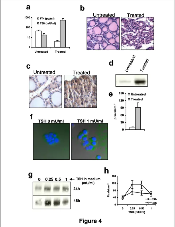
Virtually all proteins involved in thyroid function are regulated by TSH. We investigated whether also sortilin undergoes this type of regulation, both in a mouse model and in FRTL-5 cells. We first studied the effects of TSH on sortilin expression in mice treated with methimazole and sodium perchlorate, which, especially if used in combination, are potent inhibitors of thyroid hormone synthesis, leading to hypothyroidism and increased TSH secretion from the pituitary, which makes it a suitable model to study protein expression in response to TSH. As shown in Fig. 4a, following treatment of mice for 5 days, there was a reduction of serum free thyroxine (FT4) and a marked increase of serum TSH. The typical morphological and
histological changes induced by TSH were observed. Gross examination of the thyroid gland revealed a slight enlargement and engorgement compared with untreated mice (not shown). At histology (Fig. 4b), there was hypertrophy of thyroid epithelial cells, reduced amounts of colloid and of the size of follicular lumina, indicating massive colloid reabsorption. Staining of thyroid epithelial cells with an anti-sortilin antibody was markedly increased in mice treated with methimazole and perchlorate compared with untreated mice (Fig. 4c), and similar results were obtained by Western blotting analysis of thyroid extracts (Fig. 4d and e). The effects of treatment on sortilin was specific, as expression of the housekeeping protein glyceraldehydes-3-phosphate-dehydrogenase (GAPDH) was unaffected (not shown), as also reported previously [100] .
We then investigated the same issue in FRTL-5 cells. Cells were cultured either in media containing various concentrations of TSH or in medium lacking TSH, for time intervals ranging from 24 to 48 hours. As shown in Fig. 4f-h, TSH up-regulated sortilin, as observed both by immunofluorescence staining and Western blotting. The effect of TSH was time-dependent, indicating that prolonged stimulation is more effective than acute stimulation (Fig. 4 g and h). The effect was seen starting at relatively low concentrations of TSH (0.25 mU/ml), and it plateaued at concentrations greater than 0.5 mU/ml (Fig. 4 g and h), indicating that the effect was somehow saturable, as expected for a receptor/ligand-mediated mechanism, as it should be in the case of TSH/TSH-receptor interactions.TSH did not affect expression of GAPDH (not shown), as also reported previously , suggesting that its effect on sortilin was specific.
Figure 4 Expression of sortilin in thyroid epithelial cells is TSH-dependent. a-e) Mice treated with methimazole and sodium perchlorate. a) FT4 and TSH serum levels; b) H&E staining of thyroid sections; c) Immunohistochemistry for sortilin in thyroid sections. Intracellular staining is increased in thyrocytes in mice treated with methimazole and perchlorate; d) Western blotting for sortilin in thyroid extracts; e) pixel densities of the bands in Western blotting experiments; f-h) FRTL-5 cells. f) Immunofluorescence staining with an anti-sortilin antibody in cells cultured for 24h in medium lacking TSH or containing TSH 1mU/ml; g) Western blotting for sortilin in extracts from cells cultured for different time periods (24-48h) in media containing various concentrations of TSH, as indicated; e) Pixel densities of the bands in Western blotting
Un tre ate d Tre ate d
d
TSH 0 mU/ml TSH 1 mU/ml 1 10 100 1000 Untreated Treated FT4 (pg/m l) TSH (m U/m l)a
b
c
Untreated Treatede
0 30 60 90 120 150 p ix e ls /c m 2 Untreated Treated Untreated Treatedf
0 40 80 120 160 0 0.25 0.50 1 TSH (mU/ml) P ix e ls /c m 2 24h 48hh
Figure 4
24h TSH in medium (mU/ml)g
0 0.25 0.5 1 48h Un tre ate d Tre ate dd
TSH 0 mU/ml TSH 1 mU/ml 1 10 100 1000 Untreated Treated FT4 (pg/m l) TSH (m U/m l)a
b
c
Untreated Treatede
0 30 60 90 120 150 p ix e ls /c m 2 Untreated Treated Untreated Treatedf
0 40 80 120 160 0 0.25 0.50 1 TSH (mU/ml) P ix e ls /c m 2 24h 48hh
Figure 4
24h TSH in medium (mU/ml)g
0 0.25 0.5 1 48hTg is a sortilin ligand
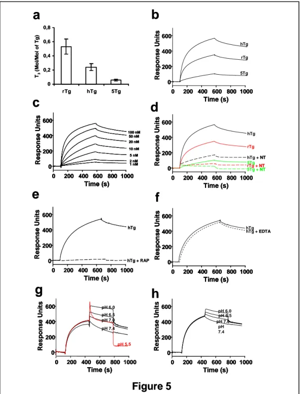
We investigated whether Tg is a sortilin ligand, using surface plasmon resonance binding assays. For this purpose, we used a purified, soluble preparation of sortilin (s-sortilin), and three purified Tg preparations, namely rat Tg (rTg), human Tg (hTg), and Tg purified from FRTL-5 cell supernatants (5Tg). As shown in Fig. 5a, the three preparations had a different hormone [(triiodo-thyronine (T3)] content. Thus, rTg was
highly hormonogenic, whereas hTg and 5Tg had a lower T3 content. This was
expected based on the knowledge that rTg was purified from the thyroids of untreated rats, that hTg was purified from the thyroids of Graves’ patients given methimazole [a potent inhibitor of hormone formation within Tg], and that FRTL-5 cells are not capable of forming hormone residues within the Tg molecules they produce. As shown in Fig 5b, all of the three Tg preparations bound to immobilized sortilin in a concentration-dependent manner and with high affinity, as indicated by the following mean Kd values: rTg: ~1 nM; hTg: ~3 nM; 5Tg: ~30 nM. The apparent different
extent of binding between the 3 Tg preparation actually reflects the different concentrations used (rTg: 25 nM; hTg: 100 nM; 5Tg: 100 nM). No binding to sorCSC3, another member of the Vps10p family, was observed (not shown). An example of the dose-dependent, high affinity pattern of binding is shown in Fig. 5c for hTg. Similar results, with different Kd values, were obtained also for rTg and 5Tg
(not shown). As shown in Fig. 5d, binding of all the three Tg preparations was inhibited, if not abolished, by the sortilin ligand neurotensin. In addition, binding was almost abolished by RAP, as shown for hTg in Fig. 5e. Because RAP binds to both
sortilin and Tg, we could not establish to what extent inhibition was due to RAP binding to sortilin, to Tg, or to both. Binding of Tg to sortilin was not affected by EDTA, indicating it is not calcium-dependent (Fig. 5f). As shown in Fig. 5g and h, binding was inhibited only at pH values ≤5.5, suggesting that probably the complex Tg-sortilin is stable in a relatively acidic environment with pH values ≥6.0.