RESEARCH ARTICLE
Mechanism and Treatment of Renal Fibrosis
LPS removal reduces CD80-mediated albuminuria in critically ill patients
with Gram-negative sepsis
X
Giuseppe Stefano Netti,
1Fabio Sangregorio,
2Federica Spadaccino,
1Francesco Staffieri,
3Antonio Crovace,
3Barbara Infante,
2Annamaria Maiorano,
2Giulia Godeas,
2Giuseppe Castellano,
4Anna Maria Di Palma,
4Clelia Prattichizzo,
1Antonella Cotoia,
5Lucia Mirabella,
5Loreto Gesualdo,
4Gilda Cinnella,
5Giovanni Stallone,
2Elena Ranieri,
1* and Giuseppe Grandaliano
2*
1Clinical Pathology Unit and Center for Molecular Medicine, Department of Medical and Surgical Sciences, University of Foggia, Foggia, Italy;2Nephrology Dialysis and Transplantation Unit, Department of Medical and Surgical Sciences, University of Foggia, Foggia, Italy;3Veterinary Surgery Unit, Department of Emergency and Organ Transplantation, University of Bari Aldo Moro, Bari, Italy;4Nephrology Dialysis and Transplantation Unit, Department of Emergency and Organ Transplantation, University of Bari Aldo Moro, Bari, Italy; and5Anesthesia and Intensive Care Unit, Department of Medical and Surgical Sciences, University of Foggia, Foggia, Italy
Submitted 17 October 2018; accepted in final form 20 January 2019 Netti GS, Sangregorio F, Spadaccino F, Staffieri F, Crovace A, Infante B, Maiorano A, Godeas G, Castellano G, Di Palma AM, Prattichizzo C, Cotoia A, Mirabella L, Gesualdo L, Cinnella G, Stallone G, Ranieri E, Grandaliano G. LPS removal reduces
CD80-mediated albuminuria in critically ill patients with Gram-negative sepsis. Am J Physiol Renal Physiol 316: F723–F731, 2019. First published January 23, 2019; doi:10.1152/ajprenal.00491.2018.— LPS-induced sepsis is a leading cause of acute kidney injury (AKI) in critically ill patients. LPS may induce CD80 expression in podocytes with subsequent onset of proteinuria, a risk factor for progressive chronic kidney disease (CKD) frequently observed after AKI. This study aimed to investigate the therapeutic efficacy of LPS removal in decreasing albuminuria through the reduction of podocyte CD80 expression. Between January 2015 and December 2017, 70 consecu-tive patients with Gram-negaconsecu-tive sepsis-induced AKI were random-ized to either have coupled plasma filtration and adsorption (CPFA) added to the standard care (n⫽ 35) or not (n ⫽ 35). To elucidate the possible relationship between LPS-induced renal damage, proteinuria, and CD80 expression in Gram sepsis, a swine model of LPS-induced AKI was set up. Three hours after LPS infusion, animals were treated or not with CPFA for 6 h. Treatment with CPFA significantly reduced serum cytokines, C-reactive protein, procalcitonin, and endotoxin levels in patients with Gram-negative sepsis-induced AKI. CPFA significantly lowered also proteinuria and CD80 urinary excretion. In the swine model of LPS-induced AKI, CD80 glomerular expression, which was undetectable in control pigs, was markedly increased at the podocyte level in LPS-exposed animals. CPFA significantly reduced LPS-induced proteinuria and podocyte CD80 expression in septic pigs. Our data indicate that LPS induces albuminuria via podocyte expression of CD80 and suggest a possible role of timely LPS removal in preventing the maladaptive repair of the podocytes and the consequent increased risk of CKD in sepsis-induced AKI.
acute kidney injury; albuminuria; CD80; chronic kidney disease; sepsis
INTRODUCTION
Acute kidney injury (AKI) in hospitalized patients has
be-come increasingly common, in particular within the intensive
care unit (ICU) population (22– 67%) and is associated with
poor long-term outcome (15, 49). Patients with severe AKI
requiring initiation of renal replacement therapy have the
highest in-hospital mortality rates, ranging from 45% to 70%
(25, 27, 32, 50). Among survivors of this high-risk population,
as many as 13–32% require dialysis at the time of hospital
discharge (2, 3, 42). Limited data are available regarding
recovery of sufficient kidney function to allow discontinuation
of dialysis therapy following hospital discharge in patients with
severe AKI (39), yet patients initiating in-hospital renal
re-placement therapy constitute a significant proportion of the
incident dialysis population each year (26, 31, 40). Several
reports in the past decade suggest a causal link between AKI
and the consequent development of progressive chronic kidney
disease (CKD) (16). Sepsis, particularly the severe form
sus-tained by Gram-negative infections, is a major cause of AKI in
ICU patients (52). Sepsis is a complex pathologic condition
arising from the host response to an overwhelming infection.
Gram-negative bacteria and the components of their walls, in
particular the lipid A-containing lipopolysaccharide (LPS),
play a major role in this setting (37). Indeed, LPS may induce
uncontrolled cytokines release and activation of coagulation on
endothelial cells leading to shock, multiple organ damage, and
even death (34).
While the pathophysiology of sepsis-induced AKI has been
widely investigated, the mechanisms underlining the transition
between LPS-mediated AKI and the onset of progressive CKD
are still poorly known.
Exposure to low-dose LPS, through direct stimulation of the
Toll-like receptor-4 (TLR-4)/CD14 receptor, rapidly
upregu-lates CD80 in podocytes in vivo, leading to nephrotic-range
proteinuria (36). CD80 is a transmembrane protein expressed
on the surfaces of B cells and other antigen-presenting cells. It
works as a costimulatory signal and modulates T-cell
activa-tion by binding to either CD28 or CTLA-4. At podocyte level,
CD80 expression, induced by various stimuli, causes actin
* E. Ranieri and G. Grandaliano contributed equally to this work. Address for reprint requests and other correspondence: G. S. Netti, Clinical Pathology Unit and Center for Molecular Medicine, Dept. of Medical and Surgical Sciences, Univ. of Foggia, Viale Luigi Pinto, 71122 Foggia, Italy (e-mail: [email protected]).
reorganization, foot process (FP) effacement, and disruption of
the slit diaphragm (SD), thereby modifying glomerular
perm-selectivity and leading to proteinuria. An increased CD80
expression at podocyte level has been reported in several
proteinuric glomerulopathies and is associated with poor
re-sponse to therapy and worse renal outcome (9, 10, 23, 35).
In the last decade, a significant improvement in treatment of
Gram-negative sepsis and septic-AKI has been obtained. Novel
blood purification approaches such as direct hemoperfusion
with polymyxin B (DHP-PMX) or citrate-based coupled
plasma filtration adsorption (CPFA) therapy have been widely
employed in ICU to treat severe sepsis and sepsis-related AKI.
Both these therapies are able to remove endotoxins by direct
adsorption of LPS onto polymyxin B-coated cartridge (48) or
by efficient removal of the LPS-adaptor protein LBP (CPFA;
Ref. 4).
This study aimed to investigate the therapeutic efficacy of
LPS removal in decreasing albuminuria through the reduction
of podocyte CD80 expression.
MATERIALS AND METHODS
Study population. The study population of the present prospective,
single center, cohort study consisted of 70 consecutive patients with AKI and Gram-negative severe sepsis undergoing coupled plasma filtration and adsorption (CPFA) added to the standard care (n⫽ 35) or not (n⫽ 35), from January 1, 2015 to December 31, 2017 at the Intensive Care Unit of University Hospital “Ospedali Riuniti” of Foggia.
The present study involving human participants was approved by the local ethical committee (Decision No. 158/CE/2014 of September 03, 2014; Ethical Committee at the University Hospital “Ospedali Riuniti” of Foggia).
All procedures performed the present study were in accordance with the ethical standards of the Declaration of Helsinki, and all the enrolled patients provided written informed consent to participate to the present study.
All of the enrolled patients were 18 yr old or older. The diagnosis of AKI was defined according to K-DIGO 2012 Guidelines (21). The diagnosis of septic shock or severe sepsis was defined according to the Third Consensus Conference on Sepsis (43) and the severity of disease was assessed by Acute Physiology and Chronic Health Eval-uation III (APACHE III) score (22). The diagnosis of Gram-negative sepsis required the presence of endotoxin levels⬎0.7. All the patients with Gram-positive sepsis were excluded from the study.
In all patients enrolled, the site of infection was identified in the first 12 h after diagnosis of sepsis. In detail, inclusion criteria were at least two of the systemic inflammatory response syndrome (SIRS) criteria and at least one organ dysfunction as defined by the American College of Chest Physicians/Society of Critical Care Medicine (ACCP/SCCM) Consensus Conference (1).
Patients were excluded from the study for the following reasons: life expectancy ⬍30 days (as assessed by the attending physician); HIV infection; uncontrolled hemorrhage within 24 h before study entry; organ transplantation or end-stage renal disease requiring he-modialysis or peritoneal dialysis before study entry; history of sensi-tivity to anticoagulant and/or extracorporeal circulation; severe throm-bocytopenia (⬍30,000 cells/mm3) and/or granulocytopenia (⬍500
cells/mm3); and an APACHE III score ⬎30, a Sequential Organ
Failure Assessment (SOFA) score ⬎12, or ⬎4 organ failures by a Goris score (13 22, 51).
A historical cohort of 24 patients with Gram-negative sepsis-induced AKI, treated with direct hemoperfusion with polymyxin B (DHP-PMX) was employed as the control group.
Extracorporeal treatment. All the patients were randomized 1:1
and assigned to receive different strategies of extracorporeal blood purification: CPFA added to the standard care (group A, n⫽ 35) or hemofiltration (group B, n⫽ 35). Patients of both groups received full intensive care management, including fluid resuscitation, vasopres-sors, antimicrobial therapy, ventilatory support, and appropriate sur-gical management, when required.
The historical cohort of 24 patients with Gram-negative sepsis-induced AKI was treated with direct hemoperfusion with polymyxin B (DHP-PMX) (group C).
CPFA was performed with the use of a four-pump monitor (Flexia, Bellco, Mirandola, Italy) consisting of a plasma filter (0.45-m2polyethersulfone) and a following absorption on an unselective
hydrophobic resin cartridge (surface of ~700 m2/g) and a final passage
of the reconstituted blood through a high-permeability 1.4-m2
poly-ethersulfone hemofilter, in which convective exchanges may be ap-plied in a postdilution fashion.
The postdilution reinfusion rate was set up to 4 l/h. The blood flow was maintained at 150 –200 ml/min, while the plasma flow was controlled by a filtration fraction ranging from 10 to 18% of blood flow (8). The reinfusion solution, which was sterile and pyrogen-free, presented the following composition (in mmol/l): 140 Na, 1.5 K, 2 Ca, 0.75 Mg, 108 Cl, 35 bicarbonate, 4 acetate, and 5.55 glucose mEq/l. The anticoagulation protocol was based on continuous citrate infusion (24). CPFA was repeated daily for the first 3 days, lasting at least 10 h/session to assure the treatment of 0.15 liter of plasma·kg⫺1·day⫺1. All patients in the historical control group underwent PMX treat-ment twice, on days 0 and 1, after diagnosis of severe sepsis. Blood flow rate was maintained at 80 –120 ml/min. Each hemoperfusion session lasted for 2 h. Heparin was used as an anticoagulant.
Serum and urinary measurements. The main clinical and laboratory
data were recorded daily. Serum levels of C-reactive protein (CRP), procalcitonin, endotoxin activity assay (EAA), and cytokines and urinary levels of creatinine, albumin, and proteins were measured at Clinical Pathology Laboratory of the University Hospital “Ospedali Riuniti” of Foggia using common laboratory assays. In detail, high-sensitive CRP was quantified by immunoturbidimetry (Beckman Coulter, Brea, CA). Procalcitonin was titrated by chemiluminescence immunoassay (VIDAS BRAHMS PCT assay; BioMerieux, Marcy-l’Etoile, France). EAA was measured by a chemiluminescent bio-assay EAA (Spectral Medical, ON, Canada). The cytokines panel detection (IL-1, IL-2, IL-6, IL-8, IL-10, TNF-␣, IL-4, IL-1␣, VEGF, IFN-␥, monocyte chemoattractant protein-1, and EGF) was performed using a Protein Biochip Array (Randox Laboratory, Crumlin, UK).
Urinary assessment of creatinine, albumin, and proteins was per-formed by routine laboratory methods. Urinary CD80 measurements were performed using a commercially available ELISA kit (Thermo Fisher, Waltham, MA) as previously described (11, 28). All the results of urine albumin, proteins, and CD80 are reported as ratio and normalized to urinary creatinine excretion.
Animal model. The animal model of endotoxemia was induced, as
previously described (4), after approval by the Ethical Committee of the Italian Ministry of Health, in 216.8⫾ 0.7-mo-old female domestic swine weighting 58.4⫾ 14.7 kg. The choice of female pigs was driven by the knowledge of a stronger activation of the innate immunity compared with male animals, as previously demonstrated (4). All the experiment procedures were performed in adherence to the National Institutes of Health Guide for the Care and Use of
Labora-tory Animals. Briefly, the animals were randomized in three groups:
control (n ⫽ 7), LPS (n ⫽ 7), and LPS ⫹ CPFA (n ⫽ 7). Under general anesthesia, LPS and LPS⫹ CPFA animals were infused with a saline solution containing 300g/kg of LPS while control animals received 10 ml of sterile saline solution. Three hours after the infusion, LPS⫹ CPFA animals were treated by CPFA as previously described for 6 h. Animals were euthanized at the end of treatment. Both control and LPS animals were euthanized 9 h after LPS infusion. A renal tissue sample was obtained after euthanization, and a portion
of each tissue specimen was immediately snap frozen in optimal cutting temperature medium (Sakura Finetek, Torrance, CA) and stored in liquid nitrogen. Urinary output was measured and urine samples were collected from all animals and stored at⫺80°C until use.
Tissue analysis. Confocal microscopy was performed on 5-
m-thick cryostat tissue sections of swine renal biopsies using a confocal laser-scanning microscope (TCS SP5; Leica Microsystems, Wetzlar, Germany), as previously described (45, 46). All the reagents were prepared in 0.05% Triton X-100-containing PBS to permeabilize cell membranes. Staining with primary hamster monoclonal anti-CD80 IgG antibody (clone 16-10A1; Thermo Fisher Scientific, San Diego, CA) and rabbit polyclonal anti-WT1 IgG antibody (clone C19; Santa Cruz Biotechnology, Santa Cruz, CA) and secondary Alexa Fluor 488-labeled goat anti-hamster IgG and Alexa Fluor 546-labeled goat anti-rabbit IgG, respectively (Molecular Probes, Eugene, OR), was performed following the manufacturers’ instructions. Nuclei were counterstained with Topro-3 (Molecular Probes). The slides were then mounted in Gel Mount (Biomed) and sealed. Specific fluorescence quantification was performed as previously described (11, 45, 46).
Statistical analysis. Results are expressed as means⫾ SD unless
otherwise stated. Statistical analyses were performed using the SPSS software (SPSS 17.0, Evanston, IL). Continuous variables were com-pared by paired or unpaired Student t-test or Mann-Whitney U-test, as appropriate. Frequencies were compared among groups by2-test. A
two-sided P⬍ 0.05 was considered statistically significant.
RESULTS
Between January 2015 and December 2017, 70 consecutive
patients met the inclusion criteria for the present study and
were assigned to two different blood purification treatments. In
detail, 35 patients with sepsis related to Gram-negative
bacte-remia were treated with CPFA added to the standard care
(group A), while the remaining 35 patients were treated only
with hemofiltration (group B).
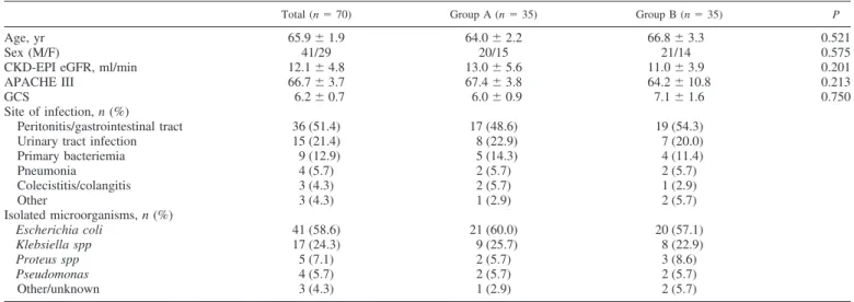
As shown in Table 1, the analysis of main clinical and
laboratory characteristics of the study population at the
begin-ning of treatment did not show statistically significant
differ-ences in age, sex distribution, and estimated glomerular
filtra-tion rate [Chronic Kidney Disease Epidemiology Collaborafiltra-tion
(CKD-EPI eGFR)], as well as in the APACHE III and Glasgow
Coma Scale (GCS) score between the two groups. Moreover
no statistical difference was observed in sites of infection and
type of isolated microorganisms (Table 1).
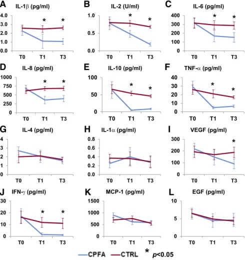
Analysis of cytokine levels at beginning of the treatment did
not show statistical differences between the two group (Fig. 1,
A–L). However, treatment with CPFA significantly reduced the
levels of IL-1
, IL-2, IL-6, IL-8, IL-10, TNF-␣, VEGF, and
IFN-
␥ already after the first treatment, as compared with group
treated with standard care (Fig. 1, A–L).
Accordingly, baseline serum CRP and Procalcitonin levels
were high in Gram-negative septic patients of two groups (Fig. 2,
A and B). Treatment with CPFA significantly reduced serum CRP
and procalcitonin levels at 1 and 3 days, as compared with control
group (Fig. 2, A and B).
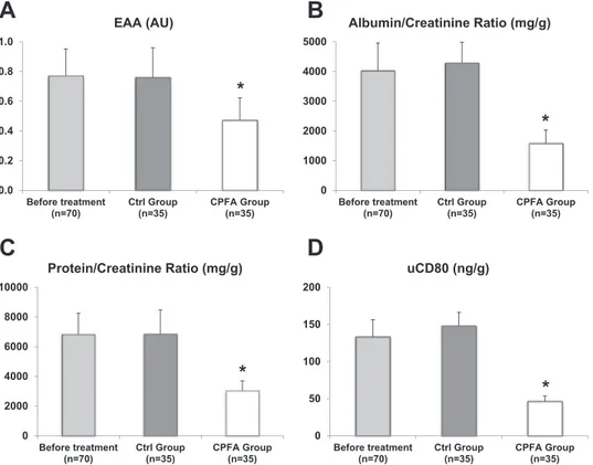
Finally, we assessed the effect of septic status and the
efficacy of proposed treatments on glomerular permeability to
proteins. Extracorporeal treatment with CPFA significantly
reduced the levels of EEA, as compared with control group
(Fig. 3A).
Baseline proteinuria and albuminuria were significantly high
in Gram-negative septic patients, but the reduction in
circulat-ing LPS levels by CPFA induced a reduction in glomerular
permeability to plasma proteins, as demonstrated by the
reduc-tion of proteinuria and albuminuria levels (Fig. 3, B and C).
Finally, we investigated the urinary excretion of CD80.
Base-line urine CD80/creatinine ratio was elevated in Gram-negative
septic patients. Removal of LPS by CPFA induced a
statisti-cally significant reduction in urinary CD80 excretion as
com-pared with control group (Fig. 3D).
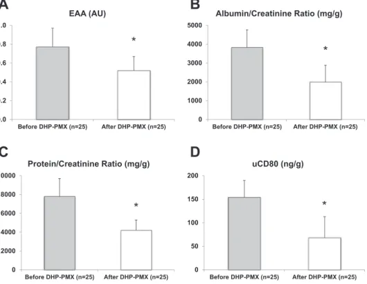
The analysis of the historical control group treated with direct
hemoperfusion with polymyxin B (DHP-PMX) confirmed the
beneficial effect of LPS removal on serum endotoxin activity and
urinary glomerular permeability to proteins, as well as on
reduc-tion of urinary CD80 excrereduc-tion (Fig. 4, A–D).
The analysis of main clinical and laboratory parameters
before and after CPFA treatment did not show any significant
difference between male and female septic patients in our study
group (data not shown).
Table 1. Main clinical and laboratory characteristics of the study population at the beginning of treatment
Total (n⫽ 70) Group A (n⫽ 35) Group B (n⫽ 35) P
Age, yr 65.9⫾ 1.9 64.0⫾ 2.2 66.8⫾ 3.3 0.521
Sex (M/F) 41/29 20/15 21/14 0.575
CKD-EPI eGFR, ml/min 12.1⫾ 4.8 13.0⫾ 5.6 11.0⫾ 3.9 0.201
APACHE III 66.7⫾ 3.7 67.4⫾ 3.8 64.2⫾ 10.8 0.213
GCS 6.2⫾ 0.7 6.0⫾ 0.9 7.1⫾ 1.6 0.750
Site of infection, n (%)
Peritonitis/gastrointestinal tract 36 (51.4) 17 (48.6) 19 (54.3)
Urinary tract infection 15 (21.4) 8 (22.9) 7 (20.0)
Primary bacteriemia 9 (12.9) 5 (14.3) 4 (11.4) Pneumonia 4 (5.7) 2 (5.7) 2 (5.7) Colecistitis/colangitis 3 (4.3) 2 (5.7) 1 (2.9) Other 3 (4.3) 1 (2.9) 2 (5.7) Isolated microorganisms, n (%) Escherichia coli 41 (58.6) 21 (60.0) 20 (57.1) Klebsiella spp 17 (24.3) 9 (25.7) 8 (22.9) Proteus spp 5 (7.1) 2 (5.7) 3 (8.6) Pseudomonas 4 (5.7) 2 (5.7) 2 (5.7) Other/unknown 3 (4.3) 1 (2.9) 2 (5.7)
Values are means⫾ SD. CKD-EPI, Chronic Kidney Disease Epidemiology Collaboration; APACHE III, Acute Physiology and Chronic Health Evaluation III; F, female; M, male; GCS, Glasgow Coma Scale.
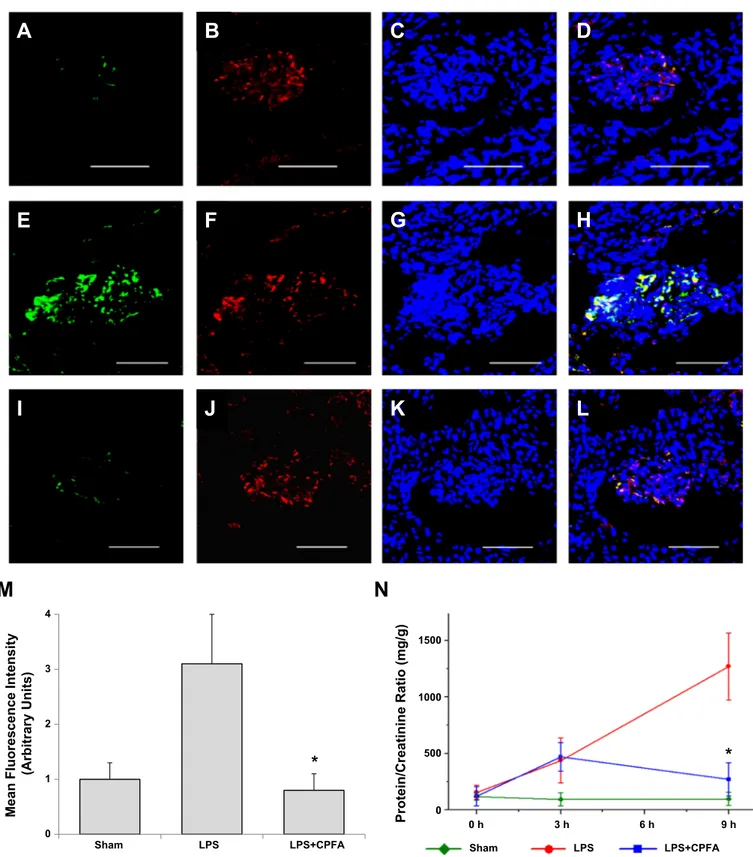
To elucidate the possible relationship between LPS-induced
renal damage and subsequent increase in glomerular
permea-bility to proteins in Gram-negative sepsis and CD80
expres-sion, a swine model of LPS-induced AKI was set up. After 3
h from LPS infusion, endotoxemic animals were treated or not
for 6 h with CPFA. Renal biopsies were performed at euthanize
nine hours after LPS infusion in all the experimental groups.
Confocal analysis of frozen renal tissues showed absence of
CD80 glomerular expression in control pigs not exposed to
LPS (Fig. 5, A–D). The experimental group exposed to LPS,
but not treated with CPFA, showed marked increase of CD80
expression at the podocyte level, as demonstrated by the
colocalization with the podocyte marker WT-1 (Fig. 5, E–H).
CPFA treatment reduced podocyte expression of CD80 after
LPS exposure, reaching a level comparable to the experimental
group not exposed to LPS (Fig. 5, I–L), as shown by the image
analysis (Fig. 5M).
Finally we evaluated the effect of CPFA treatment on
LPS-induced increase in glomerular permeability to proteins.
CPFA treatment, while reducing LPS levels and inhibiting
CD80 induction at the podocyte level, was also able to reduce
glomerular permeability to proteins (Fig. 5N, blue line).
0.0 1.0 2.0 3.0 4.0 T0 T1 T3 IL-1β (pg/ml) 0.0 0.5 1.0 T0 T1 T3 IL-2 (U/ml) 0 100 200 300 400 T0 T1 T3 IL-6 (pg/ml) 0 250 500 750 1000 T0 T1 T3 IL-8 (pg/ml) 0 50 100 T0 T1 T3 IL-10 (pg/ml) 0 10 20 30 40 T0 T1 T3 TNF-α (pg/ml) 0 1 2 3 4 T0 T1 T3 IL-4 (pg/ml) 0.0 0.2 0.4 0.6 0.8 T0 T1 T3 IL-1α (pg/ml) 0 100 200 300 T0 T1 T3 VEGF (pg/ml) 0 10 20 30 T0 T1 T3 IFN-γ (pg/ml) 0 500 1000 1500 T0 T1 T3 MCP-1 (pg/ml) 0 5 10 T0 T1 T3 EGF (pg/ml)
*
*
*
*
*
*
*
*
*
*
p<0.05*
*
*
*
*
*
A B C
D E F
G H I
J K L
Fig. 1. Effects of extracorporeal treatments [coupledplasma filtration and adsorption (CPFA)] on cytokine removal. A–F, I, and J: Gram-negative septic patients treated with CPFA (blue line) showed a significant reduction of most of cytokine levels (IL-1, IL-2, IL-6, IL-8, IL-10, TNF-␣, VEGF, and IFN-␥) as compared with control group (red line) (*P⬍ 0.05 control group vs. CPFA group). G, H, K, and L: no statistically significant differences of cytokine levels were observed within the control group (red line) before and after standard therapy (IL-4, IL-1␣, MCP-1, and EGF).
0 50 100 150 200 250 300 350 T1 T2 T3 CRP (mg/dl) 0 10 20 30 40 50 60 70 PCT (ng/ml)
*
p<0.05*
*
*
*
A B
T1 T2 T3Fig. 2. Effects of extracorporeal treatments coupled plasma filtration and adsorption (CPFA) on markers of inflamma-tion and sepsis. A and B: Gram-negative septic patients treated with CPFA (blue line) showed a significant reduc-tion of most of serum C-reactive protein (CRP; A) and procalcitonin (PCT; B) levels as compared with control group (red line) (*P⬍ 0.05 control group vs. CPFA group), while no statistically significant differences of cytokine levels were observed within the control group (redline) before and after standard therapy. T, time in days.
DISCUSSION
In the present study, we demonstrated for the first time that
LPS exposure in Gram-negative sepsis may induce
albumin-uria via podocyte expression of CD80. Selective removal of
LPS reduced both albuminuria and CD80 expression in the
experimental model as well as in the clinical setting, thus
suggesting a possible role of this therapeutic approach in
preventing the increased risk of progressive CKD in patients
with septic AKI.
Among ICU patients, AKI is common, particularly the form
associated with sepsis, and may lead to poor long-term
out-comes (6, 38). Indeed, several observational studies report an
increased incidence of progressive CKD among patients with a
previous AKI. Current clinical practice guidelines recommend
that patients should be followed-up for at least 3 mo to assess
whether they may have developed CKD (21). Despite these
recommendations, many patients with AKI do not receive a
follow-up assessment nor do they receive appropriate care
when kidney function has not recovered (14, 41), resulting in
lost opportunities to intervene and potentially improve
long-term outcomes (12). Many recent studies aimed to identify and
screen patients at high risk of developing progressive CKD to
improve outcomes for patients following AKI (7, 17). To this
aim several prediction models have been built up,
encompass-ing both clinical and laboratory variables (i.e., sex, age, and
baseline serum creatinine). Among them, albuminuria has been
proposed as an early marker of septic AKI (30) but also as a
risk factor associated with lower rate of AKI recovery at 30
days after discharge (29). Recently, an investigation on a large
Canadian cohort of patients confirmed albuminuria as one of
the risk factors independently associated with CKD onset after
AKI (18).
Our data confirm that during septic AKI due to
Gram-negative infection, there is an increase glomerular permeability
with subsequent albuminuria both in the experimental model
and in the clinical clinical setting. In patients who survive after
an AKI episode, albuminuria may represent a risk factor of
progression toward CKD, as previously reported (33).
The pathophysiology of albuminuria onset during sepsis
is still largely unclear. It has been suggested that the
sepsis-related proinflammatory state alters tubular handling
of filtered albumin and promotes albuminuria (20).
More-over, the release of several proinflammatory cytokines into
the systemic circulation during sepsis might lead to a loss of
endothelial cells barrier integrity and subsequent capillary
leak. According to this hypothesis, albuminuria represents
0 1000 2000 3000 4000 5000 Before treatment (n=70) Ctrl Group (n=35) CPFA Group (n=35) Albumin/Creatinine Ratio (mg/g) 0 2000 4000 6000 8000 10000 Before treatment (n=70) Ctrl Group (n=35) CPFA Group (n=35) Protein/Creatinine Ratio (mg/g) 0.0 0.2 0.4 0.6 0.8 1.0 Before treatment (n=70) Ctrl Group (n=35) CPFA Group (n=35) EAA (AU) 0 50 100 150 200 Before treatment (n=70) Ctrl Group (n=35) CPFA Group (n=35) uCD80 (ng/g)
B
A
D
C
*
*
*
*
Fig. 3. Effects of coupled plasma filtration and adsorption (CPFA) on markers of Gram-negative infection and glomerular permeability. A: extracorporeal treatment with CPFA significantly reduced the levels of endotoxin activity assay [EAA; 0.77⫾ 0.18 vs. 0.47 ⫾ 0.15 arbitrary units (AU), *P ⬍ 0.05], while no differences were observed in the control group (0.77⫾ 0.18 vs. 0.76 ⫾ 0.20 arbitrary units, P ⫽ 0.85). B and C: reduction in circulating LPS levels by CPFA treatment induced a reduction in glomerular permeability to plasma proteins, as demonstrated by the reduction of proteinuria and albuminuria levels (6,834.6⫾ 1,413.3 vs. 3,031.9 ⫾ 670.8 mg/g of creatinine and 4,025.7 ⫾ 935.5 vs. 1,581.7 ⫾ 449.5 mg/g of creatinine for proteinuria and albuminuria before and after treatment, respectively, *P ⬍ 0.05); for instance, standard therapy did not affect glomerular permeability to proteins in the control group (6,834.6⫾ 1,413.3 vs. 6,851.5 ⫾ 1,621.8 mg/g of creatinine for proteinuria before and after treatment, P ⫽ 0.61; 4,025.7 ⫾ 935.5 vs. 4,287.9 ⫾ 829.3 mg/g of creatinine for albuminuria before and after treatment, P⫽ 0.84). D: finally, the removal of LPS by CPFA induced a statistically significant reduction in urinary CD80 excretion in Gram-negative patients (133.2⫾ 23.1 vs 46.5 ⫾ 7.0 ng/g of creatinine for urinary CD80 before and after treatment, respectively, *P ⬍ 0.05, while no differences were observed after treatment in the control group (133.2⫾ 23.1 vs 148.1 ⫾ 17.8 ng/g of creatinine for urinary CD80 before and after treatment, P⫽ 0.96).
the glomerular manifestation of this enhanced capillary
permeability (19).
Another possible mechanism responsible of albuminuria is
podocyte disruption and apoptosis. Podocytes have a peculiar
structure and the slit diaphragms (SD) on the membranes of
their FPs play an essential role in the glomerular filtration
barrier. There is now growing evidence that several SD- and
FP-associated molecules, including nephrin, podocin, and
CD2-associated protein (CD2AP), are significantly
downregu-lated during septic AKI (36).
More generally, LPS exposure may induce renal
dysfunc-tion and consequent maladaptive repair of septic AKI
through suppression of mitochondrial biogenesis. In a
mouse model of LPS-induced AKI, endotoxin exposure was
able to disrupt mitochondrial homeostasis by
downregula-tion of peroxisome proliferator-activated receptor
␥
coacti-vator-1
␣ (PGC-1␣) and activation of the TLR-4/MEK/ERK
pathway in the renal cortex (44).
The exposure to low-dose LPS in an animal model of sepsis,
through direct stimulation of the TLR-4/CD14 receptor at
podocyte level, may rapidly induce CD80 expression leading
to nephrotic-range proteinuria (36). Indeed, CD80 expression
on podocytes induces actin reorganization, FP effacement, and
disruption of the SD complex due to sequestration of nephrin,
CD2AP, and ZO-1, thereby modifying glomerular
permselec-tivity and leading to proteinuria.
In our experimental model, exposure to LPS induced CD80
expression at podocyte level and an increase of proteinuria,
while in septic patients a Gram-negative infection induced a
significant urinary excretion of albumin and CD80. A selective
removal of LPS with blood extracorporeal treatment not only
reduced EAA but also decreased proteinuria along with CD80
expression and urinary excretion both in the animal model and
in the clinical setting.
Of note this result was obtained not only in experimental
group treated with CPFA but also in the historical control
group treated with DHP-PMX. This finding underlines the
beneficial effects of LPS removal, regardless of extracorporeal
treatment choice, on serum endotoxin activity and urinary
glomerular permeability to proteins, as well as on reduction of
urinary CD80 excretion.
Our observation strongly suggests a link in experimental and
clinical settings of a causal link between endotoxemia, CD80
expression by podocytes, and new onset albuminuria. LPS may
also induce podocyte disruption through an indirect
mecha-nism. It has been reported that during LPS-mediated sepsis,
there is an increase of IL-1
 and TNF-␣ released from
acti-vated macrophages infiltrating the glomeruli (19). These
cyto-kines may directly suppress with a paracrine effect nephrin
expression in podocytes, through the loss of nucleus-localized
WT1, a transcriptional factor for upregulating nephrin gene
(47). In our cohort of patients, the blood extracorporeal
puri-fication significantly reduces circulating levels of both IL-1

and TNF-
␣, among other proinflammatory cytokines. Thus we
cannot exclude that the effect of LPS on CD80 expression and
albuminuria may not be mediated by these two cytokines.
Whether LPS induces albuminuria through a direct or
indi-rect action, our data clearly support the hypothesis that LPS
effects on podocytes may represent a potential mechanism
involved in maladaptive repair underlying the progression of
AKI toward CKD (5). In this perspective, our observation may
suggest that a timely removal of LPS through DHP-PMX
and/or CPFA may reduce both urinary albumin and CD80
excretion, thus preventing the increased risk of progressive
CKD in patients with septic AKI.
The sample size of our clinical study and the lack of
follow-up data due to high mortality observed in our study
group may significantly limits our observations, although their
0 1000 2000 3000 4000 5000 Before DHP-PMX (n=25) After DHP-PMX (n=25) Albumin/Creatinine Ratio (mg/g) 0 2000 4000 6000 8000 10000 Before DHP-PMX (n=25) After DHP-PMX (n=25) Protein/Creatinine Ratio (mg/g) 0.0 0.2 0.4 0.6 0.8 1.0 Before DHP-PMX (n=25) After DHP-PMX (n=25) EAA (AU) 0 50 100 150 200 Before DHP-PMX (n=25) After DHP-PMX (n=25) uCD80 (ng/g)
B
A
D
C
*
*
*
*
Fig. 4. Effects of direct hemoperfusion with polymyxin B (DHP-PMX) on markers of Gram-negative infection and glomerular permeabil-ity. A: extracorporeal treatment with DHP-PMX significantly reduced the levels of endotoxin activity assay [EAA; 0.77⫾ 0.20 vs. 0.52 ⫾ 0.15 arbitrary units (AU), *P⬍ 0.05]. B and C: the reduction in circulating LPS levels by DHP-PMX treatment induced a reduction in glomer-ular permeability to plasma proteins, as demon-strated by the reduction of proteinuria and albu-minuria levels (7,804.6⫾ 1,911.3 vs. 4,209.1 ⫾ 1,080.9 mg/g of creatinine and 3,825.7⫾ 935.1 vs. 1,889.7⫾ 889.5 mg/g of creatinine for pro-teinuria and albuminuria before and after treat-ment, respectively, *P⬍ 0.05. D: finally, the removal of LPS by DHP-PMX or CPFA in-duced a statistically significant reduction in uri-nary CD80 excretion in Gram-negative patients (154.2⫾ 36.1 vs. 68.2 ⫾ 45.1 ng/g of creatinine for urinary CD80 before and after treatment, respectively, *P⬍ 0.05).
A
B
C
D
E
F
G
H
I
J
K
L
0 1 2 3 4Mean Fluorescence Intensity
(Arbitrary Units)
*
N
M
500 1500 0 1000 9 h 0 h 3 h 6 h Protein/Creatinine Ratio (mg/g)*
Sham LPS LPS+CPFA Sham LPS LPS+CPFAFig. 5. Coupled plasma filtration and adsorption (CPFA) treatment reduces podocyte expression of CD80 and urinary protein excretion after LPS exposure in a pig model of acute renal damage. A–D: control pigs, which were not exposed to LPS (sham), did not express CD80 at podocyte levels. After 3 h from LPS infusion, endotoxemic animals were treated or not for 6 h with CPFA. E–H: the experimental group exposed to LPS, but not treated with CPFA, showed marked increase of CD80 expression (green) at the podocyte level, as demonstrated by the colocalization with the podocyte marker WT-1 (red). I–L: PFA treatment was able to reduce podocyte expression of CD80 (green) after LPS exposure, reaching a level comparable to the experimental group not exposed to LPS. Nuclei were stained with To-pro-3 (bar⫽ 50 m). M: analysis of mean fluorescence intensity (MFI) confirmed a reduction of CD80 expression after CPFA treatment in LPS-exposed pigs as compared with untreated septic group (0.8⫾ 0.3 vs. 3.1 ⫾ 0.9 arbitrary units for CPFA-treated septic pigs and LPS-exposed pigs, respectively; *P ⬍ 0.02). N: treatment with CPFAwas also able to reduce glomerular permeability to proteins in LPS-exposed pigs [268.8 ⫾ 217.8 vs. 1,270.9⫾ 895.7 mg/g of creatinine for proteinuria between CPFA-treated septic pigs (blue line) and LPS-exposed untreated pigs (red line), *P ⬍ 0.05].
confirmation in the animal model may support our conclusions.
The strength of our swine model is represented by its several
advantages over any rodent model used so far. Its main limit,
on the other hand, is represented by the challenge to induce a
multimicrobial sepsis, since pigs are particularly sensible to the
severe hemodynamic instability featuring this model of severe
sepsis.
Nevertheless, we believe that further clinical investigations
are warranted to confirm our data.
ACKNOWLEDGMENTS
We thank Leonarda Varraso from the Nephrology, Dialysis, and Transplan-tation Unit of the University of Foggia, and Clara Divella from the Nephrol-ogy, Dialysis, and Transplantation Unit of the University of Bari for excellent technical assistance.
GRANTS
This work was supported by the Italian Ministry of Health (Giovani Ricercatori 2011-2012 granted to G. S. Netti and G. Castellano), and Apulia Region (Technology Cluster Project “Precious” granted to L. Gesualdo, G. Cinnella, and G. Grandaliano). The article has been published with a contri-bution from 5x1000 IRPEF funds in favor of the University of Foggia, in memory of Gianluca Montel.
DISCLOSURES
No conflicts of interest, financial or otherwise, are declared by the authors.
AUTHOR CONTRIBUTIONS
G.S.N. conceived and designed research; G.S.N., F. Sangregorio, A. Cro-vace, B.I., A.M., G. Godeas, G. Castellano, A.M.D.P., A. Cotoia, and L.M. performed experiments; G.S.N., F. Spadaccino, and C.P. analyzed data; F. Sangregorio, F. Spadaccino, B.I., A.M., G. Godeas, C.P., A. Cotoia, and L.M. interpreted results of experiments; F. Spadaccino and C.P. prepared figures; G.S.N., L.G., and G. Cinnella drafted manuscript; G.S., E.R., and G. Granda-liano edited and revised manuscript; G.S., E.R., and G. GrandaGranda-liano approved final version of manuscript.
REFERENCES
1. ACCP/SCCM. American College of Chest Physicians/Society of Critical Care Medicine Consensus Conference: definitions for sepsis and organ failure and guidelines for the use of innovative therapies in sepsis. Crit Care Med 20: 864 –874, 1992. doi:10.1097/00003246-199206000-00025. 2. Bagshaw SM, Laupland KB, Doig CJ, Mortis G, Fick GH, Mucenski
M, Godinez-Luna T, Svenson LW, Rosenal T. Prognosis for long-term
survival and renal recovery in critically ill patients with severe acute renal failure: a population-based study. Crit Care 9: R700 –R709, 2005. doi:10. 1186/cc3879.
3. Bagshaw SM, Uchino S, Kellum JA, Morimatsu H, Morgera S, Schetz
M, Tan I, Bouman C, Macedo E, Gibney N, Tolwani A, Oudemans-van Straaten HM, Ronco C, Bellomo R; Beginning and Ending Supportive Therapy for the Kidney (B.E.S.T. Kidney) Investigators.
Association between renal replacement therapy in critically ill patients with severe acute kidney injury and mortality. J Crit Care 28: 1011–1018, 2013. doi:10.1016/j.jcrc.2013.08.002.
4. Castellano G, Stasi A, Intini A, Gigante M, Di Palma AM, Divella C,
Netti GS, Prattichizzo C, Pontrelli P, Crovace A, Staffieri F, Fiacca-dori E, Brienza N, Grandaliano G, Pertosa G, Gesualdo L. Endothelial
dysfunction and renal fibrosis in endotoxemia-induced oliguric kidney injury: possible role of LPS-binding protein. Crit Care 18: 520, 2014. doi:10.1186/s13054-014-0520-2.
5. Chawla LS, Eggers PW, Star RA, Kimmel PL. Acute kidney injury and chronic kidney disease as interconnected syndromes. N Engl J Med 371: 58 –66, 2014. doi:10.1056/NEJMra1214243.
6. Coca SG, Singanamala S, Parikh CR. Chronic kidney disease after acute kidney injury: a systematic review and meta-analysis. Kidney Int 81: 442–448, 2012. doi:10.1038/ki.2011.379.
7. Coleman EA, Berenson RA. Lost in transition: challenges and opportu-nities for improving the quality of transitional care. Ann Intern Med 141: 533–536, 2004. doi:10.7326/0003-4819-141-7-200410050-00009.
8. Formica M, Inguaggiato P, Bainotti S, Wratten ML. Coupled plasma filtration adsorption. Contrib Nephrol 156: 405–410, 2007. doi:10.1159/ 000102131.
9. Garin EH, Diaz LN, Mu W, Wasserfall C, Araya C, Segal M, Johnson
RJ. Urinary CD80 excretion increases in idiopathic minimal-change
disease. J Am Soc Nephrol 20: 260 –266, 2009. doi:10.1681/ASN. 2007080836.
10. Garin EH, Mu W, Arthur JM, Rivard CJ, Araya CE, Shimada M,
Johnson RJ. Urinary CD80 is elevated in minimal change disease but not
in focal segmental glomerulosclerosis. Kidney Int 78: 296 –302, 2010. doi:10.1038/ki.2010.143.
11. Gigante M, Lucarelli G, Divella C, Netti GS, Pontrelli P, Cafiero C,
Grandaliano G, Castellano G, Rutigliano M, Stallone G, Bettocchi C, Ditonno P, Gesualdo L, Battaglia M, Ranieri E. Soluble serum␣Klotho
is a potential predictive marker of disease progression in clear cell renal cell carcinoma. Medicine (Baltimore) 94: e1917, 2015. doi:10.1097/MD. 0000000000001917.
12. Goldstein SL, Jaber BL, Faubel S, Chawla LS; Acute Kidney Injury
Advisory Group of American Society of Nephrology. AKI transition of
care: a potential opportunity to detect and prevent CKD. Clin J Am Soc Nephrol 8: 476 –483, 2013. doi:10.2215/CJN.12101112.
13. Goris RJ, te Boekhorst TP, Nuytinck JK, Gimbrère JS. Multiple-organ failure. Generalized autodestructive inflammation? Arch Surg 120: 1109 – 1115, 1985. doi:10.1001/archsurg.1985.01390340007001.
14. Harel Z, Wald R, Bargman JM, Mamdani M, Etchells E, Garg AX,
Ray JG, Luo J, Li P, Quinn RR, Forster A, Perl J, Bell CM.
Nephrologist follow-up improves all-cause mortality of severe acute kidney injury survivors. Kidney Int 83: 901–908, 2013. doi:10.1038/ki. 2012.451.
15. Hoste EA, Clermont G, Kersten A, Venkataraman R, Angus DC, De
Bacquer D, Kellum JA. RIFLE criteria for acute kidney injury are
associated with hospital mortality in critically ill patients: a cohort anal-ysis. Crit Care 10: R73, 2006. doi:10.1186/cc4915.
16. Ishani A, Nelson D, Clothier B, Schult T, Nugent S, Greer N, Slinin Y,
Ensrud KE. The magnitude of acute serum creatinine increase after
cardiac surgery and the risk of chronic kidney disease, progression of kidney disease, and death. Arch Intern Med 171: 226 –233, 2011. doi:10. 1001/archinternmed.2010.514.
17. James M, Bouchard J, Ho J, Klarenbach S, LaFrance JP, Rigatto C,
Wald R, Zappitelli M, Pannu N. Canadian Society of Nephrology
commentary on the 2012 KDIGO clinical practice guideline for acute kidney injury. Am J Kidney Dis 61: 673–685, 2013. doi:10.1053/j.ajkd. 2013.02.350.
18. James MT, Pannu N, Hemmelgarn BR, Austin PC, Tan Z, McArthur
E, Manns BJ, Tonelli M, Wald R, Quinn RR, Ravani P, Garg AX.
Derivation and external validation of prediction models for advanced chronic kidney disease following acute kidney injury. JAMA 318: 1787– 1797, 2017. doi:10.1001/jama.2017.16326.
19. Kato T, Mizuno S, Kamimoto M. The decreases of nephrin and nuclear WT1 in podocytes may cause albuminuria during the experimental sepsis in mice. Biomed Res 31: 363–369, 2010. doi:10.2220/biomedres.31.363. 20. Kato T, Mizuno-Horikawa Y, Mizuno S. Decreases in podocin, CD2-associated protein (CD2AP) and tensin2 may be involved in albuminuria during septic acute renal failure. J Vet Med Sci 73: 1579 –1584, 2011. doi:10.1292/jvms.11-0203.
21. Kidney International Supplements. KDIGO Clinical Practice Guideline for Acute Kidney Injury (Online). https://www.kisupplements.org/issue/ S2157-1716(12)X7200-9 [31 January 2018].
22. Knaus WA, Wagner DP, Draper EA, Zimmerman JE, Bergner M,
Bastos PG, Sirio CA, Murphy DJ, Lotring T, Damiano A, Harrell FE Jr. The APACHE III prognostic system. Risk prediction of hospital
mortality for critically ill hospitalized adults. Chest 100: 1619 –1636, 1991. doi:10.1378/chest.100.6.1619.
23. Ling C, Liu X, Shen Y, Chen Z, Fan J, Jiang Y, Meng Q. Urinary CD80 levels as a diagnostic biomarker of minimal change disease. Pediatr Nephrol 30: 309 –316, 2015. doi:10.1007/s00467-014-2915-3.
24. Livigni S, Bertolini G, Rossi C, Ferrari F, Giardino M, Pozzato M,
Remuzzi G; GiViTI: Gruppo Italiano per la Valutazione degli Inter-venti in Terapia Intensiva (Italian Group for the Evaluation of Interventions in Intensive Care Medicine) is an independent collabo-ration network of Italian Intensive Care units. Efficacy of coupled
plasma filtration adsorption (CPFA) in patients with septic shock: a multicenter randomised controlled clinical trial. BMJ Open 4: e003536, 2014. doi:10.1136/bmjopen-2013-003536.
25. McCarthy JT. Prognosis of patients with acute renal failure in the intensive-care unit: a tale of two eras. Mayo Clin Proc 71: 117–126, 1996. doi:10.4065/71.2.117.
26. Mendelssohn DC, Curtis B, Yeates K, Langlois S, MacRae JM,
Semeniuk LM, Camacho F, McFarlane P; STARRT Study investiga-tors. Suboptimal initiation of dialysis with and without early referral to a
nephrologist. Nephrol Dial Transplant 26: 2959 –2965, 2011. doi:10.1093/ ndt/gfq843.
27. Metnitz PG, Krenn CG, Steltzer H, Lang T, Ploder J, Lenz K, Le Gall
JR, Druml W. Effect of acute renal failure requiring renal replacement
therapy on outcome in critically ill patients. Crit Care Med 30: 2051– 2058, 2002. doi:10.1097/00003246-200209000-00016.
28. Netti GS, Prattichizzo C, Montemurno E, Simone S, Cafiero C, Rascio
F, Stallone G, Ranieri E, Grandaliano G, Gesualdo L. Exposure to
low-vs iso-osmolar contrast agents reduces NADPH-dependent reactive oxy-gen species oxy-generation in a cellular model of renal injury. Free Radic Biol Med 68: 35–42, 2014. doi:10.1016/j.freeradbiomed.2013.11.016. 29. Neyra JA, Li X, Yessayan L, Adams-Huet B, Yee J, Toto RD; Acute
Kidney Injury in Critical Illness Study Group. Dipstick albuminuria
and acute kidney injury recovery in critically ill septic patients. Nephrol-ogy (Carlton) 21: 512–518, 2016. doi:10.1111/nep.12637.
30. Neyra JA, Manllo J, Li X, Jacobsen G, Yee J, Yessayan L; AKICI
Study Group. Association of de novo dipstick albuminuria with severe
acute kidney injury in critically ill septic patients. Nephron Clin Pract 128: 373–380, 2014. doi:10.1159/000368902.
31. O’Hare AM, Batten A, Burrows NR, Pavkov ME, Taylor L, Gupta I,
Todd-Stenberg J, Maynard C, Rodriguez RA, Murtagh FE, Larson EB, Williams DE. Trajectories of kidney function decline in the 2 years
before initiation of long-term dialysis. Am J Kidney Dis 59: 513–522, 2012. doi:10.1053/j.ajkd.2011.11.044.
32. Palevsky PM, Zhang JH, O’Connor TZ, Chertow GM, Crowley ST,
Choudhury D, Finkel K, Kellum JA, Paganini E, Schein RM, Smith MW, Swanson KM, Thompson BT, Vijayan A, Watnick S, Star RA, Peduzzi P; VA/NIH Acute Renal Failure Trial Network. Intensity of
renal support in critically ill patients with acute kidney injury. N Engl J Med 359: 7–20, 2008. doi:10.1056/NEJMoa0802639.
33. Parr SK, Matheny ME, Abdel-Kader K, Greevy RA Jr, Bian A, Fly J,
Chen G, Speroff T, Hung AM, Ikizler TA, Siew ED. Acute kidney
injury is a risk factor for subsequent proteinuria. Kidney Int 93: 460 –469, 2018. doi:10.1016/j.kint.2017.07.007.
34. Ramnath RD, Ng SW, Guglielmotti A, Bhatia M. Role of MCP-1 in endotoxemia and sepsis. Int Immunopharmacol 8: 810 –818, 2008. doi:10. 1016/j.intimp.2008.01.033.
35. Reiser J, Gupta V, Kistler AD. Toward the development of podocyte-specific drugs. Kidney Int 77: 662–668, 2010. doi:10.1038/ki.2009.559. 36. Reiser J, von Gersdorff G, Loos M, Oh J, Asanuma K, Giardino L,
Rastaldi MP, Calvaresi N, Watanabe H, Schwarz K, Faul C, Kretzler M, Davidson A, Sugimoto H, Kalluri R, Sharpe AH, Kreidberg JA, Mundel P. Induction of B7-1 in podocytes is associated with nephrotic
syndrome. J Clin Invest 113: 1390 –1397, 2004. doi:10.1172/JCI20402. 37. Ronco C, Brendolan A, Dan M, Piccinni P, Bellomo R, De Nitti C,
Inguaggiato P, Tetta C. Adsorption in sepsis. Kidney Int Suppl 76:
S148 –S155, 2000. doi:10.1046/j.1523-1755.2000.07619.x.
38. Sawhney S, Marks A, Fluck N, Levin A, McLernon D, Prescott G,
Black C. Post-discharge kidney function is associated with subsequent
ten-year renal progression risk among survivors of acute kidney injury. Kidney Int 92: 440 –452, 2017. doi:10.1016/j.kint.2017.02.019. 39. Schmitt R, Coca S, Kanbay M, Tinetti ME, Cantley LG, Parikh CR.
Recovery of kidney function after acute kidney injury in the elderly: a systematic review and meta-analysis. Am J Kidney Dis 52: 262–271, 2008. doi:10.1053/j.ajkd.2008.03.005.
40. Schoonover KL, Hickson LJ, Norby SM, Hogan MC, Chaudhary S,
Albright RC Jr, Dillon JJ, McCarthy JT, Williams AW. Risk factors
for hospitalization among older, incident haemodialysis patients. Nephrol-ogy (Carlton) 18: 712–717, 2013. doi:10.1111/nep.12129.
41. Siew ED, Peterson JF, Eden SK, Hung AM, Speroff T, Ikizler TA,
Matheny ME. Outpatient nephrology referral rates after acute kidney
injury. J Am Soc Nephrol 23: 305–312, 2012. doi:10.1681/ASN. 2011030315.
42. Silvester W, Bellomo R, Cole L. Epidemiology, management, and outcome of severe acute renal failure of critical illness in Australia. Crit Care Med 29: 1910 –1915, 2001. doi: 10.1097/00003246-200110000-00010.
43. Singer M, Deutschman CS, Seymour CW, Shankar-Hari M, Annane
D, Bauer M, Bellomo R, Bernard GR, Chiche JD, Coopersmith CM, Hotchkiss RS, Levy MM, Marshall JC, Martin GS, Opal SM, Ruben-feld GD, van der Poll T, Vincent JL, Angus DC. The Third International
Consensus Definitions for Sepsis and Septic Shock (Sepsis-3). JAMA 315: 801–810, 2016. doi:10.1001/jama.2016.0287.
44. Smith JA, Stallons LJ, Collier JB, Chavin KD, Schnellmann RG. Suppression of mitochondrial biogenesis through toll-like receptor 4-de-pendent mitogen-activated protein kinase kinase/extracellular signal-reg-ulated kinase signaling in endotoxin-induced acute kidney injury. J Phar-macol Exp Ther 352: 346 –357, 2015. doi:10.1124/jpet.114.221085. 45. Stallone G, Cormio L, Netti GS, Infante B, Selvaggio O, Fino GD,
Ranieri E, Bruno F, Prattichizzo C, Sanguedolce F, Tortorella S, Bufo P, Grandaliano G, Carrieri G. Pentraxin 3: a novel biomarker for
predicting progression from prostatic inflammation to prostate cancer. Cancer Res 74: 4230 –4238, 2014. doi:10.1158/0008-5472.CAN-14-0369. 46. Stallone G, Matteo M, Netti GS, Infante B, Di Lorenzo A, Prattichizzo
C, Carlucci S, Trezza F, Gesualdo L, Greco P, Grandaliano G.
Semaphorin 3F expression is reduced in pregnancy complicated by pre-eclampsia. An observational clinical study. PLoS One 12: e0174400, 2017. doi:10.1371/journal.pone.0174400.
47. Takano Y, Yamauchi K, Hayakawa K, Hiramatsu N, Kasai A,
Oka-mura M, Yokouchi M, ShitaOka-mura A, Yao J, KitaOka-mura M.
Transcrip-tional suppression of nephrin in podocytes by macrophages: roles of inflammatory cytokines and involvement of the PI3K/Akt pathway. FEBS Lett 581: 421–426, 2007. doi:10.1016/j.febslet.2006.12.051.
48. Tani T, Hanasawa K, Endo Y, Yoshioka T, Kodama M, Kaneko M,
Uchiyama Y, Akizawa T, Takahasi K, Sugai K. Therapeutic apheresis
for septic patients with organ dysfunction: hemoperfusion using a poly-myxin B immobilized column. Artif Organs 22: 1038 –1044, 1998. doi:
10.1046/j.1525-1594.1998.06086.x.
49. Thakar CV, Christianson A, Freyberg R, Almenoff P, Render ML. Incidence and outcomes of acute kidney injury in intensive care units: a Veterans Administration study. Crit Care Med 37: 2552–2558, 2009. doi:10.1097/CCM.0b013e3181a5906f.
50. Uchino S, Kellum JA, Bellomo R, Doig GS, Morimatsu H, Morgera S,
Schetz M, Tan I, Bouman C, Macedo E, Gibney N, Tolwani A, Ronco C; Beginning and Ending Supportive Therapy for the Kidney (BEST Kidney) Investigators. Acute renal failure in critically ill patients: a
multinational, multicenter study. JAMA 294: 813–818, 2005. doi:10.1001/ jama.294.7.813.
51. Vincent JL, de Mendoncxa A, Cantraine F, Moreno R, Takala J,
Suter PM, et al. Use of the SOFA score to assess the incidence of organ
dysfunction/failure in intensive care units: results of a multi-centric, prospective study. Crit Care Med 26: 1793–1800, 1998. doi:10.1097/ 00003246-199811000-00016.
52. Zarjou A, Agarwal A. Sepsis and acute kidney injury. J Am Soc Nephrol 22: 999 –1006, 2011. doi:10.1681/ASN.2010050484.