UNIVERSITÀ DEGLI STUDI DI ROMA
"TOR VERGATA"
FACOLTA' DI SCIENZE MATEMATICHE FISICHE E
NATURALI
DOTTORATO DI RICERCA IN BIOLOGIA CELLULARE E
MOLECOLARE
XXII CICLO
“Characterization of the Salmonella enterica
zinc import apparatus and of its relevance in the
host-pathogen interaction”.
PATRIZIA PETRARCA
A.A. 2009/2010
Docente Guida: Prof. A. Battistoni
Coordinatore: Prof. G. Cesareni
Summary
The ability of bacteria to colonize specific environments relies on their ability to obtain adequate supplies of the nutrients that are indispensable for their growth. Of particular relevance for human and animal health is to understand how bacterial pathogens face the problem of nutrient limitation in the infected host, in which several essential elements are not freely available for infectious microorganisms. In this respect, the recruitment of transition metals is a particularly challenging problem for bacterial pathogens, as these elements are usually present in forms that are not easily available for infectious microorganisms. For this reason, the sophisticated strategies adopted by pathogens to obtain iron have been the focus of intense investigations since a long time. Although the relevance of other metals in the host-pathogen interaction is usually considered as less important with respect to iron, evidences are accumulating that also the mechanisms ensuring the efficient uptake of zinc plays a critical role during bacterial infections.
Salmonella enterica, as well as many other Gram-negative bacteria, responds to zinc deficiency by producing the high affinity zinc uptake transporter ZnuABC. This complex belongs to the ABC transporters family and is constituted by three proteins: ZnuA, ZnuB and ZnuC. ZnuB is a membrane permease, ZnuC is the ATPase component, whereas ZnuA is a periplasmic metallochaperone which efficiently captures zinc in this cellular compartment and then delivers it to ZnuB. The expression of the znuABC operon is regulated by the metallated form of Zur, a dimeric protein which binds two zinc ions and thus represses znuABC transcription.
To investigate the relevance of zinc in host-pathogen interactions, we have constructed Salmonella enterica mutant strains in which the znuA gene, which encodes the periplasmic component of the ZnuABC high-affinity zinc transporter, was deleted. This mutation does not alter the ability of Salmonella to grow in rich media but drastically reduces its ability to multiply in media containing low levels of zinc (minimal medium) or in rich medium supplemented whit chelating agents (EDTA or TPEN). In agreement with this phenotype, ZnuA accumulates only in bacteria cultivated in environments poor in zinc. In spite of the nearly millimolar intracellular concentration of zinc, we have found that znuA is highly expressed in intracellular salmonellae recovered either from cultivated cells or from the spleens of infected mice. We have also observed that znuA mutants are impaired in their ability to grow in Caco-2 epithelial cells and
mutants was observed in susceptible (BALB/c) or Salmonella-resistant (DBA-2) mice infected intraperitoneally or orally.
To better understand bacterial responses to zinc deficiency, we have also investigated the role of ZinT, a periplasmic protein with a putative role in zinc homeostasis, in Salmonella. We have found that zinT expression is regulated by Zur and parallels that of ZnuA and ZnuB. Despite ZinT contributes to Salmonella growth in media poor of zinc, disruption of zinT does not significantly affect virulence in mice. The role of ZinT became clear using strains expressing a mutated form of ZnuA lacking a characteristic histidine-rich domain. In fact, Salmonella strains producing this modified form of ZnuA exhibited a ZinT-dependent capability to import zinc either in vitro or in infected mice, suggesting that ZinT and the histidine-rich region of ZnuA have redundant function. The hypothesis that ZinT and ZnuA cooperate in the process of zinc recruitment is supported by the observation that they form a stable binary complex in vitro. Although, the presence of ZinT is not strictly required to ensure the functionality of the ZnuABC transporter, our data suggest that ZinT facilitates metal acquisition during severe zinc shortage.
In conclusion, this study establishes that there is a stringent control of zinc availability in eukaryotic tissues which is critically important to limit the ability of bacterial pathogens to multiply within the infected host and that functionality of the ZnuABC transporter is critical for maximizing zinc import in such a hostile environment. Moreover, we have shown that in Salmonella there is an additional protein (ZinT) which interacts with ZnuA and increases its ability to bind zinc. As ZnuABC is the only high affinity Zn(II) transporter in bacteria and there are no homologs in the mammalian hosts, these observations indicate that the mechanisms of zinc import could be a privileged target for novel antibacterial therapies.
Index
pag.
1. Introduction... 7
1.1 Salmonella infections... 7
1.2 Salmonella strategies to evade host defences... 11
1.3 Host response to Salmonella infection... 13
1.4 Infection and nutrient: the case of iron... 16
1.5 Zinc and infection... 18
1.6 Zinc homeostasis in bacteria... 19
1.7 Role of ZnuABC in bacterial virulence... 24
1.8 ZnuA: periplasmic component of ZnuABC transporter... 25
1.9 ZnuB and ZnuC proteins... 28
1.10 ZinT: a periplasmic protein with a putative role in zinc impor... 28
2 Aims of the project... 31
3. Materials and Metods... 33
3.1
Materials... 33
3.2 Methods... 41
4.Results... 49
4.1 znuA is required for Salmonella enterica growth in poor zinc environments... 49
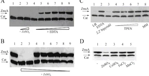
4.2 Expression of znuA is up-regulated when zinc is scarcely available... 53
4.3 ZnuA contributes to Salmonella multiplication within eukaryotic cells and accumulates in intracellular environments.. 55
4.4 A functional ZnuABC transporter is essential for Salmonella pathogenicity... 59
4.5 znuA and zinT distribution in eubacteria... 63
4.6 ZinT is not involved in cadmium resistance... 66
4.7 zinT is induced under zinc starvation and belongs to the Zur-regulated operon... 69
4.8 Consequences of zinT deletion on Salmonella growth... 73
4.9 ZinT role in Salmonella pathogenicity... 74
4.10 ZinT and ZnuA form a binary complex in vitro... 79
5. Conclusions... 85
6. References... 90
1. Introduction
1.1
Salmonella infections
Salmonella species are facultative intracellular Gram-negative bacteria
causing a wide spectrum of diseases in humans and animals ranging from self-limiting gastroenteritis to life-threatening systemic infections. Although more than 2000 different Salmonella serotypes have been so far identified, only a limited number of Salmonella enterica serovars are recognized as important human or animal pathogens. The majority of these pathogenic serovars cause acute gastroenteritis characterized by a short incubation period and a predominance of intestinal over systemic symptoms. However, a small number of serotypes typically cause severe systemic disease characterized by septicaemia, fever and/or abortion. In any case, the type of disease caused by a specific Salmonella strain usually depends on the combination of the serotype and the host involved, so that a particular
Salmonella serovar may cause a severe systemic disease in an animal species
and be clinically asymptomatic in another species. This is very clear for host adapted strains such as S. enterica serovar Typhi or Pullorum, which cause systemic diseases only in humans and poultry, respectively, but also for serovars able to infect a broad range of different animals. For example, the S.
enterica serovars Typhimurium (S. Typhimurium) and Enteritidis (S.
Enteritidis) cause diverse diseases in different animal species. In calves, S. Typhimurium (Petrie et al., 1977.) causes enterocolitis, and the animals can succumb to dehydration (Tsolis et al., 1999). In newly hatched chicks, serovars Enteritidis and Typhimurium cause systemic disease and diarrhea, whereas older chickens are asymptomatic carriers (Barrow et al., 1987; Barrow et al., 1987.). In immunocompetent humans, serovars Enteritidis and Typhimurium cause localized self-limiting enterocolitis, whereas systemic disease may develop in immunocompromised individuals (Tsolis et al., 1999.). Finally, serovars Enteritidis and Typhimurium cause a systemic typhoid feverlike disease in susceptible mouse strains, but no diarrhea. For this reason murine infection model with Salmonella enterica serovar Typhimurium have been useful for understanding the immune response to protect humans against S. Typhi. The mechanisms determining which type of disease is caused in which host by serovars Enteritidis and Typhimurium are still poorly understood.
Salmonella is typically acquired by the oral ingestion of contaminated food
or water. After entering the small intestine, it traverses the intestinal mucous layer and evades killing by digestive enzymes, bile salts, secretory IgA, antimicrobical peptide and other innate immune in order to obtain access to the underlying epithelium (Haraga et al, 2008). Salmonella penetrates the intestinal barrier preferentially using the follicle associated epitelium overlying Peyer’s patches and in particular exploits M cells (Jones et al, 1994), which are specialized epithelial cells that sample intestinal antigens through pinocytosis and transport them to lymphoid organs underlying Peyer’s patches. M cells are typically characterised by sparse, irregular microvilli on their apical surface and by a basolateral cytoplasmic invagination that forms a pocket harbouring lymphocytes and occasional macrophages (Jepsona and Clarkb, 2001).
When S. Typhimurium comes in contact with these cells it can bypass surface receptors and manipulate the host cytoskeleton directly through the injection of an array of bacterial effector molecules into the cytoplasm of the infected host cells. The combined action of these effector proteins triggers more dramatic reorganization of the actin cytoskeleton, resulting in intense membrane ruffling and subsequent bacteria internalization. (Ly and Casanova, 2007).
Salmonella can penetrate also via enterocytes (Bolton et al, 1999 and
Tam et al, 2008) or isolated lymphoid structure in witch are present M cells (Halle et al, 2006). Bacteria are present in this non-Peyer’s patch lymphoid structure after oral infection of mice and thus it can be used for bacterial exit from the intestinal lumen (Mowat et al., 2003).
Moreover, dendritic cells residing in the intestinal lamina propria might provide another gateway into the organism. These cells have been shown to extend dendrites through the epithelia lining into the intestinal lumen, allowing the direct sampling of antigens as well as pathogens (Niess and Reinecker, 2006)
Bacterial translocation via villus epithelial cells themselves might also be a way for microbes to enter the lamina propria (Fig. 1.1). Although the epithelium overlying villi is less favorable to bacterial penetration than that overlying Peyer’s patches, passage between epithelial cells, perhaps after bacteria-mediated destruction of epithelial layer integrity, could possibly occur (Tam et al, 2008).
Following passage through the epithelium of the Peyer’s patch, virulent
Salmonella strains enter the environment of the follicle dome, which is
populated with host lymphocytes and macrophages. To move into deeper tissue, these bacteria must be able to avoid and/or survive the
oxygen-Introduction
dependent and oxygen-independent killing mechanisms of professional phagocytes following internalization.(Jones and Falkow, 1996).
While bacteria which penetrate the intestinal epithelium via Peyer’s patches or isolated lymphoid tissues land directly in a lymphoid organ, bacteria that enter the lamina propria must find their way to the mesenteric lymph node. Salmonella can reach the mesenteric lymph node via the lymph as free bacteria or be transported by cells, presumably dendritic cells (Tam et
al, 2008).
Once the epithelial barrier has been breached, Salmonella serotypes that are associated with systemic illness are phagocytosed by intestinal macrophages. Internalized bacteria remain enclosed in a membrane-bound vacuole, referred to as the Salmonella-containing vacuole (SCV), which is modified by bacteria to prevent its maturation or fusion with lysosomal compartments. Bacterial directed maturation of the SCV leads to the formation of a protect intracellular niche that is permissive for bacterial replication (Ly and Casanova, 2007). Replication of pathogenic Salmonella into the host cells may lead to the death of these cells and to a subsequent release of bacteria, some of which may reach the blood via the thoracic duct. In addition pathogenic bacteria can also disseminate inside cells. These findings are supported by the observation that Salmonella is associated with phagocytes in the blood within minutes after oral infection (Worley et al, 2006). When Salmonella reaches the blood is disseminate in several additional tissue including the spleen, liver and bone marrow. In these tissues Salmonella is present inside dendritic cells, monocytes, macrophages and neutrophils (Tam et al, 2008).
Figure 1.1: Entry and capture of orally acquired Salmonella. Salmonella can invade
using different mechanisms, which are indicated with numbers. Within Peyer’s patches (PP),
Salmonella can traverse the intestinal barrier through: (1) M cells in the follicle-associated
epithelium (FAE); (2) epithelial cells forming the FAE, in particular after bacteria compromise M cells and the intestinal barrier (Jones et al, 1994); (3) Dendritic cells (DCs), could capture Salmonella (Niess and Reinecker, 2006) . Once bacteria cross the FAE they can be captured by DCs located in the subepithelial dome (SED). DCs containing Salmonella can then initiate an adaptive immune response by stimulating T cells in the PP (marked T in the figure) or migrate to the mesenteric lymph node (MLN) to initiate adaptive immunity. In addition, Salmonella can cross the intestinal barrier through solitary intestinal lymphoid tissue (SILT) (Halle et al, 2007), probably in a similar manner as in PP, as represented in (4). At villi, Salmonella can enter in different ways: (5) through M cells; (6) captured by DCs extending dendrites; or (7) passing through or between compromised epithelial cells. After bacteria reach the lamina propria, they can access the MLN. Finally, bacteria may be able to reach the blood stream, presumably transported by CD181 phagocytes, as shown in (8). In addition to being transported to the MLN via lymph within DCs, it remains possible that free bacteria could be transported in lymph. (9) Bacteria can exit the MLN as free bacteria or possibly associated with cells and seed other tissues in the body (Adapted from Tam et al, 2008).
Introduction
1.2 Salmonella strategies to evade host defences
The evolution of S. Typhimurium ability to resist against many of the host defence mechanism encountered in the lumen of the inflammed intestine has been driven by the acquisition of key virulence determinants through horizontal gene transfer (Baumler, 1997).
A remarkable feature of S. enterica in fact, is the presence of a large number of pathogenicity islands (PAI). PAI are distinct, relatively large chromosomal regions harbouring virulence genes that are present in pathogens but absent in benign relatives. PAI are characterized by a base composition different from the core genome and are often associated with transfer RNA (tRNA) genes and mobile genetic elements, like insertion sequence (IS) elements, transposons or phage genes.
The genome of Salmonella contains several of these clusters of genes, which are denominated Salmonella pathogenicity islands (SPIs). In general,
Salmonella pathogenicity island 1 (SPI-1) encodes genes necessary for
invasion of intestinal epithelial cells and induction of intestinal secretory and inflammatory responses. In contrast, Salmonella pathogenicity island 2 (2) encodes genes essential for intracellular replication. Both 1 and SPI-2, encode specialized devices for the delivery of virulence proteins into host cells, termed type III secretion systems (TTSSs) (Ohl and Miller, 2001). These systems appear to exert their function at different stages of the pathogenic cycle. While the SPI-1-encoded system is required to initiate intestinal infection, the type III secretion system encoded within SPI-2 appears to be required for the establishment of systemic infection (Zhoua and Galánb, 2001).
The SPI-1-encoded type III secretion system forms a needle-like complex that is responsible for the injection of bacterial effectors proteins into the host cell cytosol. Some of these effector proteins have been shown to be directly or indirectly involved in the regulation of the actin cytoskeleton dynamics (Ly and Casanova, 2007). For example, SipA promotes actin filament polymerization by reducing the monomer concentration necessary for filament assembly, enhancing the filament building activity of the host protein fimbrin and potentiating the nucleating activity of the C-terminal domain of SipC. SipC is a component of the bacterial translocon and contains two membrane-spanning domains (a 120-amino acid N-terminus and the 209-amino acid C-terminus) which extend into the host cytoplasm.
Moreover, the C-terminus portion promotes actin nucleation and effectors translocation (Ly and Casanova, 2007).
The Salmonella type III secretion system encoded by SPI-2, has been identified by virtue of its large impact on S. typhimurium virulence. In fact, the ability of S. typhimurium strains deficient in the SPI2-encoded T3SS to induce salmonellosis in mice is significantly decreased (Shea et al, 1996). SPI2 mutant strains attenuation is correlated with unability to proliferate within the organs of infected animals (Shea et al, 1996), and with reduced survival and proliferation inside cells.
Functional SPI-2 genes are clustered within six large transcriptional units. Thirty-one potential open reading frames on the SPI-2 region encode proteins that are directly involved in the assembly and regulation of the T3SS. The Ssa proteins are involved in the assembly of the syringe-like type III secretion injectisome. A set of nine Ssa proteins forms the injectisome core. Transport of some effectors through the injectisome is facilitated by formation of a complex between an effector and a chaperone encoded in SPI-2. For example, SscB, a protein encoded immediately upstream of sseF, acts as a chaperone for SseF (Waterman and Holden, 2003).
After phagocytosis, Salmonella undergoes extensive surface remodeling, as has been shown for the lipid A component of lipopolysaccharide (LPS) during growth within macrophages (Heithoff et al, 1999; Gibbons et al, 2005). Bacterial molecules that the host can recognize as indicators of infection, such as the SPI1 T3SS and flagellin, are repressed and the LPS structure is altered (Miao et al, 2006; Ernst et al, 2001). Some of the specific surface modifications include: decreasing the length of the O antigen, which is the repeating carbohydrate polymer of LPS; alterations to the number of acyl chains in the structure of the lipid A component of LPS; and changes in the protein content of the outer membrane, the inner membrane and the peptidoglycan layer (Guo et al, 1997; Gunn et al, 1998; Guo et al, 1998; Hilbert et al, 1999). Synthesis of enzymes that allow bacteria to cope with oxidative and nitrogenous radicals also occurs. Microarray studies have shown that up to 919 S. typhimurium genes are differentially regulated in response to the phagosomal environment, demonstrating that dramatic transcriptional and post-translational changes occur when Salmonella makes the transition from a nutrient-rich extracellular environment to the intracellular environment (Eriksson et al, 2003).
The observation that disruption of any single gene encoding for SPI2 T3SS effector protein reduces S. typhimurium virulence to a much lower extent than deletion of the entire SPI2 T3SS suggests that many effectors function cooperatively to exert their effects on the host cell. Furthermore, the
Introduction
deletion or mutation of many effector genes has no virulence phenotype, suggesting that their functions might be redundant. Early studies of SPI2 T3SS effectors, which primarily focused on determining their subcellular localization in mammalian cells, revealed that they might have specific targeting sequences that direct their localization to endosomal compartments, the Golgi apparatus, the actin cytoskeleton and the microtubule network. These observations indicated that components of these host-cell structures were the intracellular targets of the SPI2 T3SS (Haraga et al, 2008).
1.3 Host response to Salmonella infection
In response to S. typhimurium, the intestinal epithelium promotes an intense inflammatory response consisting largely of the migration of polymorphonuclear leukocytes toward and ultimately across the epithelial monolayer into the intestinal lumen (Gewirts et al, 1999).
In addition, direct contact between bacteria and host cells can result in the production of neutrophil chemoattractants known as CXC chemokines. However, in vivo these mediators are substantially produced by epithelial cells located in the crypts and at the base of villi, areas of the epithelium that are not invaded by S. Typhimurium.
The first significant interactions between bacteria and the host occur at the lymphoid follicles of the intestine. (Jones, 1997).
Experiments performed to determine whether the presence of invasive S. typhimuhum in the murine intestine has an effect on the cellular composition of Peyer's patches showed that the presence of the invasive pathogen caused a number of different host responses in the Peyer's patches (Savidge et al. 1991). The total number of M cells increased in the follicle-associated epithelium as compared to uninfected mice, the average crypt depth lengthened, and the rate of enterocyte migration from the intestinal crypts increased. In addition, the numbers of CD4+ cells increased and CD8+ cells decreased. These results indicate that the damage elicited by invasive
Salmonella induces a host response in the Peyer's patch tissue. Although the
pathogen causes significant damage to the epithelium of lymphoid follicles at early points of infection, the findings of this study indicate that the host quickly replaces cells that have been damaged and/or destroyed. In addition, it is clear that the immune system is activated by and responds to the presence of Salmonella by activating both humoral and cellular immunity (Mastroeni et al. 1993).
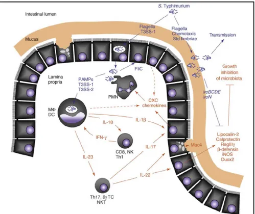
Another major aspect of the antimicrobial innate immune response encountered during acute inflammation is the production of epithelial-derived antimicrobial proteins and peptides. An important cytokine for orchestrating this arm of the response is IL-22, which induces expression in epithelial cells of host defense effector molecules that seem to be directed against luminal bacteria (Figure 1.2 and Santos et al, 2008). During
Citrobacter rodentium infection of mice, the IL-23/IL-22 axis is required for
expression in the colonic mucosa of calprotectin, an antimicrobial protein mediating zinc deprivation, and RegIIIg (regenerating islet-derived 3 gamma), a bactericidal C-type lectin (Zheng et al, 2008). RegIIIg expression in the cecal mucosa is markedly increased by an IL-23 dependent mechanism during S. Typhimurium infection of streptomycin-pretreated mice (Godinez et al, 2009). Pretreatment of mice with streptomycin renders them susceptible to serovar Typhimurium colitis that closely resembles the inflammatory responses observed in the human colon and animal models for intestinal salmonellosis (Barthel et al, 2003). Secretion of RegIIIg into the intestinal lumen of wild type mice contributes to clearance of luminal bacteria, including Listeria monocytogenes and vancomycin-resistant
Enterococcus, but this response is absent in mice deficient for myeloid
differentiation primary response protein 88 (MyD88), an adaptor protein for all Toll Like Receptors except TLR3 (Brandl et al, 2007).
In vitro, IL-22 induces the expression in human colonic epithelial cells of
inducible nitric oxide synthase (iNOS), mucin (MUC4) and lipocalin-2, an antimicrobial protein that prevents bacterial iron acquisition (Raffatellu et al, 2009). Whereas lipocalin-2 secretion is induced upon IL-22 stimulation, S. Typhimurium infection of colonic epithelial cell lines does not induce expression, suggesting that activation of this epithelial antimicrobial response requires paracrine IL-22 signaling rather than direct interaction of bacteria with enterocytes. In vivo, the epithelial cells of the ileal mucosa of rhesus macaques produce large quantities of lipocalin-2 in response to S. Typhimurium infection, resulting in accumulation of this antimicrobial in the intestinal lumen (Raffatellu et al, 2009). Other antimicrobial proteins and peptides whose transcripts are prominently induced in the intestinal mucosa during S. Typhimurium infection include iNOS, calprotectin, MUC4, dual oxidase 2 (Duox2) and bovine enteric b defensin (Godinez et al, 2008; Raffatellu et al, 2008; Raffatellu et al, 2007).
Introduction
Figure 1.2: The inflammation-adapted pathogenic lifestyle of S. Typhimurium. The
schematic shows host factors (red) and bacterial factors (blue) contributing to the development of acute intestinal inflammation within hours after S. Typhimurium infection. The bacterium initiates intestinal inflammation through direct interaction with host cells, resulting in a release of cytokines, such as IL-18 and IL-23. The cytokines IL-18 and IL-23 help to amplify responses in tissue by stimulating T cells to produce IFN-g (which activates macrophage antimicrobial activities), IL-17 (which orchestrates neutrophil barrier function) and IL-22 (which stimulates epithelial antimicrobial responses). The ability to grow on mucus carbohydrates and its resistance against antimicrobials (e.g. lipocalin-2 resistance mediated by the iroBCDEN genes) enable S. Typhimurium to benefit from the changes encountered in the inflamed intestine, resulting in its luminal outgrowth and enhanced transmission. CD8, CD8+ ab T cell; DC, dendritic cell; MF, macrophage; NK, natural killer cell; NKT, natural killer T cell; PAMPs, pathogen associated molecular patterns; PMN, neutrophil; dg TC, dg T cell; Th1, CD4+ ab memory type-1 T-helper cell; Th17, CD4+ ab memory type-17 T-helper cell (Adapted by Santos et al, 2008)
A variety of molecules in the mammalian intestine can impair growth and survival of microbes in this environment. One source of inhibitory molecules stems from many of the colonizing microbes themselves. Bacteriocins, including colicins, microcins, lantibiotics and others, are proteinaceous toxins that inhibit the growth of related bacteria (Baba et al, 1998). These molecules probably contribute to niche protection and inhibit colonization by potential pathogens. The other source of inhibitory molecules are products of the host, some of which are host defense molecules per se, whereas others have primary function in nutrient absorption. For example, bile salts and hydrolytic enzymes required for digestion are toxic for microbes and might contribute to controlling microbial proliferation and survival. Within the group of primary host defense molecules, epithelial cells make various peptides along the intestinal tract, some of which are constitutively expressed whereas others can be transcriptionally induced. In the small intestine, Paneth cells are the source of abundant quantities of antimicrobials in most mammals (Porter et al, 2002).. Among the most abundant and extensively studied antimicrobials of Paneth cells are the defensins. Constitutively expressed at high levels, defensins typically have membrane-targeted antimicrobial activity against a wide range of bacteria, some fungi and parasites. Transgenic and knockout mouse models have provided evidence that defensins contribute to the host defense against bacterial pathogens (Salzman et al, 2003; Wilson et al, 1999). Other secretory granule-associated constitutive antimicrobials produced by these cells are lysozyme and secretory phospholipase A2. Paneth cells also make some inducible antimicrobials, including RegIIIg, a C-type lectin with selective activity against Gram-positive bacteria (Cash et al, 2006; Brandl et al, 2007) In the colon, epithelial cells inducibly express a collection of antimicrobial peptides and proteins, including Resistin-like beta (RELM-b), RegIIIg, calprotectin, and b-defensins (Santos et al, 2009).
1.4 Infection and nutrient: the case of iron
The interaction between pathogenic bacteria and their host is determined by survival strategies on both sides, including competition for essential nutrients. In fact, deprivation of nutritive resource has an important role in host defences, but, during evolution, pathogenic bacteria have developed strategies to access specific nutrients from the host (Ratledge and Dover, 2000; Schaible and Kaufmann 2004; Schaible and Kaufmann, 2005).
Introduction
An instructive example for nutritive host-pathogen competition is represented by the mutual requirement for iron.
Iron is an essential growth factor for bacteria and parasites but it is also required for the host metabolism and other important host functions. In the host, essentially all the available iron is bound to specific proteins, such as transferrin, lactoferrin and ferritin, or is complexed to haem within haemoproteins (Hentze et al, 2004; Kaplan et al, 2002).
Host cells respond directly to invasive pathogens by altering their iron status. For example, macrophages, which are the principal cells that sequester invading bacteria, produce key proteins that alter their own iron status, by sequestering iron in the intravacuolar compartments colonized by invading bacteria. Such proteins are the natural resistance-associated macrophage proteins, of which two related forms are known, Nramp1 and Nramp2 (Schaible and Kaufmann, 2005).
As iron is an essential element for its growth and survival, Salmonella has evolved several strategies to access mammalian iron resources. Through secretion of siderophores, Salmonella is able to bind ferric iron with high affinity. Iron-bound siderophores are then internalized by outer membrane receptors of Salmonella, such as IroN, FepA and Cir (Hantke et al., 2003; Rabsch et al., 2003). After binding to their specific receptors at the outer bacterial membrane, iron-bound siderophores are delivered to periplasmatic-binding proteins for shuttling through the periplasmatic space and finally taken up in the cytoplasm using ABC transporters, such as FepBCDG and iroC (Nairts et al, 2007). Moreover, S. typhimurium expresses another protein named Feo as a mechanism for ferrous iron uptake, which is important during conditions characterized by low oxygen tension (Hantke, 2001).
1.5 Zinc and infection
Zinc is an essential trace element which is required by all living cells as it plays key roles in a very large number of molecular processes. It is a component of more than 300 enzymes from all six major functional classes, where it has catalytic, structural, or regulatory roles. Zinc-dependent biological functions include DNA replication, RNA transcription, signal transduction, enzymatic catalysis, redox regulation, cell proliferation, cell differentiation, and apoptosis (Overbeck and Haase, 2008).
The importance of zinc for human health is demonstrated by the clinical manifestations associated with zinc deficiency, which comprise growth retardation, thymic atrophy, hypogonadism, infertility, dermatitis, delayed wound healing, alopecia, poor pregnancy outcomes, teratology, anorexia, diarrhea, and increased susceptibility to infectious diseases caused by bacterial, viral, and fungal pathogens. (Overback, 2008)
Study performed in a mouse model showed that after infection with
Salmonella enterica serovar Typhimurium the concentration of zinc in the
plasma decreases significantly, byincreases in the liver (Rishi et al, 2007). A parallel reduction of iron content in plasma, a critical aspect of the acute phase response to bacterial infections, has been observed in response to a wide range of bacterial pathogens. This redistribution of zinc between tissues is regulated by cytochines, in particular IL-6, which stimulates the removal of this metal from plasma. It has been shown that IL-6 induces the expression of the zinc transporter Zip 14, thereby increasing zinc uptake into hepatocytes (Liuzzi et al, 2005). IL-6 also upregulates the zinc binding protein metallothionein and increases cellular zinc in hepatocytes (Schroeder
et al, 1990). Experiments with metallothionein knockout mice confirmed
that during endotoxin-induced inflammation, metallothionein is required for zinc sequestration in the liver, leading to a significant reduction in plasma zinc levels (Philcox et al, 1995). The significance of the accumulation of hepatic zinc has not yet been explained but the most common thought is that it is useful for the synthesis of zinc-proteins of acute phase and the synthesis and maturation of immune cells. This hypothesis is supported by the observation that slight zinc deficiency causes a significant depression of the immune defences (Ibs and Rink, 2003).
In addition, zinc affects several aspects of monocytes signal transduction and secretion of pro-inflammatory cytokines. In fact, it has been shown that zinc supplementation has been shown to reduce the production of tumor
Introduction
necrosis factor (TNF)-α and interleukin (IL)-1β in healthy human subjects (Ibs and Rink, 2003).
It is known that an increased hepatic concentration of zinc during infection is associated with beneficial responses, such as an increased hepatic synthesis of acute-phage globulins and an increased protection against hepatocellular damage from bacterial endotoxin. Zinc may protect against endotoxin-containing bacteria (e.g., S. typhimurium and E. coli) by stabilizing lysosomes or inhibiting lysosomal proteases (Bradley and Kluger, 1984) Among the immune cells that are affected by zinc deficiency, T lymphocytes seem to have the highest susceptibility. In fact,. zinc deficiency reduces the number of peripheral and thymic T cells, their proliferation in response to phytohemagglutinin, and the functions of helper and cytotoxic T cells, but also acts indirectly by reducing the levels of active serum thymulin (Overbeck et al, 2008). At the molecular level, zinc stimulates the autophosphorylation of the protein tyrosine kinase Lck by non-covalent interaction with the cytoplasmic tails of CD4 and CD8, leading to T cell activation. As a result, the delayed-type hypersensitivity reaction is usually reduced in zinc-deficient individuals (Salgueiro et al, 2000; Turner et al, 1990).
Moreover, other cells are also affected, leading to reduced antibody production and compromised function of cells of the innate immune system, such as natural killer cell activity, cytokine production by monocytes, and the chemotaxis and oxidative burst of neutrophil granulocytes (Ibs and Rink, 2003; Rink and Gabriel 2001).
Another important aspect is the interaction between inflammation and zinc. Pro-inflammatory cytokines have a direct influence on zinc homeostasis. It has been shown that IL-6 induces the expression of the zinc transporter Zrt- and Irt-like protein ZIP14, thereby increasing zinc uptake into hepatocytes. (Overback et al, 2008)
1.6 Zinc homeostasis in bacteria
Although zinc plays an essential role in a large number of biological processes, it can be toxic at high concentrations, and, therefore, intracellular zinc levels must be carefully regulated.
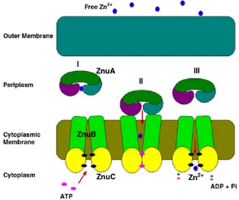
Zinc homeostasis in bacteria is achieved by balancing export systems and uptake systems. In Gram-negative bacteria, as shown in figure 1.3, zinc ions are mainly transported into the cell via ZnuABC (an ABC-type transporter)
and ZupT (a zinc permease), whereas they are removed from the cell via ZntA (a P-type ATPase) and ZitB (a cation diffusion facilitator).
Schematic representations of E. coli zinc homeostasis system and the in vitro sub-processes 1.3: A schematic graph depicts the Zn2+ homeostasis system in E. coli.
Extracellular Zn2+ enters the cytoplasm through ZnuABC and ZupT(Patzer and Hantke, 1998 Grass et al, 2002). In the presence of zinc, Zur binds to the znu operator and represses the transcription of znuACB gene cluster (Patzer and Hantke, 2000). Excess intracellular zinc ions are exported by ZntA and ZitB (Rensing et al, 1997; Chao and Fu, 2004; Grass et al, 2001). Intracellular zinc can bind with protein ZntR and convert it into a strong transcriptional activator of the zntA gene (Brocklehurst et al, 1999). The cytoplasmic zinc trafficking may involve chaperonelike proteins (Outten and Halloran, 2001). Abbreviations used in this graph are as follows: Zur* (active Zur); ZntR* (active ZntR); C? (zinc chaperone whose existence is still under debate) (Outten and Halloran, 2001), (Adapted by Cui et al 2008).
Whereas intracellular zinc is nearly millimolar, transcription of zinc uptake or efflux machinery are triggered by femtomolar zinc concentrations in vitro, i.e. six orders of magnitude less than one atom per cell. This is not consistent with a cytosolic pool of free zinc and suggests an extraordinary intracellular zinc-binding capacity. This evidence has led to hypothesize the presence of zinc chaperones in the cytoplasm, although the existence of this kind of proteins is still under debate (Outten and Halloran, 2001).
Introduction
1.6.1 Export system
The ZntA protein from E. coli belongs to the family of P-type ATPases and confers resistance to cadmium and zinc (Rensing et al, 1997). P-type ATPases transport metal ions across membranes against a concentration gradient by utilizing the energy liberated from ATP hydrolysis.
Zinc efflux through ZntA is regulated by ZntR, a zinc-responsive MerR-like transcriptional regulator (Brocklehurst et al, 1999). The binding of zinc-bound ZntR to the promoter introduces conformational changes in the DNA, which apparently make the promoter a better substrate for RNA polymerase, thus strongly activating the transcription of the zntA gene and increasing the efflux of zinc from the cell.
Another system for zinc export is constituted by ZitB (Chao and Fu, 2004; Grass et al, 2001).This protein belongs to the Cation Diffusion Facilitator family (CDF), an ubiquitous family of metal transporters found in prokaryotes and eukaryotes (Paulsen et al, 1997).
An E. coli strain disrupted only in zitB did not exhibit decreased zinc tolerance, perhaps because its absence was balanced by the activity of ZntA. It is likely that ZitB contributes to zinc homeostasis at low concentrations of zinc, while ZntA is required for growth at higher and more toxic metal concentrations (Hantke, 2001).
1.6.2 Import system
ZupT is a low affinity zinc transporter and represents the first identified bacterial member of the ZIP (ZRT IRT-like Protein) family. To assess the physiological role of ZupT Grass and collaborators have generated an E. coli mutant strain devoid of the zupT gene and a strain deleted in both zupT and the znuABC operon. Whereas the deletion of zupT only slightly affected bacterial growth under zinc deficiency, the growth of the double mutant was severely inhibited by the presence of EDTA. This inhibition was more pronounced for the double mutant than for the mutant lacking znuABC. Addition of zinc but not of nickel, copper, or cadmium alleviated this inhibition, indicating that zupT is responsible for zinc uptake (Grass et al, 2002).
When bacteria grow in condition of zinc deprivation the metal is transporter into the cytoplasm via ZnuABC, that is the only high affinity zinc transporter in Gram-negative bacteria (Patzer and Hantke, 1998). This transporter belongs to the family of ABC transporter and like all the member
of this family, consists of three components: ZnuA, ZnuB and ZnuC. ZnuB is a membrane permease, ZnuC is the ATPase component of the transporter, whereas ZnuA is a periplasmic metallochaperone, which efficiently captures zinc in this cellular compartment and then delivers the metal to the transmembrane component of the transporter (Cui et al, 2008).
Figure 1.4: Schematic representation of ZnuABC transporter. ZnuA captures zinc in
the periplasmic space (I) and then deliver it to ZnuB (II), the membran permease,that after ZnuC mediated ATP hydrolisis, transfer this metal into the cytoplasm (III) (Adapted from Chandra et al, 2007).
The genes encoding for these proteins are organized in a single operon in which the genes znuA and znuCB are transcribed divergently and the genes are separated by an unusually short intergenic region of 24 base pairs (Patzer and Hantke, 1998). In addition, the coding region of znuC gene is partially overlapped to znuB gene. znuA is upstream yebA, a gene encoding a hypothetical zinc-dependent protease with unknown function.
The ZnuABC transporter was identified for the first time in E. coli (Patzer and Hantke, 1998) and then also in Salmonella (Campoy et al, 2002).
It has been shown that an E. coli strain bearing mutations in the znuABC operon is unable to grow in a medium supplemented with the metal chelating agent EGTA at a 0.5 mM concentration, whereas the wild type is able to
Introduction
grow. Supplementation of the medium with zinc restores the growth of the mutant strain, which, in presence of zinc, grows as well as wild type strain (Patzer and Hantke, 1998).
Investigations initially carried out in E. coli and then confirmed in other microorganisms have established that zinc homeostasis is finely controlled by the coordinated activity of import and export systems regulated by Zur and ZntR, two metalloproteins able to regulate gene transcription depending on their metallation state (Patzer and Hantke, 1998; Outten et al., 1999). Zur controls the expression of a few genes involved in bacterial response to zinc shortage, whereas ZntR regulates the expression of the zinc efflux pump ZntA. It is worth observing that, while the intracellular zinc concentration is rather constant and independent of the culture medium (close to 200 µM), both these regulators are able to respond to femtomolar variations in the intracellular concentration of free zinc (Outten and O’Halloran, 2001). These observations give emphasis to the dynamic nature of metal homeostasis and suggest that very small alterations in the intracellular zinc concentration may have a relevant influence on cellular physiology.
To identify the zinc-dependent regulator of the znu genes in E. coli, constitutive mutants were isolated and tested for complementation by a gene bank of E. coli. A complementing gene, yjbK of the E. coli genome, was identified and named zur (for zinc uptake regulation). The Zur protein shows 27% sequence identity with the iron regulator Fur. High affinity 65Zn2+
transport of the constitutive zur mutant is ten fold higher than that of the uninduced parental strain (Patzer and Hantke 1998).
Using an in vivo titration assay, the nucleotide binding site affording Zur regulation was narrowed down to a 31-bp region in the promoter region of
znuA and znuCB. This location was confirmed by DNase I footprinting
assays. Zinc chelators completely inhibit DNA binding of Zur, and addition of zinc in low concentrations enhances binding (Patzer and Hantke, 2000).
Zur protected a 23-bp palindrome GAAATGTTATAWTATAACATTTC on each strand of the znu operator upstream of the znuA gene (Patzer and Hantke, 2000). This footprint resembles that of typical DNA binding dimers, such as classical helix-turn-helix proteins. Analysis of the mutant Zur proteins suggested an aminoterminal DNA contact domain around residue 65 and a carboxy-terminal dimerization and zinc-binding domain. The repressing activity of the Zur protein is Zn2+ specific since addition of Cd2+, Hg2+, Pb2+, Mn2+, Fe2 +, and Cu2 + led to a derepression of the Znu transport system in vivo (Patzer and Hantke 2000).
In addition to the genes encoding for the proteins forming the ZnuABC transporter, Zur directly regulates one or more genes encoding paralogs of
ribosomal proteins (Panina et al., 2003; Akanuma et al., 2006; Shin et al., 2007). The Zur-regulated ribosomal proteins lack a zinc-binding motif that is present in their paralogs, which are normally produced in zinc-replete conditions. The insertion of these proteins in ribosomes during zinc starvation likely facilitates growth by reducing the zinc requirements of bacterial cells.
Studied carried out in Bacillus subtilis have shown that there are two types of L31 protein, RpmE and YtiA. These proteins exchange in the ribosome in response to zinc concentration. During the growth in zinc repleted condition RpmE is present in the ribosome. Under this condition, the zinc containing Zur protein binds to the Zur box of the ytiA gene, thereby repressing its expression. Under condition of zinc starvation, RpmE would not contain the zinc ion and would thus no longer be stable in the cell. However, in the absence of zinc, Zur is unable to bind to the “Zur box,” and derepression of ytiA occurs leading to YtiA incorporation in the ribosomes in placeof RpmE. . Since the newly synthesized YtiA has a higher affinity for the ribosome than RpmE, YtiA can be efficiently incorporated into the ribosome and actively displaces bound RpmE. RpmE thus released is then degraded by an unknown protease(s). On the other hand, under these conditions newly synthesized RpmE would not be able to bind zinc and would thus be unstable. In this model, zinc plays an important role in regulating the alternation between two types of L31 protein. Since ribosomes are highly abundant in the cell, this alternation may be virtually able to increase the concentration of zinc ions which are available for other zinc-binding proteins in the cell. Therefore, this regulatory system would contribute to the zinc homeostasis in the cell under zinc-deficient conditions, as proposed by Panina and collaborators (Panina et al., 2003).
1.7 Role of ZnuABC in bacterial virulence
ZnuABC disruption reduces the ability of Gram-negative bacteria to grow in zinc-depeleted media. In addition, it has been shown that pathogenicity of some bacteria is dramatically affected by ZnuABC inactivation (Campoy et
al, 2002; Chen et al, 2001; Garrido et al, 2003; Kim et al, 2003; Kim et al,
2004; Lewis et al, 1999; Lu et al, 1997, Yang et al, 2006).
The first study that has shown that ZnuA can play an important role in the infection process has been conducted on the bacterial pathogen H. ducreyi Lewis and coworkers demonstrated that mutation in the znuA gene causes a
Introduction
significantly reduction of virulence in the temperature-dependent rabbit model. This decreased virulence was not observed when the znuA mutant was complemented with the wild-type H. ducreyi znuA gene provided in
trans (Lewis et al, 1999).
A similar result was obtained for P. multocidawhere it was demonstrated the importance of znuA and znuBC genes in virulence in a mouse model of infection (Garrido et al, 2003).
In B. abortus it was shown that deletion of znuA causes a growth reduction in zinc depleted medium and failure to replicate in Hela cells and mouse bone marrow-derived macrophages. Transformation of a mutant strain with a plasmid containing znuA gene restored the growth in zinc-chelated medium and replication into Hela cells and macrophages.
No relevant differences in bacterial internalization and phagosome-lysosome fusion after uptake in vivo were detected, indicating that the ZnuABC zinc uptake systems has important roles for virulence and contributes to utilization of nutrients required for intracellular growth, but does not affect bacterial internalization or intracellular trafficking of B. abortus (Kim et al, 2004). In addition, Yang and collaborators using a B. abortus znuA mutant strain showed that its virulence in BALB/c mice was attenuated (Yang et al, 2006).
Studies carried out in S. typhimurium showed that the virulence of either orally or intraperitoneally inoculated znuC mutant strain is significally decreased with respect to the wild-type strain. (Campoy et al, 2002)
All these data, while referring to a limited number of bacterial species, show that ZnuABC is important not only for bacterial growth, but also for virulence of pathogenic bacteria.
1.8 ZnuA: periplasmic component of ZnuABC transporter
ZnuA is the periplasmic component of ZnuABC transporter, which efficiently captures zinc in the periplasmic space and then delivers the metal to the transmembrane component of the transporter (ZnuB).
This protein is homologous to other proteins involved in the transport of metal ions in both Gram-positive and Gram-negative bacteria (Claverys et al, 2001). Proteins homologous to E. coli ZnuA are for example ZnuA of
Sinechocystis 6803, TroA of Treponema pallidum and PsaA of Streptococcus pneumonia
All these proteins belong to a large family of binding proteins that recognize either zinc, manganese, or iron as their substrate. Binding proteins of ABC
transporters has been grouped into eight clusters (Tam & Saier 1993), but since these metal-binding proteins has new characteristics, they were defined as the cluster 9 family of binding proteins by Dintilhac and coworkers (Dintilhac et al, 1997). This cluster comprises two metal-binding receptor families with primary specificities for zinc and manganese (Claverys, 2001). Structural information is currently available for seven metal transporters of the cluster 9 family: the Zn2+-bound Mn-transporter PsaA (Lawrence et al., 1998) and AdcAII (Loisel et al., 2008) from S. pneumoniae, ZnuA from E.
coli (ZnuA-Ec; Li & Jogl, 2007; Chandra et al., 2007, Yatsunyk et al, 2008),
ZnuA from Synechocystis 6803 (ZnuA-Syn; Banerjee et al., 2003) and TroA from Treponema pallidum (Lee et al., 1999), the Mn2+-bound MntC from
Synechocystis 6803 (Rukhman et al., 2005) and the more recent structurally
characterized laminin binding protein (Lbp) from S. pyogenes (Linke et al., 2009).
The overall architecture of these proteins is similar to that of other periplasmic ligand binding proteins (PLBPs) from other ABC transporter systems. A substrate binding cleft is located between two domains connected by a flexible hinge region. PLBPs are proposed to function by a ‘‘Venus-fly-trap’’ mechanism with an open, solvent-accessible ligand-free state and a closed ligand-bound state that are in kinetic equilibrium. Binding of the substrate shifts the equilibrium toward the closed conformation (Liliya et al, 2008).
ZnuA has the same structure with a “C clamp” shape that is composed of two (β/)4 domains related by a pseudo 2-fold symmetry (Banerjee et al,
2003).
The crystal structure from Synechocystis 6803 ZnuA (ZnuA_Syn) shows that the metal-binding site lies in the cleft between the two domains and is comprised of three histidine residues and one water molecule. The crystal structure from E. coli ZnuA (ZnuA_Ec) revealed that the zinc ion could be coordinated by three histidine residues (His78, His161, His225) and one glutamate (Glu77) (Li and Jogl, 2007; Yatsunyk et al, 2008). The metal coordination by Glu77 was unexpected based on sequence alignments of cluster 9 SBPs and previous structure determinations.
Least-squares superposition of the ZnuA_Ec metal-binding site with that of ZnuA_Syn illustrates that Glu77 occupies the fourth metal-coordination site. This residue is unique in the E. coli protein and replaces a hydrophobic residue (proline) in the ZnuA_Syn structure. Despite the unusual location of this residue, the coordinating oxygen atom is placed in a very similar position to the solvent water in ZnuA_Syn or to the oxygen of Asp279 in TroA or Asp295 in MntC (Li and Jogl, 2007). However, the Glu77 ligand
Introduction
was not observed in a distinct crystal structure of the same proptein, suggesting that his role is less important than that of the three conserved histidine ligands (Chandra et al., 2007).
ZnuA has a highly charged and mobile loop that protrudes from the protein in the vicinity of the metal binding site. This region is not found in proteins that bind manganese
The function of this loop remained unknown but it was proposed to play a role in periplasmic metal acquisition (Banerjee et al, 2003), in the regulation of the ABC permease activity through zinc sensing (Wei et al, 2007), and/or in the ZnuA/ZnuB interaction (Claverys, 2001). There are limited experimental data to support these hypotheses, however. Notably, a number of Zn2+/Cd2+ P-type ATPases such as E. coli ZntA and Anabaena PCC sp. 7120 AztA (Liu et al, 2005) contain a similar loop, implying a common role for this motif in Zn2+ transport (Liliya et al, 2008).
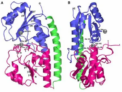
Figure 1.5: Crystal structure of zinc-bound ZnuA. a) Ribbon diagram of ZnuA
showing the N-terminal domain in pink, the C-terminal domain in blue, and the connecting α-helix in green. The zinc ions are represented as gray spheres. The ends of the disordered His-rich loop are indicated by asterisks. b) Viewed approximately 90° around the vertical axis from the orientation in a (Adapted fromYatsunyk et al, 2008).
1.9 ZnuB and ZnuC proteins
The transmembrane component of ZnuABC transporter is codified by the
znuB gene that after its activation led to a production of a 27.7 kDa protein.
Due to the difficulties in purifying membrane proteins, there is no structural information about this protein. The only information available comes from studied carried out in E. coli in which the authors applied PhoA/GFP fusion approach to derive topology models for almost the entire E. coli inner membrane proteome (Daley et al, 2005). PhoA and GFP have opposite activity profiles: PhoA is active only in the periplasm of E. coli, whereas GFP is fluorescent only in the cytoplasm. When fused in parallel to the C terminus of a membrane protein, PhoA and GFP can accurately report on which side of the membrane the C terminus is located.
These studies revealed that the C-terminal domain of ZnuB is located in the cytoplasm (Daley et al, 2005).
The third component of the complex is ZnuC, this protein has a molecular weight of about 27.9 kDa. Very little is known about this protein, but its sequence is homologous to that of ATP-ase component of other ABC transporters.
The sequence of ZnuC contain the highly conserved Walker A and B consensus motifs for nucleotide binding and the ‘LSGGQ’ motif, the diagnostic signature sequence of ABC proteins.
Interestingly, the C-terminal region of ZnuC is characterized by a region rich in histidine and acidic amino acids.Therefore, it is tempting to speculate that the C-terminal region of ZnuC binds zinc and regulates the ATPase activity on the cytosolic side of the ABC transporter. At high cytosolic concentrations of zinc, metal could bind to the C-terminus of ZnuC and inhibit the import of further zinc (Wei et al, 2007).
1.10 ZinT: a periplasmic protein with a putative role in zinc
import
Gram-negative bacteria have two compartments in which zinc is needed: the cytoplasm and the periplasm, in which several enzymes require zinc as a cofactor. Some representative examples from E. coli are: Cu,ZnSOD, PhoA, FtsK, YebA, YodA and ZnuA (Hantke, 2005).
Introduction
Several of these enzymes seem to obtain their zinc cofactor in the periplasm and, therefore, under zinc-limiting conditions the availability of zinc for these enzymes, could be limited by the activity of the ZnuABC high-affinity zinc transporter.
This possibility is supported by studies carried out in our laboratory, which have demonstrated that periplasmic zinc-binding proteins effectively compete for metal binding when zinc availability is low (Berducci et al, 2004). The availability of zinc in the periplasmic space of E. coli has been investigated using a mutant Cu,Zn superoxide dismutase whose dimerization is triggered by zinc binding. This mutant enzyme accumulates in the monomeric form when wild type cells are grown in minimal medium, but assembles in the dimeric form when it is produced in the same medium by a mutant strain lacking the periplasmic zinc metallochaperone ZnuA (Berducci
et al, 2004).
After this observation it was hypothesized that the presence of zinc-binding chaperones in the periplasm that might balance the activity of ZnuA by delivering zinc to apo-enzymes in need of their cofactor (Hantke, 2005). It was suggested that a putative candidate for this chaperone function in E. coli could be a protein named ZinT, which was previously identified as a cadmium-induced protein and therefore related to cadmium resistance.
Using a bioinformatics approach, upstream zinT gene was identified an additional putative Zur binding site (Panina et al., 2003).
Subsequent studies on E. coli have demonstrated that zinT is modulated in bacterial cells exposed to low pH (Birch et al, 2003; Kannan et al, 2008), to copper ions (Kershaw et al, 2007), to the transition metals chelator TPEN (Sigdel et al, 2006). Recent study show that the expression of zinT is induced in media containing very low levels of zinc and that the zinT gene is regulated by Zur (Graham et al, 2009).
ZinT crystal structure, which has been solved in the presence of different metal cofactors (cadmium, zinc or nickel) (David et al, 2002, David et al, 2003), revealed that two zinc binding site are present in the structure. One zinc ion is coordinated by His144 and His155, whereas the other interacts with His153, His193, and the caboxyl moiety of Glu189 (Fig. 1.6 B).Several water molecules lie in close contact with the zinc ions, but the authors are not able to give a precise description of the coordination geometry (David et
al, 2003). The three-dimensional structure of YodA consists of two domains:
an antiparallel, up-down β-barrel flanked by one α-helix (the “calyx” domain), and a helical domain that opens out at the side of the calyx β-barrel (the “helix” domain) (David et al, 2003).
Studied carried out on Enterohemorrhagic E. coli have shown that in this bacterial species a portion of YodA is secreted and that the strain lacking
zinT is unable to adhere to Hela cells. Secretion of YodA by E. coli K-12 has
not been observed, perhaps because the genes encoding the E. coli K-12 T2SS are usually transcriptionally silenced.
Although these investigations suggest that ZinT is involved in zinc homeostasis the exact function of this protein has not been elucidated. Interestingly, ZinT structure (David et al, 2002, David et al, 2003) shows a very high homology to a domain of AdcA, a component of an ABC transporter involved in zinc acquisition in Streptococcus pneumoniae (Dintilhac et al, 1997, Panina et al, 2003). As the N-terminal portion of AdcA is homologous to ZnuA, this observation strongly suggests that ZinT could cooperate with ZnuA in zinc uptake within the periplasmic space.
A B
Figure. A) Schematic views of the YodA molecule and its metal binding sites. The
“calyx” domain is in brick red (β-barrel) and cyan (helices), whereas the helical domain are colored in green (helices). The three histidines and the metal ion in the central metal-binding site are highlighted. B) Detail of the metal-binding site in the zinc crystal forms. (Adapted from David et al, 2003).
Characterization of the Salmonella enterica zinc import apparatus
2 Aims of the project
The ability of bacteria to colonize specific environments relies on their ability to obtain adequate supplies of the nutrients that are indispensable for their growth. Of particular relevance for human and animal health is to understand how bacterial pathogens face the problem of nutrient limitation in the infected host, in which several essential elements are not freely available for infectious microorganisms. Between essential nutrients there are transition metals, which are used by all organisms as cofactors in a large number of proteins. It is well known that iron availability in eukaryotic tissues is carefully controlled by sequestration mechanisms involving proteins which remove iron from the cellular and extracellular compartments accessible to bacteria. Thus, pathogenic bacteria have evolved sophisticated strategies to acquire and utilize host iron, involving the production of molecules (siderophores, hemophores and membrane associated pumps) characterized by an extraordinary high iron affinity. The outcome of the competition for iron between the host cell and the microorganism is considered one of the most important factors which determine the ability of pathogens to multiply and cause the disease.
Although iron is traditionally considered the most important trace metal involved in the host-pathogen interaction, some recent studies have suggested that also the efficient uptake of other divalent metals plays a critical role in infection and has a major role in virulence. In particular, a few observations carried out in different bacterial pathogens have suggested that zinc is not freely available within the infected host. However, there is still some resistance in considering zinc availability as an element able to limit bacterial pathogenicity, because its concentration in all cells and in plasma is quite high.
In this context, the general purpose of this project has been to clarify the mechanisms of zinc import under conditions of metal deprivation in vitro and to analyze the importance of zinc uptake in bacterial infections. This problem has been investigated in S. Typhimurium, a very interesting model microorganism, which is genetically well characterized and can be easily manipulated to generate useful mutant strains. Moreover, this bacterium can readily multiply either in laboratory conditions or in cellular or animal models and is commonly considered an excellent tool to investigate the infectious processes mediated by facultative intracellular pathogens.
To investigate the importance of zinc in the host-pathogen interaction, we have investigated the contribution of ZnuABC, the unique high affinity zinc transporter present in Gram-negative bacteria, to S. Typhimurium survival and multiplication in zinc-poor media, within eukaryotic cells and in infected mice. Moreover, we have also investigated the role in zinc uptake and bacterial virulence of ZinT, a periplasmic protein with a putative role in metal homeostasis, and of the histidine-rich domain of ZnuA.
On the whole, the results reported in this study demonstrate that despite the apparent elevated concentration of zinc within the host tissues, the functionality of the ZnuABC is critical to ensure Salmonella ability to efficiently multiply within infected mice and to cause disease. In addition, we have shown that ZinT and the His-rich domain of ZnuA are two structurally distinct elements with apparently redundant roles, which enhance metal recruitment during conditions of severe zinc shortage.
These findings suggest an evident parallelism between the mechanisms of iron and zinc sequestration in the host-pathogen relationships and shed new light on the complex functions of zinc in vertebrate and bacterial physiology.
Materials and Methods
3 Materials and Metods
3.1 Materials
Antibiotics for bacteria growth were provided by Sigma, they have been sterilized by filtration and stored in aliquots at -20 °C. Antibiotics were used at the following concentration: kanamycin 50 μg/ml, chloramphenicol 30 μg/ml and ampicillin 100 μg/ml. Antibiotics for cell culture were provided by Eurobio and stored in aliquots at -20 °C.
Restriction enzymes, alkalin phosphatase and T4 DNA ligase were provided by New England Biolabs; the Taq DNA polimerase (Expand TM) was provided by Roche.
3.1.1 Bacterial strains
PP116 zinT::cam electroporation of fragment [oli178-179 pKD3] in MA6926 pKD46;
verified by PCR (K3/oli181) camR PP118 zinT::cam znuA::kan transduction P22(SA123) on PP116;
kanR
PP120 znuA-3xFLAG-kan zinT::cam transduction P22(SA140) on PP116; camR
PP125 zinT-scar† electroporation of pCP20 in PP116;
camS
PP126 znuA::3xFLAG-scar† zinT::scar† electroporation of pCP20 in PP120;
camS
PP127 zur::kan electroporation of fragment [oli184-185 pKD4] in MA6926 pKD46;
verified by PCR (oli177/K1); kanR PP128 znuA::3xFLAG-scar† zinT::scar†
ilvI::Tn10dTac-cat::3xFLAG-kan
transduction P22(SA140) on PP126; camR
PP130 zinT::camznuA[deltaloop] transduction P22(PP116) on SA287;
S. enterica strains Relevant genotype Source/Tecnique (oligonucleotides
and template plasmid) serovar
Typhimurium
MA6926 Wild type L. Bossi MA6926(pKD46) wild type harbouring plasmid
pKD46
L. Bossi MA7223 ilvI3305::Tn10dTac-cat-
43::3xFLAG-kan
Uzzau et al, 2001 MA7225 ilvI3305::Tn10dTac cat Uzzau et al, 2001
SA123 znuA::kan Lab collection
SA140 znuA::3xFLAG-kan
ilvI::Tn10dTac-cat::3xFLAG-kan
Lab collection SA150 znuA::cam Lab collection
SA176 znuA ::scar † Lab collection
SA182 znuABC::kan Lab collection
SA229 yebA::cam electroporation of fragment [oli167-168 pKD3] in MA6926 pKD46;
verified by PCR (oli169/K1) camR SA233 znuAΔloop yebA::cam see Materials and Methods section
SA287 znuAΔloop yebA::scar† electroporation of pCP20 in SA233;
camS SA288 znuA::3xXFLAG-scar†
ilvI::Tn10dTac-cat::3xFLAG-scar†
electroporation pCP20 in SA140; kanS
PP101 znuB::3xFLAG-kan electroporation of fragment [oli172-173 pSUB11] in MA6926 pKD46;
Materials and Methods yebA::scar† camR PP131 znuA::3xFLAG-scar† zur::kan ilvI::Tn10dTac-cat::3xFLAG-scar† transduction P22(PP127) on SA288; kanR
PP132 zinT::3xFLAG-scar zur::kan transduction P22(PP127) on PP129;
kanR
PP134 zinT::3xFLAG-kan electroporation of fragment [oli182-195 pSUB11] in MA6926 pKD46;
verified by PCR (oli180/K4) kanR PP137 zinT::3xFLAG-kan znuA::cam transduction P22(PP134) on SA150;
kanR
PP138 zinT::3xFLAG-scar† electroporation pCP20 in PP134; kanS PP141 znuA::3xFLAG-kan
zinT::3xFLAG-scar†
transduction P22(SA140) on PP138; kanR
serovar Enteritidis
LK5 Wild type L. Bossi SA157 znuA::kan Lab collections
†The term “scar” refers to the DNA sequence remaining after excision of antibiotic-resistance
cassette following homologous recombination between two flanking FRT mediated by a recombinase encoded by plasmid pCP20 (Datsenko et al, 2001).
3.1.2 Eukaryotic cell lines
Mouse macrophage cell lines J774 A.1 (ATCC # TIB-67) and TIB-63. Human epithelial colorectal adenocarcinoma cells CaCo2.
Differentiated THP-1 monocytes.
3.1.3 Mice
Balb/c and DBA2 females.
3.1.4 Liquid culture medium for bacteria growth
Luria-Bertani Broth
Bacto tryptone 10 g/l yeast extract 5 g/l
Minimal medium (E50X) MgSO4 anhydrous 0.04 g/l citric acid 2 g/l K2HPO4 anhydrous 10 g/l NaNH4HPO4 · 4H2O 3.5 g/l glucose 2 g/l
Dissolve in water, heating the solution, cool and then add a drop of chloroform.
Tris minimal medium
Tris-HCl pH 7.2 120 mM K2HPO4 0.017 g/l MgCl2 2.03 g/l NH4Cl 1.06 g/l NaSO4 0.44 g/l CaCl2 0.06 g/l NaCl 4.68 g/l KCl 1.48 g/l glucose 1.98 g/l SOC
Mix in pairs 1:1 SOB 2X with salts 2X
SOB 2X Salts 2X
Bacto tryptone 40 g/l MgCl2 20 mM
yeast extract 10 g/l MgSO4 20 mM
NaCl 20 mM glucose 0.8 % KCl 5 mM P22 broth LB 100 ml E 50X 2 ml glucose 1 ml P22 0.1 (about 109 pfu/ml)
Materials and Methods
3.1.5 Solid culture medium for bacteria growth
Agar
agar 15 g/l
Dissolved in LB broth and then autoclaved for 20 minutes at 120 °C. Soft agar
agar 7 g/l
Dissolved in LB broth and then autoclaved for 20 minutes at 120 °C. Green Plates glucose 7.6 g/l Bacto tryptone 8.1 g/l yeast extract 1 g/l NaCl 5 g/l Agar 15 g/l Metyl blue 0.066 g/l Azarin yellow GG 4.68 g/l KCl 0.630 g/l