Selected Topics:
Neurological Emergencies
INFECTIVE ENDOCARDITIS PRESENTING WITH INTRACRANIAL BLEEDING
Andrea Morotti,MD,*Massimo Gamba,MD,†Paolo Costa,MD,*Loris Poli,MD,*Nicola Gilberti,MD,† Ilenia Delrio,MD,†Dikran Mardighian,MD,‡Roberto Gasparotti,MD,‡Alessandro Padovani,MD,PHD,*andAlessandro Pezzini,MD*
*Dipartimento di Scienze Cliniche e Sperimentali, Clinica Neurologica, Universita` degli Studi di Brescia, Brescia, Italy, †Dipartimento di Scienze Neurologiche e della Visione, Neurologia Vascolare, Spedali Civili di Brescia, Brescia, Italy, and ‡Dipartimento di Diagnostica per Immagini,
Neuroradiologia, Universita` degli Studi di Brescia, Brescia, Italy
Corresponding Address: Andrea Morotti,MD, Dipartimento di Scienze Cliniche e Sperimentali, Clinica Neurologica, Universita` degli Studi di Brescia, Piazzale Spedali Civili 1, 25123 Brescia, Italy
, Abstract—Background: Infective endocarditis (IE) can be complicated by intracranial bleeding (ICB) caused by different pathologic mechanisms. The occurrence of ICB in patients with IE significantly influences therapeutic deci-sions and has a negative impact on outcome. Case Report: We describe the clinical courses of 3 patients with aortic prosthetic valve IE presenting with ICB. Patients 1 and 2 experienced subarachnoid hemorrhage (SAH) and intrace-rebral hemorrhage (ICH), respectively, caused by rupture of an intracranial infectious aneurysm (IIA). Both under-went endovascular treatment of IIA with good outcome. In patient 3, ICB was the hemorrhagic conversion of an acute ischemic lesion from septic brain embolization. In the sub-acute phase of the disease, aortic valve replacement was per-formed, with excellent outcome. Why Should an Emergency Physician be Aware of This?: ICB is a relevant complication and sometimes the first clinical feature of IE. Imaging of brain vessels should be performed to investigate the patho-logic mechanism underlying ICB. The prevalence of IIA is probably underestimated and may influence the therapeutic strategy. Cerebrovascular imaging may therefore also be considered in asymptomatic subjects with left-sided IE. Withdrawal of anticoagulant treatment and delay of cardiac surgery are recommended in all cases of IE complicated by ICB. Because of the impact of ICB on IE management and outcome, a high level of clinical suspicion and prompt
recognition and treatment of this complication are neces-sary. Ó 2016 Elsevier Inc. All rights reserved.
, Keywords—infectious aneurysm; infective endocarditis; intracerebral hemorrhage; subarachnoid hemorrhage
INTRODUCTION
Up to 40% of patients with infective endocarditis (IE) develop central nervous system involvement(1). Stroke is the most frequent neurologic complication of IE and the main cause of death after heart failure; it usually oc-curs in the early phase of the disease and in >50% of these patients represents the presenting symptom (2–4). In particular, intracranial bleeding (ICB) accounts for 30% of neurologic complications and has a negative impact on IE prognosis(1,5). Several mechanisms explain the occurrence of ICB in patients with IE(6). We describe 3 cases of IE presenting with different types of ICB.
CASE REPORTS Patient 1
A 35-year-old man on oral anticoagulant therapy (OAT) for a mechanical prosthetic aortic valve presented with a thunderclap headache after a 3-day fever. Reprints are not available from the authors.
RECEIVED: 10 December 2015; FINAL SUBMISSION RECEIVED: 24 February 2016; ACCEPTED: 5 April 2016
1
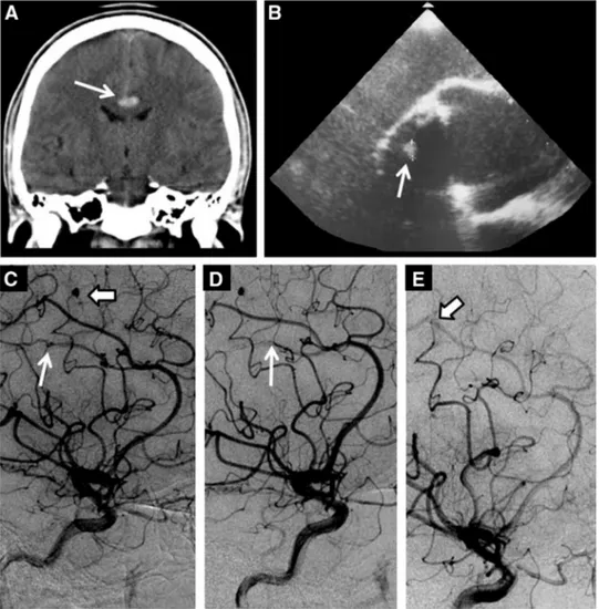
Neuroimaging revealed a subarachnoid hemorrhage (SAH; Figure 1A) from a ruptured right pericallosal aneurysm and an unruptured aneurysm of the middle ce-rebral artery (MCA;Figure 1C). Laboratory findings re-vealed an elevated C-reactive protein level (457 nmol/L [normal, 0–48 nmol/L]) and Enterococcus faecalis was isolated from blood cultures. Prosthetic valve vegetation was identified on transesophageal echocardiography (TEE;Figure 1B) and a diagnosis of IE was made. OAT was promptly discontinued and the patient had good re-covery after treatment of the ruptured aneurysm by endo-vascular occlusion of the parental artery (Figure 1D) and antibiotic therapy. Three months postdischarge, despite appropriate prolonged antimicrobial treatment, the ruptured aneurysm of the distal right MCA was un-changed. Therefore, the patient underwent endovascular
treatment by glue embolization of the parental artery (Figure 1E), with excellent outcome.
Patient 2
A 60-year-old man was admitted to our division for a right parieto-occipital intracerebral hemorrhage (ICH;
Figure 2A). His vital signs and a general examination were unremarkable except for fever with chills. The pa-tient’s medical history was significant for the presence of a mechanical prosthetic aortic valve and OAT. Cerebral computed tomography angiography (CTA) and subse-quent digital subtraction angiography (DSA) revealed the presence of an intracranial infectious aneurysm (IIA) originating from the right calcarine artery (Figure 2B–D), located at the base of the hematoma. IE
Figure 1. Coronal computed tomography scan of the brain showing subarachnoid hemorrhage in the supracallosal cistern (ar-row, A) and transesophageal echocardiography with evidence of vegetation over the mechanical prosthetic aortic valve (ar(ar-row, B). Digital subtraction cerebral angiography revealing a ruptured aneurysm of the right pericallosal artery (thin arrow, C) and an aneurysm in the M4 division of the right middle cerebral artery (thick arrow, C). Digital subtraction cerebral angiography showing treatment of the infectious aneurysms with endovascular glue embolization of the parental artery (thin arrow, D and thick arrow, E).
was suspected. Based on the simultaneous presence of fe-ver, IIA, and a mechanical prosthetic valve, according to Duke Criteria, a diagnosis of possible IE was made, in spite of the negative results of TEE and blood cultures
(4). Empiric antibiotic therapy was administered and endovascular occlusion of the IIA by glue embolization of the parental artery was successfully performed (Figure 2E), with good long-term outcome.
Patient 3
A 44-year-old man undergoing OAT for a mechanical prosthetic aortic valve was admitted with a 1-week his-tory of fever with mild weight loss. Blood tests revealed the presence of normocytic anemia with significant eleva-tion of the inflammatory markers (C-reactive protein, 1761 nmol/L [normal, 0–48 nmol/L]). The day after the admission the patient experienced a sudden onset of slurred speech and left hemiparesis. A CT scan of the brain was initially negative, but a follow-up CT scan re-vealed the presence of multifocal acute ischemic lesions (Figures 3A and 3B), with hemorrhagic transformation (Figure 3C). A CTA scan was unremarkable. The patient
underwent TEE with evidence of multiple aortic valve vegetations with severe valvular regurgitation and para-valvular abscess. Staphylococcus aureus was isolated from blood cultures, and the diagnosis of IE was made. Antibiotic treatment was started and OAT was withdrawn. Two months later, after complete disappearance of the ICB, aortic valve replacement was performed, with excel-lent outcome.
DISCUSSION
ICB is not an uncommon finding in patients with IE and can arise from variable pathologic mechanisms, such as hemorrhagic conversion of ischemic stroke, rupture of IIA, or pyogenic arteritis(7). In our case series, patients 1 and 2 experienced ICB from IIA rupture, while cerebral hemorrhage in patient 3 was caused by the hemorrhagic transformation of an acute embolic ischemic lesion.
IIA formation is the rarest neurologic complication of IE, occurring in 2% to 4% of cases(4,8). IIA are caused by septic embolization from infected heart structures and S aureus and viridans streptococci are the pathogens most frequently involved (9). Development of IIA is an Figure 2. Axial computed tomography scan of the brain showing a large right parieto-occipital intracerebral hemorrhage (A). Computed tomography angiography showing an intracranial infectious aneurysm located at the base of the intracranial hema-toma (arrows, B and C). Cerebral digital subtraction angiography revealing an aneurysm of the distal segment of the right calcar-ine artery before (arrow, D) and after (arrow, E) treatment by endovascular occlusion of the parental artery.
uncommon finding in enterococcal endocarditis (10). Most patients with IIA present with fever, associated with headache, seizures, mental status impairment, or focal neurologic signs caused by an aneurysm rupture in the brain parenchyma or subarachnoid space(8). Fever and acute onset headache should raise the suspicion of IIA rupture in all patients with ICH or SAH, especially in those with predisposing conditions for IE, such as pros-thetic heart valves.
Hemorrhagic conversion of a cerebral infarction was reported to be the most frequent cause of ICB in patients with IE(7). Overall,S aureus infection, prosthetic valve endocarditis, and OAT are the major risk factors for cere-bral hemorrhagic complications of IE(5,11).
Although non-contrast CT scan of the brain is the preferred and most feasible initial imaging study in crit-ical patients with IE and neurologic symptoms, cerebral vessel imaging is mandatory in all subjects(1,12). CTA and magnetic resonance angiography have excellent sensitivity for IIAs >5 mm; however, DSA is still considered the gold standard for the detection of IIA. IIAs are frequently multiple and have a predilection for the distal branches of the MCA. Unlike degenerative aneurysms, IIA size is not a predictor of rupture, and bleeding in the brain parenchyma is a common finding
(8,9,13).
Magnetic resonance imaging (MRI) is the best imag-ing study to detect hemorrhagic conversion of an acute ischemic lesion. Moreover, systematic brain MRI evalua-tion of IE patients revealed that up to 80% of patients develop asymptomatic brain parenchymal lesions, such as small acute embolic ischemic lesions or microbleeds
(14). However, the clinical impact of microbleed detec-tion is still unclear, and asymptomatic central nervous system involvement has not been shown to have any
influ-ence on IE prognosis and management(1,2). Therefore, a routine brain MRI scan in all patients with IE is currently not recommended(1,12). In addition, MRI compatibility of the prosthetic device is a practical limitation in several cases.
Conversely, systematic brain vessel imaging with CTA may be considered in asymptomatic subjects with left-sided endocarditis(13). The prevalence of IIA is probably underestimated because small aneurysms may remain si-lent and resolve with antibiotic treatment (12,13). The identification of unruptured IIAs might have important clinical implications and guide the therapeutic strategy. First, the presence of IIA is included in the minor criteria for the diagnosis of IE, and IIA detection may therefore help in cases with nonspecific clinical presentation (4,13). Second, if early cardiac surgery is absolutely necessary, preoperative endovascular IIA treatment may be considered in order to prevent intraoperative IIA rupture(12). Third, endovascular treat-ment is warranted for ruptured IIA but also for IIA not resolved with appropriate medical treatment. Early iden-tification of clinically silent IIA is therefore crucial and needs close imaging follow-up(12,13).
Surgical treatment of IIA and evacuation of the intra-cranial hematoma is usually reserved for bleeding with severe mass effect and intracranial hypertension(8,9,13). Heart failure is the first cause of death in IE patients and the main indication for cardiac surgery in the acute phase of the disease(12). In subjects with IE complicated by ICB, cardiac surgery should be delayed for at least 4 weeks and all antithrombotic medications should be discontinued(3,12,13).
Early administration of empiric antibiotic therapy re-duces the incidence of all cerebrovascular complications and is therefore warranted in all IE cases (12,13,15). Figure 3. A computed tomography scan of the brain showing multifocal acute ischemic lesions (arrows, A and B) and right frontal infarction with hemorrhagic transformation (C).
Finally, in patients with ischemic stroke related to IE, the potential benefits of intravenous thrombolysis administration should be balanced with the significantly increased risk of hemorrhagic transformation(16). WHY SHOULD AN EMERGENCY PHYSICIAN BE
AWARE OF THIS?
ICB is a common finding in IE, frequently occurring in the early phase of the disease and being the presenting sign in a relevant proportion of subjects. Various patho-logic mechanisms can lead to ICB in subjects with IE, and brain vessel imaging is recommended in all patients with ICB in the setting of IE. The prevalence of IIA is probably underestimated and has a significant influence on IE management. Systematic imaging screening for IIA may be considered also in asymptomatic patients with left-sided endocarditis (13,17). Antithrombotic drugs should be discontinued and cardiac surgery delayed for at least 1 month in patients with an ICB caused by IE. Clinicians should therefore be aware of ICB in the setting of IE, in order to ensure prompt recognition and treatment of this dreadful complication.
REFERENCES
1. Sonneville B, Mourvillier B, Bouadma L, Wolff M. Management of neurological complications of infective endocarditis in ICU pa-tients. Ann Intensive Care 2011;1:10.
2. Derex L, Bonnefoy E, Delahaye F. Impact of stroke on therapeu-tic decision making in infective endocarditis. J Neurol 2010;257: 315–21.
3. Novy E, Sonneville R, Mazighi M, et al. Neurological complica-tions of infective endocarditis: new breakthroughs in diagnosis and management. Med Mal Infect 2013;43:443–50.
4. Hoen B, Duval X. Infective endocarditis. N Engl J Med 2013;368: 1425–33.
5. Garcı´a-Cabrera E, Ferna´ndez-Hidalgo N, Almirante B, et al. Neuro-logical complications of infective endocarditis: risk factors, outcome, and impact of cardiac surgery: a multicenter observational study. Circulation 2013;127:2272–84.
6. Chaudhary G, Lee JD. Neurologic complications of infective endo-carditis. Curr Neurol Neurosci Rep 2013;13:380.
7. Masuda J, Yutani C, Waki R, Ogata J, Kuriyama Y, Yamaguchi T. Histopathological analysis of the mechanisms of intracranial hemorrhage complicating infective endocarditis. Stroke 1992;23:843–50.
8. Kannoth S, Thomas SV. Intracranial microbial aneurysm (infectious aneurysm): current options for diagnosis and management. Neuro-crit Care 2009;11:120–9.
9. Peters PJ, Harrison T, Lennox JL. A dangerous dilemma: manage-ment of infectious intracranial aneurysms complicating endocardi-tis. Lancet Infect Dis 2006;6:742–8.
10. Ferna´ndez Guerrero ML, Goyenechea A, Verdejo C, Fernandez Roblas R, de Go´rgolas M. Enterococcal endocarditis on native and prosthetic valves. Medicine (Baltimore) 2007;86:363–77. 11. Tornos P, Almirante B, Mirabet S, Permanyer G, Pahissa A,
Soler-Soler J. Infective endocarditis due toStaphylococcus aureus. Arch Intern Med 1999;159:473–5.
12. Habib G, Lancellotti P, Antunes MJ, et al. 2015 ESC Guidelines for the management of infective endocarditis. The Task Force for the Management of Infective Endocarditis of the European Society of Cardiology (ESC). Eur Heart J 2015;36:3075–128.
13. Baddour LM, Wilson WR, Bayer AS, et al. Infective endocarditis in adults: diagnosis, antimicrobial therapy, and management of complications. a scientific statement for healthcare professionals from the American Heart Association. Circulation 2015;132: 1435–86.
14. Duval X, Lung B, Klein I, et al. Effect of early cerebral magnetic resonance imaging on clinical decisions in infective endocarditis: a prospective study. Ann Intern Med 2010;152: 497–504.
15. Dickerman SA, Abrutyn E, Barsic B, et al. The relationship be-tween the initiation of antimicrobial therapy and the incidence of stroke in infective endocarditis: an analysis from the ICE Prospective Cohort Study (ICE-PCS). Am Heart J 2007;154: 1086–94.
16. Asaithambi G, Adil MM, Qureshi AI. Thrombolysis for ischemic stroke associated with infective endocarditis results from the nation-wide inpatient sample. Stroke 2013;44:2917–9.
17. Ducruet AF, Hickman ZL, Zacharia BE, et al. Intracranial infec-tious aneurysms: a comprehensive review. Neurosurg Rev 2010; 33:37–46.