UNIVERSITY OF CATANIA -MEDICAL SCHOOL,
INTERNATIONAL PHD PROGRAM IN NEUROPHARMACOLOGY, XXVIIIth EDITION UNIVERSITY OF BORDEAUX -DOCTORAL SCHOOL OF LIFE AND HEALTH SCIENCES,
SPECIALITY NEUROSCIENCE
R
OLE OF THE CENTRAL SEROTONIN2B RECEPTOR IN THE REGULATION OFASCENDING DOPAMINERGIC PATHWAYS
:
RELEVANCE FOR THE TREATMENT OF SCHIZOPHRENIA AND DRUG ADDICTION
PHD THESIS
CÉLINE DEVROYE
THESIS DIRECTOR (BORDEAUX):PR UMBERTO SPAMPINATO THESIS CO-DIRECTOR (CATANIA):PR FILIPPO DRAGO COORDINATOR :PR SALOMONE SALVATORE
The present work was carried out in the Team of Physiopathology of Addiction and Traumatic Memory, directed by Dr Pier-Vincenzo Piazza (INSERM U1215, Neurocenter Magendie, Bordeaux, France), and was supported by a fellowship from the International Ph.D. program in Neuropharmacology of the University of Catania, Medical School (Catania, Italy).
C
OMPOSITION OF THE JURYPr Marina Ziche (examiner). Dpt Life Sciences, University of Siena, via A. Moro 2, 53100 Siena, Italy
Pr Marco Andrea Riva (examiner). Dpt Pharmacological and Biomolecular Sciences, University of Milan, via Balzaretti 9, 20133 Milan, Italy
Pr Gianni Sava. Dpt Life Sciences, University of Trieste, via L. Giorgieri 5, 34127 Trieste, Italy
Pr Umberto Spampinato. University of Bordeaux, 146 rue Léo Saignat, 33077 Bordeaux, France
R
EMERCIEMENTSJe dois tout d’abord remercier mon directeur de thèse, le professeur Umberto Spampinato, aux côtés duquel j’ai beaucoup appris au cours des six dernières années. Merci à lui pour sa présence et pour son indéfectible persévérance dans l’enseignement de l’écriture et de la rigueur. Je lui suis reconnaissante de m’avoir accompagnée tout au long du chemin et de m’avoir inculqué non seulement l’opiniâtreté mais aussi la confiance en mon propre jugement. Au-delà de toute considération scientifique, cette expérience m’a surtout permis de mieux me connaître moi-même.
Je remercie évidemment Adeline Cathala, avec qui j’ai partagé nombre d’expériences techniques et humaines. Merci pour son soutien, son écoute et son aide indispensable. Avec elle, il va sans dire qu’au souvenir des longues heures passées à faire la guerre à la chromatographie, lesquelles m’auront enseigné la patience dans son sens le plus abouti, s’ajoutent ceux des voyages, de la course aux buffets, et bien d’autres encore.
J’exprime ma gratitude au docteur Pier-Vincenzo Piazza pour m’avoir accueillie au sein de son laboratoire, me permettant ainsi de travailler dans d’excellentes conditions. A cet égard, je remercie tous les membres de l’équipe pour leur disponibilité et leur bienveillance. Merci à Jean-François pour nos réunions (entre deux portes de l’animalerie) portant sur nos velléités de collaboration scientifique (un jour peut-être !), à Monique et Guillaume pour leurs précieux conseils, à Jess, Miguel, Agnès, Prisca, Lucie, Cédric pour leur bonne humeur, inaltérable…Bref, je serai plus exhaustive en remerciant l’ensemble du rez-de-chaussée, que j’ai rejoint bien tard d’un point de vue géographique, mais qui a souvent contribué à rendre ces journées au laboratoire plus légères.
Je remercie également l’Université de Catane d’avoir financé cette thèse. Je suis en particulier reconnaissante envers mon cotuteur, le professeur Filippo Drago, ainsi que le coordinateur du programme international de PhD en
Neuropharmacologie, le professeur Salvatore Salomone, de m’avoir accordé leur confiance pour mener à bien ce travail. Je remercie également les professeurs Ziche, Riva et Sava, d’avoir accepté d’assister à la présentation de ce travail. Merci au professeur Filippo Caraci pour son aide lors de mes échanges avec l’Université de Catane, et à Barbara Di Marco, avec qui j’ai eu le plaisir de travailler et de partager des moments à la fois bordelais et siciliens pendant six mois.
Il est bien évident que, sans le soutien de mes proches, cette expérience de vie aurait été tout autre. Aussi, j’ai bien conscience que les mots ne suffiront pas pour remercier mes parents, et leur exprimer combien je suis chanceuse de les compter parmi mes soutiens les plus assidus, et les plus infaillibles. Pourtant, je suppose qu’ils ont souffert, au même titre que d’autres, de supporter le récit interminable de mes semaines passées au labo. Je remercie également ma sœur, pour la fréquence de nos échanges à propos de tout et de rien, pour sa présence, tout simplement. Merci évidemment à ma grand-mère, ma tante, mon oncle, toute ma famille, pour ces précieux moments passés ensemble, qui m’ont rappelé combien le travail n’est qu’un moyen, et que bien d’autres choses sont fondamentales.
Je ne trouverai pas non plus les mots justes pour faire honneur au soutien de mes amis. Alors, simplement, un immense merci à Marion, Mathieu, Laurie, Maude, Simon, Marine et ma zoude. Sans vous, je n’ose pas imaginer de quelle manière j’aurais traversé ces quelques années. Nos moments de partage, les rires tout comme les violents coups de cafard, font partie de mes souvenirs les plus marquants et les plus enrichissants de cette période. Merci d’avoir su quoi dire lorsque j’en avais besoin, et surtout merci d’avoir été là. Un grand merci également à Eric, pour nos pauses café toujours trop courtes, et pour sa mise à l’épreuve de mon pessimisme récurrent. Merci aussi à Marie et Yvan, pour tous ces moments précieux passés en leur compagnie à des années lumières de tout le reste.
Enfin, il peut dorénavant croire que je m’emploie toujours à garder le meilleur pour la fin, je tiens à remercier celui qui m’a plus que quiconque aidée au cours des quatre dernières années. Merci, infiniment, à toi qui a dû absorber les moindres détails du récit de ma vie au labo, chaque soir, sans jamais céder à l’impulsion de les réduire à de simples détails. A toi qui m’a portée, avec ton humour, ton optimisme et la justesse de tes réflexions. Merci pour ta patience, ta force, et ta présence, toujours indispensables dans les meilleurs comme dans les mauvais instants. Grandir à tes côtés est sans détour la meilleure expérience de ma vie.
TABLE OF CONTENTS
LISTOFPUBLICATIONSANDSCIENTIFICCOMMUNICATIONS 11
LISTOFFIGURESANDTABLES 13
LISTOFABBREVIATIONS 15
ABSTRACT 17
RESUME 19
INTRODUCTION 21
I. Anatomy of central dopaminergic and serotonergic systems 23
II. Therapeutic relevance of the interaction between central dopaminergic and serotonergic systems
27
A. Schizophrenia 27
B. Drug addiction: focus on cocaine 33
III. The serotonin2B receptor 39
A. Localization 39
B. Molecular properties 40
C. Pharmacology 45
D. Functional aspects 49
1. Regulation of cell differentiation and proliferation 49
2. Regulation of the gastrointestinal tract 49
3. Regulation of the vascular function 50
4. Regulation of the pulmonary function 51
6. Regulation of the central nervous system 53
6.1. Regulation of serotonin transport 53
6.2. Regulation of astrocytes 54
6.3. Regulation of the dopaminergic network 54
6.3.1. Neurochemical aspects 54 6.3.2. Behavioral aspects 56 6.4. Therapeutic implications 58 6.4.1. Schizophrenia 58 6.4.2. Drug addiction 60 6.4.3. Depression 61 IV. Aims 63 PUBLICATIONS 67
I. Article 1: Differential control of dopamine ascending pathways by serotonin2B receptor antagonists: new opportunities for the treatment
of schizophrenia. Devroye et al., 2016, Neuropharmacology 109, 59-68.
69
II. Article 2: A functional interplay with serotonin1A receptors drives
central serotonin2B receptor-opposite controls of mesocortical and
mesoaccumbal dopaminergic pathways. To be submitted.
111
III. Article 3: Central serotonin2B receptor blockade inhibits
cocaine-induced hyperlocomotion independently of changes of subcortical dopamine outflow. Devroye et al., 2015, Neuropharmacology 97, 329-337.
141
DISCUSSION 181
CONCLUSIONS 193
11
L
IST OF PUBLICATIONS AND SCIENTIFIC COMMUNICATIONSPublications
*Devroye, C., Filip, M., Przegaliński, E., McCreary, A.C., Spampinato, U., 2013. Serotonin2C receptors and drug addiction: focus on cocaine. Exp. Brain Res. 230,
537-545.
*Cathala, A., Devroye, C., Maitre, M., Piazza, P.V., Abrous, D.N., Revest, J.M., Spampinato, U., 2014. Serotonin2C receptors modulate dopamine transmission in the
nucleus accumbens independently of dopamine release: behavioral, neurochemical and molecular studies with cocaine. Addict. Biol. 20, 445-457.
*Devroye, C., Cathala, A., Maitre, M., Piazza, P.V., Abrous, D.N., Revest, J.M., Spampinato, U., 2015. Serotonin2C receptor stimulation inhibits cocaine-induced Fos
expression and DARPP-32 phosphorylation in the rat striatum independently of dopamine outflow. Neuropharmacology 89, 375-381.
Devroye, C., Cathala, A., Di Marco, B., Caraci, F., Drago, F., Piazza, P.V., Spampinato,
U., 2015. Central serotonin(2B) receptor blockade inhibits cocaine-induced hyperlocomotion independently of changes of subcortical dopamine outflow. Neuropharmacology 97, 329-337.
Devroye, C., Cathala, A., Haddjeri, N., Rovera, R., Vallée, M., Drago, F., Piazza, P.V.,
Spampinato, U., 2016. Differential control of dopamine ascending pathways by serotonin2B receptor antagonists: new opportunities for the treatment of schizophrenia.
Neuropharmacology 109, 59-68.
Devroye, C., Haddjeri, N., Cathala, A., Rovera, R., Drago, F., Piazza, P. V.,
Spampinato, U. A functional interplay with serotonin1A receptors drives central
serotonin2B receptor-mediated opposite controls of mesocortical and mesoaccumbal
dopaminergic pathways. To be submitted.
12
Scientific communications
Oral communications
Devroye, C., Cathala, A., Maitre, M., Piazza, P.V., Abrous, D.N., Revest, J.M.,
Spampinato, U. 5-HT2C receptors modulate DA transmission in the nucleus accumbens
and the striatum: studies with cocaine. Neuropathology and Neuropharmacology of monoaminergic systems, CM1103 Action annual conference, October, 8-10th, 2014,
Bordeaux, France.
Devroye, C., Cathala, A., Drago, F., Piazza, P.V., Spampinato, U. Serotonin2B
receptor-dopamine interaction: new opportunities for improved treatment of schizophrenia. Mediterranean Neuroscience Society - MNS 2015, June, 12-15th, 2015, Cagliari, Italy.
Poster communications
Devroye, C., Cathala, A., Drago, F., Piazza, P.V., Spampinato, U. Blockade of central
serotonin2B receptors reduces cocaine-induced hyperlocomotion independently of
subcortical DA outflow. European College of Neuropsychopharmacology - ECNP 2014, October, 18-21st, 2014, Berlin, Germany.
Cathala, A., Devroye, C., Maitre, M., Piazza, P.V., Abrous, D.N., Revest, J.M., Spampinato, U. Serotonin2C receptor stimulation inhibits dopamine transmission in the
nucleus accumbens independently of dopamine release: studies with cocaine. ECNP 2014, October, 18-21st, 2014, Berlin, Germany.
Devroye, C., Cathala, A., Drago, F., Piazza, P.V., Spampinato, U. Serotonin2B receptor
antagonists reduce cocaine-induced hyperlocomotion: a possible post-synaptic interaction. ECNP workshop for junior scientists, March, 12-15th, 2015, Nice, France.
13
L
IST OF FIGURES AND TABLESFigures
Figure 1: Distribution of DA neuron cell groups in the adult rodent brain 24
Figure 2: Distribution of noradrenergic and adrenergic neurons and their major projections in the rat brain
25
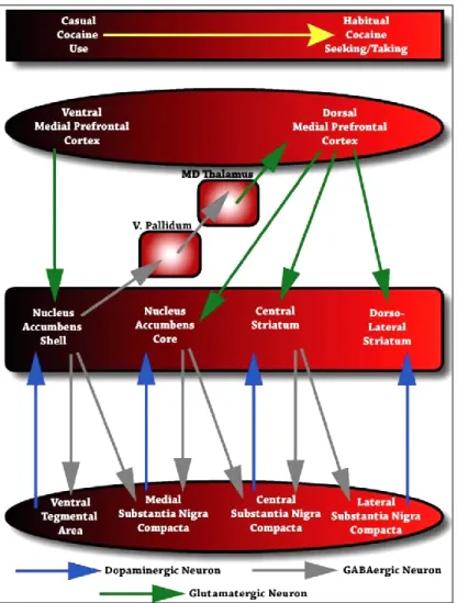
Figure 3: Distribution of 5-HT neuron cell groups in the adult rat brain 26
Figure 4: Anatomical circuits proposed by Pierce and Vanderschuren (2010) to underlie the progressive recruitment of more dorsal regions of the striatum during the transition from casual cocaine use to habitual cocaine seeking and taking
37
Figure 5: Prototypical signaling enzyme linkages of the G-protein-coupled 5-HT receptors
44
Figure 6: Schematic representation of the differential regulation of ascending DA pathways by 5-HT2BRs
184
Figure 7: Schematic representation of the functional interplay between the 5-HT2BR and the 5-HT1AR controlling mesocorticolimbic DA pathways
187
Figure 8: Time course effect of the administration of 5-HT2BR antagonists
on cocaine-induced increase in DA outflow in the mPFC
191
Figure 9: Schematic representation of the possible circuits underlying 5-HT2BR-mediated control of cocaine-induced hyperlocomotion
14
Tables
Table 1: Criteria for schizophrenia in the Diagnostic and Statistical Manual of Mental Disorders
31
Table 2: Behavioral models classically used to assess the efficacy of APDs against positive, negative and cognitive symptoms of schizophrenia, as well as their propensity to induce EPS
32
Table 3: Signaling characteristics of human 5-HT receptors 43
Table 4: Binding affinities of some antagonist ligands at human recombinant 5-HT2 receptors
46
Table 5: Binding affinities of some agonist ligands at human recombinant 5-HT2 receptors
15
L
IST OF ABBREVIATIONS5-HT serotonin
APD antipsychotic drug DA dopamine
DAG diacyl glycerol
DARPP-32 DA and cyclic 3’-5’ adenosine monophosphate-regulated phosphoprotein DAT dopamine transporter
DRN dorsal raphe nucleus
DSM diagnostic & statistical manual of mental disorders EPS extrapyramidal side effect
GI gastrointestinal ICC interstitial cells of cajal
IP3 inositol 1,4,5-trisphosphate MAPK mitogen-activated protein kinase MDMA 4-methylenedioxymethamphetamine
mPFC medial prefrontal cortex NAc nucleus accumbens
NAD nicotinamide adenine dinucleotide NET noradrenaline transporter
NMDA N-methyl-D-aspartate NO nitric oxide
NOR novel object recognition PAH pulmonary arterial hypertension PIP2 phosphatidylinositol 4,5-bisphosphate PKC protein kinase C
PLA2 phospholipase A2
PLC phospholipase C PCP phencyclidine
ROS reactive oxygen species SERT serotonin transporter
16
SNc substantia nigra pars compacta SSRI selective serotonin reuptake inhibitors
VIC valvular interstitial cells VTA ventral tegmental area
17
A
BSTRACTFour years ago, at the beginning of my thesis in Neuropharmacology, the functional role of the central serotonin2B receptor (5-HT2BR) remained poorly
investigated. Indeed, in light of the relatively recent discovery of its presence in the mammalian brain, as compared to other 5-HT receptors, only few studies had explored its impact within the central nervous system. Interestingly, it had been shown that 5-HT2BRs, while having no effect at the level of the
nigrostriatal dopaminergic (DA) pathway, afford a tonic excitatory control on the activity of the mesoaccumbal DA tract. This differential influence on subcortical DA brain regions had led to the proposal that 5-HT2BR antagonists
may be a useful tool for improved treatment of DA-related disorders requiring an independent modulation of the activity of ascending DA pathways, such as schizophrenia. However, the effect of 5-HT2BR blockade at the level of the
mesocortical DA pathway, which plays a pivotal role in the therapeutic benefit of atypical antipsychotic drugs (APDs), had never been studied. In addition, analysis of the literature revealed that 5-HT2BR blockade suppresses
amphetamine and 3,4-methylenedioxymethamphetamine-induced neurochemical and behavioral responses, suggesting that this receptor may also be a relevant pharmacological target for treating drug addiction. Nevertheless, its possible implication in the effects induced by cocaine, one of the most worldwide abused drugs, remained unknown.
Thus, the aim of the present thesis was to study the regulatory control exerted by the 5-HT2BR on both basal and cocaine-induced stimulation of DA activity,
in order to evaluate its therapeutic relevance for improved treatment of schizophrenia and drug abuse and dependence. To this purpose, we assessed the effects of potent and selective 5-HT2BR antagonists (RS 127445 and LY
266097) on DA activity, by using biochemical, electrophysiological and behavioral approaches in rats.
18
In a first group of experiments, we found that 5-HT2BRs exert a tonic inhibitory
control on DA outflow in the medial prefrontal cortex (mPFC). This finding, by showing that 5-HT2BRs afford differential controls over the three ascending DA
pathways, indicates that 5-HT2BR antagonists display an ideal pattern of effects
to restore normal DA function in schizophrenia. Accordingly, 5-HT2BR
antagonists were efficient in several behavioral models aimed at predicting APD efficacy, and had no effect in a behavioral task reflecting APD propensity to induce motor side effects. In a second group of experiments performed to determine the mechanisms underlying the differential control exerted by 5-HT2BRs on DA activity, we demonstrated that 5-HT2BR antagonist-induced
opposite effects on DA ouflow in the mPFC and the nucleus accumbens (NAc) involve a functional interplay with 5-HT1ARs expressed in the mPFC. Finally,
we found that 5-HT2BR blockade suppresses cocaine-induced hyperlocomotion.
This effect, which occurs independently from changes of DA outflow in the NAc and the striatum, where DA activity is tightly related to cocaine-induced behavioral responses, likely involves a post-synaptic interaction in subcortical DA brain regions.
To conclude, the work accomplished over the past four years provides substantial information with regards to the functional role of 5-HT2BRs in the
regulation of the activity of ascending DA pathways. In addition, while improving the understanding of the interaction between DA and 5-HT systems, the present findings altogether highlight the therapeutic potential of 5-HT2BR
19
R
ESUMEIl y a quatre ans, lorsque j’ai commencé ma thèse en Neuropharmacologie, le rôle fonctionnel du récepteur serotoninergique2B (5-HT2B) central n’était guère
connu. En effet, compte tenu de la mise en évidence de son expression dans le cerveau relativement récente par rapport aux autres récepteurs à la 5-HT, peu d’études avaient porté sur son impact au sein du système nerveux central. En particulier, il était établi que les récepteurs 5-HT2B, sans effet au niveau de la
voie dopaminergique (DA) nigrostriée, sont capables d’exercer un contrôle tonique excitateur sur l’activité de la voie DA mésoaccumbale. Sur la base de cette régulation différentielle des voies DA sous-corticales, il avait été proposé que les antagonistes du récepteur 5-HT2B pourraient constituer des outils
pharmacologiques pertinents pour le traitement des pathologies liées à une dysfonction du système DA et requérant une modulation indépendante des voies DA ascendantes, telles que la schizophrénie. Cependant, l’effet du blocage des récepteurs 5-HT2B au niveau de la voie DA mésocorticale, laquelle joue un rôle
pivot dans le bénéfice thérapeutique des antipsychotiques (APs) atypiques, n’avait jamais été exploré. De plus, l’analyse de la littérature avait révélé que le blocage du récepteur 5-HT2B réduit les réponses neurochimiques et
comportementales induites par l’amphétamine et la 3,4-méthylènedioxyméthamphétamine, suggérant que ce récepteur pourrait également représenter une cible pharmacologique intéressante pour le traitement de l’addiction. Néanmoins, la possible implication de ce récepteur dans les effets de la cocaïne, l’une des drogues les plus consommées au monde, restait alors inconnue.
Ainsi, l’objectif de cette thèse était d’étudier l’influence exercée par le récepteur 5-HT2B sur l’activité DA basale et activée par la cocaïne, afin de mieux évaluer
le potentiel thérapeutique de ce récepteur dans le traitement de la schizophrénie et de l’addiction. A cette fin, nous avons étudié les effets de deux antagonistes puissants et sélectifs du récepteur 5-HT2B (RS 127445 et LY 266097) sur
20
l’activité DA, en utilisant des approches biochimiques, électrophysiologiques et comportementales chez le rat.
Un premier groupe d’expériences a mis en évidence l’existence d’un contrôle tonique inhibiteur exercé par le récepteur 5-HT2B sur la libération de DA dans le
cortex préfrontal médian (CPFm). Ce résultat, démontrant que les récepteurs 5-HT2B régulent de manière différentielle les trois voies DA ascendantes, indique
que les antagonistes du récepteur 5-HT2B présentent un profil d’action idéale
pour restaurer une fonction DA normale chez les patients schizophrènes. En accord avec cette proposition, les antagonistes 5-HT2B se révèlent efficaces dans
plusieurs modèles classiquement utilisés pour prédire l’efficacité des APs, alors qu’ils n’ont pas d’effet dans une tâche comportementale prédisant la tendance des APs à induire des effets secondaires moteurs. Un second groupe d’expériences visant à étudier les mécanismes sous-tendant le contrôle différentiel exercé par le récepteur 5-HT2B sur l’activité DA montre que les
effets opposés induits par les antagonistes 5-HT2B sur la libération de DA dans
le CPFm et le noyau accumbens (NAc) résultent d’une interaction fonctionnelle avec les récepteurs 5-HT1A exprimés dans le CPFm. Enfin, nous avons
également démontré que le blocage du récepteur 5-HT2B supprime
l’hyperlocomotion provoquée par la cocaïne. Cet effet, qui se produit indépendamment de la libération de DA dans le NAc et le striatum, où l’activité DA est étroitement liée aux effets comportementaux induits par la cocaïne, implique une interaction post-synaptique dans les régions DA sous-corticales. En conclusion, le travail accompli au cours des quatre années passées apporte des informations substantielles quant au rôle fonctionnel du récepteur 5-HT2B
dans la régulation des voies DA ascendantes. En outre, l’ensemble de nos résultats permet non seulement d’améliorer la compréhension de l’interaction des systèmes DA et 5-HT, mais aussi de mettre en avant le potentiel thérapeutique des antagonistes du récepteur 5-HT2B pour le traitement de la
23
I. Anatomy of central dopaminergic and serotonergic systems
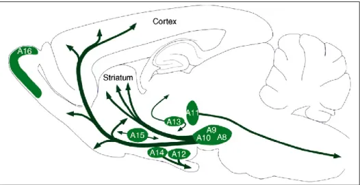
Following the first demonstration by Arvid Carlsson (awarded with the Nobel Prize in 2000 for his work in the field) of the existence of catecholamine-containing neurons in the late 1950s (Carlsson et al., 1958), the possible immunochemical identification of dopamine (DA) neurons in the central nervous system has permitted the localization and subsequent classification of mesotelencephalic DA cells into 10 distinct groups, reconciled into three main systems (Dahlström and Fuxe, 1964; Figure 1):
- the mesencephalic DA system, involving areas A8, A9 and A10 (areas A1 to A7 being noradrenergic nuclei; Figure 2)
- the diencephalic DA system, involving areas A11, A12, A13, A14 and A15 - the retinal DA tract, involving areas A16 and A17
The majority of central DA neurons originates in the mesencephalic DA system. While the substantia nigra pars compacta (SNc, A9) and the retro-rubral area (A8) mainly project to the dorsal striatum (caudate nucleus and putamen), most of the DA neurons emerging from the ventral tegmental area (VTA, A10) innervate the olfactory tubercle, the ventral striatum (namely the nucleus accumbens, NAc) and the frontal cortex, thereby indicating the existence of two sub-systems, the nigrostriatal and the mesocorticolimbic DA tracts (Fallon and Moore, 1978). This classification is of course a simplistic one, considering the substantial overlap of DA fibers in the mesencephalic DA complex (Björklund and Dunnett, 2007). However, on the basis of the most representative DA projection sites and their distinct functional properties, it is generally considered that the DA network falls out into three major ascending pathways: the nigrostriatal, the mesocortical and the mesoaccumbal DA tracts (Tzschentke, 2001; Björklund and Dunnett, 2007). In addition, for the sake of clarity, the terms “dorsal striatum” and “ventral striatum” are usually replaced by “striatum” and “NAc”, respectively.
Introduction
24
Figure 1: Distribution of DA neuron cell groups in the adult rodent brain. The DA
neurons in the mammalian brain are localized in nine distinctive cell groups, distributed from the mesencephalon to the olfactory bulb, as illustrated schematically, in a sagittal view, in the adult rat brain. The principal projections of DA cell groups are illustrated by arrows. DA, dopamine. Drawing adapted from Björklund and Dunnett, 2007.
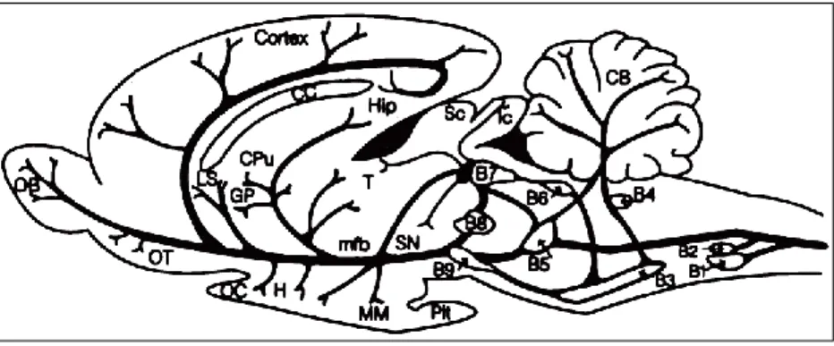
The mid-twentieth century has also witnessed the discovery of serotonin (5-HT) in significant amounts in the central nervous system (Amin et al., 1954). Histochemical fluorescence, radioautography and immunocytochemistry studies led to the classification of 5-HT neurons in 9 clusters designated as B1 to B9, all present in the brain stem (Azmitia and Segal, 1978; Jacobs and Azmitia, 1992; Figure 3):
- the nuclei B1 to B4 form the inferior group (descending pathways) - the nuclei B5 to B9 form the superior group (ascending pathways)
The superior group includes the dorsal raphe nucleus (DRN; the largest nucleus of this group, containing half of its 5-HT neurons), the median raphe nucleus, the caudal linear nucleus and the prosupralemniscus nucleus. In keeping with the extensive branching of 5-HT neuronal processes, these structures provide a widespread innervation of the forebrain (hippocampus, hypothalamus, SNc,
25 medial mammillary nucleus, lateral septum, thalamus, amygdala, cortex...), via different fiber tracts, of which the medial forebrain bundle is the most prominent (Azmitia and Segal, 1978).
Interestingly, DA brain regions (SNc, VTA) as well as their terminal fields (striatum, NAc and frontal cortex; see above) are intensively innervated by the 5-HT system (Azmitia and Segal, 1978). This anatomical promiscuity of DA and 5-HT neurons, along with the importance of DA ascending pathways in several pathological conditions (see section II), has raised and maintained, over the past 40 years, the interest of neuroscience researchers for the potential role of the fourteen 5-HT receptor subtypes in the regulation of the DA network activity.
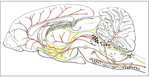
Figure 2: Distribution of noradrenergic and adrenergic neurons and their major
projections in the rat brain (drawing on the midsagittal plane of the brain). The noradrenaline system is represented by solid triangles and lines in red, and the adrenaline system is represented by open triangles and lines in green. A1, A2, A4, A5, and A7 are noradrenergic neurons; C1, C2, and C3, are adrenergic neurons; LC, locus coeruleus; SC, subcoeruleus area. Drawing adapted from Kvetnansky et al., 2009.
Introduction
26
Figure 3: Distribution of 5-HT neuron cell groups in the adult rat brain. A longitudinal
view of rat brain with a schematic representation of the localization of the 5-HT cell bodies in the raphe nuclei and in the brain stem reticular formation, and the corresponding classification into B-groups and their projections. CB, cerebellum; mfb, medial forebrain bundle; CC, corpus callosum; OB, olfactory bulb; Cpu, caudate putamen; OC, optic chiasma; GP, globus pallidus; OT, olfactory tubercle; H, hypothalamus; Pit, pituitary gland; Hip, hippocampal region; Sc, superior colliculus; lc, locus coeruleus; SN, substantia nigra; LS, lateral septal nucleus; T, thalamus; MM, medial mammilary nucleus. Drawing from Waldinger et al., 1998.
27
II. Therapeutic relevance of the interaction between central dopaminergic and serotonergic systems
A. Schizophrenia
The word "schizophrenia", introduced for the first time in 1911 by a Swiss psychiatrist, Eugen Bleuler, comes from the Greek roots “schizo” (split) and “phrene” (mind), to describe the fragmented thinking of people suffering from this neuropsychiatric disorder. Schizophrenia is a chronic, severe, and disabling mental disorder characterized by deficits in thought processes, perceptions, and emotional responsiveness. The illness occurs in less than 1 percent of the general population, and is favoured by genetic and environmental factors as well as specific brain chemistry or substance use (van Os and Kapur, 2009). Although the definitions and diagnosis criteria are constantly evolving along the different versions of the Diagnostic and Statistical Manual of Mental Disorders (DSM; Table 1), three core features emerge in the etiology of schizophrenia: positive (i.e. hallucinations, delusions), negative (i.e. social interaction deficits, blunted affect) and cognitive (i.e. working and reference memory deficits, executive function impairments, decreased vigilance) symptoms (Newman-Tancredi and Kleven, 2011; Meltzer, 2013).
Several hypothesis have been advanced with regards to the implication of specific neurotransmitters, such as DA, glutamate or 5-HT (Rastogi et al., 1981; Carlsson et al., 1999; Svensson, 2003; Moghaddam and Javitt, 2012; Halberstadt and Geyer, 2013), in the pathophysiology of schizophrenia. Although there may be a shared responsibility of several neuronal networks, in view of their close interactions in the brain (Azmitia and Segal, 1978; Carlsson et al., 2000; Delille et al., 2013), we will focus our attention on the DA hypothesis of schizophrenia, which remains one of the most prominent theory in the field. The first clues supporting this hypothesis were brought by the discovery that the DA-D2 receptor (DA-D2R) antagonist chlorpromazine
Introduction
28
increase DA (such as amphetamine and methylphenidate) provoke and/or exacerbate psychotic episodes in humans (Delay et al., 1952; Smith and Davis, 1977; Lieberman et al., 1984, 1987; Kollins et al., 2001). Today, it is generally accepted that this multimodal symptomatology results, at least in part, from an imbalance in central DA neurotransmission. Specifically, a DA hyperfunction of the mesoaccumbal pathway would lead to positive symptoms, while a DA hypofunction of the mesocortical pathway could be responsible for the expression of negative and cognitive symptoms (Svensson, 2000; Newman-Tancredi and Kleven, 2011).
The advent of schizophrenia pharmacotherapy coincides with the ability of chlorpromazine and haloperidol (another DA-D2R antagonist) to alleviate
positive symptoms. However, these treatments (later renamed “typical” antipsychotics drugs, APDs) fail to alleviate negative and cognitive symptoms (Newman-Tancredi and Kleven, 2011). In addition, they exhibit a marked propensity to induce extrapyramidal side effects (EPS), related to their blockade of DA activity in the nigrostriatal DA ascending pathway (Shapira et al., 2006). Thus, the need for treatments with a larger therapeutic window and reduced EPS risk gave birth to a second generation pharmacotherapy, the atypical APDs, of which clozapine is the prototype. These compounds are “selectively nonselective drugs” (Roth et al., 2004; Csermely et al., 2005; Wong et al., 2010), displaying antagonist properties towards the DA-D2R (as this feature
appears fundamental to alleviate positive symptoms), together with agonist or antagonist properties towards other pharmacological targets, such as 5-HT, adrenergic, muscarinic or histaminergic receptors (Newman-Tancredi and Kleven, 2011).
Noteworthy, most of atypical APDs target the 5-HT1AR, 5-HT2AR, 5-HT2BR and
the 5-HT2CR, and sometimes interact with the 5-HT6R and the 5-HT7R
(Newman-Tancredi and Kleven, 2011). Previous studies have shown that the atypicality of APDs (low EPS incidence and improved therapeutic effect with regards to typical APDs) would be related to their low affinity towards
DA-29 D2Rs together with high 5-HT2AR antagonist properties (Newman-Tancredi and
Kleven, 2011). It is also well-established that 5-HT1ARs are key targets for the
therapeutic benefit of atypical APDs. Indeed, microdialysis and knock-out studies have shown that activation of this receptor is necessary for the increase in cortical DA outflow induced by atypical APDs (Ichikawa et al., 2001; Díaz-Mataix et al., 2005), an effect which could be related to their ability to alleviate the cognitive and negative symptoms of schizophrenia. Actually, several findings suggest that the therapeutic benefit of atypical APDs partly results from a cooperation between 5-HT2ARs and 5-HT1ARs. As a matter of fact,
administration of a 5-HT2AR antagonist, at a dose which has no effect by itself,
potentiates 5-HT1AR agonist-induced increase in DA outflow in the medial
prefrontal cortex (mPFC, Ichikawa et al., 2001).
That 5-HT receptors could be interesting targets for the improved treatment of schizophrenia is strengthened by previous findings supporting the involvement of the 5-HT system in the pathophysiology of schizophrenia. First, in schizophrenic patients, many 5-HT receptors, as well as the 5-HT transporter (SERT), tryptophan hydroxylase and monoamine oxidase proteins show alterations in terms of mRNA or protein levels in different brain regions, as well as specific protein sequences (single nucleotide polymorphisms; Bennett et al., 1979; Baou et al., 2015). In addition, low cortical 5-HT levels have been associated with positive symptoms (Rasmussen et al., 2010). Whether these alterations are the cause or the consequence of a DA imbalance in the central nervous system remains unclear. However, it is noteworthy that both the DA and the 5-HT systems may be involved in both the treatment and the development of schizophrenia.
It must be borne in mind that it is crucial to develop new APDs. Indeed, although it is better tolerated with regards to typical APDs, the treatment of schizophrenia with the available atypical APDs is often associated with important side effects (as sedation, weight gain, and metabolic disturbances), related to their multitarget properties, in particular their affinity for
Introduction
30
histaminergic and muscarinic receptors, as well as 5-HT2CRs (Nasrallah, 2008).
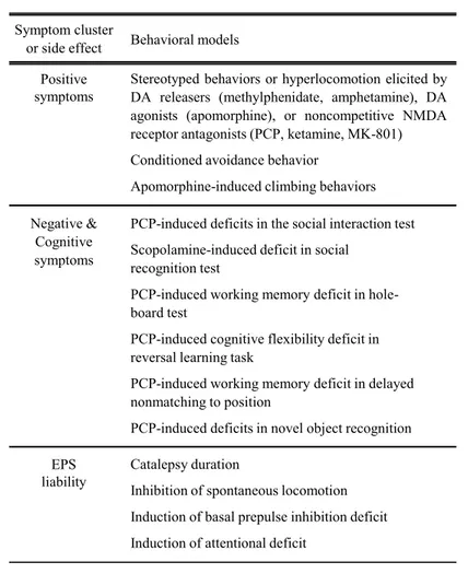
Also, a substantial number of patients fail to respond to this pharmacotherapy, especially in terms of negative and cognitive symptoms, which may be due at least in part to the heterogeneity of the disease. From a preclinical point of view, developing behavioral models can be difficult because of the typical human nature of some of the symptoms observed in schizophrenia (for example flattened affect). In addition, it is, at the moment, impossible to reconcile all the features of schizophrenia in a unique animal model, because of the multimodal nature of schizophrenia. Instead, numerous behavioral models in the primate or the rodent have been developed to predict the ability of a given compound to alleviate either the positive, cognitive or negative symptoms of schizophrenia, or their propensity to induce EPS (Table 2). On the basis of these limitations, the only strategy to identify a potential candidate for treating schizophrenia would be to assess its effects in numerous models of the different symptom clusters of schizophrenia.
31
Table 1: Criteria for schizophrenia in the Diagnostic and Statistical Manual of Mental
Introduction
32
Table 2: Behavioral models classically used to assess the efficacy of APDs against
positive, negative and cognitive symptoms of schizophrenia, as well as their propensity to induce EPS. APD, antipsychotic drug; DA, dopamine; NMDA, N-methyl-D-aspartate; PCP, phencyclidine; EPS, extrapyramidal side effect. Adapted from
Newman-Tancredi and Kleven, 2011. Positive symptoms Negative & Cognitive symptoms EPS liability
Stereotyped behaviors or hyperlocomotion elicited by DA releasers (methylphenidate, amphetamine), DA agonists (apomorphine), or noncompetitive NMDA receptor antagonists (PCP, ketamine, MK-801) Conditioned avoidance behavior
Apomorphine-induced climbing behaviors PCP-induced deficits in the social interaction test Scopolamine-induced deficit in social
recognition test
PCP-induced working memory deficit in hole-board test
PCP-induced cognitive flexibility deficit in reversal learning task
PCP-induced working memory deficit in delayed nonmatching to position
PCP-induced deficits in novel object recognition Catalepsy duration
Inhibition of spontaneous locomotion Induction of basal prepulse inhibition deficit Induction of attentional deficit
Symptom cluster
33
B. Drug addiction: focus on cocaine
Substance use disorders (drug abuse and dependence) are classically defined as chronically relapsing disorders characterized by the compulsion to seek and take a drug, loss of control in limiting intake, and emergence of a negative emotional state reflecting a motivational withdrawal syndrome when access to the drug is prevented (Koob and Volkow, 2010).
As opportunities to use and abuse drugs have increased dramatically during the past 50 years, so has the research on addiction. A first step in the understanding of the neurobiological substrates of drug addiction was taken with the identification of a reward circuitry (a reward being a class of unconditioned motivational stimuli provided with hedonic properties that can act as positive reinforcers), which drives the reinforcing effects of drugs of abuse (Koob and Volkow, 2010). In particular, previous intracranial brain stimulation or self-administration studies have shown that the most sensitive sites of this network belong to the mesocorticolimbic tracts (Olds and Milner, 1954; Crow, 1973; Wise, 1978; Kornetsky and Esposito, 1979). Also, increased DA in the NAc is the hallmark of all drugs abused by humans (Di Chiara and Imperato, 1988), and has been suggested to be the starting point of psychostimulant drug-induced rewarding effects (Koob and Volkow, 2010). Altogether, these findings demonstrate that the DA ascending pathways play a critical role in the behavioral and neurochemical effects of psychostimulant drugs. Noteworthy, this network can be critical not only for the acute reward properties of psychostimulant drugs but also for the long-term neuroadaptative changes in the brain responsible for the ingrained behaviors characterizing drug addiction (Grüsser et al., 2004; Bossert et al., 2009; McClernon et al., 2009; Pierce and Vanderschuren, 2010). Indeed, several studies have shown that the transition from goal-directed (occasional and controlled drug use) to habitual forms of cocaine seeking/taking and chronic relapse is mediated by the gradual
Introduction
34
recruitment of ventromedial-to-dorsolateral striatal DA regions with prolonged drug self-administration (Pierce and Vanderschuren, 2010; Figure 4).
Cocaine has become one of the most commonly used drugs in the world. It is a severe public health problem, with major somatic, psychological, psychiatric, socio-economic and legal implications. However, there is no approved medication for the treatment of cocaine abuse and dependence. As a member of the psychostimulant drug class [with nicotine, amphetamine, methamphetamine, and 4-methylenedioxymethamphetamine, (MDMA)], cocaine-induced short-term effects include mental alertness, energy, euphoria, mood elevation, decreased appetite, dilated pupils, and increased body temperature, heart rate and blood pressure. Following chronic exposure, tolerance to the feeling of energy and euphoria occurs, and cocaine use leads to addiction, paranoia, irritability, restlessness, auditory hallucinations and mood disturbances (Filip et al., 2010). From an experimental point of view, there are several behavioral responses induced by cocaine in laboratory animals that partly resemble some of the symptoms seen in humans (acute or chronic psychomotor stimulation, subjective effects, rewarding/reinforcing properties, and relapse). Thus, over the past 20 years, numerous behavioral models have been developed, the most frequently used including evoked locomotor hyperactivity, conditioned locomotion, sensitization, drug discrimination and reinstatement of seeking behavior (Filip et al., 2012).
It is well-established that cocaine blocks the DA transporter (DAT) and the SERT, as well as the norepinephrine transporter (NET) to a lesser extent (Koe, 1976). Several findings suggest that DAT blockade is required for the reinforcing effects of cocaine. Indeed, cocaine self-administration is inhibited in DAT knock-out mice (Thomsen et al., 2009), while it is not disrupted by the systemic administration of 5-HT (Porrino et al., 1989; Peltier and Schenk, 1991) or noradrenergic antagonists (De Wit and Wise, 1977). In addition, it has been shown that genetic ablation of SERT in both rats and mice fails to reduce cocaine-induced place preference (Sora et al., 1998; Homberg et al., 2008).
35 Nevertheless, other findings question the importance of DAT blockade in the expression of cocaine-induced behaviors. Thus, in DAT knock-out mice, cocaine-conditioned place preference is maintained (Sora et al., 1998) and cocaine self-administration remains unaltered (Rocha et al., 1998), which contrasts with other findings (Thomsen et al., 2009). Interestingly, the ability of cocaine to target the SERT could be a key factor in the expression of its behavioral effects. Indeed, 5-HT outflow is increased in the NAc of rats during cocaine-induced hyperlocomotion (Broderick et al., 2004), and reduced during withdrawal from cocaine self-administration (Parsons et al., 1996). Furthermore, it has been shown that cocaine-conditioned place preference is decreased in SERT knock-out mice (Hall et al., 2009), which also contrasts with previous reports (Sora et al., 1998; Homberg et al., 2008). Thus, no clear conclusion can be drawn with regards to the relative contribution of each transporter to the behavioral effects of cocaine. However, it should be emphasized that compensatory mechanisms may be triggered by the chronic absence of a given transporter. In this regard, Di Chiara and coworkers have shown that despite the genetic ablation of the DAT, cocaine is able to increase NAc DA outflow by blocking the NET (Carboni et al., 2001), which acts as an alternative site for DA clearance from the extracellular compartment (Carboni et al., 1990; Tanda et al., 1997). These findings provide an explanation for the persistence of cocaine reinforcement in DAT knock-out mice. Finally, a previous study has shown that the DAT, the SERT and the NET all participate to cocaine-induced conditioned locomotion in mice, albeit to a different degree and at different stages of the development of this behavior, thereby suggesting the polygenic nature of cocaine reward mechanisms (Hall et al., 2009). Whatever the neurochemical effects underlying the behavioral effects of cocaine may be, the present observations altogether suggest the tight cooperation of DA and 5-HT systems in the control of cocaine rewarding properties. Accordingly, numerous studies have shown that 5-HT receptors are
Introduction
36
able to control cocaine-induced neurochemical and behavioral effects (Bubar and Cunningham, 2008; Howell and Cunningham, 2015).
Although the 5-HT system appears to play a key role in the neurochemical and behavioral responses of cocaine, clinical tests have failed to demonstrate a clear therapeutic effect of SERT antagonists (Howell and Cunningham, 2015). This may be due to the fact that blockade of the SERT, by increasing 5-HT outflow, triggers the modulation of all 5-HT receptors regardless of their subtype and different implications in the behavioral effects of cocaine. When investigating the impact of specific 5-HT subtypes in behavioral models of drug dependence, preclinical studies have permitted to identify the 5-HT1BR (antagonists),
5-HT2AR (antagonists), 5-HT2CR (agonists) and 5-HT3R (antagonists) as
promising candidates for improved treatment of cocaine addiction (Filip et al., 2010). Finally, a recent paper, demonstrating the existence of a synergistic efficacy of a 5-HT2AR antagonist plus a 5-HT2CR agonist to attenuate relapse
factors (impulsivity and cue reactivity), suggests the therapeutic relevance of a bifunctional ligand as an anti-addiction pharmacotherapy, with improved efficacy, potency and selectivity over individual molecules (Cunningham et al., 2013).
37
Figure 4: Anatomical circuits proposed by Pierce and Vanderschuren (2010) to
underlie the progressive recruitment of more dorsal regions of the striatum during the transition from casual cocaine use to habitual cocaine seeking and taking.
39
III. The serotonin2B receptor
The 5-HT receptors encompass fourteen subtypes, reconciled into seven receptor families (5-HT1-7) on the basis of their molecular properties (Table 3
and Figure 5). All 5-HT receptors belong to the seven transmembrane spanning receptor family, more commonly referred to as G-protein-coupled receptors, except the 5-HT3R subtype which is a ligand-gated ion channel (Hannon and
Hoyer, 2008). The 5-HT2BR is the most recent addition to the 5-HT2R family
(Foguet et al., 1992; Kursar et al., 1992), which includes the 5-HT2A and the
5-HT2C subtypes.
A. Localization
The 5-HT2BR (formerly called 5-HT2FR) was first cloned and characterized in
the rat stomach fundus (Foguet et al., 1992; Kursar et al., 1992), then in the mouse (Loric et al., 1992) and in the human (Kursar et al., 1994; Schmuck et al., 1994; Bonhaus et al., 1995). Its presence has been demonstrated in various peripheral tissues, such as the liver, kidney, heart, uterus, trachea and small intestine (Foguet et al., 1992a, 1992b; Kursar et al., 1994; Schmuck et al., 1994; Bonhaus et al., 1995; Fiorica-Howells et al., 2000). Specifically, several studies have shown that it is expressed in rat and pig blood vessels (Ullmer et al., 1995; Watts and Thompson, 2004), in pig heart valves (Fitzgerald et al., 2000) and in the smooth muscle cells of the stomach fundus and myenteric neurons of the intestine of mouse and rat (Fiorica-Howells et al., 2000). In humans, it has been found in endothelial and smooth muscle cells of the pulmonary vasculature (Ullmer et al., 1995), in liver Kupffer cells and tumor-associated macrophages (de Las Casas-Engel et al., 2013), in heart valves (Fitzgerald et al., 2000) and in the longitudinal and circular smooth muscles as well as in the myenteric nerve plexus of the colon (Borman et al., 2002).
Introduction
40
The first studies investigating 5-HT2BR expression in the mammalian brain have
failed to detect the presence of these receptors (Foguet et al., 1992; Kursar et al., 1992; Pompeiano et al., 1994), probably because of the low sensitivity of the employed techniques at that time. Later, several mRNA expression and in-situ hybridization studies have shown that the 5-HT2BR is actually expressed in
the spinal cord (Helton et al., 1994) as well as in several regions of the central nervous system, such as the frontal cortex, lateral septum, dorsal hypothalamus, medial amygdala, DRN, locus coeruleus, hippocampus and cerebellum (Duxon et al., 1997a; Bonaventure et al., 2002). However, there is a paucity of information regarding its cellular localization in the central nervous system. What is known so far is that 5-HT2BRs are expressed in primary astrocyte
cultures from mouse neocortex (Li et al., 2008) and from rat cortex, hippocampus and brainstem (Hirst et al., 1998; Sandén et al., 2000), although a previous study has failed to detect them in rat brain astrocytes (Duxon et al., 1997a). In addition, their presence has been detected in SERT-expressing primary neurons from mouse embryonic raphe nuclei (Launay et al., 2006), and in mouse post-natal microglia (Kolodziejczak et al., 2015).
B. Molecular properties
Studies in cells expressing 5-HT2BRs have shown that this receptor is
functionnally coupled to the protein Gq/11 (Kursar, 1992; Wainscott et al., 1993;
Kursar et al., 1994; Schmuck et al., 1994; Cox and Cohen, 1995; Ellis et al., 1995; Loric et al., 1995; Launay et al., 1996; Cussac et al., 2002, 2008). Activation of Gq/11 stimulates phospholipase C (PLC), which cleaves the
phosphatidylinositol 4,5-bisphosphate (PIP2) into diacyl glycerol (DAG) and
inositol 1,4,5-trisphosphate (IP3). Thereafter, IP3 activates its receptors, in
particular calcium channels of the endoplasmic reticulum (Figure 5). The resulting increase in intracellular calcium combined with the activation of protein kinase C (PKC, by DAG and its translocation to the membrane) triggers
41 various intracellular changes (i.e. apoptosis, regulation of enzyme activity, permeability of ion channels, regulation of the activity of ion pumps and components of the cytoskeleton), leading to numerous physiological responses (such as fertilization, cell growth, transformation, secretion, smooth muscle contraction, sensory perception and neuronal signaling; Berridge, 1993). However, studies in tissue preparations, demonstrating that 5-HT2BR
stimulation-induced responses in the stomach fundus (contraction) or the jugular vein (relaxation) occur independently from PIP2 hydrolysis, have
suggested that other signalization pathways may be responsible for 5-HT2B
R-mediated increased intracellular calcium (Cox and Cohen, 1995; Ellis et al., 1995). For instance, as suggested by Cox and Cohen (1995), this effect could involve the second messenger cyclic ADP-ribose (a metabolite of NAD+), which is able to mobilize intracellular calcium stores (Galione, 1992). However, there is no data in the literature with regards to a possible interaction between cyclic APD-ribose and 5-HT2BRs. A more plausible mechanism could involve a
direct effect of PKC, which can be activated independently of PIP2 hydrolysis
(Whetton et al., 1988), without translocation to the membrane (Heidenreich et al., 1990; Deisher et al., 1993), and has been shown to participate to the 5-HT2BR-mediated contractile effect of the rat stomach fundus (Cox and Cohen,
1995). Finally, it should be noted that the 5-HT2BR is also able to stimulate
L-type voltage dependent calcium channels, thereby suggesting the role of extracellular calcium in its functional effects (Cox and Cohen, 1995). Further studies are needed to clarify the exact mechanisms involved in the 5-HT2B
R-induced increase of intracellular calcium.
Additional intracellular signalization pathways recruited by the 5-HT2BR have
been reported in the literature, although these couplings, observed in cell lines expressing 5-HT2BRs, need to be further confirmed in tissue preparations. Thus,
nitric oxide (NO), which induces the relaxation of various blood vessels, has been shown to behave as a signaling effector of the 5-HT2BR in LMTK
Introduction
42
capacity to differentiate into neuronal 5-HT cells expressing the 5-HT2BR),
5-HT activates the ras protein which in turn stimulates the mitogen-activated protein kinase (MAPK) pathway (Launay et al., 1996), responsible for the regulation of the expression of genes involved in proliferation, differentiation, apoptosis, and synaptic plasticity (Girault, 2007). Finally, previous studies in 1C11 cell lines or in mesoblastic cells converting into osteocytes have demonstrated that the 5-HT2BR can control arachidonic acid release through the
activation of phospholipase A2 (PLA2; Tournois et al., 1998; Locker et al.,
2006). Activation of PLA2 by 5-HT2BRs can also lead to reactive oxygen
species (ROS) production through NADPH oxidase activation in 1C11 cells, an effect which could trigger the degradation of 5-HT (Pierce et al., 2005; Schneider et al., 2006).
43
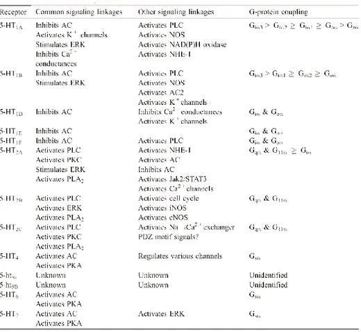
Table 3: Signaling characteristics of human 5-HT receptors. The table is not
all-encompassing. 5-HT, serotonin; AC, adenylyl cyclase; cNOS, constitutive NOS; ERK, extracellular signal-regulated kinases; iNOS, inducible NOS; Jak, Janus kinase; NHE, Na+ /H+ exchange; NOS, nitric oxide synthase; PDZ, PS-95 discs-large ZO-1; PKA, protein kinase A; PLC, phospholipase C; PKC, protein kinase C; PLA2, phospholipase
A2; STAT, signal transducers and activators of transcription. Adapted from Raymond et al., 2001.
Introduction
44
Figure 5: Prototypical signaling enzyme linkages of the G-protein-coupled 5-HT
receptors. 5-HT1Rs typically inhibit AC through pertussis toxin-sensitive G-proteins of
the Gi family, whereas 5-HT4Rs, 5-HT6Rs, and 5-HT7Rs typically stimulate AC through
Gs family G-proteins. Activation of AC results in increased production of cAMP, leading to activation of PKA. 5-HT2Rs activate PLC-β through Gq/11 family
G-proteins, resulting in accumulation of PIP2 to IP3 and DAG. Generation of IP3 results in
elevation of intracellular Ca2+ levels, whereas DAG activates the Ca2+ and phospholipid-dependent protein kinase (PKC). 5-HT2Rs also typically activate PLA2 through
G-proteins to increase the accumulation of AA. 5-HT, serotonin; AC, adenylyl cyclase; cAMP, cyclic AMP; PKA, protein kinase A; PLC, phospholipase C; PIP2,
phosphatidylinositol 4,5-bisphosphate; IP3, inositol trisphosphate; DAG, diacylglycerol;
PKC, protein kinase C; PLA2, phospholipase A2; AA, arachidonic acid. Drawing from Raymond et al., 2001.
45
C. Pharmacology
The first efforts aimed at exploring the role of 5-HT2BRs in physiological or
pathological states have been hampered by the lack of selective and potent ligands. Indeed, most agents initially failed to discriminate the 5-HT2BR from
the 5-HT2AR and the 5-HT2CR, especially the latter, with which it shares a high
degree of sequence homology and very similar pharmacology (Foguet et al., 1992a, 1992b; Kursar et al., 1992; Wainscott et al., 1993). Thus, in keeping with the first belief that 5-HT2BRs were absent in the central nervous system
(see above), it is noteworthy that several central functions may have been illegitimately ascribed to the 5-HT2CR in the past.
In the mid-1990s, SB 204741 [N-( l-Methyl-5-indolyl)-N'-(3-methyl-5-isothiazoly1)urea] was introduced as a 5-HT2BR antagonist (Forbes et al., 1995).
This compound, while displaying a higher selectivity for the 5-HT2BR over the
5-HT2CR, the 5-HT2AR and numerous other receptors, has a relatively low
affinity for the 5-HT2BR (Bonhaus et al., 1995; Forbes et al., 1995; Baxter,
1996). Thereafter, two studies identified LY 266097 [1-[(2-Chloro-3,4-dimethoxyphenyl)methyl]-2,3,4,9-tetrahydro-6-methyl-1H-pyrido[3,4-b]indole] and RS 127445 [2-amino-4-(4-fluoronaphth-1-yl)-6-isopropylpyrimidine] as potent and high-affinity antagonists (pKi = 9.8 for LY 266097 and 9.5 for RS 127445), with more than 100-fold and 1000-fold selectivity for the 5-HT2BR
over the other 5-HT2R subtypes, respectively (Audia et al., 1996; Bonhaus et
al., 1999). These compounds are at the moment the best 5-HT2BR antagonists
available (Table 4).
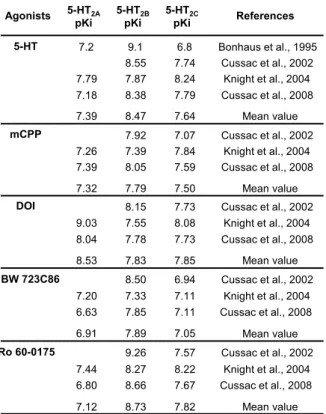
Identifying a proper 5-HT2BR agonist is still a challenge today (Table 5). The
compound BW 723C86 [(α-methyl-5-(2-thienylmethoxy)-1H-indole-3-ethanamine] is classically used to assess the effects of 5-HT2BR stimulation
(Gobert et al., 2000; Auclair et al., 2010), but it is only a preferential agonist. Indeed, this compound displays a poor affinity and low selectivity for the 5-HT2BR (Baxter, 1996; Kennett et al., 1997, 1998). In addition, there are
Introduction
46
substantial differences in BW 723C86 affinities for 5-HT receptors depending on the species or tissue preparations in which its binding properties are assessed (Baxter, 1996). Finally, it should be borne in mind that Ro 60-0175 [(αS-6-chloro-5-fluoro-α-methyl-1H-indole-1-ethanamine], initially characterized as a highly selective 5-HT2CR agonist (Martin et al., 1998), could be a good
5-HT2BR agonist. Indeed, Ro 60-0175 was discovered to be a potent (pEC50 =
9.05) and high efficacy agonist (79%) at 5-HT2BRs (Porter et al., 1999), with
better affinity and functional selectivity over the 5-HT2CR (Cussac et al., 2002).
Once again (see above), it is therefore possible that several effects of Ro 60-0175 reported in the literature and attributed to the 5-HT2CR may actually result
from 5-HT2BR stimulation.
Table 4: Binding affinities (pKi) of some antagonist ligands at human recombinant
5-HT2 receptors. SB 204741 LY 266097 RS 127445 Bonhaus et al., 1999 Knight et al., 2004 Mean value Cussac et al., 2002 Mean value Mean value Knight et al., 2004 Cussac et al., 2002 Antagonists 5-HT2A References pKi 6.30 6.16 7.71 < 5 5-HT2B pKi 9.5 8.97 9.70 6.90 7.29 9.23 9.75 7.09 5-HT2C pKi 6.4 6.33 7.17 5.56 5.67 6.36 7.39 5.61 6.03 < 5 Audia et al., 1996 7.71 9.80 7.61
47
Table 5: Binding affinities (pKi) of some agonist ligands at human recombinant 5-HT2
receptors. Ro 60-0175 BW 723C86 DOI mCPP 5-HT Bonhaus et al., 1995 Cussac et al., 2002 Knight et al., 2004 Cussac et al., 2008 Mean value Cussac et al., 2002 Mean value Knight et al., 2004 Cussac et al., 2008 Mean value Cussac et al., 2002 Knight et al., 2004 Cussac et al., 2008 Cussac et al., 2002 Knight et al., 2004 Cussac et al., 2008 Cussac et al., 2002 Knight et al., 2004 Cussac et al., 2008 Mean value Mean value Agonists 5-HT2A References pKi 7.2 7.79 7.18 7.26 7.39 9.03 8.04 7.20 6.63 7.44 6.80 7.39 7.32 8.53 7.12 6.91 5-HT2B pKi 9.1 8.55 7.87 8.38 7.92 7.39 8.05 8.15 7.55 7.78 7.33 8.50 7.85 8.27 9.26 8.66 8.47 7.79 7.83 8.73 7.89 5-HT2C pKi 6.8 7.74 8.24 7.79 7.07 7.84 7.59 7.73 8.08 7.73 7.11 6.94 7.11 8.22 7.57 7.67 7.64 7.50 7.85 7.82 7.05
49
D. Functional aspects
1. Regulation of cell differentiation and proliferation
Interestingly, the 5-HT2BR is involved in the proper development or the
modulation of the number of various cell subtypes in the body, thereby suggesting that this receptor may play important roles in the onset of peripheral functions or in their regulation during adulthood. In particular, pharmacological and knock-out studies have shown that 5-HT2BR modulation impacts on the
differentiation and/or proliferation of mouse enteric neurons (Fiorica-Howells et al., 2000), hepatocytes (Lesurtel et al., 2006) and cardiomyocytes (Nebigil et al., 2000, 2003), as well as on the number of retinal cells in the Xenopus (De Lucchini et al., 2003, 2005). In addition, 5-HT2BRs may participate to the
proper wiring of neuronal networks by regulating the motility of microglial processes in the postnatal mouse (Kolodziejczak et al., 2015).
2. Regulation of the gastrointestinal tract
Since the discovery and characterization of 5-HT2BRs in the rat stomach fundus,
several studies have attempted to determine the exact role of these receptors in the regulation of the gastrointestinal (GI) tract. Thus, it was found that stimulation of 5-HT2BRs triggers a contractile effect on smooth muscle
preparations isolated from the rat stomach fundus and maintained in organic bath solutions (Baxter et al., 1994; Cox and Cohen, 1995), as well as on intestinal smooth muscles in humans (Borman and Burleigh, 1995). Another substantial aspect of the involvement of 5-HT2BRs in the regulation of the GI
function relies on their ability to modulate the proliferation of the interstitial cells of cajal (ICC), which are expressed all along the GI tract and determine the frequency of contraction of its smooth muscle cells (Tharayil et al., 2010). Specifically, in mice, 5-HT2BRs promote the ICC turnover both in vitro and in
Introduction
50
2007; Tharayil et al., 2010). Interestingly, these findings have shed light on the therapeutic potential of 5-HT2BR ligands in pathological conditions depending
on a decreased number of ICCs or a disrupted ICC network, such as slow transit constipation, diabetic gastroparesis, or pseudo obstruction (Kenny et al., 1998; He et al., 2000; Lyford et al., 2002; Forster et al., 2005). Finally, it is noteworthy that 5-HT2BRs may participate to the maturation of the GI tract, as
suggested by a previous study showing that 5-HT promotes the in vitro development of enteric neurons through 5-HT2BR stimulation (Fiorica-Howells
et al., 2000).
3. Regulation of the vascular function
The involvement of 5-HT2BRs in the control of vascular function was
introduced with the discovery of its relaxing effect on the jugular vein (Ellis et al., 1995). This effect has been subsequently attributed to the ability of 5-HT2BRs to trigger the release of NO, although this signalization pathway has
been observed only in vitro (Manivet et al., 2000). In this context, it is noteworthy that 5-HT2BRs may play an important role in migraine headache.
Indeed, a previous study has suggested that migraine headache would result, at least in part, from the formation of NO triggered by the activation of 5-HT2BRs
located on the endothelial cells of human meningeal blood vessels (Schmuck et al., 1996). Specifically, the release of NO, which by itself induces migraine headaches in susceptible humans (Olesen et al., 1994), would stimulate the trigeminal nerve (Wei et al., 1992). Stimulation of this nerve would provoke blood vessel dilatation and protein leakage, leading to inflammation within the dural vasculature (Moskowitz, 1993). On the other hand, 5-HT2BR stimulation
has been shown to induce a contractile effect on the renal artery isolated from the rat and maintained in an organic bath (Watts and Thompson, 2004), suggesting that 5-HT2BRs may play an important role in the regulation of blood
5-51 HT2BRs control renal sympathoexcitation and sympathoinhibition in the in vivo
rat (Knowles and Ramage, 1999, 2000).
4. Regulation of the pulmonary function
Pulmonary arterial hypertension (PAH) is a progressive disease, characterized by sustained elevation of pulmonary arterial pressure associated with abnormal vascular proliferation, neomuscularization of small pulmonary arteries with intimal thickening, leading to right ventricle failure and death (Rhodes et al., 2009). Interestingly, it has been shown that 5-HT2BR activation is critical for the
development of PAH in mice. Specifically, 5-HT2BRs regulate the
differentiation and proliferation of bone-marrow stem cells, which are known to participate in the development of PAH and pulmonary vascular remodeling (Launay et al., 2012). In line with these results, 5-HT2BR expression is
increased in pulmonary arteries of both humans and mice suffering from PAH (Launay et al., 2002). In addition, it has been demonstrated that all the physiological features of the chronic-hypoxic-mouse model of PAH (i.e. increase in pulmonary blood pressure and in lung remodeling, associated with an increase in vascular proliferation, elastase activity and transforming growth factor-beta levels) are no longer observed in the 5-HT2BR knock-out mouse
(Launay et al., 2002). Similarly, pharmacological blockade of 5-HT2BRs with
selective antagonists counteracts monocrotaline-induced muscularization of pulmonary arterioles and perivascular fibrosis in the rat lung (Zopf et al., 2011), as well as the increase in pulmonary arterial pressure in mice challenged with hypoxia (Dumitrascu et al., 2011). Altogether, these findings suggest that the use of 5-HT2BR antagonists may be a valuable therapeutic approach for the