Pediatr Radiot (1988) 19:41-44
Pediatric
Radiology
© Springer-Verlag 1988
Chondrodystrophic dwarfism
and multiple malformations in two sisters
U. E. Pazzaglia 1, L. Pedrotti 1, G.
Beluffi 2 and L. Ceciliani t
1 Clinica Ortopedica dell'Universitfi di Pavia, and
2 Servizio di Radiodiagnostica, Sezione di Radiopediatria, I. R. C. C.S. Policlinico San Matteo, Pavia, Italy
Abstract.
A genetic skeletal dysplasia with dwarf-
ism, scoliosis and multiple skeletal defects was ob-
served
in
two sisters. Only nine cases with similar
features have been reported in the literature.
A syndrome presenting with dwarfism, spondylo-
metaphyseal abnormalities and advanced bone age
was reported by Neimann and colleagues in 1964 [1]
and by Desbuquois and colleagues in 1966 [2].
Other cases with similar features were described by
Piussan and colleagues [3], who distinguished this
condition from Larsen's Syndrome.
We report two new observations of this rare dis-
ease.
Laboratory tests and urinary mucopolysaccharides were nor- mal; caryogram was normal, 46 XX.
The congenital displacement of the hips was treated success- fully with traction and piaster casts.
After discharge the patient was not seen again until she was 19 years old. The facial features were even more evident and the scoliosis had progressed to reach more than 150 degrees; her height was 85 cm (Fig.l). Intelligence was normal, although she was inclined to refuse inter-personal relationship because of her physical handicaps.
Skin fibroblast cultures and lysosome hydrolases were nor- mal.
Case 2
Third daughter of the same parents came to our observation at the age of four years. Another sister, aged five, was normal.
Case reports
Case I
Was the first daughter of consanguineous parents. The father had a short stature, but no cases of dwarfism were observed in the family pedigree.
The pregnancy and the delivery were uneventful; a mild bilat- eral club foot was observed at birth and it was treated by simple manipulation.
We first observed the child at the age of three years because of bilateral hip dislocation and scoliosis.
The head was abnormally large with protrusion of the frontal bones; the fontanels were closed; the face showed protruding eyes, saddle nose and hypertrophy of the upper jaw and zygo- matic bones. The mandible, on the contrary, was small and the teeth were normal. The neck was short. The trunk was deformed by scoliosis. Height was 60 cm and there was a symmetrical shortening of both upper and lower limbs. Dislocation of the heads of the radii limited supination of the elbows. The hands were short and squat.
The hips were displaced; hyperlaxity of the knees was also observed.
42 U.E. Pazzaglia et al, : Chondrodystrophic dwarfism
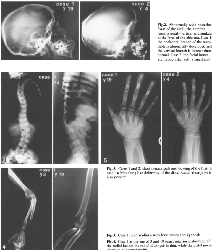
Fig.2. Abnormally wide posterior fossa of the skull; the anterior fossa is nearly vertical and sunken at the level of the chiasma. Case 1: the horizontal branch Of the man- dible is abnormally developed and the vertical branch is thinner than normal. Case 2: the thcial bones are hypoplastic, with a small and
Fig. 5. Cases I and 2: short metacarpals and bowing of the first. In case 1 a Madelung-like deformity of the distal radius-ulnar joint is also present
Fig.3. Case 2: mild scoliosis with four curves and kyphosis Fig.4. Case 1 at the age of 3 and t9 years: anterior dislocation of the radial heads; the radial diaphysis is thin, while the distal meta- physis is of normal width
The child was 58 cm tall; her head was large and her face presented the same characteristics as the first sister (Fig.l). Her neck was short and scotiosis was present. The upper limbs were short with some degree of flexion and pronation of the elbows; displacement of the radial head was observed on both sides. The hands had short fingers. The hips were displaced but they could be
reduced easily with flexion and abduction. The lower limbs were short. Intelligence was normal. Cariogram was 46 XX, normal.
Laboratory tests, urinary mucopolysaccharides and hydro- lases tested in serum and in cultured fibroblasts were normal. The child was kept in a divaricator for one year but no improve- ment of the hip laxity was obtained.
U. E, Pazzaglia et al. :S Chondrodystrophic dwarfism 43
Fig.6. Case 1 at the age of 3 years and case 2 at the age of 4 years: wide acetabula with an horizontal and irregular border; valgus of the proximal femora and irregular epiphyseal centers (hypoplastic on the medial side)
Fig.7. Cases 1 and 2: short and squat first ray of the feet with varus first metatarsal. Case 1 : irregular metatarsals with shortening of the 4th, 5th right and 3rd, 4th, 5th left. Pseudo-cystic cavities are present in the distal ends of metatarsals
Radiographic features
The skull presented in both sisters an abnormally wide posterior fossa; the anterior fossa was nearly vertical and sunken at the level of the chiasma. The parietal bones were thicker than normal and the facial bones were hypoplastic, with a small and short mandible. In the older sister, at the end of the skeletal growth, the horizontal branch of the mandible appeared abnormally de- veloped and the vertical branch was thicker than normal (Fig. 2). The younger sister had a mild scoliosis (less than 30 °) with four curves and a kyphosis with apex at the 12th dorsal and 1st lumbar vertebrae. These vertebral bodies had a hypoplastic supe- rior edge (Fig.3).
In the older sister a single curve scoliosis of 65 ° was present at the age of three, and it had increased to more than 160 ° at ma- turity. The deformity of the chest made difficult to evaluate the morphology of the ribs in this girl, which presented irregular cal- cification of the chondro-sternal cartilages; in the younger sister, on the contrary, they were abnormally thick at the lateral and anterior segments.
The upper limb bones showed a mesomelic shortening; the radius had a thin diaphysis contrasting with the normal width of the distal metaphysis and a bowed proximal end. In both sisters the radial heads were dislocated anteriorly and the distal radio- ulnar joints had a Madelung-like deformity (Fig. 4).
Carpal ossification centers were normal for the age. All meta- carpals were short, with some shorter than the others; the first was also bowed. Proximal and middle phalanges were normal, while the distal had an enlarged base (Fig. 5).
The pelvis was anteverted. Acetabula were wide, horizontal and with an irregular border.
The proximal epiphyseat center of the femurs were hypoplas- tic on the medial side and in valgus. The great trochanter ossifi- cation center had an irregular shape (Fig. 6).
The lower limb bones were short, with discrepancy between tibia and fibula (the latter exceeding the former by about 1 cm),
The diaphysis of the femur was thin, in contrast with the nor- mal width of distal metaphyses, which were osteopenic. In the older sister this osteopenia had produced a crushing of the tibial plates.
44
Proximal femoral metaphysis presented short neck and varus deformity.
In both sisters, at the age of three and four respectively, the feet h a d a short and squat first ray, with varus of the 1st metatar- sal. At the end of skeletal growth in the older sister many irregu- larities of metatarsals were observed, with a shortened first ray, but with a still more considerable shortening of the right 4th, 5th and left 3rd, 4th, 5th metatarsals.
Pseudo-cystic cavities resembling those observed in multiple exostosis disease, were present at the distal ends of these short- ened metatarsals.
The hallux was shorter than the other toes; the right 3rd, 4th, 5th and left 3rd, 4th phalanges had excess ossification nuclei. The middle phalanx of the left 5th toe had a n hour-glass like shape (Fig.7).
The clavicles were short, the scapulae had a squared shape with hypoplastic glenoid.
Discussion
The clinical and radiographic features of these two
sister are peculiar and we found remarkable diffi-
culty in classifying them among skeletal dysplasias.
The similarities of the pathological characteristics
and consanguinity of parents supported the hy-
pothesis of a genetic dysplasia.
Cases with similar features have been reported
by Desbuquois et al. [2] as "Chondrodystrophic
dwarfism with anarchical ossification and multiple
malformations" and under other denomination by
Neimann et al. [1] and by Piussan et al. [3].
The two sister reported here had many features
in common with the cases already reported in the
literature, namely short stature, osteoporosis, sco-
liosis, dysmorphic vertebral bodies, epiphyseal and
metaphyseal abnormalities, hyperlaxity of the
joints, dysplasia of the acetabula and dislocation of
the hips. However, ulnar deviation of the hands,
and early appearance of tarsal and carpal centers
U. E. Pazzaglia et al.: Chondrodystrophic dwarfism
observed in other cases were not present. On the
contrary the profile of the skull, the bowing of
proximal radius with dislocation of the radial head
and the Madelung-like deformity of the distal
radio-carpal joint were peculiar features.
To our knowledge only nine cases of this dys-
plasia have been hitherto reported: three siblings
described by Piussan et al. [3] had consanguineous
parents. The occurrence of the disease in two other
sisters with consanguineous parents further support
the autosomal recessive mode of transmission.
Case no. 1 is the oldest described in the literature
and also the one with the longest follow-up.
Differential diagnosis is with the Larsen syn-
drome.
Acknowledgements. The
authors are grateful to Dr. P. Maroteaux for the useful diagnostic indication and for the references of pub- lished cases.References
1, Neimann N, Pierson M, Manciaux M, Sapetier J (1964) A propos d'un nouveau cas de nanisme diastrophique. Arch Fr P~diatr 2I : 957
2. Desbuquois G, Grenier B, Michel J, Rossignol C (1966) Nanisme chondrodystrophique avec ossification anarchique et polymalformations chez deux soeurs. Arch Fr Prdiatr 23: 573 3. Piussan C, Maroteaux P, Castroviejo I, Risbourg B (1975) Dys-
plasie osseuse avec nanisme et alterations squelettiques dif- fuses. Six observations. Arch Fr Prdiatr 32:541
Accepted: 6 December 1987 Prof. U.E. Pazzaglia
Clinica Ortopedica dell'Universitfi I. R. C. C.S. Policlinico San Matteo Via Tarmelli 3
1-27100 Pavia Italy
Literature in pediatric radiology (continued from p.27)
Chirurgie P£'diatrique (Paris)
Ostrome ostroide du col f~moral de l'enfant et l'adolescent. Longis, B. et al. (Serv. de Chirurg. Prd, CHU Dupuytren, 2, ave. Alexis-Carrel, F-87 000 Limoges, France) 29, 24 (1988)
Les drcollements 6piphysaires spontan~s chez l'enfant porteur d'une spina bifida. Michel, F. et al. (Centre Livet, H6pital de Lyon, 8, rue de Mar- gnolles, F-69 300 Caluire, France) 29, 29 (1988)
Epiphysiolyse externe avec ,,caput valga". Brichaux, J.C. et al. (Diard, F., Serv. de Rad., Hfpital des Enfants, 168 cours de l'Argonne, F-33077 Bordeaux Cedex, France) 29, 39 (1988)
Journal de Radiologie (Paris)
Empreinte ou compression trachrale lat~rale due au tronc artrriel brachio- crphalique chez l'enfant. Ghidalia, S. et al. (Sew. de Rad., Pav. de la M+re et de l'Enfant, CHRU de Nantes, BP 1005, F-44035 Nantes, France) 09, 205 (1988)
Les calcifications laryngo-trachro-bronchiques idiopathiques chez l'en- fant. Apropos de deux observations. Thollot, F. et al. (Serv. de Rad.,
Hfip. d'Enfants 5, atlre du Morvan F-54500 Vandoeuvre, France) 69, 217 (1988)
Journal of Neuroradioiogy (Paris)
Oligodendrogliomes thalamiques chez l'enfant: TDM et +volution clini- que. Nov, A.A. et al. (Dep. of Rad., Group Health Cooperative, Central Hosp., 200 15th Ave. East, Seattle, WA 98112, USA) 15, 23 (1988) Diagnostic tomodensitomrtfique dans soixante cas d'abc+s crrrbral de pe-
tite taille. Wu Yujin et al. (Serv. de Neurolog., Hebei Provincial Hosp., Jichang Road, Shijiazhuang, Hebei Province, People's Rep. of China) 15, 77 (1988)
P~iatrie (Lyon)
Hydroprricarde nronatal par cathrter 6picutanro-cave en silicone. Don- nou-Da Lage, M.D. et al. (Guillois, B., Serv de reanimation prd. et de nronatologie, CHU morvan, F-29279 Brest Cedex, France) 43, 23 (1988)