Alma Mater Studiorum – Università di Bologna
DOTTORATO DI RICERCA IN
Scienze Biomediche e Neuromotorie
Ciclo XXX
Settore Concorsuale: 06/D6-Neurologia
Settore Scientifico Disciplinare: MED26-Neurologia
FLOW CYTOMETRY ANALYSIS OF T CELL SUBSETS IN
CEREBROSPINAL FLUID AND PHERIPERAL BLOOD OF
NARCOLEPSY TYPE 1 PATIENTS WITH LONG LASTING
DISEASE
Presentata da: Dott.ssa Moresco Monica
Coordinatore Dottorato
Supervisore
Prof. Lucio Ildebrando Cocco
Prof. Giuseppe Plazzi
2
INDEX:
ABSTRACT………...P 4 1. CHAPTER 1, Introduction………...P 5 1.1 NORMAL SLEEP STRUCTURE………...P 6 1.2 NARCOLESPY TYPE 1 DISORDER………..…...…P 6 1.3 EPIDEMIOLOGY………...P 7 1.4 SYMPTOMS………...P 7 1.5 THE HYPOCRETIN SYSTEM IN NT1………...…P 9 1.6 ENVIRONMENTAL FACTORS AND GENETIC EVIDENCES
SUPPORTING THE AUTOIMMUNE HYPOTESIS OF NT1……..….…..P 10 1.7 HUMORAL IMMUNITY AND ROLE OF AUTOANTIBODY
IN NT1………...…………...P 12 1.8 T CELL MEDIATED IMMUNITY……….…...P 15 1.9 THE CELLULAR BASIS OF IMMUNOLOGICAL MEMORY –
CENTRAL MEMORY AND EFFECTOR MEMORY
T CELL SUBSETS………...P 17 1.10 NATURAL KILLER CELL MEDIATED IMMUNITY……….…...P 20 1.11 IMMUNITY AND AUTOIMMUNITY IN
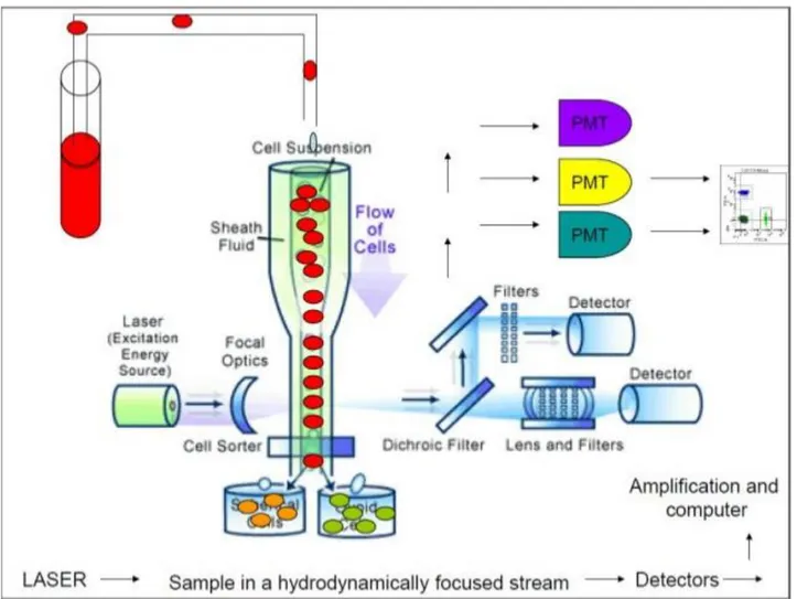
CENTRAL NERVOUS SYSTEM……….………..P 22 1.12 AIMS……….…..…P 24 1.13 REFERENCES………....P 25 2. CHAPTER 2, Materials and Methods………....…...P 37 2.1 PATIENTS AND CONTROLS……….…..…..P 38 2.2 COLLECTION OF CSF CELLS AND PBMCs………..……..P 38 2.3 HLA-DQB1*06:02 ALLELE TYPING………...…...P 39 2.4 FLOW CYTOMETRY ANALYSIS………..….…...P 41 2.4.1 THE FLUIDIC SYSTEM………..……...….…..P 42 2.4.2 LIGHT SOURCES……….……...….…P 43 2.4.3 THE PHYSICAL PARAMETERS………..……...…....P 44 2.4.4 FLUORESCENCE AND COMPENSATION………...….…P 45 2.4.5 DATA COLLECTION AND DISPLAY……….…...P 46 2.4.6 IMMUNOPHENOTYPING………....….…...…P 46 2.4.7 FLOW CYTOMETRY ANALYSIS IN NT1………...…....P 47 2.5 STATISICAL ANALYSIS………...……...P 51
3
2.6 REFERENCES………...…..P 52 3. CHAPTER 3, Results……….…...P 53 3.1 DEMOGRAPHIC CHARACTERISTIC IN NT1 AND HC GROUP...P 54 3.2 CSF T CELLS AND PBMCs IN NT1 VERSUS HLA DQB1*06:02
NEGATIVE HC………...P 54 3.3 CSF/PBMC FOLD-CHANGE IN NT1 VERSUS
HLA DQB1*06:02 NEGATIVE HC………...P 56 3.4 CSF VERSUS PBMC T CELL SUBSETS IN NT1 AND
HLA DQB1*06:02 NEGATIVE HC………...…P 57 4. CHAPTER 4, Discussion………...P 60 4.1 CONCLUSION………..……..P 63 4.2 REFERENCES………..……...…..P 64
4
ABSTRACT
Background: Type 1 Narcolepsy (NT1) is a central hypersomnia linked to the destruction of hypocretin-producing neurons. A great body of genetic and epidemiological data points to a likely autoimmune disease aetiology. Recent reports have characterized peripheral blood T-cell subsets in NT1, whereas data regarding the cerebrospinal fluid (CSF) immune cell composition are lacking. In this study, we aim to identify the T- and NK- cell subsets in NT1 patients with long-term disease course.
Methods: Immune cell subsets from CSF and peripheral blood mononuclear cell (PBMC) samples were analysed by flow cytometry in two age-, sex and BMI-balanced groups of 14 NT1 patients versus 14 healthy controls. The frequency of CSF cell groups were compared with PBMCs. Non-parametric tests were used for statistical analyses.
Results: NT1 patients did not show significant differences of CSF immune cell subsets compared to controls, despite a trend towards higher CD4+ terminally differentiated effector memory T cells. T cells preferentially displayed a memory phenotype in the CSF compared to PBMCs. Furthermore, a reduced frequency of CD4+ terminally differentiated effector memory T cells and an increased
frequency of Natural Killer CD56bright cells were observed in PBMCs from patients compared to
controls. Finally, the ratio between CSF and peripheral CD4+ terminally differentiated effector
memory T cells showed a two-fold increase in NT1 patients versus controls.
Conclusions: Significant differences in PBMCs and CSF immune cell profile were found between NT1 patients versus controls. These differences may reflect differences in HLA status, or be primary or secondary to hypocretin deficiency in narcolepsy.
5
CHAPTER 1
Introduction
6
1.1 NORMAL SLEEP STRUCTURE
Normal sleep is divided into two phases: non–rapid eye movement (NREM) and rapid eye movement (REM) sleep. NREM sleep is further distributed into progressively deeper stages of sleep: stage N1, stage N2, and stage N3. As NREM stages progress, stronger stimuli are required to result in an awakening. Stage REM sleep is characterized by tonic and phasic components. The phasic component is a sympathetically driven state of rapid eye movements, muscle twitches, and respiratory variability. Tonic REM is a parasympathetically driven state with no eye movements. Moreover, the REM period length and density of eye movements increases throughout the sleep cycle (Berry et al. 2012). Waking usually transitions into light NREM sleep. NREM sleep typically begins in the lighter stages N1 and N2, and progressively deepens to slow wave sleep as evidenced by higher-voltage delta waves. N3 (slow wave sleep) is present when delta waves account for more than 20% of the sleep electroencephalogram (EEG). REM sleep follows NREM sleep and occurs 4-5 times during a normal 8-hour sleep period. The first REM period of the night may be less than 10 minutes in duration, while the last may exceed 60 minutes. The NREM-REM cycles vary in length from 70-100 minutes initially to 90-120 minutes later in the night.
Usually, N3 sleep is present more in the first third of the night, whereas REM sleep predominates in the last third of the night. This can be helpful clinically as NREM parasomnias such as sleep walking typically occur in the first third of the night with the presence of N3 sleep. This contrasts with REM sleep behaviour disorder (RBD), which typically occurs in the last half of the night (Berry et al. 2012).
1.2 NARCOLESPY TYPE 1 DISORDER
Narcolepsy is a life-long, underrecognized sleep disorder that was firstly described by Westphal in 1877 and Gelineau in 1880 (Schenck et al. 2007). It has a complex symptomatology that reflects the partial or global intrusion of REM sleep into wakefulness, causing excessive daytime sleepiness (EDS), cataplexy (i.e. sudden loss of muscle tone during wakefulness triggered by strong emotions), hypnagogic hallucinations, sleep paralysis, and disrupted nocturnal sleep (Dauvilliers et al. 2007; Abad & Guilleminault, 2009; Roth et al. 2013). While EDS with recurrent sleep attacks is the most common feature, cataplexy is pathognomonic, allowing us to clinically distinguish narcolepsy with cataplexy from narcolepsy without cataplexy. Along with the discovery of hypocretin deficiency as causal for narcolepsy, the disorder has been recently divided in narcolepsy type 1 (NT1), characterized by low cerebrospinal fluid (CSF) hypocretin-1 levels and cataplexy in most of these cases, and in narcolepsy type 2 (NT2), characterized by normal CSF hypocretin-1 levels and absence of cataplexy. The hypocretin neurons main function is to sustain wakefulness and suppress rapid eye
7
movement sleep (Bonnavion & de Lecea 2010), explaining why a loss of these neurons causes the narcoleptic phenotype. In addition to the lack of signal from preprohypocretin in the brains of NT1 patients, signals from the colocalizing proteins dynorphin and neuronal activity-regulated pentraxin have also been found lacking (Blouin et al. 2005; Crocker et al. 2012). Interestingly, nearby and intermingled melanin concentrating hormone-producing neurons were found to be intact (Peyron et al. 2000). The specific loss of hypocretin neurons in NT1 is now thought to result from an autoimmune attack on these neurons.
1.3 EPIDEMIOLOGY
The prevalence of NT1 is 0.02% to 0.05% (Silber et al. 2002). Globally, the prevalence varies from highest in Japan (0.16%) to lowest in Israel (0.0002%) (Longstreth et al. 2007). NT1 is prevalent in 0.02% to 0.18% in the United States and Western European populations (Darien 2014).
Narcolepsy onset usually arises in adolescence or young adulthood with a bimodal distribution: two peaks have been described, at the age of around 15 and 35 years, respectively (Dauvilliers et al. 2001; Dauvilliers et al. 2007).
The exact prevalence of NT2 is uncertain (Westchester, 2014). Cases of narcolepsy without cataplexy represent 15% to 25% of the clinic narcoleptic population. The age of onset mirrors that of NT1.
1.4 SYMPTOMS
The detrimental effect of narcolepsy on daily functioning and activities, psychosocial functioning, quality of life, work performances, and familial life are well described in the literature on adult patients (Ingravallo et al. 2012). Failure to recognize symptoms may delay diagnosis from 8.7 years up to 22.1 years (Thorpy & Krieger et al. 2014; Luca et al. 2013). Historically, narcolepsy was considered a disorder of adulthood, but accumulating evidence indicates that the disease most often starts during childhood / adolescence (Thorpy & Krieger et al. 2014), ages characterized by specific peculiarities in the clinical phenotype (Dahmen et al. 2001; Ohayon et al. 2005).
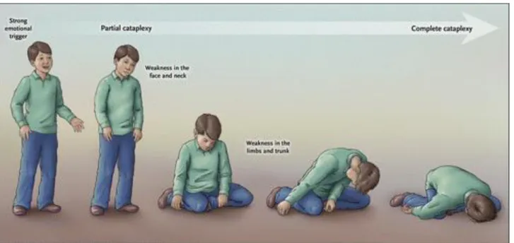
Extended sleep time and weight gain with rapid increase in body mass index (BMI) at disease onset are often noted in adult and even more in children patients (Rocca et al. 2015; Pizza et al. 2013; Dahmen et al. 2011), while the association with precocious puberty can obviously occur only in children especially with very early disease onset (Plazzi et al. 2006; Poli et al. 2013). Cataplexy in adults is characterized by paroxysmal episodes of loss of muscle tone triggered by strong, mainly positive, emotions. Typical cataplectic triggers are indeed laughing, sharp remarks, telling or listening to jokes, being tickled or tickling others, surprise, chuckling, feeling angry, stressed, or
8
embarrassed/ashamed (Overeem et al. 2011). The cataplexy childhood phenotype, especially close to the disease onset, often consists in a complex movement disorder with persistent hypotonia and prominent orofacial involvement (jaw opening, eyelid drooping, head rolling, or tongue thrusting movements), also called “cataplectic facies” even in the absence of emotional triggers (Rocca et al. 2015, Pizza et al. 2013).
Figure 1: cataplexy features in NT1 patient (Scammel 2015).
Cognitive dysfunction includes difficulty focusing, thinking, and concentrating along with automatic behaviors (U.S. Food and Drug Administration 2013; Maski et al. 2017). Hyperactivity, aggressive behavior, problems interacting with peers, and psychopathological features may be additional clinical features very relevant in children affected by NT1 (Filardi et al. 2017). Moreover, nocturnal behavioral and movement disorders have been recently described as additional features in pediatric NT1 patients (Antelmi et al. 2017).
Narcolepsy in children represents a diagnostic challenge because of its variability in the presenting signs, and for the scarce awareness of the disease among physicians (Wise & Lynch, 2001). Associated symptoms such as cataplectic attacks may be confused with atonic seizures, especially in children under 5 years old (Guilleminault & Pelayo,
2000
), or even with neuromuscular diseases (Plazzi et al. 2011). However, the preservation of consciousness despite EDS and the link to emotions remain the distinctive features of cataplexy. Sleep paralysis can be accompanied by hypnagogic hallucinations that consist in vivid visual or auditory experiences usually of benign content. Nevertheless, both these signs can be unrecognized or misinterpreted by young children or their parents (Yoss & Dali 1960).9
Narcolepsy diagnosis is based on the combination of clinical assessment and objective confirmation by means of neurophysiological and biological examinations. The neurophysiological disease hallmark is the early appearance of REM sleep at sleep onset, named sleep onset REM period (SOREMP), a finding described in 1960 by Vogel (1960), that rapidly became the distinctive disease fingerprint along with the standardization of the Multiple Sleep Latency Test as in-laboratory diagnostic procedure to characterize EDS disorders in sleep laboratories worldwide (Littner et al. 2005). However, the discovery of the pathogenic relevance of hypocretin-1 as unique biological disease marker coupled with the increasing availability of cerebrospinal hypocretin-1 assay was recently integrated in the latest diagnostic requirements in the International Classification of Sleep Disorders, third edition (ICSD-3) (Darien 2014). Indeed, diagnostic criteria for narcolepsy now require the combination of different symptoms and objective neurophysiological or biological evidence; the ICSD-3 (Darien 2014) provides the diagnostic criteria that must be met for NT1 disease, i.e. the patient has EDS occurring for at least three months, and the presence of one or both of the following:
1. Cataplexy, and a mean sleep latency of ≤ 8 minutes and two or more SOREMPs on a multiple sleep latency test (MSLT). A SOREMP (within 15 minutes of sleep onset) on the preceding nocturnal polysomnogram may replace one of the SOREMPs on the MSLT.
2. CSF hypocretin-1 concentration, measured by immunoreactivity (RIA assay), is either ≤ 110 pg/mL or <1/3 of mean values obtained in normal subjects with the same standardized assay.
1.5 THE HYPOCRETIN SYSTEM IN NT1
The hypocretins are two peptides, hypocretin 1 (also named A) and hypocretin 2 (or orexin-B), generated from a single preprohypocretin peptide synthesized by a small number of neurons restricted to the lateral hypothalamus and perifornical brain area (de Lecea, 2012; Sakurai et al. 1998). In contrast, hypocretin axons are found throughout the central nervous system (CNS), with innervation of the hypothalamus, locus coeruleus, raphe, midline thalamus, all levels of spinal cord, sympathetic and parasympathetic centres, and many other brain regions (Peyron et al. 1998; van den Pol 1999). Two G protein–coupled receptors that respond to the hypocretins have been identified (Sakurai et al. 1998). In parallel to the wide distribution of axons, the two-hypocretin receptors show a widespread and heterogeneous pattern of expression throughout the CNS (Trivedi et al. 1998). Hypocretin raises synaptic activity in neurons of the hypothalamus (de Lecea et al. 1998) and locus coeruleus (Hagan et al. 1999; Horvath et al. 1999) and can act on postsynaptic receptors to increase cytosolic calcium or at presynaptic receptors on axon terminals to enhance release of glutamate and
10
GABA (van den Pol et al. 1998). The fact that hypocretin can enhance activity of either excitatory or inhibitory neurons suggests that the peptide could ultimately increase or decrease the activity of innervated brain circuits (van den Pol 2000).
The potential importance of hypocretin neurons in preventing NT1 was first suggested by the finding that narcoleptic dogs have mutations on the hypocretin receptors 2 (Lin et al. 1999). Even though different mutations were found in each of two narcoleptic breeds of dogs, in each breed the mutation was localized to the hypocretin receptor 2, rendering it non-functional. Parallel studies in hypocretin knockout mice revealed a similar narcoleptic phenotype (Chemelli et al. 1999), indicating that loss of either the peptide or one of the two peptide receptors results in narcolepsy in animal model (van den Pol 2000).
Following studies based on immunocytochemistry and in situ hybridization indicate that there is a substantially decreased number of neurons producing hypocretin in the hypothalamus of human NT1 patients (Peyron et al. 2000; Thannickal et al. 2000). Thannickal et al. show that the number of hypocretin neurons is decreased by about 90% in four narcoleptic human brains. In parallel, Peyron et al. show an absence of hypocretin mRNA from the hypothalamus, and the loss of hypocretin 1 and 2 peptides from extrahypothalamic hypocretin target regions of six narcoleptic brains. Both studies of narcoleptic brains report no decreased number of neurons containing melanin-concentrating hormone, a neuroactive peptide found in cells in the same hypothalamic region of hypocretin-producing neurons. This finding suggests a very selective absence of hypocretin neurons, rather than a general loss of neurons from the hypothalamus (van den Pol 2000) led to the hypothesis that narcolepsy is an autoimmune driven process within the hypothalamus.
1.6
ENVIRONMENTAL
FACTORS
AND
GENETIC
EVIDENCES
SUPPORTING THE AUTOIMMUNE HYPOTESIS OF NT1
Autoimmune disease result from the interaction between environmental factors and a susceptible genetic background. It has become increasingly clear that environmental factors play an important role in the development and progression of a variety of autoimmune conditions (Molina & Shoenfeld, 2005). Currently the most important environmental factor for NT1 development is the influenza A H1N1 virus. Following the 2009-2010 campaign of pandemic H1N1 flu vaccination using Pandemrix, an AS03 (squalene, alpha-tocopherol) H1N1 vaccine developed in response to the 2009 H1N1 influenza pandemic, a significant six to nine fold increase in the risk of developing NT1 was reported in northern of Europe (Partinen et al. 2012), and a peak in the incidence of H1N1 infection in late 2009 was followed by a threefold increase in NT1 incidence in China (Han et al. 2011). A
11
phenomenon that was followed by a decrease in cases in the later years (Han et al. 2013). Intriguingly, the vast majority (>95%) of patients diagnosed with NT1 following the 2009 pandemic were not vaccinate for H1N1, indicating that naturally occurring influenza-A infections may increase the susceptibility of developing this disorder. Further reports discovered an association in Sweden, Finland, Ireland and in the United Kingdom subsequent the AS03-adjuvanted vaccine and an increased risk of developing NT1 especially in individuals between 5 and 19 years of age (Nohynek et al. 2012; Mahlios et al. 2013). On the other hand, there have been no reported increases in the incidence of NT1 in Italy, or the Netherlands, though the reason behind this phenomenon remains unclear (Wijnans et al. 2012; Mahlios et al. 2013). There is also evidence of an association with other common upper airway infections such as beta haemolytic streptococcal infections (Aran et al. 2009; Longstreth 2007), and lastly, the incidence rate of NT1 onset significant increase in the months following winter-related infections with a seasonal patterns (Han et al. 2011). Streptococcal infections have also been associated with several neurological autoimmune disorders, suggesting that certain immunological responses to infection may result in specific neurological damage (Mahlios et al. 2013; Snider & Swedo, 2003). Moreover, further epidemiological studies have revealed that individuals infected with streptococcus are more than five times as likely to develop NT1 (Koepsell et al. 2010).
The first suggestion, before any epidemiological data were reported, that NT1 might be an autoimmune disorder, came from HLA typing studies. In humans, more than 20 polymorphic HLA genes encode multiple subtypes of Major Histocompatibility Complex (MHC) class I and II proteins that present foreign peptides to T-cell during infections, triggering immune responses (Kornum et al. 2011). NT1 has the strongest HLA associations known, with more than 99% of patients carrying HLA DQB1*06:02. (Mignot et al. 2001). Among HLA class II genes, the DRB1*15:01-DQA1*01:02-DQB1*06:02 is the disease-associated haplotype in NT1. In European populations, the three alleles of this haplotype are in complete linkage disequilibrium, so that any single allele is representative of the haplotype (Mignot et al. 2001; Hor et al. 2010; Tafti et al. 2014). Studies including molecular genotyping of HLA class I genes (A, B, and C) in large populations with NT1 are lacking. However, a recent study used molecular HLA class I typing in 304 patients and 304 controls and imputed class I genotypes from previous genome-wide (GW) association studies in White and Chinese populations. This study found, in addition to DPB1, significant associations between narcolepsy and HLA-A*11:01, B*35:03, and B*51:01 (Ollila et al. 2015). HLA class II molecules are constitutively expressed only by antigen-presenting cells such as dendritic cells, macrophages, or B cells, while HLA class I molecules are ubiquitously expressed by nearly all nucleated cells.
12
Unique to NT1 is the genetic association with a polymorphism in the T cell receptor alpha (TCRα) gene, which encodes the alpha chain of the αβ-heterodimer of the T cell receptor on both CD4+ and cytotoxic CD8+ T cells, which can bind to both MHC-I and MHC-II molecules (Hallmayer et al.
2009). The TCR consist of α and β chain and similarly to the immunoglobulin loci, DNA recombination occurs within the TCR loci to create an enormous diversity in the receptors expressed and leads to generation of a unique repertoire of TCR bearing T-cells maybe with autoreactive potential (Krangel 2009; Kornum et al. 2011).
Furthermore, a genetic association of NT1 with purinergic receptor subtype 2Y11 (P2RY11) was discovered, and since this receptor is highly expressed on cytotoxic lymphocytes suggests an involvement of immune responses in aetiology of the disease (Kornum et al. 2011). Another different mutation in the DNA Methyltransferase 1 (DNMT1) gene led to narcolepsy with or without cataplexy in the context of two different autosomal dominant progressive neurodegenerative disorders, named hereditary sensory and autonomic neuropathy with dementia and hearing loss type IE (HSAN-IE), and autosomal dominant cerebellar ataxia, deafness and narcolepsy (ADCA-DN) (Winkelmann et al. 2012; Moghadam et al. 2014).
As DNMT1 is located in extremely close proximity to P2RY11 (within 30 kb), it is possible that the genetic association in sporadic cases and the rare autosomal dominant ADCA-NC/HSAN-IE disease could be linked somehow (Singh et al. 2013).
Complementing this observation DNMT1 is highly expressed in immune system cells, and hypo methylation has been shown to be involved in other autoimmune diseases such as lupus (Singh et al. 2013). DNMT1 also plays an important role in differentiation of CD4+ T cells into T regulatory cells
by relieving repression of Foxp3 expression following TCR stimulation (Josefowicz et al. 2009). A possibility would be that an absence of T regulatory cells with specificity toward hypocretin neurons could result in autoimmunity.
In one more study, Faraco et al. (2013) used the Immunochip, an SNP chip designed to further fine map various autoimmune disease loci, and found that variants in two additional loci, Cathepsin H and Tumor necrosis factor super family member 4 (TNFSF4, also called OX40L), attained genome-wide significance (Faraco et al. 2013). As these genes are important for antigen processing and downstream T cell stimulatory effects, these findings further implicate the process of antigen presentation by HLA Class II APCs to T cells in the context of NT1 (Singh et al. 2013).
13
The ultimate proof for autoimmunity would be the demonstration of T cell reactivity or autoantibodies directed towards hypocretin-producing neurons. Despite wide research, findings have been either controversial or negative. Post-mortem studies have only been conducted using brains from the late stages of NT1. While one study found an upregulation of glial fibrillary acidic protein (GFAP), a marker of astrogliosis, another study did not find any signs of inflammation (Peyron et al. 2000; Thannickal et al. 2003). Older studies have typically searched for a serum or CSF autoantibodies, but these studies have only found the examined autoantibodies in a minority of patients (Overeem et al. 2008). Moreover, reports of CSF and serum samples from patients sampled closer to the time of diagnosis have also shown varying results, possibly because of the latency between symptoms onset and clinical diagnosis (Degn and Kornum 2015; Kornum et al. 2015). For example, two studies have reported changed levels of the main subclasses of total IgG in NT1 and idiopathic hypersomnia (HI) (Tanaka & Honda 2010
)
and increased serum levels of total but not free IgG and IgM antibodies against hypocretin-1 in NT1, NT2 and HI diseases (Deloumeau et al. 2010). In 2010, the presence of autoantibodies against tribble homologues 2 (TRIB2) was detected in 14% of European patients versus 5% of controls (Cvetkovic-Lopes et al. 2010), a finding replicated in other patient populations (Kawashima et al. 2010; Toyoda et al. 2010). However, TRIB2 is expressed not only by hypocretin-producing neurons, but also present in many other cell populations, both in CNS and in the periphery, including immune cells (Kornum et al. 2011; Sung et al. 2006; Eder et al. 2008). Thus, TRIB2 autoantibodies are unlikely to be causative of the specific neuronal destruction; they could be reflective of a downstream effect that develops secondary to the neuronal destruction or they could be present before the disease in some individuals, causing higher risk of developing NT1 (Degn & Kornum 2015; Kornum et al. 2011). Another study discovered brain-specific autoantibodies in serum from NT1 patients (Bergman et al. 2014) do not target hypocretin neurons at all. Curiously, these autoantibodies were also detected in the serum of patients with other sleep disorders, and even in some healthy controls (Bergman et al. 2014). Finally, recent report investigated the presence of antibodies to the hypocretin receptor-2 in NT1 and NT2 patients, the results showed only 5% of patients positive for hypocretin receptor-2 autoantibodies with negative associations concluding that antibodies are not common in NT1 (Giannoccaro et al. 2017).This development in NT1 research be like that of Rasmussen’s encephalitis, an MHC class I-restricted CD8+ mediated epilepsy syndrome. Initial research on Rasmussen’s encephalitis was dominated by the detection of autoantibodies, with the identification of glutamate receptor 3 antibodies in a few patients (Wiendl et al. 2001) and later of α7 nicotinic acetylcholine receptors and Munc18-1 autoantibodies (Degn & Kornum 2015; Watson et al. 2011; Alvarez-Barón et al. 2008). Nevertheless, plasmapheresis was only effective in a few patients, and, similarly, the autoantibodies were only
14
identified in serum from a low fraction of patients (Varadkar et al. 2014). In neuromyelitis optica, antibodies against aquaporin 4 are identified in 60-80% of patients, and these respond well to intravenous immunoglobulin (IvIg) therapy (Jarius et al. 2014), suggesting a pathogenic role of aquaporin 4 autoantibodies. Because none of the NT1 related autoantibodies have been found in more than a small percentage of the patients, and since the clinical response to IvIg treatment is unpredictable at best (Knudsen et al. 2012), the role of CNS autoantibodies in NT1 disease remain unclear. Autoantibodies might be produced as part of secondary immune response to the initial neuronal damage in NT1, or they might increase the risk of developing NT1 by a yet unknown mechanism. As in the case of Rasmussen’s encephalitis, NT1 is more likely caused by T cell, which is supported by the fact that other disorders involving a loss of neurons have also been shown to involve autoimmune T cells (Degn & Kornum 2015).
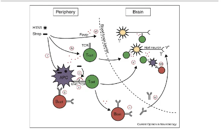
Figure 2: possible pathway for a role of influenza or streptococcus infections in the development of NT1. A H1N1 influenza or streptococcus pyogenes infection could stimulate autoreactive T cells or B cells via several possible mechanisms. Selected resting autoreactive T cells and B cells may have reactivity towards hypocretin-producing cells, having escaped negative selection. These could be activated in the following ways: (i) Molecular mimicry, T cells. Antigens from the virus or bacteria are presented by, for example, MHC DQA1*01:02-DQB1*06:02 on an antigen presenting cell (APC). A T cells recognized the antigen and is activated. The same T cells migrates into the brain were it
15
recognized a hypocretin-cell specific antigen (cross-reactivity) inducing autoimmune process. (ii) Molecular mimicry, B cells. An autoreactive B cell can be triggered if it also recognizes an antigen from the microorganism. This process requires signals from activated T cell (T helper). (iii) Superantigens from streptococcus cross-link the MHC and TCR molecules independent of specific antigen specificity activating the autoreactive T cells. (iv) Bystander activation. Resting autoreactive cells are activated because of general immune activation independent of specific antigen. (v) Lymphocyte migration to CNS. Once activated, the T cells can migrate to the brain, depending on the type of T cell a variety of mechanisms could account for the autoimmune attack. (vi) Opening of the blood brain barrier. The immune response to infection also normally includes fever and other factors that can do the blood brain barrier more penetrant to lymphocytes and will allow antibodies to access to CNS. (vii) Production of autoantibodies can also occur as a secondary response to hypocretin producing-cell death via antigen presenting cells from the brain that have phagocytosed the dead neurons (Kornum et al. 2011).
1.8 T CELL MEDIATED IMMUNITY
T cell mediated immunity is an adaptive process that eliminates viral, bacterial, parasitic, or malignant cells by producing antigen-specific T lymphocytes. This type of immunity can also lead to the development of autoimmune disorders when the wrong recognition of a self-antigen occurs. The specificity of T lymphocytes is based on the recognition of antigenic peptides via the T cell receptor (TCR) receptor. Each T cell expresses on its surface a unique TCR because of the recombination processes that sort these highly variable receptors and the selection that occurs during the maturation of the lymphocytes in thymus. The pool of mature T cells leaving the thymus therefore has a vast repertoire of specific antigen receptors (Broere et al., 2011). The thymus selection process leads to the appearance of T cells with two types of TCRs. Most of them express a TCR formed by α and β chains. Lymphocytes carrying this TCR can be subdivided into several subgroups based on surface markers and different functional properties. Only a small fraction of T cells leaving thymus and expressing a TCR formed by the γ and δ chains, these lymphocytes appear much less heterogeneous than those with a TCR αβ and reside in the skin and in certain mucous surfaces, appear to have a role in the initial response to microbial invasions.
The mature T cells that leave the thymus, also known as naïve T cell (which have not yet encountered the antigen), circulate through the blood and the lymphatic system in order to concentrate in the secondary lymphoid organs. In these secondary lymphoid organs, namely: spleen, lymph nodes, and lymphoid tissues associated with mucous membranes (such as tonsils) that initiate the immune response mediated by T cells. These naïve cells reside in the spleen for a few hours and in the lymph
16
nodes for about a day, before leaving the latter through the splenic vein or efferent lymph vessels to reach the bloodstream again. Through the circulation, naïve T cells reach a new lymphoid organ, repeating this cycle until they will be activated by the encounter of the antigen or will die. Inside secondary lymphoid organs, antigenic peptides are presented to naïve T cells by antigen presenting cells (APCs). In particular, DCs (Dendritic Cells) are the most efficient APC cells as they provide T lymphocytes also costimulatory signals necessary for their activation. DCs capture antigens in the non-lymphoid tissues of the body, and then migrate into the secondary lymphoid organs. These cells processed the antigen to provide antigenic peptides exposed on the cell surface in the context of the MHC complex (Broere et al. 2011). The MHC complex is a set of surface proteins essential for the immune system, whose main function is to bind antigens derived from pathogenic agents and expose them to the cell surface to allow recognition by appropriate T cells. There are two main classes of MHC molecules: the MHC class I, which is present on all the nucleated cells; and MHC class II that is expressed only by "professional" APC cells, such as B cells, macrophages, and dendritic cells. Naïve T cells recognize the antigen presented by MHC through TCR binding. CD4+ T cells recognize the antigen in the context of class II MHC molecules, while T CD8+ lymphocytes bind antigenic peptides presented by class I MHC molecules. Recruitment of TCR triggers an intracellular signalling cascade leading to naïve T cell activation. Activated T cell proliferates rapidly (i.e. clonal expansion) and migrates through the tissues to the inflamed site for exert their functions. In response to antigen stimulation, CD4+ T cells differentiate into T helper (Th)-1 or Th2 cells. Th1 cells produce signature
cytokine INF-γ, whereas Th2 cells produce IL-4, IL-5, IL-13, and IL-10. CD4+ T helper lymphocytes
produce cytokines that can stimulate the production of antibodies by B cells and the actions of other T lymphocytes. They are essential in the activation and growth of cytotoxic T cells, and in maximizing bactericidal activity of phagocytes such as macrophages.
Cytotoxic CD8+ T cells are very effective in destroying directly infected or malignant cells that present the antigen. Virtually, every cell in the body expresses the class I MHC and is therefore able to present antigenic peptides thus becoming a potential target of cytotoxic CD8+ T cells. These CD8+ T cells are able to destroy more than onetarget cell, while saving the adjacent healthy cells. Two are the major cytotoxicity mechanisms activated by CD8+ T cells: the release of lithic granules, and apoptosis induced by the Fas receptor, both triggered by antigen recognition via the TCR. Lithic granules are secreted lysosomes containing granzyme, and perforin. Granzymes are inducing apoptosis molecules on target cells. The Fas receptor (also known as CD95) is part of Tumor Necrosis Factor (TFN) superfamily; it is expressed on target cells and has an intracellular death domain capable of inducing caspase-dependent apoptosis when the receptor binds its ligand. Recruiting TCR
17
increases expression of Fas ligand (also called CD95L) on the surface of cytotoxic cells, and the binding between CD95 and CD95L induces the death of target cells. The cytotoxic T CD8+
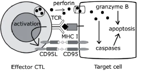
lymphocytes do not require costimulatory signals in addition to antigen recognition to perform their effector function, and consequently they can destroy a wide variety of target cells that exhibit exogenous antigens (Broere et al. 2011). Possible mechanisms of CD8+ T cell mediated cytotoxicity are shown in Figure 3.
Figure 3. Mechanisms that mediate the cytotoxicity of T CD8+ lymphocytes: 1) Secrete of lithic
granules containing perforin and granzyme B. Perforin creates pores on the target cell membrane allowing granzyme B to activate caspases inducing cell apoptosis. 2) Interaction between CD95 (Fas) and CD95L (Fas ligand). Activation of T cell through TCR induces expression of CD95L binding CD95 to target cells causing death (Broere et al. 2011).
Most effector T cells disappear once the antigenic agent is eliminated, but a small portion of them remain and form the pool of memory T cells. Contrasting the naïve cells that live for a few months, and the effector cells that disappear after the pathogen's elimination, memory T cells can survive for years in the lymphoid organs and peripheral tissues. These lymphocytes can be easily activated and immediately acquire effective functions in peripheral tissues, while encountering clonal expansion in the lymphoid organs. This provides a rapid immune response (secondary response) when an antigen to which the organism has already been exposed appears again. Memory cells respond much more rapidly to the antigen than T naïve cells, so in the case of infection they help to eliminate the pathogen at early stages by preventing the spread of the disease (Broere et al. 2011).
1.9 THE CELLULAR BASIS OF IMMUNOLOGICAL MEMORY - CENTRAL
MEMORY AND EFFECTOR MEMORY T CELL SUBSETS
18
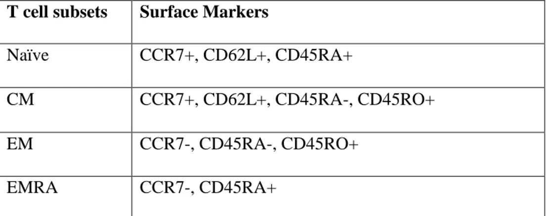
Following antigen encounter and subsequent resolution of the immune response, a single naïve (N) T cell is able to generate multiple subsets of memory T cells with different phenotypic and functional properties and gene expression profiles. Single-cell technologies, first flow cytometry, have revealed the complex heterogeneity of the memory T cell compartment and its organization into subsets. However, a consensus has yet to be reached, regard nomenclature and phenotypic level, on the identification of these T cell subsets. Recent studies indicate that memory T lymphocytes contain distinct populations of central memory (CM) and effector memory (EM) cells characterized by distinct homing capacity and effector functions. After positive and negative selection T cells are released from the thymus as mature, N T cells harbouring a given epitope specificity. In response to antigen (Ag) encounter, N T cells proliferate and differentiate into effector cells, the vast majority of which migrate to peripheral tissues and inflamed sites to facilitate destruction of infected targets (Zhang & Bevan, 2011). Following Ag clearance, for example that in smallpox vaccination, more than 95% of the effector cells die while a small pool of T cells ultimately develops into long-lived memory T cells (Hammarlund et al. 2003; Mahnke et al. 2013). Single cell technologies, like flow cytometry; have revealed the complex heterogeneity of the T cell compartment and its organization into subsets. Together, monoclonal antibodies and flow cytometry have allowed the phenotypic examination of single cells level within heterogeneous cellular population, and the identification and isolation of certain cell types from biological fluid or tissue. CM- and EM-T cells were initially defined in the human system based on two criteria: 1) the absence or presence of immediate effector function and, 2) the expression of homing receptors that allow cells to migrate to secondary lymphoid organs versus non-lymphoid tissues (Sallusto et al. 1999; Sallusto et al. 2004). Human T CM cells are CD45R0+ memory cells that constitutively express CCR7 and CD62L, two receptors also found of N T cells, required for cells extravasation through endothelial tissue and migration to T cell areas of secondary lymphoid organs (Campbell et al. 1998; Förster et al. 1999). CD45R0 and CD45RA correspond to the high and low molecular weight protein products of splice variants of CD45 gene, respectively (Terry et al. 1988; Mahnke et al. 2013). When comparing to N T cells, CM cells have higher sensitivity to antigenic simulation, are less dependent to co-stimulation and upregulate CD40L (a protein primarily expressed on activated T cells) providing a more effective stimulatory feedback to dendritic cells (DC) and B cells. Following TCR triggering, CM T cells produce mainly Interleukin (IL)-2, but after proliferation, they quickly differentiate to effector cells and produce large amounts of Interferon-γ (INF-γ) or IL-4 (Sallusto et al. 2004). Human EM T cells are memory cells that have lost the constitutive expression of CCR7, are heterogeneous for CD62L expression, and display characteristic sets of chemokine receptors and adhesion molecules that are required for homing to inflamed tissues (see Table 1). EM T cells are characterized by rapid effector functions (Lanzavecchia
19
& Sallusto, 2000; Sallusto et al. 2004). CD8+ T EM cells carry the larger amount of perforin, and
both CD4+ and CD8+ produce INF-γ, IL-4, and IL-5 within hours following antigenic stimulation.
Some CD8+ and with less frequency, CD4+ EM cells express CD45RA and are defined as terminally
differentiate EM (EMRA) cells. EMRA T cells have mostly been studied in the CD8+ T cell compartment, where they are found at appreciable frequencies in most individuals (Tian et al. 2017). By contrast, the frequency of CD4+ EMRA T cells varies drastically between individuals ranging from <0.3% to nearly 18% of total CD4+ T cells in an apparently healthy population, and their functional role is less clear (Tian et al. 2017). In addition to exhibiting CD45RA+ and CCR7- phenotype, CD4+ EMRA T cells have also been characterized by decreased expression of CD27 and CD28, as well as increased expression of CD57 and effector molecules such as perforin and granzyme B that resemble more terminally differentiated state (Tian et al. 2017). In addition, the CD8+ EMRA T cells carry the largest amount of perforin.
Therefore, the EM T cell pool contains T helper 1, T helper 2, and cytotoxic T lymphocytes (CTL). The relative proportions of CM T cells and EM T cells in peripheral blood vary in the CD4+ and CD8+ compartments; CM cells are more prevalent in CD4 T, whereas EM cells are more frequent in CD8+ lymphocytes. Within the tissues however the different T cell subsets shown characteristic distribution patterns (Sallusto et al. 2004). CM T cells are enriched in lymph nodes and tonsils, while lung, liver, and gut contain greater proportion of EM T cells (Campbell et al. 2001). In antigen-primed individuals, tetanus toxoid specific CD4+ T cells can be detected in circulating CM and EM T cells
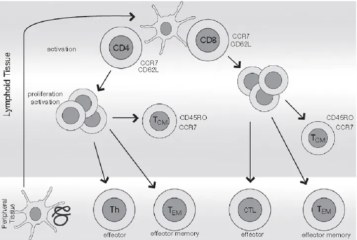
up to 10 years after anti-immunization (Sallusto et al. 1999). The development of T cell memory subsets is shown in Figure 4.
T cell subsets Surface Markers
Naïve CCR7+, CD62L+, CD45RA+
CM CCR7+, CD62L+, CD45RA-, CD45RO+ EM CCR7-, CD45RA-, CD45RO+
EMRA CCR7-, CD45RA+
20
Figure 4.Development of T cell subsets. APC cells capture antigens in peripheral tissues and migrate to secondary lymphoid organs, where N T cells are activated by the recognition of the MHC-peptide complex. They proliferate and differentiate themselves into effective cells or memory cells. Both effective CD4+ cells (Th, T helper) and CD8+ efficacious (CTL, cytotoxic) migrate to peripheral tissues to perform their function. The memory cells themselves are subdivided into CM T cells expressing CCR7 and recirculating between lymph nodes, or in EM T cells that do not express CCR7 and migrate to peripheral tissues (Broere et al. 2011).
1.10 NATURAL KILLER CELL MEDIATED IMMUNITY
Natural Killer (NK) cells are effector lymphocytes of the innate immune system that control several types of tumours and microbial infections by limiting their spread and subsequent tissue damage. Recent studies highlights the fact that NK cells are also regulatory cells engaged in reciprocal interactions with DCs, macrophages, T cells, and endothelial cells. NK cells were recognized as a separate lymphocyte lineage, with both cytotoxicity and cytokine-producing effector functions (Trinchieri, 1989). In fact, NK cells can limit or exacerbate immune responses; they can regulate the immune response by killing APCs or over-activated T cells or by producing anti-inflammatory cytokines (such as IL-10) to prevent too strong inflammatory response (Vivier et al. 2011; Zhang et al. 2006). Nevertheless, the role of NK cells is not helpful in all situations. Over activation or
21
dysfunction of NK cells may be associated with pathogenesis of some diseases. For example, NK cells are found to act as a two edged weapon and play opposite roles with both regulatory and inducer activity in autoimmune diseases (Perricone et al. 2008; Zhang & Tian, 2017). NK cells are heterogeneous cell population and divided into different subsets based on their surface phenotype or cytokine secretion pattern. So far, various human NK subsets with distinct phenotypic and functional properties have been identified and they usually represent distinct stages of a linear development process. Furthermore, different NK cells are found to locate in several immune organs and anatomy sites, demonstrating that the microenvironment influences the differentiation and function of NK subsets (Zhang & Tian, 2017).
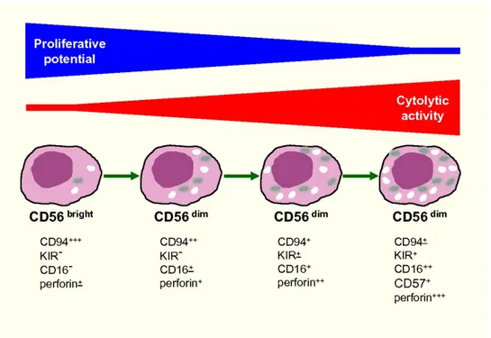
It is widely accepted that human NK cells can be divided into two subsets (showed in Figure 5) based on their cell-surface density of CD56: CD56dim NK and CD56bright NK cells (Cooper 2001; Cooper et al. 2001). In peripheral blood lymphocyte populations, approximately 10% of NK cells express high levels of CD56 (CD56bright CD16dim/-), while the more abundant NK cell subset expressed CD56 at low density (CD56dim CD16+). CD56dim CD16+ NK cells exert higher cytotoxicity and express higher levels of Ig-like NK receptors than CD56bright NK cell subset, yet CD56bright NK cells exert relatively lower cytotoxic capacity but can produce abundant amounts of cytokines such as INF-γ, TNF-α, IL-10, IL-13, and granulocyte-macrophage colony-stimulating factor (GM-CSF). The CD56dim CD16+ NK subset is predominant in peripheral blood, whereas CD56bright CD16dim/- NK cells are more abundant in secondary lymphoid tissues, particularly rich in immune tolerance organs, such as the liver and uterus (Cooper et al. 2001; Zhang & Tian, 2017). The distribution of NK cell subpopulations in the various tissues is determined by chemokine receptors on NK cells and chemokines secreted in sites of tissues. CD56bright NK cells express CCR7, which promotes entry into secondary lymphoid
organs and contributes to migration toward sites of infection or inflammation. CD56bright NK cells also express high levels of CD62L a receptor required for lymphocytes homing to secondary lymphoid organs.
CD56bright NK cells are a regulatory NK subset and play major roles in maintaining immune homeostasis in both physiological and pathological conditions by secreting immune-regulatory cytokines. However, they can also become cytotoxic upon appropriate activation through inflammatory cytokines or triggering of co-activating receptors, thus can mediate killing of target immune cells such as autologous activated T cells or immature DCs for regulation of immune response. CD56dim NK cells are cellular cytotoxic killer cells and exert major early immune-surveillance against infected or malignant cells. They are also potent producers of cytokines (INF-γ and TNF-α) upon recognition of susceptible target cells (Zhang & Tian, 2017).
22
Figure 5. Model of human NK-cell differentiation (Moretta 2010).
NK-T cells are another subset of NK cells and they are a population of autoreactive cells that mediate both protective and regulatory immune functions. NK-T cells express an invariant TCRα chain (Vα24-JαQ in humans) and recognize glycolipid antigens in association with the MHC class I-like molecules CD1d.
NK-T cells are different from functionally differentiated conventional αβ T cells in that they are autoreactive and produce both Th1 and Th2 cytokines, including IL-4, IL-10 and INF-γ, upon stimulation with their ligands (Taniguchi et al. 2003; Seino & Taniguchi, 2005). NK-T cells appear to have distinct functional capabilities; they have been shown to mediate both protective and regulatory immune functions. These includes tumour rejection, protection against infectious microbes, maintenance of transplant tolerance, and inhibition of autoimmune disease development (Taniguchi et al. 2003). NK-T cell functions and or cytokine profiles can be altered by modulation of TCR signalling. Research reported that the frequency of NK-T cell stimulation through the TCR affects their function (Kojo et al. 2005). Suggesting that the intensity of TCR signals influences the action of NK-T cells in a manner similar to the effect of TCR avidity on positive and negative selection of developing T cells in the thymus (Starr et al. 2003; Seino & Taniguchi, 2005).
1.11 IMMUNITY AND AUTOIMMUNITY IN CENTRAL NERVOUS SYSTEM
The CNS was considered an immunologically privileged compartment and protected from autoimmune process by both the blood-brain barrier (BBB) and by the lack of MHC class I expression
23
on neurons. Actually, this is known to not be the case. Research has shown that memory T cells cross the BBB and monitor the CNS by interacting with antigen-presenting DCs in the perivascular and subarachnoid space where they can be reactivated (Ransohoff & Engelhardt, 2012; Degn & Kornum, 2015). The fifth lumbar of the spinal cord has been suggested as a site of entrance; T cells can also enter the CNS through the choroid plexus (Ransohoff & Engelhardt, 2012).
The surveillance of the CNS by T cells is crucial for protecting the brain from infections; inhibition of T cells entry in humans can lead to progressive multifocal leukoencephalopathy caused by an uncontrolled infection by the Cunningham (JC) virus, which is asymptomatic in immunocompetent individuals. In general, virus specific T cells play a central role in the control of many neurotropic viruses, such as West Nile virus and herpes simplex virus type 1. For example, in mice and humans, herpes simplex virus enters in a lifelong latent state within infected sensory neurons. Activated CD8+ T cells found in direct contact with infected neurons keep the virus in a latent state without causing the cells lysis (Degn & Kornum, 2015).
Another CNS disease, the paediatric autoimmune neuropsychiatric disorders associated with streptococcal infections, also known as “PANDAS,” is associated with obsessive-compulsive and tic disorders. The streptococcal infections may trigger an autoimmune reaction that exacerbates these conditions. Recurrent streptococcal tonsillitis is one of the frequent infections associated with PANDAS condition (Heubi & Shott, 2003).In the past, other neurological disorders including narcolepsy-like symptoms have been associated with H1N1 infections, most notably in the contest of the 1918 Spanish flu pandemic. Coincident with this epidemic a smaller one, called Encephalitis Lethargica (EL), a disease characterized by hypersomnolence and posterior hypothalamic lesions, occurred (von Economo 1931; Kornum et al. 2011). Although EL has largely disappeared, cases are still occasionally reported, and interestingly were found to be associated with higher ASO titers (Lopez-Alberola, 2009; Dale et al. 2001). Furthermore, autoimmune T cell responses directed against antigens that are derived from the CNS are thought to trigger several diseases, including multiple sclerosis, neuromyelitis optica and acute disseminated encephalomyelitis. Multiple sclerosis is thought to occur in genetically predisposed individuals following exposure to an environmental trigger that activates myelin- specific T cells, which allows the T cells to cross the BBB (Goverman, 2009). Reactivation of the T cells by CNS-resident APCs that present myelin antigens triggers the recruitment of innate immune cells, which have important roles in mediating demyelination and axonal damage (Goverman, 2009). Finally, relative recent studies suggest that psychiatric disorders (such as major depressive disorder, bipolar disorder and schizophrenia) could be considered as inflammatory conditions of CNS via microglial activation (Réus et al. 2015).
24
1.12 AIMS
Researches have investigated immune cell phenotype subsets in various CNS autoimmune diseases. In multiple sclerosis, patients have decreased frequency of EM and EMRA in CD4+ and CD8+ T cells, a finding present at onset and that persists throughout the clinical course (Pender et al. 2014; Teniente-Serra et al. 2016). Another larger study found a higher percentage of EM T cells in various CNS inflammatory syndromes in comparison to controls (Mullen et al. 2012).
In this study, we used flow cytometry to analyze the distribution of CD4+ T cells, CD8+ T cells, and NK cells in the peripheral blood and, for the first time, in the CSF of NT1 patients compared to age and gender balanced healthy controls, differing for the presence of HLA DQB1*06:02 allele. Finally, we analyzed T cell subsets differences between CSF and peripheral blood in NT1 patients versus controls.
25
1.13 REFERENCES
Abad V, Guilleminault, Ch. [Sleep and psychiatry]. Zh Nevrol Psikhiatr Im S S Korsakova, (2009) 109:102-8.
Alvarez-Barón E, Bien CG, Schramm J, Elger CE, Becker AJ, Schoch S. Autoantibodies to Munc18, cerebral plasma cells and B-lymphocytes in Rasmussen encephalitis. Epilepsy Res. (2008) 80:93-7. doi:10.1016/j.eplepsyres.2008.03.007.
Antelmi E, Pizza F, Vandi S, et al. The spectrum of REM sleep-related episodes in children with type 1 narcolepsy. Brain. (2017) 140:1669-1679.
Aran A, Lin L, Nevsimalova S, et al. Elevated anti-streptococcal antibodies in patients with recent narcolepsy onset. Sleep. (2009) 32:979-83.
Bergman P, Adori C, Vas S, et al. Narcolepsy patients have antibodies that stain distinct cell populations in rat brain and influence sleep patterns. Proc Natl Acad Sci U S A. (2014) 111:E3735-44. doi: 10.1073/pnas.1412189111.
Berry RB, Budhiraja R, Gottlieb DJ, et al. American Academy of Sleep Medicine. Rules for scoring respiratory events in sleep: update of the 2007 AASM Manual for the Scoring of Sleep and Associated Events. Deliberations of the Sleep Apnea Definitions Task Force of the American Academy of Sleep Medicine. J Clin Sleep Med. (2012) 8:597-619.
Blouin AM, Thannickal TC, Worley PF, Baraban JM, Reti IM, Siegel JM. Narp immunostaining of human hypocretin (orexin) neurons: loss in narcolepsy. Neurology. (2005) 65:1189-92.
Bonnavion P, de Lecea L. Hypocretins in the control of sleep and wakefulness. Curr Neurol Neurosci Rep. (2010) 10:174-9. doi: 10.1007/s11910-010-0101-y.
Broere F, Apasov SG, Sitkovsky MV, van Eden W. T cell subsets and T cell-mediated immunity. Chapter A2, Principles of Immunopharmacology: 3rd revised and extended edition. Springer Basel AG, Basel, Switzerland, 2011, pp 15-27.
26
Campbell JJ, Bowman EP, Murphy K, et al. 6-C-kine (SLC), a lymphocyte adhesion-triggering chemokine expressed by high endothelium, is an agonist for the MIP-3beta receptor CCR7. J Cell Biol. (1998) 141:1053-9.
Campbell JJ, Murphy KE, Kunkel EJ, et al. CCR7 expression and memory T cell diversity in humans. J Immunol. (2001) 166:877-84.
Center for Drug Evaluation and Research (CDER), U.S. Food and Drug Administration (FDA). The Voice of the Patient. A series of reports from the US Food and Drug Administration’s (FDA’s) Patient Focused Drug Development Initiative; Narcolepsy Public Meeting; September 24,(2013). http://www.fda.gov/downloads/ForIndustry/UserFees/PrescriptionDrugUserFee/UCM402907.pdf.
Chemelli RM, Willie JT, Sinton CM, et al. Narcolepsy in orexin knockout mice: molecular genetics of sleep regulation. Cell. (1999) 98:437-51.
Cooper MA, Fehniger TA, Caligiuri MA. The biology of human natural killer-cell subsets. Trends Immunol. (2001) 22:633-40.
Cooper MA, Fehniger TA, Turner SC, et al. Human natural killer cells: a unique innate immunoregulatory role for the CD56(bright) subset. Blood. (2001) 97:3146-51.
Crocker A, España RA, Papadopoulou M, et al. Concomitant loss of dynorphin, NARP, and orexin in narcolepsy. Neurology. (2005) 65:1184-8.
Cvetkovic-Lopes V, Bayer L, Dorsaz S, Maret S, Pradervand S, Dauvilliers Y. Elevated Tribbles homolog 2-specific antibody levels in narcolepsy patients. J Clin Invest. (2010) 120:713-9. doi: 10.1172/JCI41366.
Dahmen N, Bierbrauer J, Kasten M. Increased prevalence of obesity in narcoleptic patients and relatives. Eur Arch Psychiatry Clin Neurosci. (2001) 251:85-9.
Dale RC, Church AJ, Cardoso F, et al. Poststreptococcal acute disseminated encephalomyelitis with basal ganglia involvement and auto-reactive antibasal ganglia antibodies. Ann Neurol. (2001) 50:588-95.
27
Darien, IL. American Academy of Sleep Medicine. International Classification of Sleep Disorders, 3rd ed. American Academy of Sleep Medicine. (2014).
Dauvilliers Y, Arnulf I, Mignot E. Narcolepsy with cataplexy. Lancet. (2007) 369:499–511.
Dauvilliers Y, Montplaisir J, Molinari N, et al. Age at onset of narcolepsy in two large populations of patients in France and Quebec. Neurology. (2001) 57:2029-33.
de Lecea L, Kilduff TS, Peyron C, et al. The hypocretins: hypothalamus-specific peptides with neuroexcitatory activity. Proc Natl Acad Sci U S A. (1998) 95:322-7.
de Lecea L. Hypocretins and the neurobiology of sleep-wake mechanisms. Prog Brain Res. (2012) 198:15-24. doi: 10.1016/B978-0-444-59489-1.00003-3.
Degn M, Kornum BR. Type 1 narcolepsy: a CD8(+) T cell-mediated disease? Ann N Y Acad Sci. (2015) 1351:80-8. doi: 10.1111/nyas.12793.
Deloumeau A, Bayard S, Coquerel Q, et al. Increased immune complexes of hypocretin autoantibodies in narcolepsy. PLoS One. (2010) 5:e13320. doi: 10.1371/journal.pone.0013320.
Eder K, Guan H, Sung HY, et al. Tribbles-2 is a novel regulator of inflammatory activation of monocytes. Int Immunol. (2008) 20:1543-50. doi: 10.1093/intimm/dxn116.
Faraco J, Lin L, Kornum BR, et al. ImmunoChip study implicates antigen presentation to T cells in narcolepsy. PLoS Genet. (2013) 9:e1003270. doi: 10.1371/journal.pgen.1003270.
Filardi M, Pizza F, Tonetti L, Antelmi E, Natale V, Plazzi G. Attention impairments and ADHD symptoms in adult narcoleptic patients with and without hypocretin deficiency. PLoS One. (2017) 12:e0182085.
Förster R, Schubel A, Breitfeld D, et al. CCR7 coordinates the primary immune response by establishing functional microenvironments in secondary lymphoid organs. Cell. (1999) 99:23-33.
28
Giannoccaro MP, Waters P, Pizza F, Liguori R, Plazzi G, Vincent A. Antibodies Against Hypocretin Receptor 2 Are Rare in Narcolepsy. Sleep. (2017) 40(2). doi: 10.1093/sleep/zsw056.
Goverman J. Autoimmune T cell responses in the central nervous system. Nat Rev Immunol. (2009) 9:393-407. doi: 10.1038/nri2550.
Guilleminault C, Pelayo R. Narcolepsy in children: a practical guide to its diagnosis, treatment and follow-up. Paediatr Drugs. (2000) 2:1-9.
Hagan JJ, Leslie RA, Patel S, et al. Orexin A activates locus coeruleus cell firing and increases arousal in the rat. Proc Natl Acad Sci U S A. (1999) 96:10911-6.
Hallmayer J, Faraco J, Lin L, et al. Narcolepsy is strongly associated with the T-cell receptor alpha locus. Nat Genet. (2009) 41:708-11. doi: 10.1038/ng.372.
Hammarlund E, Lewis MW, Hansen SG, et al. Duration of antiviral immunity after smallpox vaccination. Nat Med. (2003) 9:1131-7.
Han F, Lin L, Li J, Dong XS, Mignot E. Decreased incidence of childhood narcolepsy 2 years after the 2009 H1N1 winter flu pandemic. Ann Neurol. (2013) 73:560. doi: 10.1002/ana.23799.
Han F, Lin L, Warby SC, et al. Narcolepsy onset is seasonal and increased following the 2009 H1N1 pandemic in China. Ann Neurol. (2011) 70:410-7. doi: 10.1002/ana.22587.
Heubi C, Shott SR. PANDAS: pediatric autoimmune neuropsychiatric disorders associated with streptococcal infections--an uncommon, but important indication for tonsillectomy. Int J Pediatr Otorhinolaryngol. (2003) 67:837-40.
Hor H, Kutalik Z, Dauvilliers Y, et al. Genome-wide association study identifies new HLA class II haplotypes strongly protective against narcolepsy. Nat Genet. (2010) 42:786-9. doi: 10.1038/ng.647.
Horvath TL, Peyron C, Diano S, et al. Hypocretin (orexin) activation and synaptic innervation of the locus coeruleus noradrenergic system. J Comp Neurol. (1999) 415:145-59.
29
Ingravallo F, Gnucci V, Pizza F, et al. The burden of narcolepsy with cataplexy: how disease history and clinical features influence socio-economic outcomes. Sleep Med. (2012) 13:1293-300. doi: 10.1016/j.sleep.2012.08.002.
Jarius S, Wildemann B, Paul F. Neuromyelitis optica: clinical features, immunopathogenesis and treatment. Clin Exp Immunol. (2014) 176:149-64. doi: 10.1111/cei.12271.
Josefowicz SZ, Wilson CB, Rudensky AY. Cutting edge: TCR stimulation is sufficient for induction of Foxp3 expression in the absence of DNA methyltransferase 1. J Immunol. (2009) 182:6648-52. doi:10.4049/jimmunol.0803320.
Kawashima M, Lin L, Tanaka S, et al. Anti-Tribbles homolog 2 (TRIB2) autoantibodies in narcolepsy are associated with recent onset of cataplexy. Sleep. (2010) 33:869-74.
Knudsen S, Biering-Sørensen B, Kornum BR, et al. Early IVIg treatment has no effect on post-H1N1 narcolepsy phenotype or hypocretin deficiency. Neurology. (2012) 79:102-3. doi:10.1212/WNL.0b013e31825dce03.
Koepsell TD, Longstreth WT, Ton TG. Medical exposures in youth and the frequency of narcolepsy with cataplexy: a population-based case-control study in genetically predisposed people. J Sleep Res. (2010) 19:80-6. doi:10.1111/j.1365-2869.2009.00756.x.
Kojo S, Seino K, Harada M, et al. Induction of regulatory properties in dendritic cells by Valpha14 NKT cells. J Immunol. (2005) 175:3648-55.
Kornum BR, Faraco J, Mignot E. Narcolepsy with hypocretin/orexin deficiency, infections and autoimmunity of the brain. Curr Opin Neurobiol. (2011) 21:897-903. doi: 10.1016/j.conb.2011.09.003.
Kornum BR, Pizza F, Knudsen S, Plazzi G, Jennum P, Mignot E. Cerebrospinal fluid cytokine levels in type 1 narcolepsy patients very close to onset. Brain Behav Immun. (2015) 49:54-8. doi: 10.1016/j.bbi.2015.03.004.
30
Krangel MS. Mechanics of T cell receptor gene rearrangement. Curr Opin Immunol. (2009) 21:133-9. doi: 10.1016/j.coi.20021:133-9.03.0021:133-9.
Lanzavecchia A, Sallusto F. Dynamics of T lymphocyte responses: intermediates, effectors, and memory cells. Science. (2000) 290:92-7.
Lin L, Faraco J, Li R, et al. The sleep disorder canine narcolepsy is caused by a mutation in the hypocretin (orexin) receptor 2 gene. Cell. (1999) 98:365-76.
Littner MR, Kushida C, Wise M, et al. Standards of Practice Committee of the American Academy of Sleep Medicine. Practice parameters for clinical use of the multiple sleep latency test and the maintenance of wakefulness test. Sleep. (2005) 28:113-21.
Longstreth WT Jr , Koepsell TD , Ton TG , Hendrickson AF , van Belle G .The epidemiology of narcolepsy. Sleep. (2007) 30:13–26.
Lopez-Alberola R, Georgiou M, Sfakianakis GN, Singer C, Papapetropoulos S. Contemporary Encephalitis Lethargica: phenotype, laboratory findings and treatment outcomes. J Neurol. (2009) 256:396-404. doi: 10.1007/s00415-009-0074-4.
Luca G, Haba-Rubio J, Dauvilliers Y, et al. European Narcolepsy Network. Clinical, polysomnographic and genome-wide association analyses of narcolepsy with cataplexy: a European Narcolepsy Network study. J Sleep Res. (2013) 22:482-95.
Mahlios J, De la Herrán-Arita AK, Mignot E. The autoimmune basis of narcolepsy. Curr Opin Neurobiol. (2013) 23:767-73. Doi: 10.1016/j.conb.2013.04.013.
Mahnke YD, Brodie TM, Sallusto F, Roederer M, Lugli E. The who's who of T-cell differentiation: human memory T-cell subsets. Eur J Immunol. (2013) 43:2797-809. doi: 10.1002/eji.201343751.
Maski K, Steinhart E, Williams D, et al. Listening to the Patient Voice in Narcolepsy: Diagnostic Delay, Disease Burden, and Treatment Efficacy. J Clin Sleep Med. (2017) 13:419-425.
31
Mignot E, Lin L, Rogers W, et al. Complex HLA-DR and -DQ interactions confer risk of narcolepsy-cataplexy in three ethnic groups. Am J Hum Genet. (2001) 68:686-99.
Moghadam KK, Pizza F, La Morgia C, et al. Narcolepsy is a common phenotype in HSAN IE and ADCA-DN. Brain. (2014) 137:1643-55. doi: 10.1093/brain/awu069.
Molina V,Shoenfeld Y. Infection, vaccines and other environmental triggers of autoimmunity. Autoimmunity. (2005) pp.235-245.
Moretta L. Dissecting CD56dim human NK cells. Blood. (2010) 116:3689-91. doi: 10.1182/blood-2010-09-303057.
Mullen KM, Gocke AR, Allie R, et al. Expression of CCR7 and CD45RA in CD4+ and CD8+ subsets in cerebrospinal fluid of 134 patients with inflammatory and non-inflammatory neurological diseases. J Neuroimmunol. (2012) 249:86-92. doi:10.1016/j.jneuroim.2012.04.017.
Nohynek H, Jokinen J, Partinen M, et al. AS03 adjuvanted A H1N1 vaccine associated with an abrupt increase in the incidence of childhood narcolepsy in Finland. PLoS One. (2012) 7:e33536. doi:10.1371/journal.pone.0033536.
Ohayon MM, Ferini-Strambi L, Plazzi G, Smirne S, Castronovo V. How age influences the expression of narcolepsy. J Psychosom Res. (2005) 59:399-405.
Ollila HM, Ravel JM, Han F, et al. HLA-DPB1 and HLA Class I confer risk of and protection from narcolepsy. Am J Hum Genet. (2015) 96:136–46.
Overeem S, Black JL 3rd, Lammers GJ. Narcolepsy: immunological aspects. Sleep Med Rev. (2008) 12:95-107. doi: 10.1016/j.smrv.2007.07.010.
Overeem S, van Nues SJ, van der Zande WL, Donjacour CE, van Mierlo P, Lammers GJ. The clinical features of cataplexy: a questionnaire study in narcolepsy patients with and without hypocretin-1 deficiency. Sleep Med. (2011) 12:12-8. doi: 10.1016/j.sleep.2010.05.010.
32
Partinen M, Saarenpää-Heikkilä O, Ilveskoski I, et al. Increased incidence and clinical picture of childhood narcolepsy following the 2009 H1N1 pandemic vaccination campaign in Finland. PLoS One. (2012) 7:e33723. doi: 10.1371/journal.pone.0033723.
Pender MP, Csurhes PA, Pfluger CM, Burrows SR. Deficiency of CD8+ effector memory T cells is an early and persistent feature of multiple sclerosis. Mult Scler. (2014) 20:1825-32. doi: 10.1177/1352458514536252.
Perricone R, Perricone C, De Carolis C, Shoenfeld Y. NK cells in autoimmunity: a two-edge weapon of the immune system. Autoimmun Rev. (2008) 7:384-90. doi: 10.1016/j.autrev.2008.03.002. Epub 2008 Mar 31.
Peyron C, Faraco J, Rogers W, et al. A mutation in a case of early onset narcolepsy and a generalized absence of hypocretin peptides in human narcoleptic brains. Nat Med. (2000) 6:991-7 doi: 10.1038/79690.
Peyron C, Tighe DK, van den Pol AN, et al. Neurons containing hypocretin (orexin) project to multiple neuronal systems. J Neurosci. (1998) 18:9996-10015.
Pizza F, Franceschini C, Peltola H. Clinical and polysomnographic course of childhood narcolepsy with cataplexy. Brain. (2013) 136:3787-95.
Plazzi G, Parmeggiani A, Mignot E, et al. Narcolepsy-cataplexy associated with precocious puberty. Neurology. (2006) 66:1577-9.
Plazzi G, Pizza F, Palaia V, et al. Complex movement disorders at disease onset in childhood narcolepsy with cataplexy. Brain. (2011) 134:3477-89.
Poli F, Pizza F, Mignot E, et al. High prevalence of precocious puberty and obesity in childhood narcolepsy with cataplexy. Sleep. (2013) 36:175-81.
Ransohoff RM, Engelhardt B. The anatomical and cellular basis of immune surveillance in the central nervous system. Nat Rev Immunol. (2012) 12:623-35. doi: 10.1038/nri3265.
33
Réus GZ, Fries GR, Stertz L, et al. The role of inflammation and microglial activation in the pathophysiology of psychiatric disorders. Neuroscience. (2015) 300:141-54. doi: 10.1016/j.neuroscience.2015.05.018.
Rocca FL, Pizza F, Ricci E, Plazzi G. Narcolepsy during Childhood: An Update. Neuropediatrics. (2015) 46:181-98.
Roth T, Dauvilliers Y, Mignot E, et al. Disrupted nighttime sleep in narcolepsy. J Clin Sleep Med. (2013) 9:955-65. doi: 10.5664/jcsm.3004.
Sakurai T, Amemiya A, Ishii M, et al. Orexins and orexin receptors: a family of hypothalamic neuropeptides and G protein-coupled receptors that regulate feeding behavior. Cell. (1998) 92:1.
Sallusto F, Geginat J, Lanzavecchia A. Central memory and effector memory T cell subsets: function, generation, and maintenance. Annu Rev Immunol. (2004) 22:745-63.
Sallusto F, Lenig D, Förster R, Lipp M, Lanzavecchia A. Two subsets of memory T lymphocytes with distinct homing potentials and effector functions. Nature. (1999) 401:708-12.
Scammell TE. Narcolepsy. N Engl J Med. (2015) 373:2654-62. doi:10.1056/NEJMra1500587.
Schenck CH, Bassetti CL, Arnulf I, Mignot E. English translations of the first clinical reports on narcolepsy and cataplexy by Westphal and Gélineau in the late 19th century, with commentary. J Clin Sleep Med, (2007) 3:301-11.
Seino K, Taniguchi M. Functionally distinct NKT cell subsets and subtypes. J Exp Med. (2005) 202:1623-6.
Silber MH , Krahn LE , Olson EJ , Pankratz VS . The epidemiology of narcolepsy in Olmsted County, Minnesota: a population-based study. Sleep. (2002) 25:197–202.
Singh AK, Mahlios J, Mignot E. Genetic association, seasonal infections and autoimmune basis of narcolepsy. J Autoimmun. (2013) 43:26-31. doi:10.1016/j.jaut.2013.02.003.