8
Chapter 1
9
The endothelium plays a crucial role in the control of vascular tone, coagulation, fibrinolysis and inflammation as well. To date, several clinical studies have focused on the assessment of endothelium-dependent vasomotion as a surrogate measure of endothelial function, and there is now extensive evidence of impaired endothelium-dependent vasodilation in patients with clinical condition, such as essential hypertension, characterized by increased risk of cardiovascular disease or in patient with overt atherosclerosis 1. Abnormal endothelium-dependent vasodilation has been documented in most conditions that are associated with atherosclerosis and therefore it is currently considered an early feature in atherogenesis. Moreover, dysfunction of vascular endothelium independently predict cardiovascular events 1, 2. However, endothelium-dependent vasodilation may not be representative of other important aspects of endothelial function, such as the regulation of fibrinolysis. This is a crucial aspect to investigate since the initiation, progression and resolution of thrombus associated with eroded or unstable coronary plaque are critically dependent on the efficacy of endogenous fibrinolysis. In particular, the acute release of tissue-type plasminogen activator (t-PA), the main activator of fibrinolysis, from the endothelium makes an important contribution to the defense against intravascular thrombosis.
Endothelial function
Role of endothelium in modulating vascular tone
The endothelium is an autocrine-paracrine organ, which controls vascular tone. Vascular endothelium produces several vasoactive factors, among which nitric oxide (NO) is the most important relaxing factor. NO is produced and released by NO synthase (NOS) from L-arginine in response to several stimuli, such as acetylcholine, bradykinin, substance P, acting on specific receptors, and by mechanical forces, such as shear stress during increased blood flow (Figure 1). NO stimulates smooth muscle guanylyl cyclase to form cyclic GMP (cGMP), which results in
10
reduction of intracellular calcium, and results in relaxation and vasodilation of vascular smooth muscle cells (VSMCs) 3.
Besides inducing vasodilation NO has numerous effects that can be regarded as protective from atherosclerosis. It prevents the adhesion of leucocytes and their migration into the arterial wall, proliferation of VSMCs, platelets adhesion and aggregation.
Prostacyclin (PGI2) is another endothelial relaxing factor which inhibits the activation of platelets. However, PGI2 is not involved in the regulation of basal tone being produced at the site of vascular injury where counteract vasoconstriction and platelets deposition.
ATG AT-I R enin N O-Sy nth ase C yclo oxigenas e N O L-A rg Renin AT-I AC E AT- II T A M T P ETB S1 TGFββββ1Th r A DP ET AT- II Ach A A S hear Stres s ••••O 2 -PGH2 TXA2
-E C-E Big-E T ET-1 ET-1 ETA ETB AT1 T X cGMP cGMP K+ PGI2 PGI2 EDHF EDHFCONTRACTION
RELAXATION
5-HT BK S1 Bk P T Th r M A T P latelets Endothelium Smooth muscle cellsFig. 1
Endothelial relaxing factors. The main relaxing factors produced by endothelial cells are the nitric oxide (NO) and the endothelium-derived hyperpolarizing factor (EDHF). NO is produced intracellularly by NO-synthase from L-arginine in responce to several endothelial agonists, acting on specific receptors, such as acetylcholine, bradykinin substance P, or by mechanical forces, such as the shear stress.
Finally, endothelial cells produce other relaxing factors, including EDHF, which causes smooth muscle cells hyperpolarization. In several experimental models and clinical conditions, such as essential hypertension, EDHF induces vasodilation as a rapid compensatory mechanism for decreased NO availability 45; 6. EDHF production involves the
11
activation of cytocrome P450 epoxygenase (CYP 2C9) which is mainly expressed whithin endothelial cells7. CYP 2C9 generates the metabolites of arachidonic acid epoxyeicosatrienoinc acids (EETs), which either initiate the endothelial cell hyperpolarization or are released from endothelial cells to stimulate K+ on vascular smooth cells 5.
Role of endothelium in modulating endogenous fibrinolysis
In healthy conditions, the endothelium prevents thrombus formation through a number of mechanisms. Thrombomodulin, protein S, heparan sulfate, proteoglycans and tissue factor pathway inhibitor are all endothelium-derived inhibitors of coagulation, whereas prostacyclin, NO and surface-bound CD-39 inhibits platelets aggregation. However, when endothelial function is perturbed, for example with injury or inflammation, it can rapidly become procoagulant by down-regulating its anticoagulant functions, inducing tissue factor expression and increasing secretion of factors such as fibronectin, von Willebrand factor, and platelets activating factor 3
A crucial aspect of endothelial function concerns the acute release of t-PA since the consequent activation of the endogenous fibrinolytic system protects the circulation from inappropriate intravascular fibrin deposition and thrombosis. After initiation of thrombus formation, the endothelium acutely releases t-PA in response to a range of factors predominantly related to the coagulation cascade, especially activated factor X ( factor Xa) and thrombin 8. Once released, t-PA catalyzes the conversion of circulating plasminogen to the active form plasmin, facilitating thrombus dissolution through the proteolysis of fibrin to soluble fibrin degradation products (Figure2).
12 Figure 2
The endothelial fibrinolytic response to luminal thrombosis. Agonist generated from coagulation cascade act on endothelial cell surface G protein-coupled receptors (GPRCs) (1) to stimulate release of tissue-type plasminogen activator (t-PA) from storage granules, a step which requires an increase in intracellular calcium (Ca++) concentration (2). Free t-PA acts on thrombus-bound plasminogen (3) to produce plasmin (4) that, in turn, degrades cross-linked fibrin into fibrin degradation products (FDPs) (5), thus dissolving the thrombus. The fibrinolytic process is inhibited by inactivation of t-PA by plasminogen activator inhibitor-1 (PAI-1) and plasmin by α2−antiplasmin.(From Oliver et al. ATVB 2005;25:2470-2479)
The conversion of plasminogen to plasmin by t-PA is highly enhanced in the presence of fibrin and at the endothelial surface, 9, 10 ensuring efficient localized activation. Since plasminogen is present at vast molar excess over t-PA in plasma, the onset and efficacy of fibrinolysis are mainly determined by the rapidity and magnitude of t-PA release.
The concentration of t-PA in human plasma is 3 to 10 ng/mL, but a relatively small portion is functionally active because of the presence of serine protease inhibitors (serpins), principally, plasminogen activator inhibitor (PAI) type 1 (PAI-1), but also PAI-2, PAI-3, a2-macroglobulin, and C1 esterase inhibitor (Figure 2) 11. The portion of active t-PA varies inversely with plasma PAI-1 concentrations, from 2% to 33% 11. The plasma half-life of t-PA is about 5 minutes, and the liver is the major site of clearance 12.
13 13
, and there is a several-fold molar excess of PAI-1 over t-PA in plasma 11. Therefore, for active unbound t-PA to reach a thrombus, rapid local release is vital, particularly because fibrinolysis is much more effective if t-PA is incorporated during, rather than after, thrombus formation 14.
Encoded by a gene on chromosome 8, t-PA is a 68 KDa serine protease of 530 amino acids and the endothelium is its principal site of generation. Endothelial cells in culture synthesize and constitutively secrete t-PA 15. t-PA is released from storage granules following several stimuli. Some authors have suggested that t-PA is stored with vWF in Weibel-Palade bodies 16
, but there are now convincing evidence that t-PA is stored in vesicle distinct from Weibel-Palade bodies 17, and this is consistent with the in vivo observation that agents stimulating t-PA release do not also release vWF 18, 19. The signaling pathways regulating stimulated secretion have not been elucidated, but G-proteins and increased intracellular calcium concentration appear to be important 17, 20. Endothelial synthesis of t-PA varies with vessels size and anatomic location. In humans, immunoreactive t-PA is present in normal endothelium of the internal mammary and coronary arteries, saphenous vein and aorta 21, 22. Release of t-PA also varies with region, the upper limbs, for example, releasing more than the lower limbs. The capacity of the endothelium to store and release can be induced for many hours without undergoing significant tachyphylaxis 23, 24 and the forearm can release up to 4.5 µg/min 24
enough for local plasma concentration to approach those achieved during systemic therapeutic thrombolysis.
Acute Endothelial t-PA Release: in vivo assessment
In humans, acute release of t-PA can be assessed systemically, for example after intravenous infusion of desmopressin 25 or bradykinin 26. However, this approach is limited by potential confounding effects, such as changes in systemic hemodynamic, clearance of t-PA and PAI-1,
14
activation of sympathetic nervous system and concomitant release of other mediators. Direct assessment of local capacity for acute t-PA release within individual vascular beds avoids these problems and is likely to better represent the defense against arterial thrombosis. Local availability of active t-PA depends on the extent of local t-PA release rather than on the amount of t-PA or PAI-1 entering the tissue in the arterial blood 27.
Regional Tissue Release
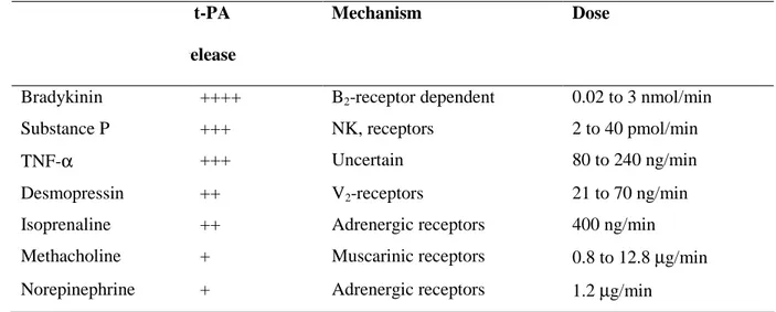
Acute t-PA release has been assessed in both the forearm and coronary circulations of humans using a number of endothelial agonists including bradykinin, substance P, desmopressin, and methacholine (Table 1)
Table 1. Substances that Stimulate Acute Tissue Plasminogen Activator (t-PA) Release in Humans
t-PA release
Mechanism Dose
Bradykinin ++++ B2-receptor dependent 0.02 to 3 nmol/min
Substance P +++ NK, receptors 2 to 40 pmol/min
TNF-α +++ Uncertain 80 to 240 ng/min
Desmopressin ++ V2-receptors 21 to 70 ng/min
Isoprenaline ++ Adrenergic receptors 400 ng/min Methacholine + Muscarinic receptors 0.8 to 12.8 µg/min Norepinephrine + Adrenergic receptors 1.2 µg/min
(modified from Oliver JJ, et al ATVB 2005;25:2470-2479)
Forearm Release
Methodology used to assess forearm t-PA release in response to intrabrachial infusions is based on the differences in plasma concentrations of t-PA between inflowing arterial and outflowing venous plasma of a single arm 28, 29. This tecnique require the infusion of drugs via
15
an intra-arterial catheter and the arteriovenous concentration gradient is calculated from blood samples taken simultaneously from this and ipsilateral venous catheter. Forearm plasma flow is calculated using forearm blood flow (FBF), measured by strain gauge pletismography, and arterial hematocrit corrected for 1% trapped plasma. Net release is calculated as the product of the arteriovenous concentration gradient and forearm plasma flow, as follows:
Net release=(Cv-Ca) x FBF x [(101-hematocrit)/100]
where Cv and Ca are the venous and arterial concentrations, respectively. The antigen concentrations of t-PA are measured using enzyme-linked immunosorbent assays 30, and net release is expressed as ng per 100 mL of forearm tissue per minute. The activity of t-PA is determined phothometrically 31 and expressed as IU per 100 mL of forearm tissue per minute. Several findings indicate that there is no demonstrable release of PAI-1 across the forearm, therefore, t-PA activity increases in parallel with t-PA antigen concentration 32, 33. There is no consensus on whether it is better to measure t-PA antigen or activity. Whereas it is only unbound t-PA that it is functionally active, ultimately the efficacy of endogenous fibrinolysis id determined by the magnitude of local t-PA release and the resultant t-PA activity at the site of thrombus.
Stimulation of t-PA Release
A number of humoral and coagulant factors cause t-PA release. The muscarinic receptor agonist metacholine induces t-PA release in the forearm. In contrast, it has been reported that acetylcholine, also a muscarinic agonist, does not stimulate t-PA release 28, 34. The reason for these difference is not clear, although varying potencies and stabilities of the respective compound may be responsible.
The adrenergic agonists norepinephrine 32 and isoproterenol 35 induce t-PA release, as does mental stress, which is associated with adrenergic activation 18, 32. These data suggest a role for the sympathetic nervous system in controlling vascular t-PA secretion. Results from
16
animal studies show that chemical sympathectomy in rats reduces basal plasma t-PA concentrations and agonist-induced t-PA release in isolated vessels 36. In addition, in a porcine model, stimulation of cardiac sympathetic nerves causes coronary t-PA release36. Bradykinin is an inflammatory vasodilator peptide and a powerful stimulant of endothelial t-PA release via B2 receptors 37. The tachykinin vasodilator, substance P, acts mainly through the neurokinin type 1 (NK1) receptor. It is a central and peripheral neurotransmitter and mediates neurogenic inflammation 38. Substance P induces t-PA release in both the forearm and coronary circulations, and is the most potent known stimulant of t-PA in humans.
The vasopressin analogue, desmopressin, is a V2 receptor agonist that releases t-PA in the forearm circulation 23, 33.
Regulation of t-PA Release
The role of NO in the mechanism of endothelial t-PA release is unclear. Substance P-induced t-PA release was inhibited by the NO synthase inhibitor, NG monomethyl-L-arginine (L-NMMA) 24. In contrast, bradykinin-induced t-PA release was unaffected by L-NMMA in one study 39 and increased by L-NMMA in another 40. Shear stress is a well-characterized stimulus for t-PA secretion from cultured endothelial cells 41. In ex vivo human conduit vessels, shear stress stimulates t-PA expression and increases its intracellular storage pool without stimulating its release 42. Consistent with this, marked increases in blood flow during infusions of vasodilators such as sodium nitroprusside and papaverine do not cause in vivo t-PA release 4344.
Clinical Relevance of Endothelial t-PA Release
Acute plaque rupture or erosion of a coronary atheromatous plaque, and the consequent thrombosis, cause the majority of sudden cardiac deaths and myocardial infarctions 45. Small areas of denudation and thrombus are commonly found on atheromatous plaques and are
17
usually subclinical 46. However, with imbalance of fibrinolytic system, atherothrombosis may propagate, leading to arterial occlusion 46. The importance of acute endogenous t-PA release is further exemplified by the high rate of spontaneous reperfusion in the infarct-related artery after acute myocardial infarction 47. Since a reduction in t-PA activity or release is associated with an increased incidence of major adverse cardiac events in patients with stable 48 or unstable angina 49 it has been suggested that coronary thrombosis critically depends on local fibrinolytic balance 50.
Basal Plasma t-PA Concentration
Several studies have investigated the relationship between basal venous t-PA antigen concentrations and subsequent coronary heart disease. In a meta-analysis of prospective studies, the risk of coronary heart disease was about 50% greater in those with plasma t-PA antigen concentrations in the highest tertile compared to those in the lowest one 51. This apparent contradiction actually reflects the concomitant increase of plasma PAI-1 concentrations and associated reduction in t-PA activity. However, whereas it is of interest, basal plasma t-PA levels do not reflect the local capacity for acute endothelial t-PA release in response to developing thrombus27. It follows that the assessment of acute endothelial t-PA release is likely to be of greater pathophysiological relevance.
Acute Endothelial t-PA Release as a Novel Measure of Endothelial Function
The endothelium has a number of important functions, including regulation of vascular tone, inflammation, coagulation and fibrinolysis as well. However, to date, most of clinical studies on endothelial function have focused on endothelium-dependent vasodilation. Although this is a widely accepted surrogate marker for the role of endothelial function in atherothrombosis 1, measuring other aspects of endothelial function, such as the capacity for t-PA release, may provide additional and novel insights.
18
Measurement of coronary t-PA release is likely to be of greatest relevance to coronary pathophysiology but can only be performed in selected patients undergoing coronary angiography. Therefore, forearm assessment is performed most widely, although this vascular bed is less susceptible to atherosclerosis and subsequent thrombosis, raising question on its validity as a surrogate for the coronary circulation. Nevertheless, consistent findings between the peripheral 52-54 and coronary circulations 43, 55 support the notion that the forearm model is a reasonable surrogate.
19
Aim of the Study
The aim of the study was to investigate the role of endothelial pathways in the regulation of endothelial t-PA release in physiological condition and in patients with essential hypertension, a clinical condition associated with increased risk of atherothrombotic events, including myocardial infarction and stroke.
To address this aim the following research questions were developed:
• To determine the possible regulatory role of NO-synthase pathway in modulating endothelial t-PA release in healthy condition and in essential hypertension, a clinical condition in which a reduced NO-availability is well documented.
• To determine the role of adrenergic stimuli on endothelial t-PA release in healthy conditions and in essential hypertension. In particular, receptor-characterization and NO-synthase pathway were assessed.
• To determine the role of the NO-independent pathways in the modulation of endothelial t-PA release. In particular, the role of cytochrome P450 2C9-derived hyperpolarizing factor in was assessed in healthy subjects and in hypertensive patients.
20 References
1. Lerman A, Zeiher AM. Endothelial function: cardiac events. Circulation. 2005;111:363-368.
2. Perticone F, Ceravolo R, Pujia A, Ventura G, Iacopino S, Scozzafava A, Ferraro A, Chello M, Mastroroberto P, Verdecchia P, Schillaci G. Prognostic significance of endothelial dysfunction in hypertensive patients. Circulation. 2001;104:191-196.
3. Furchgott RF, Zawadzki JV. The obligatory role of endothelial cells in the relaxation of arterial smooth muscle by acetylcholine. Nature. 1980;288:373-376.
4. Campbell WB, Gebremedhin D, Pratt PF, Harder DR. Identification of epoxyeicosatrienoic acids as endothelium-derived hyperpolarizing factors. Circ Res. 1996;78:415-423.
5. Gauthier KM, Edwards EM, Falck JR, Reddy DS, Campbell WB. 14,15-epoxyeicosatrienoic acid represents a transferable endothelium-dependent relaxing factor in bovine coronary arteries. Hypertension. 2005;45:666-671.
6. Taddei S, Ghiadoni L, Virdis A, Buralli S, Salvetti A. Vasodilation to bradykinin is mediated by an ouabain-sensitive pathway as a compensatory mechanism for impaired nitric oxide availability in essential hypertensive patients. Circulation. 1999;100:1400-1405. 7. Taddei S, Versari D, Cipriano A, Ghiadoni L, Galetta F, Franzoni F, Magagna A, Virdis A,
Salvetti A. Identification of a cytochrome P450 2C9-derived endothelium-derived hyperpolarizing factor in essential hypertensive patients. J Am Coll Cardiol. 2006;48:508-515.
8. Emeis JJ. Regulation of the acute release of tissue-type plasminogen activator from the endothelium by coagulation activation products. Ann N Y Acad Sci. 1992;667:249-258. 9. Hoylaerts M, Rijken DC, Lijnen HR, Collen D. Kinetics of the activation of plasminogen by
21
10. Plow EF, Felez J, Miles LA. Cellular regulation of fibrinolysis. Thromb Haemost. 1991;66:32-36.
11. Nordenhem A, Wiman B. Tissue plasminogen activator (tPA) antigen in plasma: correlation with different tPA/inhibitor complexes. Scand J Clin Lab Invest. 1998;58:475-483.
12. Narita M, Bu G, Herz J, Schwartz AL. Two receptor systems are involved in the plasma clearance of tissue-type plasminogen activator (t-PA) in vivo. J Clin Invest. 1995;96:1164-1168.
13. Ehrlich HJ, Gebbink RK, Keijer J, Linders M, Preissner KT, Pannekoek H. Alteration of serpin specificity by a protein cofactor. Vitronectin endows plasminogen activator inhibitor 1 with thrombin inhibitory properties. J Biol Chem. 1990;265:13029-13035.
14. Fox KA, Robison AK, Knabb RM, Rosamond TL, Sobel BE, Bergmann SR. Prevention of coronary thrombosis with subthrombolytic doses of tissue-type plasminogen activator. Circulation. 1985;72:1346-1354.
15. van den Eijnden-Schrauwen Y, Kooistra T, de Vries RE, Emeis JJ. Studies on the acute release of tissue-type plasminogen activator from human endothelial cells in vitro and in rats in vivo: evidence for a dynamic storage pool. Blood. 1995;85:3510-3517.
16. Huber D, Cramer EM, Kaufmann JE, Meda P, Masse JM, Kruithof EK, Vischer UM. Tissue-type plasminogen activator (t-PA) is stored in Weibel-Palade bodies in human endothelial cells both in vitro and in vivo. Blood. 2002;99:3637-3645.
17. Emeis JJ, van den Eijnden-Schrauwen Y, van den Hoogen CM, de Priester W, Westmuckett A, Lupu F. An endothelial storage granule for tissue-type plasminogen activator. J Cell Biol. 1997;139:245-256.
18. Jern C, Selin L, Tengborn L, Jern S. Sympathoadrenal activation and muscarinic receptor stimulation induce acute release of tissue-type plasminogen activator but not von Willebrand factor across the human forearm. Thromb Haemost. 1997;78:887-891.
22
19. Coulet F, Blons H, Cabelguenne A, Lecomte T, Lacourreye O, Brasnu D, Beaune P, Zucman J, Laurent-Puig P. Detection of plasma tumor DNA in head and neck squamous cell carcinoma by microsatellite typing and p53 mutation analysis. Cancer Res. 2000;60:707-711.
20. Knop M, Gerke V. Ca2+ -regulated secretion of tissue-type plasminogen activator and von Willebrand factor in human endothelial cells. Biochim Biophys Acta. 2002;1600:162-167. 21. Salame MY, Samani NJ, Masood I, deBono DP. Expression of the plasminogen activator
system in the human vascular wall. Atherosclerosis. 2000;152:19-28.
22. Steins MB, Padro T, Li CX, Mesters RM, Ostermann H, Hammel D, Scheld HH, Berdel WE, Kienast J. Overexpression of tissue-type plasminogen activator in atherosclerotic human coronary arteries. Atherosclerosis. 1999;145:173-180.
23. Wall U, Jern C, Jern S. High capacity for tissue-type plasminogen activator release from vascular endothelium in vivo. J Hypertens. 1997;15:1641-1647.
24. Newby DE, Wright RA, Dawson P, Ludlam CA, Boon NA, Fox KA, Webb DJ. The L-arginine/nitric oxide pathway contributes to the acute release of tissue plasminogen activator in vivo in man. Cardiovasc Res. 1998;38:485-492.
25. Allen RA, Kluft C, Brommer EJ. Effect of chronic smoking on fibrinolysis. Arteriosclerosis. 1985;5:443-450.
26. Brown NJ, Nadeau JH, Vaughan DE. Selective stimulation of tissue-type plasminogen activator (t-PA) in vivo by infusion of bradykinin. Thromb Haemost. 1997;77:522-525. 27. Hrafnkelsdottir T, Gudnason T, Wall U, Jern C, Jern S. Regulation of local availability of
active tissue-type plasminogen activator in vivo in man. J Thromb Haemost. 2004;2:1960-1968.
28. Brown NJ, Gainer JV, Stein CM, Vaughan DE. Bradykinin stimulates tissue plasminogen activator release in human vasculature. Hypertension. 1999;33:1431-1435.
23
29. Jern C, Selin L, Jern S. In vivo release of tissue-type plasminogen activator across the human forearm during mental stress. Thromb Haemost. 1994;72:285-291.
30. Declerck PJ, Alessi MC, Verstreken M, Kruithof EK, Juhan-Vague I, Collen D. Measurement of plasminogen activator inhibitor 1 in biologic fluids with a murine monoclonal antibody-based enzyme-linked immunosorbent assay. Blood. 1988;71:220-225. 31. Wiman B, Lindahl T, Almqvist A. Evidence for a discrete binding protein of plasminogen
activator inhibitor in plasma. Thromb Haemost. 1988;59:392-395.
32. Jern S, Selin L, Bergbrant A, Jern C. Release of tissue-type plasminogen activator in response to muscarinic receptor stimulation in human forearm. Thromb Haemost. 1994;72:588-594.
33. Wall U, Jern S, Tengborn L, Jern C. Evidence of a local mechanism for desmopressin-induced tissue-type plasminogen activator release in human forearm. Blood. 1998;91:529-537.
34. Rosenbaum DA, Pretorius M, Gainer JV, Byrne D, Murphey LJ, Painter CA, Vaughan DE, Brown NJ. Ethnicity affects vasodilation, but not endothelial tissue plasminogen activator release, in response to bradykinin. Arterioscler Thromb Vasc Biol. 2002;22:1023-1028. 35. Stein CM, Brown N, Vaughan DE, Lang CC, Wood AJ. Regulation of local tissue-type
plasminogen activator release by endothelium-dependent and endothelium-independent agonists in human vasculature. J Am Coll Cardiol. 1998;32:117-122.
36. Wang Y, Jiang X, Hand AR, Gilles C, Kirk J, Cone RE, O'Rourke J. Additional evidence that the sympathetic nervous system regulates the vessel wall release of tissue plasminogen activator. Blood Coagul Fibrinolysis. 2002;13:471-481.
37. Hall JM. Bradykinin receptors. Gen Pharmacol. 1997;28:1-6.
38. Newby DE, Stewart A, Witherow FN, Grieve S, Dawson P, Fox KA, Webb DJ, Ludlam CA. Local and systemic effects of intra-arterial desmopressin in healthy volunteers and patients
24
with type 3 von Willebrand disease. Role of interleukin-6. Thromb Haemost. 2000;84:195-203.
39. Brown NJ, Gainer JV, Murphey LJ, Vaughan DE. Bradykinin stimulates tissue plasminogen activator release from human forearm vasculature through B(2) receptor-dependent, NO synthase-independent, and cyclooxygenase-independent pathway. Circulation. 2000;102:2190-2196.
40. Smith DT, Hoetzer GL, Greiner JJ, Stauffer BL, DeSouza CA. Endothelial release of tissue-type plasminogen activator in the human forearm: role of nitric oxide. J Cardiovasc Pharmacol. 2003;42:311-314.
41. Diamond SL, Sharefkin JB, Dieffenbach C, Frasier-Scott K, McIntire LV, Eskin SG. Tissue plasminogen activator messenger RNA levels increase in cultured human endothelial cells exposed to laminar shear stress. J Cell Physiol. 1990;143:364-371.
42. Sjogren LS, Gan L, Doroudi R, Jern C, Jungersten L, Jern S. Fluid shear stress increases the intra-cellular storage pool of tissue-type plasminogen activator in intact human conduit vessels. Thromb Haemost. 2000;84:291-298.
43. Matsumoto T, Minai K, Horie H, Ohira N, Takashima H, Tarutani Y, Yasuda Y, Ozawa T, Matsuo S, Kinoshita M, Horie M. Angiotensin-converting enzyme inhibition but not angiotensin II type 1 receptor antagonism augments coronary release of tissue plasminogen activator in hypertensive patients. J Am Coll Cardiol. 2003;41:1373-1379.
44. Minai K, Matsumoto T, Horie H, Ohira N, Takashima H, Yokohama H, Kinoshita M. Bradykinin stimulates the release of tissue plasminogen activator in human coronary circulation: effects of angiotensin-converting enzyme inhibitors. J Am Coll Cardiol. 2001;37:1565-1570.
45. Burke AP, Farb A, Malcom GT, Liang YH, Smialek J, Virmani R. Coronary risk factors and plaque morphology in men with coronary disease who died suddenly. N Engl J Med. 1997;336:1276-1282.
25
46. Ibanez B, Vilahur G, Badimon JJ. Plaque progression and regression in atherothrombosis. J Thromb Haemost. 2007;5 Suppl 1:292-299.
47. Armstrong PW, Baigrie RS, Daly PA, Haq A, Gent M, Roberts RS, Freeman MR, Burns R, Liu P, Morgan CD. Tissue plasminogen activator: Toronto (TPAT) placebo-controlled randomized trial in acute myocardial infarction. J Am Coll Cardiol. 1989;13:1469-1476. 48. Held C, Hjemdahl P, Rehnqvist N, Wallen NH, Bjorkander I, Eriksson SV, Forslund L,
Wiman B. Fibrinolytic variables and cardiovascular prognosis in patients with stable angina pectoris treated with verapamil or metoprolol. Results from the Angina Prognosis study in Stockholm. Circulation. 1997;95:2380-2386.
49. Hoffmeister HM, Jur M, Ruf-Lehmann M, Helber U, Heller W, Seipel L. Endothelial tissue-type plasminogen activator release in coronary heart disease: Transient reduction in endothelial fibrinolytic reserve in patients with unstable angina pectoris or acute myocardial infarction. J Am Coll Cardiol. 1998;31:547-551.
50. Rosenberg RD, Aird WC. Vascular-bed--specific hemostasis and hypercoagulable states. N Engl J Med. 1999;340:1555-1564.
51. Lowe GD, Danesh J, Lewington S, Walker M, Lennon L, Thomson A, Rumley A, Whincup PH. Tissue plasminogen activator antigen and coronary heart disease. Prospective study and meta-analysis. Eur Heart J. 2004;25:252-259.
52. Witherow FN, Dawson P, Ludlam CA, Fox KA, Newby DE. Marked bradykinin-induced tissue plasminogen activator release in patients with heart failure maintained on long-term angiotensin-converting enzyme inhibitor therapy. J Am Coll Cardiol. 2002;40:961-966. 53. Newby DE, Wright RA, Labinjoh C, Ludlam CA, Fox KA, Boon NA, Webb DJ.
Endothelial dysfunction, impaired endogenous fibrinolysis, and cigarette smoking: a mechanism for arterial thrombosis and myocardial infarction. Circulation. 1999;99:1411-1415.
26
54. Duffy GP, Berry DJ, Rowland C, Cabanela ME. Primary uncemented total hip arthroplasty in patients <40 years old: 10- to 14-year results using first-generation proximally porous-coated implants. J Arthroplasty. 2001;16:140-144.
55. Newby DE, McLeod AL, Uren NG, Flint L, Ludlam CA, Webb DJ, Fox KA, Boon NA. Impaired coronary tissue plasminogen activator release is associated with coronary atherosclerosis and cigarette smoking: direct link between endothelial dysfunction and atherothrombosis. Circulation. 2001;103:1936-1941.