UNIVERSITY OF SIENA
DEPARTMENT OF BIOTECHNOLOGY, CHEMISTRY AND PHARMACY
DOTTORATO DI RICERCA IN SCIENZE CHIMICHE E FARMACEUTICHE
XXXIII CICLO
DOCTORATE IN CHEMICAL AND PHARMACEUTICAL SCIENCES
XXXIII CYCLE
COORDINATOR: Prof. Maurizio Taddei
Use and characterisation of free or
immobilised enzymatic systems for the
synthesis and functionalisation of novel
materials
SSD: CHIM/02TUTOR: CANDIDATE:
Prof. Rebecca Pogni Jessica Costa
ACADEMIC YEAR: 2020/2021
Firmato digitalmente da: COSTA JESSICA Firmato il 12/02/2021 14:03 Seriale Certificato: 80941106738351767014465678551648208000 Valido dal 11/02/2021 al 11/02/2024 ArubaPEC S.p.A. NG CA 3
PRODUCTION AND CHARACTERISATION OF BIOMATERIALS
3.1 Pigments production 65
3.2 Uv-Vis and FT-IR characterisation of melanin pigments 66 from bacterial and enzymatic synthesis
3.3 S-,X- and Q-Band Multifrequancy EPR analysis of 68 Melanin pigments
3.4 Multifrequency CW EPR power saturation and Q-Band 73 pulse EPR relaxation studies on melanin pigments
3.5 Homogentisic Acid and Gentisic Acid biosynthesized 79 pyomelanin mimics
CONCLUSIONS 92
ABBREVIATIONS 95
REFERENCES 98
SCIENTIFIC PRODUCTION LIST 119
INTERNATIONAL PEER REVIEWED
the material can plays a crucial role in the immobilisation process and the properties of the produced catalytic system.
In this thesis we have chosen two different supports: super paramagnetic nanoparticles for both the enzymes used and the chitosan beads as an alternative support for chitinase.
Magnetic nanoparticles show interesting properties for enzymatic immobilisation, they can be obtained with small size, increasing the yield of enzymatic immobilisation and above all, the reaction products can be easily recovered applying an external magnetic field.
Magnetic nanoparticles were prepared following the traditional method of co-precipitation of Fe2+ and Fe3+ ions. In addition, the MNPs surface was coated with (3-aminopropyl) triethoxysilane (APTES), so the amino groups on their surface allow the attack of linker such as glutaraldehyde, to facilitate the enzymatic binding. This immobilisation process was successfully used for chitinase, obtaining a high immobilisation yield and increasing enzymatic stability. Different was for laccase, which having a different catalytic mechanism a revision of the synthesis has been attempt. The use of the magnetic nanoparticles obtained with the traditional method hampered the detection of stable radical species formed during the catalytic mechanism as it happens for the oxidation product of 2,2'-azino-bis (3-ethylbenzothiazolin-6-sulfonic acid) (ABTS), the standard compound used to test the enzyme activity. Changing some synthetic parameters, the new magnetic nanoparticles were produced and characterised. In fact nanoparticles with a lower aggregation state and a smaller hydrodynamic diameter were obtained and tested without any interference with the ABTS substrate.
Chitinase was also immobilised on chitosan beads/Macro-Spheres (CMS), as this support is completely atoxic and so most suitable for application in food industries.
The presence of active amino groups in deacetylated GlcNAc units of chitosan also enables the binding of the linker (glutaraldehyde and genipin) and then of the enzyme. The goal of this part of the thesis was to attempt the immobilisation of Chitinase on different supports, MNPs and CMS, for the efficient production of COS.
1.2 Laccase
Laccases (benzenediol: oxygen oxidoreductase. EC 1.10.3.2) are multicopper- containing oxidoreductases found in higher plants, bacteria, fungi, insects and lichens.
They find application in several industrial sectors such as food, pulp and paper, textile and cosmetics [8]. The potential applications of laccases in numerous and different biocatalytic processes for industry and environmental solutions has increased the interest in understanding their structure and mechanism of action. Laccase typically comprises four copper ions in their catalytic site. These coppers are classified as type 1 (T1), type 2 (T2) and binuclear type 3 (T3), according to their spectroscopic behaviour and role in catalysis. The initial electron acceptor in laccase-catalyzed oxidation is copper T1 located in the cavity close to the enzyme surface. The reduction of copper T1 is a rate-limiting step in the reactions catalysed by laccase [9]. In general, laccases oxidize a wide range of substrates, typically substituted phenols and aromatic amines, which are transformed into free radicals. Unstable chemical products and primarily generated free radicals commonly start other reactions, leading to complex chemical transformations of biological relevance such as lignin synthesis and degradation [10,11].
The overall laccase reaction involves one electron, sequential oxidations of four molecules of reducing substrates, concurrently with two double electron reductions of oxygen atoms into their respective H2O molecules
(Figure 1). This process is accompanied by a catalytic exchange of 4 H+ equivalents [12]. It was also observed that substrates of laccase, such as 2,2'-azino-bis (3-ethylbenzothiazolin-6-sulfonic) (ABTS), radical 2,2,6,6-tetramethylpiperidine 1-oxyl (TEMPO) or violuric acid (VA) can act as mediators speeding up the catalytic reaction on several substrates and making possible the oxidation of compounds that otherwise would not be the substrate of this enzyme. Furthermore, the cationic ABTS radical, formed by laccase, has been completely characterised by EPR spectroscopy and the analysis confirmed that ABTS+. is the more stable radical [13,14].
found in bacteria, viruses, fungi and plants and they participate in physiological processes such as nutrition, parasitism, morphogenesis and immunity [21–23].
They are specific for the β(1à4) glycosidic bonds present in chitin [24]. Chitin represents the second most abundant polysaccharide in nature after cellulose, as it is one of the main components of the exoskeleton of insects and crustaceans and it is also presents in some fungi. It is formed by N-acetyl-β-D-glucosamine (GlcNac) units linked by type β(1à4) glycosidic bonds [25–27].
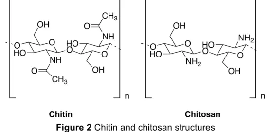
Chitin has strong inter- and intra-molecular hydrogen bonding, which provides its insoluble property in inorganic and organic solvents. For that its deacetylated form, chitosan, is more widely used for industrial applications (Figure 2) [28]. Chitosan is a linear polysaccharide that contains copolymers of D-glucosamine (deacetylated units) and N-acetyl-D-glucosamine (acetylated units) linked by β (1à4) glycosidic bonds [29]. The deacetylation of chitosan is generally defined as the glucosamine/N-acetyl glucosamine ratio, which goes up as chitin is converted to chitosan. Therefore, when the percentage of N-acetyl glucosamine is higher than glucosamine, the biopolymer is called chitin and when the percentage of glucosamine exceeds N-acetyl glucosamine the compound is called chitosan [30].
Figure 2 Chitin and chitosan structures
Thanks to their properties such as biocompatibility, non-toxicity, biodegradability and biological activities such as antimicrobials and antioxidants, these biopolymers can be considered as a new source of
O O O OH NH CH3 OH NH CH3 O HO HO O O n Chitin O O O OH NH2 OH NH2 O HO HO n Chitosan
functional materials. In fact, they are used in many sectors like pharmaceuticals, biomedical, cosmetic, food, textiles, agriculture, paper and enzymatic immobilisation [31].
Chitinases are responsible for the biotransformation of chitin or chitosan into chitooligosaccharides. According to their catalytic action, chitinases have been divided into two groups:
i. Endo-chitinases: cleave chitin at internal sites;
ii. Exo-chitinases: slit chitin from its reducing or non-reducing end.
The hydrolysis of chitin yields a series of chitooligosaccharides (COS) containing random GlcNAc and D-glucosamine (GlcN) units. Three families of COS can be differentiated: fully acetylated chitooligosaccharides (faCOS) formed exclusively by GlcNAc, partially acetylated chitooligosaccharides (paCOS) composed of GlcN and GlcNAc and fully deacetylated chitooligosaccharides (fdCOS) formed exclusively by GlcN. The bioactivity of COS is well reported, in particular their anti-inflammatory, neuroprotective, antibacterial, antiviral, antihypertensive and antitumor properties. The size of COS defined by the degree of polymerization (DP), degree of deacetylation (DD) and pattern of acetylation (PA) exert a notable influence on their properties [32,33].
This thesis is focused on the use of a specific exo-chitinase Chit42 from fungus Trichoderma harzianum cloned in Pichia pastoris (in collaboration with the group at UAM (Spain)). This enzyme hydrolysed chitin and chitosan with a low DD giving rise to mixtures enriched in
faCOS and paCOS, respectively [34].
Chitinases have shown immense potential for a wide range of applications involving food and medicine. Applications using chitinase-based processing are economic, eco-friendly, and non-hazardous.
Chitinases can be used for a number of human healthcare applications such as the production of ophthalmic preparations with microbicides. Thanks to its antifungal activity, chitinase can be combined with
antifungal drugs and used as a therapeutic treatment for various fungal infections [35,36].
Chitinases are also attaining prominence in the field of biotechnology applied in waste management and pest control in agriculture [22].
1.4 Enzymatic immobilisation
As reported in the previous paragraphs, enzymes find useful applications in different fields. However, two critical parameters that limit their application are quantity and quality. For industrial-scale processes, enzymes in tons and their extreme purity are required. This leads to an increase in the cost of the process. One of the solutions made to solve these problems was enzymatic immobilisation [37].
In the last two decades, the immobilisation of enzymes is an important challenge in biotechnology. The aim of this technique is to obtain reusable and stable enzymes with resistance to different reaction environment. Immobilisation improves many properties of enzymes such as pH tolerance, heat stability, functional stability and performance in organic solvents, furthermore stabilizing the structural rigidity of enzyme prevents his dissociation and inactivation [38,39].
The methods of enzyme immobilisation can be divided into three categories:
i. Binding to a support ii. Entrapment
iii. Cross-linking
Binding to support can be physical, ionic and covalent. Generally, the stronger is the covalent binding because physical and ionic are too wear to keep the enzyme fixed under extreme industrial conditions. However, covalent binding has a disadvantage, linking the enzyme with support can change the active conformation losing its activity.
Entrapment via inclusion of an enzyme in a polymer network requires the synthesis of polymeric matrix in the presence of the enzyme. The
physical forces are not enough to retain the enzyme so an additional covalent attachment is often required.
Cross-linking of enzyme aggregates is used to prepare carrierless macroparticles. The loss of activity is inevitable due to the difficult accessibility of some of the enzyme situated deeply within the carrier pores, inaccessible to the substrate. However, this approach offers clear advantages: highly concentrated enzyme activity in the catalyst, high stability and low production costs owing to the exclusion of an additional carrier [40,41].
From literature, many techniques were used for chitinase and laccase immobilisation [37,42].
Chitinases were immobilised on silica gel by physical adsorption, on chitin and chitosan by covalent binding, on cellulose and Amberlite by ionic bonding. The obtained results show that chitosan, Amberlite and chitin had the highest immobilisation yield and the immobilisation on chitosan beads increases the activity of enzyme immobilised compared to the free enzyme [43,44]. In addition it was discovered that the immobilisation of chitosanase in chitin powder by cross-linking improves the enzyme properties [45].
Chitinase was also immobilised on calcium alginate, agar-agar, magnetic nanoparticles and silica gel beads and the immobilised chitinase expressed higher stability compared to the free one. It is an important factor for industrial applications [42,46].
Laccase was immobilised on Amberlite by covalent binding, the immobilised biocatalyst displays an improved thermal and storage stability paired with a good performance for the reusability [47]. Silica-based supports are widely studied for the immobilisation of laccases. The mesoporous silica materials act as good carriers for adsorption due to their pore size ordered structure, high surface area and high thermal stability. Good results were obtained immobilising laccases on mesoporous silica by physical adsorption and covalent bonding [48]. A wide variety of supports such as cellulose, agarose, collagen, inorganic carriers like glass, silica, metallic nanoparticles of gold, silver are widely used for laccase immobilisation [49].
The type of support material used plays a crucial role in the immobilisation process due to the strong effect of these materials on the properties of the produced catalytic system. The main required features of support materials for effective enzyme immobilisation products are: thermal and chemical stability, insolubility under reaction conditions, high affinity to enzymes, regeneration and reusability, availability and less price, presence of reactive functional groups and biocompatibility [50,51].
The support structure and immobilisation technique are major factors limiting the immobilisation process. Many techniques and carriers for immobilisation have been developed over the years; however, there is no universal method of immobilisation or ideal support for all enzymes and applications. Both have advantages and disadvantages, which need to be balanced through the optimisation process [52].
In this thesis, magnetic nanoparticles and chitosan beads as supports were investigated for the immobilisation of laccase and chitinase. The aim of the research was to optimize the immobilisation process in order to have advantages in the industrial application of these enzymes.
1.5 Magnetic nanoparticles
Nanoscience is one of the most important research in modern science. The use of nanoparticles offers major advantages due to their unique size and physicochemical properties. In fact, before focusing on the use of nanoparticles as a support for immobilisation, it is necessary to analyse their characteristics.
1.5.1 Physicochemical properties of magnetic nanoparticles
The term “nanoparticles” refers to materials with at least one dimension between approximately 1 and 100 nanometers (nm). Magnetic nanoparticles Fe3O4 (MNPs) are a class of nanoparticles that can be
Five basic types of magnetism can be described: ferromagnetism, paramagnetism, diamagnetism, antiferromagnetism and ferrimagnetism. In ferromagnetic materials, an atom has a net magnetic moment due to unpaired electrons. The material is composed of domains each containing large numbers of atoms whose magnetic moments are parallel producing a net magnetic moment of the domain that points in some direction. The magnetic moments of the domains are randomly distributed giving a zero net magnetic moment on the material. When the ferromagnetic material is placed in a magnetic field, the magnetic moments of the domains align along the direction of the applied magnetic field forming a large net magnetic moment.
In paramagnetic materials, an atom has a net magnetic moment due to unpaired electrons but magnetic domains are absent. When the paramagnetic material is placed in a magnetic field, the magnetic moment of the atoms aligns along the direction of the applied magnetic field forming a weak net magnetic moment.
In diamagnetic materials, atoms have no unpaired electrons, which results in zero net magnetic moment. These materials display a very weak response against the applied magnetic field due to the realignment of the electron orbits when a magnetic field is applied.
Antiferromagnetic materials are compounds of two different atoms that occupy different lattice positions. The two atoms have magnetic moments that are equal in magnitude and opposite in direction, which results in zero net magnetic moment.
Ferrimagnetic materials, such as magnetite Fe3O4, are also compounds
of different atoms residing on different lattice sites with antiparallel magnetic moments. However, in these materials, the magnetic moments do not cancel out since they have different magnitudes which result in a net spontaneous magnetic moment [54].
An important aspect of magnetic nanoparticles is their surface. As the size of the particles decreases, the ratio of the surface area to volume of the particle increases. For nanoparticles, this ratio becomes significantly large causing a large portion of the atoms to reside on the surface compared to those in the core of the particle.
The large surface-volume ratio of the nanoparticles is the key factor to the physical, chemical and mechanical properties compared to those of the corresponding bulk material [55].
Furthermore, it was shown that the magnetic moment per atom and the magnetic anisotropy of nanoparticles could be different than those of a bulk specimen. It is well established that bulk ferrimagnetic material is composed of small regions, called magnetic domains. In each domain, the magnetic moments of atoms are aligned in one direction giving a net magnetization of each domain. The directions of magnetization of the domains are different. Hence the net magnetization of a magnetic material resulted from the addition of the different magnetization of all domains. It was found that magnetic domains in ferrimagnetic crystals have a minimum size, around 100 nm, below which the ferrimagnetic material cannot split up further into domains and are called single domain particles. Thermal energy plays a major role in the magnetic instability of single domain magnetic particles [56].
1.5.2 Magnetic nanoparticles synthesis
MNPs of different types and sizes are now being synthesized via several physical and chemical methods. The most common methods including co-precipitation, hydrothermal synthesis, thermal decomposition, microemulsion and sonochemical synthesis can all be directed to the synthesis of high quality iron oxide nanoparticles [57]. The co-precipitation technique is probably the simplest and most efficient chemical pathway to obtain magnetic particles. Iron oxide nanoparticles are usually prepared by an aging stoichiometric mixture of ferrous and ferric salts in aqueous medium.
Fe3+ + Fe2+ + 8OH- à Fe3O4↓ + 4 H2O (1)
According to the thermodynamics of this reaction, complete precipitation of Fe3O4 should be expected at a pH between 8 and 14, with a
stoichiometric ratio of 2:1 (Fe3+/Fe2+) in a non-oxidizing oxygen environment. Magnetite (Fe3O4) is not very stable and is sensitive to
oxidation because it is transformed into maghemite (γ-Fe2O3) in the
presence of oxygen.
The main advantage of the co-precipitation process is that a large number of nanoparticles can be synthesized.
A wide variety of factors can be adjusted in the synthesis of iron magnetic nanoparticles to control the size, magnetic characteristics or surface properties. A number of studies have dealt with the influence of these different factors. The size and shape of the nanoparticles can be tailored with relative success by adjusting pH, ionic strength, temperature, nature of the salts (perchlorates, chlorates, sulfates and nitrates) or the Fe3+/Fe2+ concentration ratio. The addition of chelating organic anions or polymer surface complexing agents during the formation of nanoparticles can help to control the size of magnetite [53,58].
It is a challenge to control the size, stability, shape, and dispersibility of nanoparticles in desired solvents. Magnetic iron oxide nanoparticles have a large surface to volume ratio and therefore possess high surface energies. Consequently, they tend to aggregate to minimize the surface energies. Moreover, the naked iron oxide NPs have high chemical activity and are easily oxidized in air, generally resulting in loss of magnetism and dispersibility. Therefore, providing proper surface coating and developing some effective protection strategies to keep the stability of magnetic iron oxide NPs is very important. The coating is designed to improve the stability and solubility of nanoparticles, increase their biocompatibility, target specificity, and to prevent agglomeration, oxidation, corrosion and toxicity [59].
A promising approach is coating magnetic nanoparticles with silica. It could shield the magnetic dipolar attraction between magnetic nanoparticles, favouring the dispersion of the nanoparticles in liquid media. Furthermore, due to the existence of many silanol groups, magnetic nanoparticles could have various functional groups. Finally, silica provides a chemically inert surface for magnetic nanoparticles in biological systems [60].
1.5.3 Application of MNPs on enzyme immobilisation
The development of nanotechnology has found employment in enzymatic immobilisation, especially the use of magnetic nanoparticles had gained significant attention due to strong magnetic properties, low toxicity and biocompatibility with biological materials [61].
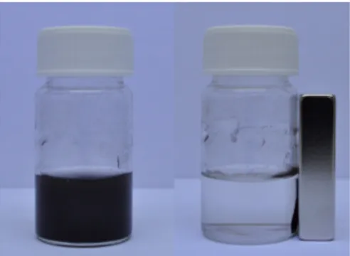
Nanoparticles present a high specific area, meaning that more enzymes can be immobilised, because it increases the available attacking surface. Furthermore, their magnetic force allows them to easily separate the final product from the reaction environment by applying an external magnetic field (Figure 3).
Figure 3 Attraction of magnetic nanoparticles applying external magnetic field
Compared with the conventional immobilisation methods, nanoparticle as support can be characterised by two main features: the composition, size and morphology of particles can be conveniently tuned by changing the reaction conditions and the uniform particles make it easy to perform the enzyme immobilisation on a large scale without using surfactants and toxic reagents. The methods developed for enzyme immobilisation on magnetic nanoparticles mainly include physical immobilisation and covalent conjugation.
Physical immobilisation can be considered the simplest functionalization method employed in protein immobilisation, as it may be easily carried out by just dipping the material into a solution containing the target biomolecules, and no additional coupling reagents, surface treatment and protein modification are required. Physical immobilisation is simple and mild, this method generally involves comparatively weak interactions such as electrostatic interactions, hydrogen bonds, van der Waals forces and hydrophobic interactions, and the binding stability of
adsorbed species is highly affected by environmental conditions (pH, temperature, ionic strength and enzyme concentration).
Covalent immobilisation is particularly attractive, as it could be carefully regulated with specific functional groups to bind to proteins. Several immobilisation protocols using covalent binding have already been developed and employed in enzyme immobilisation [62].
For example, Laccase, aminoacylase and lipase were immobilised via covalent bonding to magnetic nanoparticles coated with 3-(aminopropyl)triethoxysilane (APTES) and glutaraldehyde. Coupling agents such as glutaraldehyde are often utilized to covalently cross-link the modified magnetic nanoparticles and enzymes because their functional group e.g.; aldehyde group can interact with both functional groups of the modified magnetic nanoparticles and proteins [63–66]. 1.6 Chitosan supports
The efficiency of the enzymatic immobilisation system depends on the properties of support material and enzyme. A number of desirable characteristics should be common to any support, such as high affinity to proteins, hydrophilicity, availability of functional groups for reactions with enzymes or for chemical modification, rigidity and mechanical stability, easy preparation, chemical and thermal stability. Furthermore, for pharmaceutical, medical and food applications, no-toxicity and biocompatibility are also required. In addition, to respect the environment, the support should be biodegradable and to make the system industrially applicable the material should be inexpensive.
So many characteristics seem difficult to have many materials that have been designed for enzymatic immobilisation, organic or inorganic, natural or synthetic. Chitosan has many of the above mentioned characteristics.
Chitosan is a natural polysaccharide, so it is biocompatible, biodegradable, physiological inertness and non-toxic. It is obtained at a relatively low cost from shells and shellfish, waste of the seafood processing industry. Chitosan has reactive amino and hydroxyl groups, amenable to chemical modifications. In addition, amino groups make chitosan a cationic polyelectrolyte, one of the few found in nature. This
characteristic gives important properties: chitosan aggregates with polyanionic compound, it is soluble in aqueous acidic media (pH < 6.5) and when dissolved has a high positive charge on –NH3+ groups that
adhere to negatively charged surfaces [67].
Chitosan is often used as support for enzyme immobilisation [68–72]. As enzyme immobilisation supports chitosan is used in the form of powder, flakes and gels. To prepare the chitosan gel need to dissolve it in dilute solutions of most organic acids, such as formic, acetic, tartaric and citric acids, to form viscous solutions that precipitate upon an increase in pH and by the formation of water-insoluble ionotropic complexes with anionic polyelectrolytes. In this way, chitosan gels in the form of beads, membranes, coatings, capsules, fibres, hollow fibres and sponges can be manufactured [73].
The methods of chitosan gel preparation described in the literature can be broadly divided into four groups: [74,75]
i. Solvent evaporation method
The method is usually applied for the preparation of membranes and films. The solution of chitosan is cast onto a plate and allowed to dry at an elevated temperature of about 65 °C. Then membrane/film is neutralized with a dilute NaOH solution and crosslinked to avoid disintegration. Enzymes could be immobilised on the film/membrane by adsorption, covalent binding or inclusion. Spray drying is a variant of the solvent evaporation method allowing the preparation of beads smaller in size than those prepared with the other methods [76].
ii. Neutralization method
This technique is used to obtain chitosan spherical beads of different sizes. These are obtained by adding a chitosan solution dropwise to a NaOH solution. The enzyme could be immobilised with the common methods: adsorption, covalent binding or inclusion [77].
iii. Crosslinking method
In this method, a chitosan solution is mixed with a crosslinking agent to obtain a gel.
The crosslinker is a bifunctional agent, it has two functions: crosslinking and activation, so the immobilisation of the enzyme on such prepared gels does not need chemical activation.
Glutaraldehyde is the common crosslinker used, due to its reliability and ease of use, but more importantly, due to the availability of amino groups for the reaction with glutaraldehyde not only on enzymes but also on chitosan [78].
iv. Ionotropic gelation method
The method is utilized chiefly for the preparation of gel beads, which is achieved by adding an anionic polyelectrolyte solution dropwise into an acidic chitosan solution. Chitosan has a cationic polyelectrolyte nature then spontaneously forms water-insoluble complexes with anionic polyelectrolytes. The anionic poly- electrolytes used include alginate, molybdate, carrageenan, xanthan, various polyphosphates and sulfates or enzymes themselves. Enzyme immobilisation is achieved by preparing an enzyme-containing anionic polyelectrolyte solution prior to gelation. The enzyme is immobilised by inclusion in the interior of the beads/capsules [79,80].
In this thesis the production of chitosan beads/Macro-Spheres (CMS) following the neutralization methods was carried out. The presence of amino groups in chitosan allows the binding of the linker and then of the enzyme. Most enzymes, such as trypsin, laccase and peroxidase have been already immobilised on CMS with successful results.[81–83] For chitinases the immobilisation process needs to be optimized because many articles use chitinase immobilised on CMS but a decrease of activity after the reuses was observed [84,85].
1.7 Enzymatic synthesis of Melanin
In paragraph 1.3 was reported that laccases are oxidases that contain several copper atoms and catalyse single‐electron oxidations of phenolic compounds with concomitant reduction of oxygen to water. Laccases are involved in various biosynthetic processes contributing to carbon recycling in land ecosystems and the morphogenesis of biomatrices, wherein low molecular weight naturally occurring phenols serve as key enzyme substrates. Laccase catalysed processes yield several types of biopolymers, including those of cuticles, lignin, polyflavonoids and melanin pigments, using natural mono‐ or poly‐ phenols as building blocks. Notably, such synthetic pathways can also reproduce physicochemical properties (e.g. those of chromophores, and radical‐scavenging, hydration and antimicrobial activities) found in natural biomaterials [86].
This part of the thesis is focused on the use of laccase to produce different melanin pigments. The latter have been compared with melanins from different sources and characterised using EPR spectroscopy.
1.7.1 Melanin
Melanin is a biopolymer present in living organisms and plants, especially it is found in the skin and hair of mammals, in the ink of sepia in bacteria, birds and insects.
Melanin is a pigment characterised by unique properties. It is easy and cheaply available, biocompatible, biodegradable, it has strong relevance in the production of biomaterials, owing to the need for biomaterials with no toxicity [87].
Melanin has the ability to absorb radiation, quench and scavenge excited molecules. This is why melanin plays an important role in protecting human skin against damage caused by UV-visible radiation [88,89].
Among its biological functions are thermoregulation, anti-oxidant and chelating action, antibiotic function. Melanin has chelating properties through the anionic functions such as the carboxyl and deprotonated
hydroxyl groups. The antibiotic properties are due to the presence of nucleophilic groups such as thiols (-SH) and amino groups (-NH2) [90].
In plant, melanin provides mechanical strength and protection from damage [91].
In human, this molecule plays an important role through its special properties and functions affecting general health, including photoprotective and immunological action. Recently, it has been demonstrated that melanin modulates cytokine production, stimulates lymphoid tissue and antibody production. Its antioxidant, anti-inflammatory, immunomodulatory, radioprotective, hepatic, gastrointestinal and hypoglycaemic benefits have only recently been recognized and studied. Finally, melanin can also be involved in nerve system protection. For instance, the melanin present in the brain (neuromelanin), specifically in the substantia nigra, helps neurons to manage the toxic presence of iron and other metals, through metal induced oxidative stress, promoting neuronal survival. This melanin plays a protective role in the skin as neuromelanin plays against several harmful factors to neurons. It is also associated with certain disorders of the nervous system such as Parkinson’s disease [92,93].
Furthermore, Melanin has special chemical-physical properties essential to its function. Melanin appears as a black solid and is almost insoluble in the aqueous and organic solvents. It presents a highly negative surface, which confers stability to the molecules by electrostatic repulsion [94].
Regarding the UV-visible absorption spectrum, melanin resembles more an inorganic semiconductor material than an organic typical chromophore. In addition, the spectrum changes during the formation of the polymer. In fact, it evolves during the oxidation and agglomeration of the melanin precursors, associated with the formation of new cross-links [95].
Melanin has the ability to convert the absorbed UV light radiation into heat [96]. The ability to absorb radiation will quench and scavenge excited molecules, promoting a photoprotective effect in melanin pigments. This characteristic is reflected in the protection of human skin by the melanocytes against UV damage [88].
Another important property is the radical scavenging and antioxidant activity that is due to the existence of DHICA monomers, by transfer of H atoms. This characteristic is attributed to the chemical structure of the monomers, DHICA being less stable and less aggregated tends to react with reactive oxygen species (ROS) generated by physiological reactions [97].
Moreover, melanin holds strong electrical and photo-conductivity. The indolic structure of melanin and the electronic delocalization are thought to be responsible for its electrical conductive properties under specific conditions of humidity, temperature and electrical fields. On the other hand, photoconductivity, which under UV or visible light absorption decreases the resistance, is influenced by the oxidation of hydroxyquinones to semiquinones, releasing mobile protons. These properties are owed to the free radicals of the quinones oxidation, which can reduce or oxidize metals [98]. Melanin is a promising coating for material technology and in the field of optoelectronics, as a large proportion of the new melanin applications rely on their efficient photon-photon coupling [99].
Melanin is a heterogeneous biopolymer, the molecular structure has not yet been univocally defined because it varies depending on polymerization conditions. In fact, it has a high molecular weight due to the oxidizing polymerization of phenolic or indolic compounds.
1.7.2 Melanogenesis
The biosynthetic process of melanin is called melanogenesis, it is a complex pathway involving a combination of enzymatic and chemical catalysed reactions. This process produces two types of melanin: eumelanin and pheomelanin formed by the conjugation of cysteine or glutathione (Figure 4) [100].
Melanogenesis is started with the oxidation of tyrosine to dopaquinone (DQ) by an oxidative enzyme called tyrosinase (TYR). The quinone produced will serve as a substrate of eumelanin and pheomelanin synthesis. Then, DQ undergoes intramolecular cyclization to produce indoline, leukodopachrome (cyclodopa). The redox reaction between
Figure 4 Melogenesis process: eumelanin and pheomelanin production. (Tyr:
Tyrosinase; Trp-1: Tyrosinase-related protein-1; Trp-2: Tyrosinase-related protein-2; DQ: dopaquinone: L-Dopa: L-3,4-dihydroxyphenylalanine; DHICA: 5,6-dihydroxy2 carboxylix acid; DHI: 5,6-dihydroxyindole; ICAQ: indole-2-carboxylic acid-5,6-quinone; IQ: indole-5,6-quinone; CD: cysteinyl-dopa; 5-S-CD: 5-S-cysteinyl-dopa; 2-S-5-S-CD: 2-S-cysteinyl-dopa.) HO COOH NH2 COOH NH2 O O L-Tyrosine Dopaquinone (DQ) HO COOH NH2 L-Dopa HO TYR TYR TYR N H HO HO COOH Leukodopachrome N H COOH ICAQ O O N H+ COOH Dopachrome N H COOH DHICA -O O TRP-2 -CO2 HO HO N H COOH HO HO TYR N H O O IQ TRP-1 EUMELANIN Cysteine HO COOH NH2 5-S-CD HO S HOOC NH2 + HO COOH NH2 2-S-CD HO S H2N COOH DQ Dopa COOH NH2 CD-quinones S HOOC NH2 + COOH NH2 S H2N COOH O O O O COOH NH2 HO + HO COOH NH2 Bebzothiazine intermediates N S S N (HOOC) (HOOC) PHEOMELANIN DHI
leukodopachrome and DQ gives rise to dopachrome and L-3,4-dihydroxyphenylalanine (L-DOPA), which is also a substrate for tyrosinase and can be oxidized to DQ. Dopachrome is gradually modified through reactions that lead to the formation of dihydroxyindole (DHI) and dihydroxyindole-2-carboxylic acid (DHICA). The following step is catalysed by TRP-2 (Tyrosinase-related protein-2), now known as dopachrome tautomerase (DCT). Finally, DHI and DHICA are oxidized by TRP-1 (Tyrosinase-related protein-1) to eumelanin. Alongside, in the presence of cysteine or glutathione, DQ is converted to 5-S-cysteinyl-dopa or glutothionyl5-S-cysteinyl-dopa. Consequently, their oxidation gives benzothiazine intermediates and at the end pheomelanin [101].
Three are the enzymes involved in the melanogenesis process, TYR, TRP-1 and TRP-2, but tyrosinase plays the most important role [102]. Tyrosinases, also called monophenol monooxygenases, are type 3 copper proteins with two copper ions in the active site. They manifest two catalytic properties: monooxygenase and oxidase activity. These actions reflect the oxidation states of the active center. They catalyse the conversion of monophenols into o-diphenols, followed by the oxidation of the o-diphenols to the corresponding quinone derivates [103,104].
1.7.3 Types of Melanin
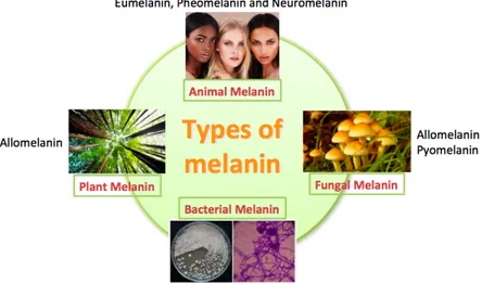
As mentioned above the structure of melanin has not been characterised because of its heterogeneity. To better comprehension of the melanin structure, should be important analyse the classification of melanin according to the source: animal melanin, plant melanin, fungal melanin and bacterial melanin (Figure 5).
Figure 5 Classification of melanin according to the source
I. Animal melanin
Melanin is the main pigment responsible for the various pigmentations found in animal and human skin, hair and eyes. Basically, most of the melanins are dark, from black to brown, but other melanins are reddish or yellowish. According to that, animal melanin is divided into two large groups: eumelanin and pheomelanin. [105,106] Eumelanin provides primarily dark colors, from brown to black. Eumelanin structure has been hampered because it is difficult to study due to its insolubility and resistance. Due to the unavailability of crystallized melanin, the structure could not be ascertained by the most classical physical methods such as X-ray diffraction. Pheomelanin is more treatable than eumelanin because can be dissolved in alkali media. The color of pheomelanin is yellowish or reddish, and it is found in relatively large quantities in red hair, freckles and feathers of fowls and other birds. The main difference of eumelanin in chemical terms is the presence of sulfur. Melanin is also found in certain regions of the brain and adrenal gland of some mammals, this particular melanin is called neuromelanin, it is a dark polymer pigment produced in specific populations of catecholaminergic neurons in the brain, mostly sited in the substantia nigra and in lower proportion in the locus coeruleus. Neuromelanin is a mixture of pheomelanin and eumelanin. Neuromelanin granules should
have pheomelanin in the core and eumelanin in the surface, which is compatible with occasional exhaustion of glutathione or cysteine reduction system during neuromelanin formation [107]. Furthermore, in mammals, melanin is formed even in some other tissues different from the skin, eye, inner ear, and brain, as in the adipose tissue [108]. Aside from mammals, bird feathers contain eumelanin and pheomelanin and the relative amounts of each pigment differ with the avian species [109]. Melanin is also found in reptiles, amphibians, and fish. These animals can change color relatively rapidly to camouflage themselves against changing backgrounds so that melanin is more versatile and offers hue due to the existence of chromatophores and iridophores in addition to melanocytes [110]. The ink in the cuttlefish contains a special type of melanin called sepiamelanin. This melanin is mostly eumelanin, as it is formed by an irregular heteropolymer of indole and carboxylated pyrrolic units with some associated protein [111]. In insects, melanins are present to protect their soft bodies by a process called sclerotization [112]. The main precursor for that process is N-acetyldopamine, which is derived from the amino acid L-tyrosine, so this melanin should be considered as a special type of eumelanin [113].
II. Plant melanin
The presence of nitrogen is important for plant growth. According to that, the amino acid L-tyrosine is not used for the synthesis of plant melanin. They employ some phenol nitrogen-free precursors, such as catechols, dihydroxynaphthalenes or other types of dihydroxybenzenes to carry out this part of its secondary metabolism. In general, the obtained melanin is a polymer without nitrogen and is generically named allomelanin. In plants, the most common precursor is just catechol, so the melanin formed is also named catechol-melanin. and the enzymatic system involved in the synthesis is named catechol oxidase [114,115].
The color of allomelanin is always from dark brown to totally black, and its structure depends on the nature of the main unit
oxidized. Some vegetables use just normal catechol, but others use different catecholic acids such as caffeic, protocatechuic, chlorogenic or gallic acids. According to that, the melanin pigment is a polymer derived from these catechols and the corresponding quinones formed by the action of catechol oxidases [116].
III. Fungal melanin
Fungal melanin is quite abundant and appears in the cell wall [117]. The precursors and the nature of the corresponding melanin structure show variability. The first characterisations suggested catechol precursors form allomelanin devoid of nitrogen by similarity to plant melanins. However, the precursor identified was not catechol, but hydroxylated naphthalene-derived molecules. In this way, a new subtype of allomelanins was described, the DHN-melanins (dihydroxynaphthalene melanin). This type of melanin is quite common in ascomycetous and some imperfect fungi. DHN-melanine has also been identified in the yeast. In basidiomycetes, melanin is formed from a benzoquinone obtained by the action of tyrosinase on the precursors, gamma-glutaminyl-4-hydroxybenzene (GHB). This synthesis could be considered specific to mushrooms and different from all other melanin pigments. Other basidiomycetes as Cryptococcus neoformans are also able to synthesize pyomelanin derivated from a fungal metabolite, homogentisic acid (HGA) [118]. The possibility to synthetize two different types of melanin depending on the environmental conditions. This characteristic was also observed in Aspergillus fumigatus, which is able to form DHN-melanin and pyomelanin starting L-tyrosine through homogentisic acid and in Aspergiullus nidulas that forms the DHN-melanin and DOPA-melanin [119–121].
IV. Bacterial Melanin
In a great number of bacterial species were found melanin pigments characterised by black or dark brown colors.
the tyrosinase, its structure, catalytic mechanism, regulation and its expression. Simultaneously, they were used to analyse the final product, melanin [122,123]. In Streptomices, melanins contain nitrogen and their structure is similar to animal eumelanin. However, there are also bacteria such as Azotobacter that can form allomelanin from catecholic precursors [124]. Some bacteria are able to produce DHN-melanin and others as
Pseudomonas are able to produce pyomelanin from
homogentisic acid, in fact, was observed that some bacteria synthesize pyomelanin in response to specific physiological conditions that are stressful for theirs. Under those conditions, there is an accumulation of homogentisic acid due to the low activity of homogentisate dioxygenase. Consequently, homogentisic is oxidised and pyomelanin is formed [116].
1.7.4 Melanin characterisation by EPR Spectroscopy
Melanin is an amorphous organic polymer with numerous intermediate free radicals trapped in its structure. These paramagnetic centers are responsible for the special paramagnetic properties of melanin. For this Electron Paramagnetic Resonance (EPR) became an important spectroscopic technique to study and characterise this polymer.
In fact, EPR spectroscopy is useful to identify and study chemicals that have one or more unpaired electrons, though their paramagnetic properties is possible to identify their structure and characteristics [125]. Moreover, EPR spectroscopy can be considered as one of the most advantageous methods to investigate melanin and to estimate the concentration of pheomelanin and eumelanin.
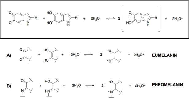
Investigation of synthetic (5-S-cysteinyldopa) polymer of pheomelanin showed that the EPR spectra of free radicals in cysteinyldopa and dopa-melanin are clearly different. The dopa-dopa-melanin radicals appear to be ‘O-C-C-O’ semiquinones (Figure 6,a) whereas cysteinyldopa melanin radicals are ‘O-C-C-N’ semiquinonimines (Figure 6,b). Consequently, ‘O-C-C-O’ semiquinones give singlet-shaped EPR spectra. This is caused by interactions of unpaired electrons with oxygen nucleus resulting in no hyperfine splitting, whereas the interaction between an
unpaired electron and nitrogen 14N nucleus (I=1) gives a triplet-shared EPR spectrum [126,127].
Figure 6 Formation of semiquinone radicals in melanin. a): eumelanin b):
pheomelanin
1.7.5 EPR Spectroscopy
Spectroscopy is the measurement and interpretation of the energy differences between the atomic and molecules states to collect information about the identity, structure and dynamics of the sample analysed.
These energy differences expressed by ΔE have an important relationship with the absorption of electromagnetic radiation. According to Planck’s law, electromagnetic radiation will be absorbed if:
ΔE = hν (2)
where h is Planck’s constant and ν is the frequency of the radiation. The absorption of energy causes a transition from a lower energy state to a higher energy state. In conventional EPR spectroscopy, ν is maintained constant and the magnetic field H is varied until the ΔE is
matched by the frequencies at which absorption occurs, through the so-called “resonance condition”.
hν = gβH
For EPR experiments, radiation in the gigahertz range (GHz) with a wavelength of a few cm is used [128].
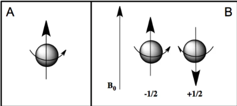
An isolated electron, without any outside forces, has an intrinsic angular moment called “spin”. An electron has a charge and the angular motion of this charge generates a magnetic field then it acts like a little bar magnet or magnetic dipole with a magnetic moment indicated with µ (Figure 7,a).
EPR spectroscopy studies the energy differences due to the interaction of unpaired electrons in the sample with a static magnetic field. This effect is called the Zeeman Effect. The magnetic field, B0, produces two
energy levels for the magnetic moment of the electron. The unpaired electron will have a state of lower and highest energy when the magnetic moment is aligned with the magnetic field (Figure 7,b).
Figure 7 a) Electron spin and magnetic moment of unpaired electron; b)
Minimum and maximum energy orientation of magnetic moment respect to magnetic field B0
The two states are labelled by the projection of the electron spin, ms, on
the direction of the magnetic field. Because the electron is a spin 1/2 particle, the parallel state is designed as ms= +1/2 and the antiparallel
state is as ms= +1/2. The energy of each orientation is the product of µ
called the Bohr magneton and ge is the spectroscopic g-factor of the free
electron and equals 2.0023192778 (≈ 2.00).
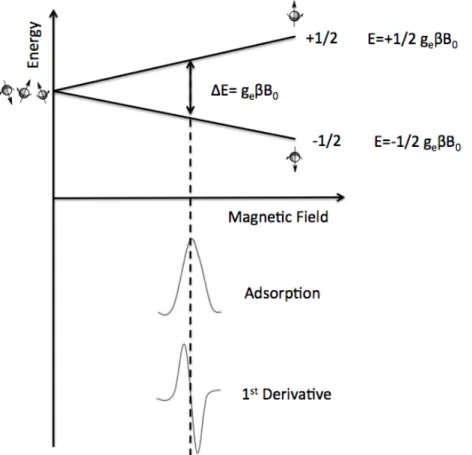
As described in figure 8, there are two energy levels for the electron in a magnetic field.
The distance between Zeeman levels is characteristic of the particular type of paramagnetic centres and is expressed by g-factor (g= hν/βB0).
The position of the middle of EPR spectrum provides information about g-value and on the type of paramagnetic centre, while the quantitative information is delivered by the field under the curve of adsorption of microwaves (the internal intensity of the signal).
Figure 8 Zeeman splitting of the energy levels of the system of unpaired spins
in the external magnetic field. Below the absorption curve and its first derivative.
In real systems, electrons are normally associated with one or more atoms. There are several important consequences of this:
i. An unpaired electron can gain or lose its angular momentum, which can change the value of g-factor. For every paramagnetic molecule, there exists a unique axis system called the principal axis system. The g-factor measured along these axes are called the principal g-factors and are labeled gx, gy and gz.
ii. Systems with multiple unpaired electrons experience electron-electron interactions that give rise to ‘fine’ structure. This is realized as zero field splitting and exchange coupling and can be large in magnitude.
iii. The magnetic moment of a nucleus with a non-zero nuclear spin will affect any unpaired electrons associated with that atom. This leads to the phenomenon of hyperfine coupling, splitting the EPR resonance signal into doublets, triplets and so forth. Additional smaller splittings from nearby nuclei is called “superhyperfine” coupling.
iv. Interactions of an unpaired electron with its environment influence the shape of an EPR spectral line.
v. These effects in an atom or molecule may not be the same for all orientations of an unpaired electron in an external field. This anisotropy depends upon the electronic structure of the atom or molecules in question, and so can provide information about atomic or molecular orbital containing the unpaired electron.
With the intensity of the applied magnetic field increasing, the energy difference between the energy levels widens until it matches with the microwave radiation, and results in the absorption of photons. This is the fundamental basis for EPR spectroscopy. EPR spectrometers typically vary the magnetic field and hold the microwave frequency. EPR spectrometers are available in several frequency ranges (Table 2).
EPR experiments often are conducted at X and, less commonly, Q bands, but the low spectral resolution over g-factor at these wavebands limits the study of paramagnetic centers with comparatively low anisotropic magnetic parameters.
Table 2 List of microwave frequencies commonly available in EPR
spectrometers
Microwave Band Frequency (GHz)
L 1.1 S 3.0 X 9.5 Q 35 W 90 J 270
Continuous wave (CW) are recorded by putting a sample into a microwave irradiation field of constant frequency and sweeping the external magnetic field B0 until the resonance condition is fulfilled.
In pulse EPR the spectrum is recorded by exciting a large frequency range simultaneously with a single power microwave pulse of a given frequency at constant magnetic field B0.
Most EPR applications still make use of continuous wave methods as the recording and interpretation of pulse EPR spectra requires sophisticated technical equipment and a more advanced theoretical background. A significant advantage of CW EPR with respect to the pulse methods is the higher sensitivity. A further limitation of pulse EPR is also the low measuring temperatures imposed by the short relaxation of the transverse magnetization involved in pulse experiments, especially for transition metal ions. CW EPR spectra on the other hand can be recorded at room temperature for a large number of spin systems, including radicals and transition metal ions. The additional information about weakly coupled nuclei and relaxation properties of the spin system that can be obtained by manipulating the spins with sequences of MW pulses explains on the other hand the efforts put into the development of new pulse methods. In fact, CW and pulse EPR are complementary and only the application of both gives a reliable picture of the spin system [129,130].
2.1 MNPs synthesis
Superparamagnetic nanoparticles Fe3O4 (MNPs) were synthetized using
chemical co-precipitation of Fe3+ and Fe2+ ions. Two different methods were using which in the following text will be called 1 and MNPs-2:
1) MNPs-1: FeCl3⋅6H2O (2,335 g, 2 eq.) and Fe(SO4)2(NH4)2SO4⋅6H2O
(1,863 g, 1 eq.) were dissolved in 50 ml ultrapure water and were left for about 30 minutes to bubble under nitrogen until the temperature reached 60 °C. Then 45 ml of a solution 1 M di NaOH was added. The reaction solution quickly changed colour and became black, the heat was turned off and was left to react under nitrogen for 1 hour. Afterwards, the Fe3O4 nanoparticles were separated from the reaction
solution with a magnet and washed several times with ultrapure water until the pH solution became neutral. The nanoparticles were dried dried under N2 flux.
2) MNPs-2: 1.62 g ferric chloride and 0.64 g ferrous chloride (ratio 2:1) were dissolved in 50 ml of bidistilled water and mixed in a flask with two necks under nitrogen. After 10 minutes, 3 ml of ammonia solution (NH3OH 25%) was added drop by drop, a black precipitate was
observed and left to react for 1 hour. Then, the nanoparticles were separated and washed with ultrapure water using several cycles of ultracentrifuge (cycle: 30000 rpm, 20 degrees and 1 hour). After that, the nanoparticles were freeze-dryer [131].
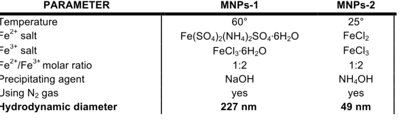
Table 3: Comparison between the MNPs-1 and MNPs-2 synthesis and
nanoparticles characteristics
PARAMETER MNPs-1 MNPs-2
Temperature 60° 25°
Fe2+ salt Fe(SO4)2(NH4)2SO4⋅6H2O FeCl2
Fe3+ salt FeCl3⋅6H2O FeCl3
Fe2+/Fe3+ molar ratio 1:2 1:2
Precipitating agent NaOH NH4OH
Using N2 gas yes yes
MNPs-1 and MNPs-2 were synthetized by co-precipitation of ferric and ferrous ions. In the co-precipitation method, two strategies are involved: a short burst of nucleation occurs when the concentration of the species reaches critical supersaturation, and then, there is a slow growth of the nuclei by diffusion of the solutes to the surface of the crystal. To produce monodisperse iron oxide nanoparticles, these two stages should be separated; i.e., nucleation should be avoided during the period of growth [53].
The co-precipitation process depends mostly on parameters such as reaction temperature, pH, nature of salts, type of precipitant agent and Fe2+/Fe3+ molar ratio. The parameters of synthesis were changed to study the effect of the correlation between those parameters and the nanoparticles size (Table 3).
The change in the reaction temperature can bring to control the size of magnetic nanoparticles. The average size and the distribution percentage size of Fe3O4 nanoparticles increase with the reaction
temperatures due to the acceleration of chemical reaction of Fe2+ and Fe3+ ions [132–134]. MNPs-1 and MNPs-2 synthetized at 60°C and 25°C respectively did not report many differences in size but the lower temperature used for the synthesis, improved the distribution percentage size, obtaining a hydrodynamic diameter of 49 nm for MNPs-2.
Another factor affecting the size of magnetic nanoparticles is the Fe2+/Fe3+ molar ratio. In both synthesis the ratio were 1:2 [135].
The presence of inert gas such as nitrogen is necessary for the production of magnetic nanoparticles because magnetite (Fe3O4) in
presence of oxygen can be oxidized into maghemite (γ-Fe2O4) [53].
The precipitant agent plays another important role in the reaction system, NaOH was used for MNPs-1 and NH4OH for MNPs-2.
NaOH has a high concentration of OH- and the higher pH value, with the same concentration of alkali, will accelerate the reaction and lead to large particle size. On the contrary, NH4OH releases OH- gradually to
make the particle size controllable and the process more stable [136]. In this context, NH4OH favours a better dispersion of MNPs-2, however, no
The ionic strength is another effect that allows the control of size and dispersion of magnetic nanoparticles. The higher ionic strength of the solution causes a reduction of zeta potential at a constant pH, resulting in lower electrostatic stability and an increase of the hydrodynamic diameter of the particles [137].
The different nature of salts for MNPs-1 and MNPs-2 synthesis affects the ionic strength, MNPs-1 have a higher ionic strength that increases their hydrodynamic diameter.
2.2 MNPs characterisation
In order to test the stability of the dispersions of MNPs-1 and MNPs-2, several solutions with different pH (from 3 to 9) were prepared, the ζ-potential and size were measured for those samples (Figure 9). The surface charge of magnetic nanoparticles depends on the pH value. In an aqueous system, the surface is covered with groups of –FeOH. At lower pH (pH < isoelectric point of MNPs), the higher content of H+ caused the protonation of OH and the formation of –FeOH2+. Instead,
increasing the pH value (pH > isoelectric point of MNPs), the OH are deprotonated forming FeO-, which gives a negative charge to the surface of the nanoparticles [138]. According to this, the z-potential of MNPs-1 showed a positive and negative charge before and after their isoelectric point. The different behaviour was observed for MNPs-2, that at high pH values they did not have a negative charge. This suggests the presences of NH4+ on the nanoparticles surface. It was assumed
that during the synthesis of MNPs conducted under alkali conditions [131], the NH4+ from precipitating agent (NH4OH) was attracted by –OH
-of nanoparticles, giving them a positive charge. This interaction was reported by Unsoy et al. in the synthesis of chitosan coated iron oxide nanoparticles, where –NH3+ groups of chitosan were attracted by –OH
-of iron oxide [139].
Another point that supports the hypothesis of the presence of NH4+ on
the nanoparticles surface is the shift of isoelectric point. From the literature, The isoelectric point of MNPs-NH2 shifts to higher pH values
absorbed at the nanoparticles surface the isoelectric point shifts to lower pH values [141].
Figure 9 MNPs pH stability. A) Size comparison between 1 and
MNPs-2 with different pH (from 4 to 9); B) ζ-potential comparison between MNPs-1 and MNPs-2 with different pH (from 4 to 9)
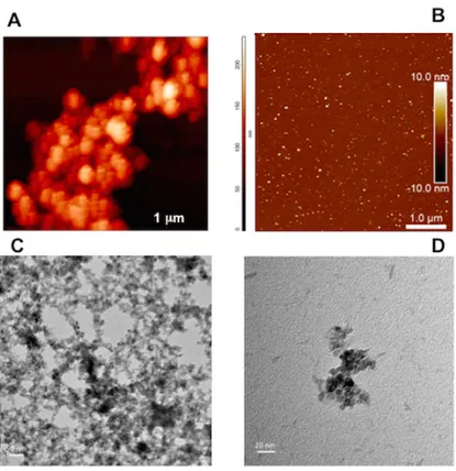
Morphology and size of nanoparticles were evaluated using AFM (Figure 10, A-B). Both nanoparticles showed a spherical shape and the average size of 2-20 nm. Furthermore, the Figure 10, B highlights the excellent dispersion of MNPs-2. TEM analysis for MNPs-2 was carried out to confirm the results obtained by AFM (Figure 10,C-D).
MNPs-1 and MNPs-2 were characterised by FT-IR (Figure 11). In the IR spectra, the broad peak around 3000-3500 cm-1 is due to the O-H stretching vibration and the peak at 1623 cm-1 can be attributed to the bending vibration of adsorbed water molecules. The presence of NH4+
on the nanoparticles surface can be confirmed by FT-IR spectroscopy. In the Figure 11, the MNPs-2 spectrum (red line) has a peak at 1427 cm-1 attributed to the deformation of NH4+ associated with a peak at
Figure 10 Characterisation of MNPs-1 and MNPs-2. A) AFM of MNPs-1; B)
AFM of MNPs-2; C) TEM of MNPs-2 in 50 nm; D) TEM of MNPs-2 in 20 nm
2.3 Laccase immobilisation on MNPs
Laccase from Trametes versicolor (Tv) was immobilised on magnetic nanoparticles following various procedures.
In MNPs-1 the laccase was immobilized through covalent bond. For this purpose the MNPs-1 were functionalized with amino groups using APTES. MNPs-1 (1 g, 4.32⋅10-3 mol) were dissolved into EtOH (332 ml) [144]. Than, APTES (8.0 ml, 0.034 mol) was added to the Fe3O4
nanoparticles solution and its pH was adjusted to 5 by the drop-wise addition of HCl 1N (34.6 ml). After stirring at 25 °C for 24 h, the amino-coated Fe3O4 nanoparticles were collected using a permanent magnet,
washed with ethanol three times and last time with ultrapure water, and then dried under nitrogen flux. The amino-coated Fe3O4 nanoparticles
were dissolved into phosphate buffer (PBS) (100 Mm, pH 7.1) and sonication was performed on the solution for 15 min. Successively, to extend the spacer and facilitate the covalent attachment of laccase, the amino groups on the magnetic nanoparticles were linked to aldehyde groups by treating them with glutaraldehyde. The linker allows the higher mobility of the enzyme in the space, favouring the reaction with the substrate [66]. The glutaraldehyde solution was added to the solution, which was stirred at 150 rpm at 25 °C for 2 hours.
The sample of MNPs-1-APTES-glutaraldehyde were collected using the permanent magnet and washed with phosphate buffer three times to remove the unreacted glutaraldehyde [63]. The laccase solution was added to MNPs-1-APTES-glutaraldehyde complexes and the solutions stirred at 150 rpm at 25 °C for 16 hours.[145] Finally, the magnetic nanoparticles Fe3O4-Laccase were separated magnetically and washed
two times with 0.5 ml of acetate buffer (100 Mm, pH 4.5) to remove the free enzyme. The washing buffer was collected for the determination of the activity. The activity of laccase immobilised on MNPs-1 was measured using the dye 3-Amino-4-hydroxybenzene sulfonic acid (0.25 mM) as a substrate in acetate buffer (pH 4.5). The formation of a coloured due to the oxidation of the substrate by the enzyme was analysed measuring the absorbance increase at 420 nm (ε/dm3 mol-1 cm-1 8600) with a UV-Vis Spectrophotometer [19].
separate the solution from the nanoparticles (2B, Scheme 2). Then, the laccase solution was added and left to react for 16 hours at 150 rpm at 25 °C. Finally, the magnetic nanoparticles with linker and laccase were separated magnetically and washed one time with 1 ml of acetate buffer (3B, Scheme 2). The washing buffer was collected to analyze the activity. The activity of immobilised laccase for the other MNPs-2 route was measured using 2,2´-azino-bis (3-ethylbenthiazoline-6-sulphonic acid) (ABTS, 0.02 mM) as substrate in acetate buffer (pH 4.5, 100 mM), and the formation of product (ABTS·+) was analyzed measuring the absorbance increasing at 420 nm (ε= 3.6 × 10⁴ M-1 cm-1).
The enzymatic immobilization was confirmed by FTIR spectroscopy. The FTIR spectra measured for the MNPs-1 functionalized with amino group (NH2-MNPs 1), for the laccase conjugated to the NH2-MNPs-1
(Laccase-MNPs-1) and for laccase are shown in Figure 12, A. The spectrum of the NH2-MNPs-1 is similar to those reported in the literature
for amino silanized magnetite NPs [148–151].
The intense band peaked at 990 cm-1 is due to the stretching of the Si-O-Si bonds of the silane coating [148,150]. The bands at ca. 1510 and 1615 cm-1 can be ascribed to NH2 involved in hydrogen bonds and to
free NH2 groups, respectively.
The changes in the FTIR spectra after laccase immobilization on the NH2-NPs are more clearly visible in the insect of Fig. 12,A, where the
region from 1300 to 1800 cm-1 is shown. The peak at 1644 cm-1 in the middle curve corresponds to the amide I vibrational mode of the amide groups whereas the band at ca. 1530 cm-1 to the amide II vibrational mode. Probably, to the last band there is also a contribution from the unreacted amino groups at the surface of the NPs. In the Figure 12,B is possible to see the spectra of each product of the various immobilization steps on MNPs-2 with and without linker. The changes in the FTIR spectra after laccase immobilization were observed in the region 1250-1750 cm-1.
conformation. Consequently the accessibility and accommodation of the substrate may be reduced [152].
Table 4 Optimization of immobilisation conditions testing different
concentration of support, laccase and glutaraldehyde. All the experiments were carried out for three times.
Support (mg) Immobilisation* yield (%) Activity§ recovery (%) MNPs-1 25 35 94 50 78 65 100 57 15 MNPs-2 (with linker) 25 17 15 50 40 14 100 25 10 MNPs-2 (without linker) 25 23 18 50 47 30 100 30 30 *
Immobilisation yield (%)= immobilised activity/ starting activity x 100; §Activity recovery (%)= observed activity/ starting activity x 100
MNPs-1 and MNPs-2 with and without linker were reused for 10 cycles. For MNPs-1, laccase maintains the same activity for 10 consecutive reuses. Instead for MNPs-2 a small decrease of the activity was observed, but than it was stabilized (Figure 13).
According to these results, for the immobilization it seems more convenient to use MNPs-2 with 0.5 mg/ml of laccase solution (87% of
Laccase concentration Immobilisation* yield (%) Activity§ recovery (%) MNPs-1 0.5 mg/ml 87 50 1 mg/ml 80 43 2 mg/ml 51 37 MNPs-2 (with linker) 0.5 mg/ml 97 83 1 mg/ml 69 65 2 mg/ml 60 53 MNPs-2 (without linker) 0.5 mg/ml 87 75 1 mg/ml 54 46 2 mg/ml 52 43
immobilization yield and 75% of activity), since it is easier to immobilize the enzyme in only one step. Moreover, it does not need the use glutaraldehyde that it is toxic.
Figure 13: Reusability of laccase immobilised on MNPs-1, MNPs-2 with and
without linker.
During the immobilization test a strange (an unexpected) behaviour was observed using ABTS as substrate. Usually, Laccase oxidises ABTS to form the stable cation radical ABTS⋅+ (Figure 14) [13].
The ABTS substrate is commonly used to test the laccase activity as the stable cationic radical formed originates a peak in the UV-vis spectrum at 420 nm which is used to follow the oxidation reaction. ABTS is added to the solution with immobilized laccase and then with magnet the solution is isolated from the MNPs with laccase. This solution is analysed by UV-vis spectrophotometry to determine by UV-visible analysis the enzyme activity.
The UV-vis analysis shows that the concentration of ABTS (responsible of the peak at 340 nm) decrease during the reaction with MNPs-1-laccase but the peak of the cation radical ABTS⋅+ at 420 nm is not
observed (Fig. 15, A).
0 1 2 3 4 5 6 7 8 9 10 0 50 100 Activity recovery (%) Number of cycles MNPs-1 MNPs-2 with linker MNPs-2 without linker