DOTTORATO DI RICERCA IN BIOLOGIA
CELLULARE E MOLECOLARE
Dipartimento di Farmacia e Biotecnologie
XXVI Ciclo
Settore Concorsuale di afferenza: 05/E2
Settore Scientifico disciplinare:
BIO/11
TITOLO TESI
Clostridium difficile toxins facilitate bacterial colonization by
modulating the fence and
gate function of colonic epithelium
Presentata da: MAGDALENA JULIA KASENDRA
Coordinatore Dottorato: Relatore:
Chiar.mo Prof. VINCENZO SCARLATO Chiar.ma Prof. VINCENZO SCARLATO Co-relatore:
Chiar.ma Dott.ssa ROSANNA LEUZZI
2
Abstract
The contribution of Clostridium difficile toxin A and B (TcdA and TcdB) to cellular intoxication has been extensively studied, but their impact on bacterial colonization remains unclear. By setting-up two- and three-dimensional in vitro models of polarized gut epithelium, we investigated how C. difficile infection is affected by host cell polarity and whether TcdA and TcdB contribute to such events. Indeed, we observed that C. difficile adhesion and penetration of the epithelial barrier is substantially enhanced in poorly polarized or EGTA-treated cells, indicating that bacteria bind preferentially to the basolateral cell surface. In this context, we demonstrated that sub-lethal concentrations of C. difficile TcdA are able to alter cell polarity by causing redistribution of plasma membrane components between distinct surface domains. Taken together, the data suggest that toxin-mediated modulation of host cell organization may account for the capacity of this opportunistic pathogen to gain access to basolateral receptors leading to a successful colonization of the colonic mucosa.
3
Acknowledgements
This work was conducted at Novartis Vaccines and Diagnostics in In vitro Cell Biology Group, headed by Marco Soriani. I am very thankful for his welcoming in the lab and providing me with the opportunity to work at the frontier of current knowledge and vaccine development. I equally thank to Nigel Minton who has awarded me with Marie Curie Fellowship and allowed me to perform my thesis project under the framework of ‘CLOSTNET’ - European Community's Seventh Framework Programme [PEOPLE-ITN-2008-237942].
I am also extremely grateful to my supervisors, Dr Rosanna Leuzzi and Prof. Vincenzo Scarlato, for all the support and guidance they have given me over the course of my PhD. Most importantly, their never-wavering optimism that has encouraged me to battle on and eventually produce a thesis of which I am proud.
I would also like to acknowledge all my lab members (past and present) of the In Vitro Cell Biology group or elsewhere at Novartis. I thank Riccardo Barrile for his very fruitful and enriching collaboration in the last few months of my thesis work. I would also like to thank to Christina Merakou, Buket Baddal, Valentina Rippa and Chara Korea for their kind-hearted help, scientific feedback, moral support and for making my PhD experience thoroughly enjoyable.
I would like to equally thank to my doctoral schools, Alma Mater Studiorum at the University of Bologna and PhD Academy at Novartis, for their active drive to provide a dynamic, interdisciplinary and exiting science education. I’m especially grateful to PhD Director Ilaria Ferlenghi for her endless support and faith in me.
To the members of my thesis defence committee. I am thankful that you kindly accepted to take precious time to judge my work.
Finally, I especially thank my boyfriend who never fails to make me smile and all my family for their unconditional love and support throughout my life and imparting me a drive to succeed.
4
Table of Contents
Abstract
_________________________________________________________________ 2Acknowledgments
_______________________________________________________ 3Table of Contents
_______________________________________________________ 4List of Figures __________________________________________________
6List of Publications
______________________________________________________ 7Chapter 1: Introduction
81.1. Clostridium difficile: emergence of a significant human pathogen ________________ 9
1.1.1. Clinical characteristics and epidemiology of C. difficile infection __________ 9
1.1.2. Toxins – major virulence factors of C. difficile _______________________ 13 1.2. Polarized epithelium – a specialized barrier against pathogens _________________ 16 1.3. In vitro tissue culture systems to study pathogens interactions with polarized
epithelium ______________________________________________________________ 17 1.4. Pathogens subvert host cell polarity: an emerging theme ______________________ 19
Aims of the study
21Chapter 2: Results
222.1. Importance of the host cell polarity for their susceptibility to C. difficile infection __ 22 2.1.1. Cell polarity significantly affects C. difficile ability to infect Caco-2 cells __ 22 2.1.2. 3D cyst model confirms the propensity of C. difficile to target basolateral membrane __________________________________________________________ 25 2.2. Effects of C. difficile toxins on the host cell polarity _________________________ 27 2.2.1. TcdA and TcdB modulate the gate function of colonic epithelium ________ 27 2.2.2. TcdA, but not TcdB, is able to modify the fence function of colonic
epithelium _________________________________________________________ 28 2.2.3. TcdA subversion of host cell polarity is not due to apoptosis ____________ 30
5
2.3. Influence of TcdA-mediated subversion of cell polarity on C. difficile ability to
colonize epithelium_______________________________________________________ 32 2.3.1. TcdA-mediated subversion of cell polarity facilitates C. difficile adhesion and translocation of Caco-2 monolayer _______________________________________ 32
Chapter 3: Discussion
36Chapter 4: Experimental Procedure
404.1. C. difficile strains and culture conditions __________________________________ 40 4.2. 2D and 3D cell cultures ________________________________________________ 40 4.3. Dextran permeability and membrane diffusion assays ________________________ 41 4.4. Immunofluorescence microscopy ________________________________________ 41 4.5. Assessment of apoptosis by TUNEL assay and active caspase-3 staining _________ 42 4.6. C. difficile adhesion and translocation assay on 2D Caco-2 monolayers __________ 42 4.7. C. difficile infection of 3D Caco-2 cysts ___________________________________ 43 4.8. Quantification of C. difficile binding to 3D Caco-2 cysts ______________________ 43 4.9. Statistical analyses ____________________________________________________ 44
6
List of Figures:
Figure 1. Rates of discharges from U.S. short-stay hospitals of patients with C. difficile-associated disease listed as primary or as any diagnosis _____________________________ 11 Figure 2. Yearly C. difficile–related mortality rates per million population, United States, 1999–2004 ________________________________________________________________ 12 Figure 3. Structure of toxin A and B from C. difficile. Toxins A and B consist of three major domains __________________________________________________________________ 14 Figure 4. Regulation of Rho GTPases and inhibition of their function by C. difficile toxins _ 15 Figure 5. Cell polarity of epithelial cells _________________________________________ 17 Figure 6. In vitro Caco-2 cells model of polarized epithelium and its optimization for the use in anaerobic conditions ______________________________________________________ 19 Figure 7. Cell polarization-state dependent C. difficile infection of Caco-2 cells _________ 23 Figure 8. Increase in C. difficile capacity to colonize and penetrate epithelium induced by EGTA treatment ___________________________________________________________ 24 Figure 9. Adhesion of C.difficile to Caco-2 AP-side-out and BL-side-out cysts __________ 26 Figure 10. Effect of TcdA and TcdB on gate function of tight junctions (TJs) determined by the paracellular passage of non-ionic tracer ______________________________________ 27 Figure 11. Effect of TcdA and TcdB on the fence function of tight junctions (TJs) _______ 29 Figure 12. Effect of sub-lethal concentrations of C. difficile toxins on Caco-2 apoptosis ___ 31 Figure 13. Effect of TcdA on 630 C. difficile colonization of colonic mucosa ___________ 33 Figure 14. Effect of TcdA on R20291 C. difficile colonization of colonic mucosa ________ 34 Figure 15. Effect of TcdA on C. difficile colonization of partially polarized Caco-2 cells __ 35 Figure 16. Effect of TcdB on 630 C. difficile colonization of colonic mucosa ___________ 35 Figure 17. Schematic diagram illustrating the proposed model of the pathogenesis of C. difficile infection ___________________________________________________________ 39
7
List of Publications:
Kasendra M, Barrile R, Leuzzi R, Soriani M: Clostridium difficile toxins facilitate bacterial colonization by modulating the fence and gate function of colonic epithelium. J Infect Dis. 2013 Dec 13.
Kovacs-Simon A, Leuzzi R, Kasendra M, Minton N, Titball RW, Michell SL. Lipoprotein CD0873 is a novel adhesion of Clostridium difficile. J Infect Dis. 2014 Jan 29.
8
Chapter 1: Introduction
Discovery and development of antimicrobial agents was undoubtedly at the forefront of research in the 20th century with one of the first and still most widely used antibiotics - penicillin. Given its potent activity against various kinds of bacteria, penicillin was immediately recognized and subsequently used as a therapeutic agent for the treatment of many infectious diseases. However the collateral biological cost of this broad-spectrum activity has been identified only recently. Unrelenting rise in the occurrence of antimicrobial resistance by many common human pathogens and decrease in the effectiveness of vancomycin, which is seen as the last line of defence against many microorganisms, have demonstrated the importance of the commensal microbiota for human health. For instance destruction of intestinal microflora by antibiotic treatment has been identified as the first and essential step in the pathogenesis of infectious diarrhoea caused by Clostridium difficile.
C. difficile is a Gram-positive, obligate anaerobic, spore-forming bacterium which has recently emerged as a significant pathogen of the human gastrointestinal tract. While it is found as a part of the commensal flora only in up to 3% of healthy adults, upon antibiotic treatment it efficiently colonizes gut leading to a broad range of disease manifestations ranging from asymptomatic carriage, mild diarrhoea to life-threating toxic megacolon and pseudomembranous colitis (PMC). [1, 2]. Acquisition of C. difficile occurs by oral ingestion of spores that resist the acidity of stomach and germinate into vegetative bacteria in the small intestine. Upon reaching colon C. difficile adheres and becomes pathogenic through production of two potent toxins, TcdA and TcdB. Both toxins belong to the family of large clostridial glucosylating toxins and inactivate host GTPases (including Rho, Rac, and Cdc42), leading to alteration of the epithelial barrier, damage to human intestinal mucosa, and inflammation of the colon [3-6]. The high rate of recurrent disease, even after repeated
9
antimicrobial treatments [7], suggests that C. difficile has evolved mechanisms to persist in the intestinal tract. This long-term establishment is likely mediated by multiple colonization strategies. While the effects of toxins on epithelial cells have been largely described in vitro, little is known about how they might benefit bacteria during the host colonization process. The recognition of Rho family GTPases as master regulators of cell polarity in eukaryotes [8] led us to hypothesize that toxin-mediated subversion of the epithelial polarity might serve as an important strategy for bacterial settlement in the gut.
In the present study, we postulate that C. difficile TcdA is able to perturb epithelial polarity by causing redistribution of plasma membrane components between distinct surface domains. As a consequence, subverted cell polarity enables bacteria to gain access to the basolateral surface where they display a preferential association. Therefore, TcdA might play an important role in the colonization of colonic epithelium not only by disrupting its barrier function but also by perturbing epithelial polarity and promoting mucosal association.
1.1.
Clostridium difficile: emergence of a significant human pathogen
1.1.1. Clinical characteristics and epidemiology of C. difficile infection
The primary clinical manifestation of C. difficile infection (CDI) is diarrhoea, accompanied by fever and abdominal pain. Continuous accumulation of C. difficile toxins in the human colon induce a damage of the intestinal mucosa and an acute inflammatory response. A sloughed-off layer of mucous, fibrin, dead epithelia and leukocytes form characteristic pseudomembraneous structures over the internal surface of the colon [9]. Prolonged infection might lead to more serious complication namely toxic megacolon, in which abnormal colon dilation is often accompanied by a paralysis of the peristaltic movements and the risk of rupture [10]. Also a kidney failure might occur in the case of severe dehydration. Any of these sequelae if left untreated can eventually result in patient’s death. Recent reports reveal also
10
the occurrence of a systemic dissemination of C. difficile toxins that leads to a severe cardiovascular damage and consequently to death [11, 12].
With the widespread of overuse and/or inappropriate use of antibiotics, C. difficile became endemic in hospitals and chronic-care facilities worldwide. Nowadays it is recognized as a major cause of up to 20-25% of cases of antibiotic-associated diarrhoea and almost 100% of PMC [13]. Susceptibility to CDI results from the perturbations of the normal gut microflora induced by the exposure to almost any antibiotic and persists until the flora is fully restored. This might occur even a long time after the end of the therapeutic treatment.
High risk for CDI was evidenced for ampicillin (amoxicillin) and cephalosporins that are widely prescribed and for which C. difficile exhibits a natural resistance [14-18]. The emergence of antibiotic-resistant strains of C. difficile during the last three decades resulted in a series of out-breaks of CDI [19]. For instance, a common use of clindamycin for the treatment of colitis gave a rise to several epidemics throughout the 1980’s and 1990’s caused by clindamycin resistant strains [20]. These strains possessed the erm gene, which confers resistance to macrolides, lincosamides and streptogramins [20, 21]. Recently fluoroquinolones have been the major class of antimicrobials implicated in CDI, since the current epidemic strain exhibit high resistance to this class of broad-spectrum antibiotics [19, 22, 23]. Fortunately, bacterial resistance to the two antibiotics primarily used for CDI treatment, vancomycin and metronizadole, has not emerged so far, although a significant increase in the amount of metronidazole required for the patient recovery has been observed [24]. In the search for additional treatment options, other drugs have been tested, such as rifaxamin, unfortunately resulting in a rapid acquisition of resistance [25]. Please refer to the review by Kelly and Lamont for the further details on the clinical treatment of CDI and its challenges [26].
11
In the recent years, the epidemiology of CDI has changed dramatically, with increases noted in the morbidity and mortality of disease in North America, Canada and Europe. In United States, national surveillance data indicate that the number of hospital discharges with CDI listed as a primary diagnosis (which refers to patients for whom CDI is responsible for the hospitalization) or any diagnosis (which reflects discharged patients for whom CDI was either a primary or a secondary diagnosis, including those patients who may have contracted CDI during the current hospitalization, as well as those who acquired it by other means) increased significantly between 2000 and 2003 (Figure 1) [27].
Figure 1. Rates of discharges from U.S. short-stay hospitals of patients with C. difficile-associated disease listed
as primary or as any diagnosis.
Source: McDonald LC, Owings M, Jernigan DB, 2006.
More recent statistics reveal even a fourfold increase in the number of hospital discharges with CDI between 1993 and 2009, increasing from approximately 85,700 cases in 1993 to 336,600 cases in 2009. Patients older than 65 years of age have been most affected, representing over two-thirds of all patients with this disease [28]. Nevertheless, CDI is
12
increasingly recognized in patient populations previously felt to be at low risk, including children, healthy peripartum women, and other healthy people with no recent healthcare contact or antimicrobial exposure [29].
In parallel with the rising incidence of CDI, a significant increase in the severity of the disease has been observed, with greater numbers of complications and mortality related to CDI. Reports of CDI outbreaks in hospitals in Quebec, Canada, and subsequently in the United States, emerged, describing severe cases associated with higher numbers of colectomies, treatment failures, and deaths than were ever reported before [19, 22, 30]. For instance, in 2004, the mortality rate of nosocomial CDI in Quebec hospitals was 6.9%, compared to 1.5% among Canadian hospitals in 1997 [30, 31]. In United States, death certificate data showed that mortality rates from CDI increased from 5.7 per million population in 1999 to 23.7 per million in 2004 (Figure 2) [32].
Figure 2. Yearly C. difficile–related mortality rates per million population, United States, 1999–2004. Source:
13
The reasons for these variations in epidemiology include changes in antimicrobial use or infection control practices, but also the emergence of a new hypervirulent strain of C. difficile, namely BI/NAP1/027. This epidemic strain was found to be associated with the outbreaks in Quebec and subsequently in Europe [22, 23, 30] It is known to produce 16-fold higher concentrations of toxin A and 23-fold higher concentrations of toxin B in vitro than toxinotype 0 strains [33]. Additionally it secretes another toxin called binary toxin, that may be associated with more severe diarrhea [34]. Therefore, the extreme virulence of the BI/NAP1/027 strain may result from a combination of different factors that include increased toxin A and B production, binary toxin, or other yet unknown determinants.
1.1.2. Toxins - major virulence factors of C. difficile
The virulence of C. difficile is conferred primarily by 2 large exotoxins, toxin A (308.0 kDa) and B (269.6 kDa), encoded by their genes, tcdA and tcdB, which are located on a 21-kilobase section of chromosomal DNA known as the pathogenicity locus (paloc). Toxin-negative C.
difficile strains are considered nonpathogenic. In addition to toxins A and B, some strains also
produce a binary toxin, encoded by ctdA and ctdB, located outside the paloc. The role of binary toxin in the pathogenesis of C. difficile remains unclear; however, its presence in BI/NAP1/027 epidemic strains has raised concerns about its synergism with toxins A and B in causing severe colitis [26].
Belonging to the family of large clostridial toxins, C. difficile toxins A and B are single-chain proteins with three functional domains: (a) the C-terminally located receptor-binding domain, (b) the middle hydrophobic domain involved in membrane translocation and (c) the N-terminal catalytic domain harbouring the glucosylotransferase activity (Figure 3) [35].
14
Figure 3. Structure of toxin A and B from C. difficile. Toxins A and B consist of three major domains.
Source Jank, T., T. Giesemann, and K. Aktories. 2007.
Both, TcdA and TcdB, catalyse the mono-O-glucosylation of the Rho GTPases (Rho, Rac, Cdc42 isoforms) at a threonine residue (Thr35/37), which is essential for their switch from ‘inactive’, GDP-bound to ‘active’, GTP-bound state [3]. Importantly, only in the ‘on’ (GTP) state, GTPases are able to recognize target proteins and generate a response until GTP hydrolysis returns the switch to the ‘off’ state. Rho proteins are involved in numerous signal processes, including regulation of actin cytoskeleton, cell cycle progression, gene transcription, proliferation and apoptosis. They also play essential roles in host–pathogen interactions by participating in epithelial barrier functions, immune cell migration, phagocytosis and cytokine production (Figure 4) [8, 36]. Therefore the inactivation of Rho proteins mediated by clostridial toxins might lead to a series of sequelae, including disaggregation of actin cytoskeleton, increased intestinal permeability, strongly enhanced cytokine production, apoptosis and even cell death.
15
Figure 4. Regulation of Rho GTPases and inhibition of their function by C. difficile toxins.
Source: Jank, T., T. Giesemann, and K. Aktories. 2007
Recently Rho family GTPases have been also recognized as major evolutionarily conserved regulators of polarity in eukaryotic cells, from yeast to mammals. Among G-proteins, RhoA, Cdc42, and Rac1, in particular, play essential roles in establishing polarity in different contexts, including asymmetric cell division, wound healing, apical–basal polarity of epithelial cells, and front–rear polarity of migrating cells [37-39]. Cell polarization, i.e., the asymmetric distribution of subcellular structures and components, is known as critical for a variety of biological processes in uni- and multicellular organisms [40].
16
1.2.
Polarized epithelium – a specialized barrier against pathogens
The polarized epithelium of human mucosa serve as a barrier against endogenous microflora and as a first line of defence from the environment and external pathogens. This barrier is mainly composed by polarized epithelial cells, specialized immune cells, and secreted mucus. Many pathogens have evolved different strategies in order to circumvent this barrier, including entering and passing through the cells or crossing through intercellular junctions [41].
The mucosal barrier encountered by C. difficile during infection – colonic epithelium - is comprised of one layer of epithelial cells with specialized and distinct apical (AP) and basolateral (BL) surfaces, separated by tight junctions (TJs) [42, 43]. The apical and basolateral membrane domains are distinguished by unique compositions of proteins and lipids, creating specific membrane domains with distinct roles in formation and maintenance of the barrier function, as well as diverse physiological functions. The apical surface faces the lumen of the cavity and contains transporters and enzymes that are specialized to interact with the external environment. Whereas the basolateral surface faces adjoining cells and the underlying basement membrane. The basolateral plasma membrane contains many transporters and receptors that are involved in the uptake of nutrients and hormones from the circulation (e.g., receptors for transferrin and low-density lipoproteins) (Figure 5A). The TJs located at the apical-most region of the lateral surface define the boundary between the apical and basolateral domains [44, 45] (Figure 5B). They play pivotal roles in tissue integrity and maintenance of cell polarity by acting like a ‘gate’ and a ‘fence’. In particular, they aim to regulate the paracellular passage of molecules, including pathogens (gate function), and to restrict the movement of plasma membrane components between apical and basolateral regions (fence function) [42, 46].
17
Figure 5. Cell polarity of epithelial cells. (A) Schematic representation of the structural and functional polar
segregation of the apical and basolateral microenvironments created by the presence of tight junction (TJ) membrane proteins. (B) Electron microscopic images of tight junctions (TJs), adherens junctions (AJs) and desmosomes. TJs are located at the most apical parts of the plasma membranes of intestinal epithelial cells, whereas AJs and desmosomes are localized in the more basal parts of the lateral membranes, as revealed by thin-section electron microscopic images (a) and freeze-fracture replica images (b). Bars, 100 nm.
Source: Tsukita, S., Y. Yamazaki, T. Katsuno, A. Tamura, and S. Tsukita. 2008
1.3.
In vitro tissue culture systems to study pathogens interactions with
polarized epithelium
Our understanding of the interactions between pathogens and the mucosal barrier has been greatly aided by the use of epithelial cell lines, including dog kidney (MDCK) cells, Calu-3 and Caco-2 cells (derived from human lung and colorectal adenocanciroma, respectively) that when grown in vitro on transwell inserts form a single confluent monolayer and recapitulate the development of polarized epithelium [47].
Transwell system allows cells to obtain nutrients from the basolateral medium and therefore to form polarized epithelium with distinct apical and basolateral surfaces and functional TJs within the first days of their growth. A fully polarized epithelium might be obtained by the continuous culture. The advantages of this in vitro system are:
- it allows a comparison of microbe interactions between the apical and basolateral surfaces, - it enables studies of pathogen–epithelial interactions with distinction for the processess of bacterial adhesion and translocation of epithelium [41] (Figure 6A).
18
All of the epithelial cell types mentioned above can also be grown as three-dimensional (3D) cysts when embedded in extracellular matrix [46] in order to resemble even more closely organs of the human body. This 3D system allows examination of pathogens interactions with the basolateral cell surface in the absence of the porous filter support [48, 49]. In summary, in vitro models of polarized epithelium provide powerful platforms to analyze host–pathogen interactions at the mucosal cell surface, which can then be further validated in the animal studies.
For the purpose of this study we have employed Caco-2 cell line, that mimic natural physiology and architecture of the colonic tissue in vitro. When grown in the presence of collagen Caco-2 cells form a polarized monolayer with dense junctional complexes in both, two- and three-dimensional settings, representing a very tight paracellular and trancellular barrier. Since C. difficile is an obligate anaerobic bacterium, our cellular model was optimized for the use in the anaerobic cabinet, by estimating the maximum time of incubaction (1h for 2D and 2h for 3D cultures) using Live/Dead viability staining (Figure 6B).
19
Figure 6 In vitro Caco-2 cells model of polarized epithelium and its optimization for the use in anarobic
conditions. (A) Schematic representation of Caco-2 monolayer grown in Transwell system and infected with bacteria (B) Survival of 2D Caco-2 cells system under anaerobic conditions. Cell viability was determined using a live/dead assay kit after 30 min, 1 h, 1.5 h and 2 h of growth under anaerobic condition. All the cells remain alive till 1 hour of incubation in anaerobic cabinet. Live cells (green) and dead cells (red).
1.4.
Pathogens subvert host cell polarity: an emerging theme
Recently an emerging theme in microbial pathogenesis is the recognition that several human mucosal pathogens, including viruses and bacteria, exploit or disrupt components of the mucosal barrier in order to facilitate colonization, to create a specialized niche for replication where they are invisbile for the host immune system, and/or to disseminate to distant tissues or to a new host. Host cell polarity is a logical target for pathogens, as its control requires constant sensing of external cues.
Here we point out some of recently described examples of pathogenic bacteria that modulate epithelial cell polarity in order to successfully colonize humans and cause a disease. For instance, an opportunistic human pathogen Pseudomona aeruginosa is able to transform apical cell surface of polarized epithelium into basolateral membrane through subversion of the PI3K/PIP3/Akt pathway, creating a local microenvironment that facilitates its colonization and entry into the mucosal barrier [50].
20
Enteropathogenic Escherichia coli, a leading cause of diarrhea in children in the developing countries, induces relocalization of basolaterally restricted proteins, such as β1-integrin to the apical cell surface providing the opprotunity for their interactions with bacterial invasins, thus driving internalization [51].
The disruption of epithelial barrier polarity by CagA has also been reported to create a nutrient-rich niche for Helicobacter pylori replication at the apical surface, allowing growth of microcolonies directly over the intercellular junctions [52].
Also several viruses, including adenovirus, papillomavirus or human T-cell leukemia virus type 1 have been shown to target and inactivate key cell polarity proteins leading to the loss of polarity and disruption of cell junctions, that might have a direct function in the pathology of cancer [53]. .
21
Aims of the study
This study aims to a better understanding of the contribution of TcdA and TcdB in the pathogenesis of C. difficile infection with a specific emphasis on their role in colonization of polarized colonic epithelium.
By the use of two and three-dimensional Caco-2 cell models, which resemble colonic tissue and mimic natural infection under anaerobic conditions, we have examined:
the importance of host cell polarity for their susceptibility to C. difficile infection, the effects of clostridial toxins on gate and fence function of TJs and
the influence of toxin A-mediated subversion of cell polarity on bacterial ability to colonize epithelium.
22
Chapter 2: Results
2.1.
Importance of the host cell polarity for their susceptibility to C.
difficile infection
2.1.1. Cell polarity significantly affects C. difficile ability to infect Caco-2 cells
To dissect the impact of the host cell polarity on C. difficile infection we employed a human colon carcinoma cell line (Caco-2) that closely resembles the natural physiology and architecture of human colonic tissue in vitro [54, 55]. Caco-2 cells were grown on a porous filter support and subsequently infected with C. difficile 630 strain either at its apical (AP) or basolateral (BL) side at increasing levels of cellular differentiation. The degree of epithelial polarity was monitored by trans-epithelial electrical resistance (TEER) and dextran flux measurements (Figure 7A). After one hour of incubation in anaerobic conditions the level of bacterial adhesion was determined by CFU counting. As shown in Fig. 1B bacteria adhere almost equally (p=0.4) to non-polarized Caco-2 cells (TEER 100 Ω.cm2) after infection from the apical or basolateral side. On the contrary, we observed a twenty times lower bacteria binding (p<0.0001) to the apical surface of fully polarized Caco-2 cells (TEER 600 Ω.cm2) compared to non-polarized cells. Overall, increasing level of polarization paralleled a rise in bacterial adhesion to the basolateral side of the cells and a significant drop in their ability to infect the apical membrane of Caco-2 cells. Moreover C. difficile displayed a preferential binding to the basolateral membrane, which was maximal (23-fold higher bacteria binding to BL cell surface compared to AP, p<0.0001) when the monolayer was infected after 21 days of growth (Figure 7B). Together these data suggest that the efficiency of C. difficile infection of epithelial cells is dependent on the state of cellular differentiation.
23
Figure 7. Cell polarization-state dependent C. difficile infection of Caco-2 cells. (A) Kinetics of TEER and
dextran permeability during 21 days of Caco-2 monolayers growth. (B) Caco-2 cells at different stages of differentiation were infected either at apical (white bars) or basolateral (black bar) side with C. difficile 630 strain. After 1 hour of incubation the number of bacteria adhering to epithelium was quantified by colony-forming units (CFU) counting and expressed as CFU/100 cells. Nonspecific background binding of C. diffficile to collagen-coated empty insert was subtracted from experimental sample counts before analysis. Results are the mean ± SD from three independent experiments performed in triplicates.
To further prove this relationship, a fully polarized Caco-2 monolayer was exposed to EGTA, a well-known calcium chelator able to disrupt E-cadherin mediated cell-cell contact, followed by C. difficile infection from the apical side. As expected, EGTA treatment caused a gradual and significant increase (from 2.6±0.6 to 14.8±3.7-fold increase compared to untreated cells) in bacterial association with Caco-2 cell surfaces (Figure 8B), which was consistent with the phenotype observed by confocal imaging (Figure 8C). This result further confirms the notion that host cell susceptibility to C. difficile infection correlates with its polarity status. Moreover, the EGTA-mediated alteration of epithelial barrier integrity was found to promote an enhanced bacterial translocation through Caco-2 monolayers, resulting in 3 to 10 times higher number of bacteria found to penetrate epithelium compared to those that remained in close contact with Caco-2 cells (Figure 8B).
24
Figure 8. Increase in C. difficile capacity to colonize and penetrate epithelium induced by EGTA treatment. (A)
Effect of EGTA on TEER and permeability to FITC-dextran (FD4) in Caco-2 monolayer. TEER is expressed as the percentage of the baseline value (before EGTA treatment). Each value represents the mean ± SD (n=3). (B) Fully polarized Caco-2 monolayers were preincubated with 4 mM EGTA for the indicated times followed by apical infection with C. difficile 630. After 1 hour of incubation the number of bacteria that adhered (white bar) or translocated across (black bar) the epithelium was quantified by CFU counting and expressed as CFU/100 cells. Values are the mean ± SD from three independent experiments performed in triplicates. *P< 0.05; **P< 0.01; *** P< 0.001.(C) Maximum projection (extended focus) images (MIPs) of adherent C. difficile 630 bacteria on control and EGTA-treated Caco-2 cells. Bacteria were labeled with an anti-whole bacteria serum and a secondary fluorescent antibody (green), cellular actin was stained phalloidin-Alexa Fluor 568 (red). Scale bars 10 µm.
25
2.1.2. 3D cyst model confirms the propensity of C. difficile to target basolateral membrane
To further demonstrate the preferential targeting of C. difficile to the basolateral surface of the mucosal barrier, a 3D intestinal epithelial cell culture system was employed [56]. Caco-2 cells were grown in Matrigel to form hollow spheres consisting of a fully-polarized monolayer surrounding a central lumen, with apical side facing the lumen and basolateral side facing the surrounding Matrigel (BL-side-out cysts). As illustrated in Figure 9A (lower panel), proteins that are typically expressed at the intestinal brush border (such as atypical protein kinase C, aPKC) were observed to localize to the cell surface lining the lumen of the sphere. Staining of zonula occludens-1 (ZO-1) was observed at the level of tight junctions in the sub-apical region of cells. This indicates that the Caco-2 cyst model recapitulates the organization of human gut with structural cell polarity, as displayed by enterocytes in the intestine. Importantly, when an antibody against the extracellular domain of integrin β1 was added during the growth, cysts with opposite polarity were formed (AP-side-out cysts) (Figure 9A, upper panel). These spheres, while less well organized, were fully polarized as BL-side-out cysts. The use of Caco-2 cysts which selectively expose outward its apical or basolateral membrane allowed us to compare bacterial interactions with distinct cellular domains. C. difficile infection of AP- and BL-out-cysts was carried out for two hours under anaerobic conditions and was followed by qualitative and quantitative examination of bacterial binding to AP versus BL side (Figure 9B and C). As shown in Figure 8B, a significantly higher number of bacteria was found to adhere to BL-side-out compared to AP-side-out cysts (77.5±6 vs 11.6±1.9 bacteria/100 cells, respectively; p< 0.0001), suggesting that receptors localized basolaterally are a preferential target of C. difficile during colonization of the human gut. .
26
Figure 9. Adhesion of C.difficile to Caco-2 AP-side-out and BL-side-out cysts. (A) Representative single x-y confocal sections of AP-side-out or BL-side-out Caco-2 cysts visualized by differential interference contrast (DIC) or stained for DNA (blue), ZO-1 (red) and a marker of the apical membrane domain: aPKC (green); (B) Number of C. difficile 630 bacteria bound (per 100 cells) to AP-side-out versus BL-side-out Caco-2 cyst. Quantification was performed on 50 cysts for each type using Imaris software. Bars indicate means and each dot represents an individual Caco-2 cyst. The p-value is derived from the Mann-Whitney test; (C) Three dimensional reconstruction of one representative cyst for each type: AP and BL-side-out cysts were infected with C. difficile 630 for 2 hours and stained for DNA (blue), actin (red) and bacteria (green) stained. Bar, 10 µm.
27
2.2.
Effects of C. difficile toxins on the host cell polarity
2.2.1. TcdA and TcdB modulate the gate function of colonic epithelium
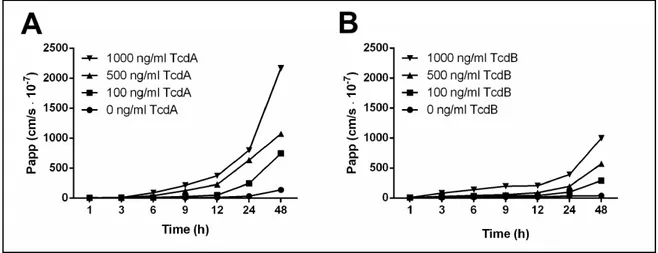
Having established that host cell polarity might influence the capacity of C. difficile to colonize and penetrate colonic epithelium, we determined whether its toxins, known to disrupt epithelial barrier integrity [57-60], are also able to modulate the epithelial fence function. Alteration of the TJs gate function in Caco-2 monolayers pre-exposed to sub-toxic concentrations of TcdA and TcdB ranging between 100 to 1000 ng/ml (as determined by measurement of ATP in metabolically active cells) was confirmed by detecting the paracellular passage of fluorescently labeled 4 kDa dextran (FD4). In accordance with previously published observations [57-60] a dose/time-dependent increase in paracellular permeability for non-ionic molecules was observed in toxin-treated cells (Figure 10A and B). Intriguingly, the extent of this enhancement differed significantly between TcdA and TcdB, amounting to 8-25-fold versus and 3-12-fold, respectively, after 24 hours of exposure.
Figure 10. Effect of TcdA and TcdB on gate function of tight junctions (TJs) determined by the paracellular
passage of non-ionic tracer. Fully polarized Caco-2 cells were exposed for indicated time to different concentrations of TcdA (A) or TcdB (B) and paracellular flux of 4 kDa FITC-dextran was measured and reported as the mean Papp values of three monolayers.
28
2.2.2. TcdA, but not TcdB, is able to modify the fence function of colonic epithelium Then we examined whether C. difficile toxins may influence the polarity of colonic mucosa by perturbing the distribution of plasma membrane components between distinct surface domains. At first, diffusion of fluorescently labeled sphingomyelin, a lipid component of plasma membrane, was visualized by confocal imaging in control and toxin-treated monolayers. As expected, in control cells the fluorescent lipid complex added apically was found to easily intercalate and diffuse within outer leaflet of the bilayer giving an evident staining of the apical membrane. On the contrary, upon EGTA (positive control) or TcdA treatment, diffusion of the lipid was no longer limited, leading to lateral and basolateral membrane labeling, as shown in confocal z-sections (Figure 11A). Slight staining of lateral membranes was also detected in cells treated with TcdB (500 and 1000 ng/ml), presumably as a result of lipid diffusion in solution across disrupted TJs. These data suggest that, beside the mutual effect of toxins on cellular permeability, alteration in TJs fence function is predominantly induced by TcdA. To further examine this specific modulation of cellular organization, changes in the polarized distribution of plasma membrane proteins in monolayers exposed to C.difficile toxins were assessed by immunofluorescence staining of apical (atypical protein kinase C, aPKC) and basolateral proteins (E-cadherin). Additionally ZO-1 labeling was simultaneously performed to reveal the precise localization of TJs. As shown in Figure 11B, Caco-2 monolayers exposed to EGTA or TcdA displayed a significant displacement of cell membrane constituents, including expansion of aPKC to lateral cell membranes and E-cadherin staining localized all around the cells. Also the distribution of ZO-1 was evidently altered. No changes in the distinct composition of plasma membrane proteins were observed in response to TcdB. Together these observations demonstrate that sub-toxic concentrations of TcdA, but not TcdB, are able to affect both barrier function (gate function) of the epithelium, as well as to subvert cell polarity (fence function). .
29
Figure 11. Effect of TcdA and TcdB on the fence function of tight junctions (TJs). Fully polarized Caco-2 monolayers were treated with indicated concentrations of toxins or with 4mM EGTA for 30 minutes or incubated in control medium. The membrane distribution of the fluorescent markers was determined by z sectioning by confocal microscopy. (A) Bodipy FL-C5-sphingomyelin/BSA complex diffusion assay. Fluorescent lipid/BSA was loaded to the apical surface. Arrows indicate lateral membrane staining. (B) Distribution of plasma membrane proteins specific for apical (aPKC, white) or basolateral (E-cadherin, green) membrane. TcdA and EGTA treated monolayers show compromised apicobasal polarity. The arrowheads denote the mislocalized membrane proteins whereas red staining indicates localization of ZO-1 in TJ region. Nuclei are stained with DAPI (blue). Bar, 10 µ m. .
30
2.2.3. TcdA subversion of host cell polarity is not due to apoptosis
Since C. difficile toxins are known to induce apoptosis in many cells types, we wanted exclude the possibility that TcdA effect on epithelial polarity may be triggered by apoptosis. Thus in order to evaluate the presence of apoptotic cells in ours assays a double staining of activated caspase-3 and TUNEL was performed (Figure 12A). Caspase-3 cleavage is an early event that initiates apoptosis whereas nuclear DNA fragmentation detected by TUNEL assay is observed at the very late states of cellular death. The percentage of active caspase-3 and TUNEL-positive cells in monolayers treated with different concentrations of TcdA or TcdB for 16 hours (the maximum incubation time used in this study) was determined and compared with the positive and negative controls (Figure 12B). Cells incubated with 2 µm staurosporine for 8 hours were used as a positive control showing high rates of apoptosis (23±0.5% of TUNEL-positive cells, 21±6% of active caspase-3-positive cells). In contrast in untreated cultures less than 1% of cells were TUNEL or active caspase-3-positive (0.5±0.3% of TUNEL- and 0.2±0.1% of active caspase-3-positive cells). Treatment with C. difficile toxins in concentrations between 100 and 1000 ng/ml did not induce a significant increase in Caco-2 apoptosis rates, even after prolonged incubation period of 16 hours. Therefore we can conclude that TcdA-induced depolarization of Caco-2 cells is not a secondary effect of apoptosis.
31
Figure 12. Effect of sub-lethal concentrations of C. difficile toxins on Caco-2 apoptosis. (A) Assessment of
Caco-2 cells apoptosis by TUNEL assay (green) and immunostaining for cleaved caspase-3 (red) after treatment with 1000 ng/ml of TcdA or TcdB for 16 hours or 2µM staurosporine (STS) for 8 hours (as a positive control). Merged microphotographs demonstrate the simultaneous presence of active caspase-3 and fragmented DNA in apoptotic cells. Nuclei were stained with Hoechst (blue). Bar, 10 µm. (B) Quantification of TUNEL and active caspase-3 positive cells using ImageJ software. Values are the mean from three independent experiments performed in triplicate.**P <0.01; ***P < 0.001.
32
2.3.
Influence of TcdA-mediated subversion of cell polarity on C. difficile
ability to colonize epithelium
2.3.1. TcdA-mediated subversion of cell polarity facilitates C. difficile adhesion and translocation of Caco-2 monolayer
Since our results suggested that cell polarity was a deterrent to C. difficile colonization of the host cell surface, and that TcdA was able to alter this epithelial function, we asked whether this activity could play a beneficial role in establishment of C. difficile infection. Being unable to compare C. difficile wild-type and toxin mutant strains in our infection assay because of drastically limited time of Caco-2 survival in anaerobic condition (less than 2 hours), we tested the effect of purified toxins on C. difficile capacity to infect human cells. Briefly, fully polarized Caco-2 monolayers were pre-exposed to different concentrations of TcdA or TcdB for 16 hours followed by incubation with C. difficile 630 at the apical side. In order to monitor toxin action in our in vitro assay, FITC-dextran was added simultaneously to the inner chamber of the Transwell system (Figure 13A). As a result striking differences were observed in the ability of C. difficile 630 strain to colonize cells in vitro in the presence or absence of TcdA (Figure 13B and C). Statistically significant increase in bacterial adhesion rates were noticed in Caco-2 monolayers pre-exposed to 500 ng/ml and 1000 ng/ml of TcdA (2.6±0.2 and 5.3±0.2-fold, respectively). The influence of the highest concentration tested on the number of adherent bacteria was comparable to the effect promoted by 30 minutes of EGTA treatment (44.4±5 bacteria/100 cells in toxin-treated monolayers versus 40.7±7 bacteria/100 cells in EGTA-exposed monolayers). However, as shown in Figure 13B, bacterial translocation is only slightly enhanced by sub-toxic doses of TcdA without reaching the levels observed upon EGTA treatment (Figure 11B).
33
Figure 13. Effect of TcdA on 630 C. difficile colonization of colonic mucosa (A) Effect of TcdA on epithelial
polarity determined by TEER and dextran flux measurements. TEER is expressed as the percentage of the baseline value (before TcdA treatment). Each value represents the mean ± SD (n=3). (B) Fully polarized Caco-2 monolayers were preincubated with indicated concentrations of TcdA for 16 hours followed by apical infection with C. difficile 630 strain. After 1 hour of incubation the number of bacteria that adhered (white bar) or translocated across (black bar) the epithelium was quantified by CFU counting and expressed as CFU/100 cells. Values are the mean ± SD from three independent experiments performed in triplicates. **P<0.01; ***P<0.001. (C) MIPs of adherent C. difficile bacteria on control and TcdA-treated Caco-2 cells. Bacteria were labeled with an anti-whole bacteria serum and a secondary fluorescent antibody (green) and cellular actin was stained phalloidin-Alexa Fluor 568 (red). Scale bars 10 µm.
34
Similar results were obtained by incubating cells with C. difficile R20291 (Stoke Mandeville), characterized as an epidemic and hypervirulent strain (Figure 14), indicating that this phenotype is not strain specific.
Figure 14. Effect of TcdA on R20291 C. difficile colonization of colonic mucosa. Fully polarized Caco-2
monolayers were preincubated with indicated concentrations of TcdA for 16 hours followed by apical infection with C. difficile R20291 (Stoke Mandeville). After 1 hour of incubation the number of bacteria that adhered (white bar) or translocated across (black bar) the epithelium was quantified by CFU counting and expressed as CFU/100 cells ± SD. Values are the mean ± SD from three independent experiments performed in triplicates. *P<0.05; **P<0.001.
Strikingly, no differences in bacterial adhesion between control and TcdA-treated cells were observed when only partially polarized (4-days-old) monolayers were infected confirming again that epithelial polarity is an important target of C. difficile pathogenesis (Figure 15).
35
Figure 15. Effect of TcdA on C. difficile colonization of partially polarized Caco-2 cells. (A) Effect of TcdA on
epithelial polarity determined by TEER and dextran flux measurements. TEER is expressed as the percentage of the baseline value (before TcdA treatment). Each value represents the mean ± SD (n=3). (B) Four days-old Caco-2 monolayers were preincubated with indicated concentrations of TcdA for 16 hours followed by apical infection with C. difficile 630 strain. After 1 hour of incubation the number of bacteria that adhered (white bar) or translocated across (black bar) the epithelium was quantified by CFU counting and expressed as CFU/100 cells ± SD. Values are the mean ± SD from three independent experiments performed in triplicates. **P<0.01; ***P<0.001.
Moreover, polarized monolayer exposure to TcdB was not able to facilitate colonization of Caco-2 cells (Figure 16), in line with the evidence that this toxin is not able to interfere with polarization status.
Figure 16. Effect of TcdB on 630 C. difficile. colonization of colonic mucosa (A) Effect of TcdB on epithelial
polarity determined by TEER and dextran flux measurements. TEER is expressed as the percentage of the baseline value (before TcdB treatment). Each value represents the mean ± SD (n=3). (B) Fully polarized Caco-2 monolayers were preincubated with indicated concentrations of TcdB for 16 hours followed by apical infection with C. difficile 630 strain. After 1 hour of incubation the number of bacteria that adhered (white bar) or translocated across (black bar) the epithelium was quantified by CFU counting and expressed as CFU/100 cells ± SD. Values are the mean ± SD from three independent experiments performed in triplicates.**P<0.1
36
Chapter 3: Discussion
An important aspect in the understanding of microbial pathogenesis is the recognition of the different strategies evolved by bacteria and viruses to circumvent or disrupt the mucosal barrier in order to facilitate colonization and/or dissemination to distal tissues. These can be achieved by a specific pathogen-mediated subversion of the host cell organization and functions [61]. In particular, a number of mucosal pathogens, including Pseudomonas aeruginosa, Helicobacter pylori and enteropathogenic Escherichia coli, have been shown to directly target various components of the polarity regulation network [51, 52, 62]. The data reported in this study postulate that this scenario may also apply to C. difficile infection with TcdA playing a major role in the alteration of epithelial cell polarity, leading to increased adhesion to the host cell surface.
By the use of epithelial Caco-2 cells grown on Transwell inserts to mimic natural architecture and tissue polarity in vitro, we found that the establishment of C. difficile infection strongly depends on the state of cellular differentiation, further reinforcing studies previously undertaken by Cerquetti and colleagues [63]. The use of the Transwell system allowed us to precisely distinguish between bacterial adhesion and translocation rates and to demonstrate that both processes are significantly enhanced in monolayers whose fence and gate function are not fully established or have been altered by calcium chelation. This is consistent with the preferential tropism of C. difficile to the basal cell surface observed in 2 and 3D Caco-2 cell models. Taken together our findings suggest that C. difficile might exploit a nutrient-rich niche located directly beneath the epithelium to persist in the human colon.
37
Although the effect of TcdA and TcdB, recognized as main virulence factors of C. difficile, on the structure of cytoskeleton and the function of TJs has been a topic of an intense study, the fundamental question of how they might benefit bacteria during host colonization remains unanswered [64, 65]. Moreover our current understanding of the molecular mechanism of C. difficile toxins action is mainly based on the in vitro studies using highly toxic concentrations that lead to increased paracellular permeability and apoptotic or necrotic cell death [58-60, 66-68]. Since toxin production in vitro usually ranges from 50 to 2000 ng/ml in 24 hours of C. difficile growth [69], it is likely that the local concentration of TcdA and TcdB in the colon of C. difficile-infected patients may reach similar levels. Based on the evidence reported in this study, we hypothesize that C. difficile may initiate its association with the colonic mucosa by producing low amounts of toxin A that could facilitate the first steps of colonization. Indeed, the alteration of plasma membrane components distribution between distinct surface domains of TcdA-treated Caco-2 cells together with the previously mentioned C. difficile preference for the basolateral membrane suggests that subversion of host cell polarity might act as a key mechanism leading to the increased bacterial binding. This notion might also explain why in vivo adhesion of an avirulent non-toxinogenic strain is facilitated by co-administration of C. difficile toxins in hamster model [70]. In this context, also the elevated rate of intestinal colonization in healthy adults infected by toxigenic C. difficile suggest that minimal level of toxin production could facilitate the colonization state without the occurrence of clinical symptoms [71, 72]. On the contrary, toxin B beside its expected influence on bacterial translocation rates does not play role in the establishment of the apical surface association consistent with its inability to modulate epithelial fence function.
38
Segregation of apical and basolateral receptors at the epithelial cell surface, and the compartmentalization of different cytoplasmic and membrane-associated signaling molecules, is crucial for the regulation of an innate immune response to pathogens in polarized epithelium and for prevention of unrestrained or prolonged inflammation. Under normal healthy circumstances several toll-like receptors (TLRs), such as TLR5, that recognize pathogen-associated molecular patterns (PAMPs) and activate innate immune pathways are physically separated from the luminal content. This allows epithelial cells to mount a rapid pro-inflammatory response only if surface or secreted components from commensal or pathogenic bacteria have breached the epithelial barrier [73]. Therefore, alterations in cell polarity induced by C. difficile toxins may allow PAMPs to access the BL surface and trigger the production of inflammatory cytokines and chemokines. Activation of different TLRs, including TLR4 and TLR5, by several C. difficile components (ie, surface layer proteins and flagellin) is known to play a pivotal role in determining the final outcome of infection [74, 75]. Notably, Yoshino and colleagues [75]recently demonstrated the contribution of clostridial toxins in this process, showing that breaching of epithelial barrier function by C. difficile toxin might lead to more severe inflammation present at the site of infection. Therefore, as postulated in this study, modulation of cell polarity by TcdA may not only facilitate colonization of the gut mucosa, but may also play a role in landscaping the immune response.
Taken together, our concept on C. difficile pathogenesis that combines the current knowledge with the results obtained in this study and depicted in the diagram (Figure 16) propose that different local levels of C. difficile toxins as well as the time of exposure might serve as a fine tuning of cell epithelial barrier ranging from mild alterations of its polarity, to induction of acute inflammation, apoptotic signaling and cell death. This specific modulation of mucosal organization and functions might facilitate C. difficile settlement in the human gut
39
and development of the chronic and persistent infection. In conclusion, we suggest TcdA-mediated subversion of the epithelial polarity as a novel strategy used by C. difficile to enhance its ability to reside in a very competitive environment of the human gut.
Figure 17. Schematic diagram illustrating the proposed model of the pathogenesis of C. difficile infection, that
have partly emerged from the experiments reported in this study. Low levels of TcdA present locally in the gut of C. difficile-infected individuals perturb epithelial polarity leading to exposure of toll-like (TLR) and other basolaterally localized receptors to luminal bacteria and their products (pathogen-associated molecular patterns, PAMPs). This promotes an increased colonization of colonic mucosa and stimulates production of inflammatory cytokines at the sites of infection. Subsequent increase in mucosal permeability, damage to the intestinal epithelial tissue and bacterial dissemination lead to development of acute and chronic inflammation, fluid secretion and diarrhea.
40
Chapter 4: Experimental Procedures
4.1. C. difficile strains and culture conditions
C. difficile strains 630 and B1/NAP1/027 R20291 were kindly provided by Nigel Minton (University of Nottingham, Nottingham, UK). Strains were grown on agar or in BHI-broth (Bacto, USA) at 37°C in anaerobic conditions. For infection studies, C. difficile strains were grown in BHI-broth until an OD600 of 0.5 (early exponential phase).
4.2. 2D and 3D cell cultures
The colon adenocarcinoma cell line Caco-2 (obtained from the American Type Culture Collection) was cultured in DMEM/F12 (Invitrogen) supplemented with 10% heat-inactivated FBS (Invitrogen), 20 mmol/L HEPES, 100 nmol/L nonessential amino acids and 100 μg/ml of penicillin/streptomycin and used between passage numbers 20–25. Cells were grown as 2D monolayers on collagen-coated Transwell inserts (3-μm-pore size, BD Biosciences) at a seeding density of 105 cells/insert and supplemented with fresh media every day. Unless stated otherwise, cells were allowed to differentiate for 21 days. To disrupt calcium-dependent intercellular junctions, monolayers were transiently exposed to 4mM EGTA [76]. Cell polarity and tight junction barrier function were verified by TEER and dextran flux measurements as described previously [77]. For 3D cell culture, Caco-2 cells were grown as cysts in Matrigel as described previously [56].
Briefly, a cell:matrigel mix containing 5.8 x 10⁴ cells/ml, 0.02 M HEPES, pH 7.4, 1 mg/ml Collagen I (Sigma-Aldrich) and 40% Growth Factor Reduced BD Matrigel™ Matrix was plated on 8-well chamber slide (BD Biosciences) incubated at 37°C, 5% CO2 for 1 hour to solidify and overlaid with complete growth media. Cysts were allowed to develop for 14-16 days at 37°C, 5% CO2, changing media every day, to form BL-side-out cysts with AP membrane facing the lumen and BL membrane facing surrounding collagen. In order to
41
produce the spheres with opposite polarity the anti-integrin beta-1 antibody AIIB2 (1:100 dilution, Developmental Studies Hybridoma Bank, University of Iowa) was added for entire culture period. Under these conditions, polarized AP-side-out cysts were formed.
4.3. Dextran permeability and membrane diffusion assays
These assays were performed as described elsewhere [78]. Briefly, 2D Caco-2 cell cultures were washed with ice-cold HBSS/HEPES buffer and labeled on the apical side with 5 μM solution of BODIPY-FL-C5-sphingomyelin/BSA complex (Molecular Probes) for 10 minutes on ice. Inserts were rinsed with ice-cold HBSS/HEPES buffer, incubated for 1 hour on ice and immediately observed by confocal microscopy. Paracellular permeability was quantified measuring the transepithelial flux of a 4 kDa fluorescein isothiocyanate (FITC)-labeled dextran (Sigma-Aldrich) [77] and expressed as apparent permeability coefficient (Papp): Papp (cm/s) = dQ / dt (1 / AC0), where dQ / dt is the permeability rate (μg/s), C0 is the initial concentration in the upper chamber (μg/ml), and A is the surface area of the membrane (cm2) [79].
4.4. Immunofluorescence microscopy
2D Caco-2 cultures pretreated with TcdA, TcdB, EGTA or medium alone were rinsed with Dulbecco’s phosphate-buffered saline (DPBS), fixed with 4% paraformaldehyde, permeabilized and blocked with 3% [wt/vol] bovine serum albumin (blocking buffer) in DPBS. Incubation with primary antibodies, including rabbit anti-ZO-1 antibody (61-7300, Invitrogen), mouse anti E-cadherin (33-4000, Invitrogen), and goat anti-PKCç antibody (sc-216-G, Santa Cruz Biotechnology), was performed either overnight at 4°C or for 2 hour at room temperature and followed by the treatment with the appropriate Alexa fluor-conjugated secondary antibody. Samples were mounted using ProLong Gold Antifade Reagent with
42
DAPI (Invitrogen) and analyzed by confocal microscopy using Zeiss LSM 710. Z-stacks 3D reconstructions were performed by Imaris software (BitPlane Inc.).
4.5. Assessment of apoptosis by TUNEL assay and active caspase-3 staining
The effect of TcdA and TcdB on Caco-2 apoptosis was assessed by immunostaining of active caspase-3 and by the recombinant terminal deoxynucleotidyl transferase (rTdT) mediated dUTP Nick End Labelling (TUNEL). Caco-2 cells grown on transwell inserts were exposed to different concentrations of TcdA or TcdB (100, 500, and 1000 ng/ml) for 16 hours. Subsequently cells were stained by TUNEL assay kit according to manufacturer’s instructions (Invitrogen). For caspase-3 activation cells were counterstained with anti-active caspase-3 rabbit polyclonal antibody (9661, Cell Signalling Technologies), followed by the appropriate Alexa fluor-conjugated secondary antibody. As a positive control Caco-2 cells were treated with 2 μM staurosporine (Sigma-Aldrich) for 8 hours. Cells were examined with a Zeiss LSM 710 confocal laser scanning microscope. Apoptosis was quantified as the percentage of cells showing TUNEL-positive nuclei and active-caspase-3.
4.6. C. difficile adhesion and translocation assay on 2D Caco-2 monolayers
2D Caco-2 cultures were transferred into anaerobic cabinet and washed once with pre-reduced HBSS. Bacteria from exponential phase cultures were pelleted, resuspended in cell culture medium and apically added to the monolayers for 1 hour. Dextran solution was applied simultaneously to monitor monolayer integrity during infection. Bacterial translocation was determined by colony-forming unit (CFU) counting of bacteria recovered from the outer chamber of the Transwell system. After extensive washings and cells lysis with 1% saponin; adherent bacteria were counted by plating appropriate dilutions on BHI-agar. Optimal multiplicity of infection (MOI of 1:20) and maximum incubation time of Caco-2 monolayers
43
in anaerobic conditions were determined in the initial experiments (data not shown). To test the effect of toxins on the capacity of C. difficile to infect epithelial cells, Caco-2 monolayers were preincubated with TcdA or TcdB (100, 500, 1000 ng/ml) for 16 hours.
4.7. C. difficile infection of 3D Caco-2 cysts
After treatment with 100 U/ml Collagenase VII (Sigma) for 15 minutes at 37°C to allow bacterial access to the cells surface, cysts were infected with 107 bacteria from exponential phase cultures and incubated for 2 hours under anaerobic conditions. Cultures were then washed three times to remove unbound bacteria, fixed with 4% paraformaldehyde for 30 minutes at room temperature and blocked with blocking buffer for 1 hour at room temperature. Incubation with primary rabbit ZO-1 antibody (Invitrogen) and goat anti-PKCç antibody (Santa Cruz) or an anti-whole bacteria serum was performed overnight at 4°C, and was followed by the incubation with fluorophore-conjugated secondary antibodies for 1 hour at room temperature. Finally, cysts were washed, mounted and analyzed by confocal microscopy. Bacterial binding to 3D cysts was quantified using Imaris, as further described in Supplementary data, on 3D reconstruction of images acquired with Zen2009 software.
4.8. Quantification of C. difficile binding to 3D Caco-2 cysts
Imaris 3D analysis software was used to quantify bacterial adhesion to 3D cysts. Briefly, image stacks obtained by confocal microscopy were reconstructed with Imaris into 3D ‘‘surface objects’’ for two microscopy channels, one for fluorescently stained bacteria, the other for DAPI-stained nuclei of Caco-2 cells. The number of ‘‘surface objects’’ for each channel was determined using a “splitting touching objects” option and size threshold criteria. Any object above the minimal (for bacterial aggregates) or below the maximal (for the nuclei of Caco-2 cells) size threshold was counted as one. 50 cysts were quantified for each type:
44
AP- and BL-side-out. Results are expressed as the number of C. difficile bound to 100 Caco-2 cells, where one sphere consists of 25-40 cells.
4.9. Statistical analyses
All experiments were performed at least three times in triplicates. Statistical analyses were performed using the non-parametric Mann-Whitney U test.