International PhD Program in Neurosciences
XXXI Cycle
PhD thesis
Kathrin Heiss
Coordinator: Prof. Salvatore Salomone Tutor: Prof. Giovanni Li Volti
Department of Biomedical and Biotechnological Sciences Section of Biochemistry.
University of Catania
TABLE OF CONTENTS
ACKNOWLEDGEMENTS ... 3
LIST OF ABBREVIATIONS ... 4
ABSTRACT ... 5
MICROGLIA ... 6
ORIGIN AND DEVELOPMENT ... 7
THE PROCESS OF ACTIVATION ... 8
MORPHOLOGY AND IDENTIFICATION OF MICROGLIA ... 10
SIGMARECEPTOR ... 13
SIGMA RECEPTOR FUNCTIONS AND STRUCTURE ... 13
SIGMA RECEPTOR ISOFORMS ... 14
CHAPTER I ... 16
CHAPTER II ... 31
REFERENCES ... 39
LIST OF ABBREVIATIONS
1,3-Di-(2-tolyl) guanidine (DTG)
4-methoxy-3-(2-phenylethoxy)-N,N-dipropylbenzeneethanamine hydrochloride (NE-100) 2,2-dithio-bis-nitrobenzoic acid (DTNB)
central nervous system (CNS) interleukin 10 (IL-10)
nitric oxide (NO)
lipopolysaccharide (LPS)
Phosphate Buffered Saline solution (PBS) reactive oxygen species (ROS)
reduced glutathione (GSH) tumor necrosis factor-α (TNF-α)
ABSTRACT
Experimental research is making considerable efforts to understand the rules that regulate the balance between toxic and protective brain innate immunity. Sigma-1 receptor is expressed in both neurons and glia is a unique class of intracellular proteins and is involved in neurodegeneration. Our aim was to evaluate the biological effects of sigma-1 selective ligands, and bifunctional sigma-1 selective ligands conjugated with lipoil function in microglia following hypoxia/reoxygenation condition. BV2 cells were exposed to 3 hours of hypoxia and 24h of reoxygenation. Cells were treated with sigma-1 agonist (+)-Pentazocine, various bifunctional sigma-1 agonists conjugated with lipoil function and with lipoic acid alone. We assessed cell viability, apoptosis, reactive oxygen species (ROS) formation, mitochondria membrane potential, and total thiol groups content (GSH). Our results showed that 24h of reoxygenation resulted in a significant decrease of cell viability and increase in apoptosis when compared to control. No significant effect of
(+)-Pentazocine and tested compounds was observed on cell viability following 24h of
reoxygenation. Furthermore, all treatments resulted in a significant decrease of ROS formation when compared to untreated cells. Finally, pharmacological treatments restored mitochondrial membrane potential when compared to the untreated group. Consistently with these results we also showed that GSH content was restored following pharmacological treatments. Our results showed that newly synthetized bifunctional sigma-1 compounds exhibited significant antioxidant activity and induce apoptosis in activated microglia thus providing a new tool for effective manipulation of brain inflammation, with the specific aim of favoring its protective arm and boosting innate neuroprotective mechanisms
G
ENERAL INTRODUCTIONM
ICROGLIAThe concept of microglia was introduced by Pio del Rio-Hortega as a defined cellular element of the central nervous system (CNS) [1]. In this article, Rio-Hortega hypothesized that microglia enters the brain during early development and these cells have an amoeboid morphology and originate from mesoderm; also Nissl considered them as mesodermal despite of the concept of an ectodermic origin of glial cells [2]. Furthermore, they use vessels and white matter as guiding structures for migration and they transform into a branched, ramified morphological phenotype in the more mature brain. He saw also that in mature brain they are found dispersed throughout the CNS with little variation: in particular, after a pathological event, these cells withstand a transformation. The transformed cells acquire amoeboid morphology similar to the observed in early development: moreover, they have the capacity to migrate, proliferate and phagocyte. These statements, even though they were formulated in 1932, are perfectly valid today and it could be possible to be found in modern textbook and journal.
Modern researches about microglia started in the late 1960s with the work of Georg Kreutzberg that introduce the facial nerve lesion model. In this model it is possible to study microglial answer to injury in tissue with an intact blood-brain barrier. [3] This method supported the possibility to distinguish responses of intrinsic microglia from invading monocyte; moreover, it helped to state the importance of microglia in degeneration and regeneration of brain.
Over the years there was an explosion of studies on cultured microglia, helped by research conducted by Costero [4]. Unfortunately, microglia recognizes the cell culture environment as foreign with the consequence that all these in vitro studies may not reflect
properties of microglia in the non-pathological brain. To date, the combination of advanced imaging protocols with the use of genetically based cell-specific markers has allowed studies of microglia in the undisturbed tissue [5, 6].
O
RIGIN AND DEVELOPMENT
The origin of microglia has been debated for a long time but today there is the general opinion that microglial cells are derived from progenitors that migrated from the periphery and from mesodermal/mesenchymal origin [7]. In rodent these cells migrate from the blood system as monocytic cells, originated by bone marrow. In postnatal development, they migrate into the brain until 10th day. The shape of these cells is amoeboid and it can be easily recognized in slices of corpus callosum [8, 9]. In adult animal there is only a little exchange between blood and brain tissue, but after blood-brain barrier damage it is possible to see a migration of a subpopulation of monocytes entering the brain and transforming in microglia [10]. This evidence conduces to the obvious conclusion that in a healthy and intact brain the microglial cells exist ad a stable population. Another kind of invasion is shown in embryonic life and it occurs from the middle of the fist trimester in human and between embryonic days 10 and 19 in rodents. [7]
There is evidence for two separate population of microglial cells: one is derived from progenitors that are of myeloid/mesenchymal origin; the second represents a transitory form of a fetal macrophage related to amoeboid microglial population as showed in postnatal brain of rodents [11].
After the invasion of brain parenchyma, microglia cells change phenotype in ramified shape. Nowadays, the evidences show much more what controls the activation of microglia instead of what converts them to ramified phenotype. Nevertheless, some cell culture studies contribute to found some interesting results such as astrocyte conditioned medium
increases ramification of blood monocyte in culture [12]. Evidences showed that a combination of astrocyte conditioned medium with ATP or adenosine produces a more extensive ramification phenotype [13]. Other possible agents of transformation can be cytokines released from astrocyte such as transforming growth factor-b (TGF-b), macrophage stimulating factor (M-CSF) and granulocyte/macrophage colony-stimulating factor (GM-CSF). [14]
T
HEP
ROCESS OFA
CTIVATION
As described before, microglia in healthy mature CNS (including brain, spinal cord, eye and optic nerve) have a ramified morphology, a small soma with fine cellular processes. This typical phenotype, different from a classic macrophage, has been associated with microglial “resting” state. Any real or potential danger to CNS, such as infection, trauma, ischemia, neurodegenerative diseases or any disturbance or loss of brain homeostasis can evoke rapid and strong changes in microglia cell shape, gene expression and functional behavior: this changes is commonly defined “microglial activation” [15-22]. Moreover, microglia is able to become motile and moves to a lesion or controls infectious invaders following chemotactic gradients. Local densities can also increase by proliferation to supply more cells for the defense against germs and to organize protection and restoration of tissue homeostasis. This induction and rearrangement releases multiple pro-inflammatory and immuno-regulatory factors and compounds that can be configured as additional elements of activation process. Microglial cells are also able to produce neurotrophic factors.
The different stages of microglial activation were defined analyzing morphological, molecular and functional characteristics in a condition of fully activated microglia presenting them-selves like other macrophages [16, 17, 19]. Until recently, microglia in
healthy adult CNS were considered inoperative due to low or absent expression of activation-associated molecules and ramified morphology [20]. However, these “resting” cells scan actively their environment and monitor extracellular space and cellular neighborhood without disturbing the delicate neuronal circuitry, ready to transform to active profile upon appearance of signs indicating a threat to the CNS [5, 6]. Hence, it is possible to state that activated microglia primarily supports and protects structural and functional integrity of the CNS. Nevertheless, while supporting this hypothesis of activity, monitoring and maintenance of CNS homeostasis, the housekeeping activity of microglia remains not observed: moreover, this physiological activity could be overlooked [19]. In case of small vascular defect or a neuronal impairment, microglia would act to protect and give trophic support reducing synaptic input via synaptic stripping [23, 24]. Nevertheless, this kind of restricted and transient activities could not be recognized: in other words, very little information is acquired about impact of daily function of microglia.
The transformation from surveillance mode to active microglia means more of a shift in activities rather than “activation” by it self: this hypothesis endorses a constant activity of microglia with no periods of inactivity. Additionally, discussing about “activation” notes that this term do not contain any information about functional orientation which plays an important role in CNS consequences [25]. Although, there is clear evidence that microglial activation is not an “all-or-none” process as well as it is not a linear pathway with a fixed outcome: indeed, activated microglia can acquire distinct functional states [19, 26-29]. Microglial activation can start with an early emergency reaction to fight an infection or to limit damage after injury: findings derive from responses to injury [30-32], ischemia [33], or autoimmune inflammation [34].
A lot is known about activation process and events affecting microglia, although there is a lack of evidence about the period after these events. Hanisch et al. demonstrate that a
terminated microglial response may leave traces and it is in the best case successful [19]. The post-activated microglia can remain not distinguishable using morphology or isolated criteria like marker staining from the other “resting” cells in near population, while long-lasting adjustments are running. Epigenetic mechanisms of long-long-lasting adjustment are already known and receptors with regulatory influences on transcriptional control have been reported: these regulations could be selective for sets of genes [35-37].
M
ORPHOLOGY AND IDENTIFICATION OF MICROGLIA
The studies about microglia morphology and its changes allowed recognizing the role in diseased CNS [19, 20]. In healthy matured tissues of the brain, spinal cord or retina microglia characteristics do not point out an immediate association with a macrophage nature.
The cellular processes proliferating from the small soma with arborization are typical for the “ramified” microglia: this term is commonly associated with “resting” microglia, showing an strong link between morphology and function [38]. The process of microglial transformation is follow by marked morphological changes: microglia reduces the complexity of shape by retracting the branches of the arborization so that they are resorbed into the cell body. It is possible to distinguish different steps and intermediate stages, including processes such as withdrawal, transition or hyper-ramification and subsequent formation of new branches; these steps along with motility and locomotion, i.e., microglial movement in the tissue [38-40]. An interesting aspect can be underlined in ageing, because of microglia gains morphological markers typical for senescence and functional deterioration [38, 41, 42].
During in vitro studies, microglial cells do not have typical ramified structure showed in normal CNS: they show heterogeneous shapes, from spindle and rod-shaped or amoeboid
to round cells; although, it is possible to impose a morphological changing by treatment with typical activating agents, such as bacterial LPS [43]. Obviously, the morphological responses is different depending on the type of insult [38, 44] and cells can acquire bushy or bi- and tripolar, spindle or rod shapes [45, 46]. The different cell shape assumed by microglia after fast and drastic transformation could indicate a requirement for functional adjustments. For example, in the cells requiring motility and directed migration it is common to find filopodia protrusion and dynamic rebuilding; likewise, phagocytotic activity in a myelin lesion is represented by the appearance of “foamy” cells with lipid- or myelin-loaded organelles in bran tissue section. Even though morphology is not always a reliable reflection of functional activity and some gene expression or activity can happen in absence of evident morphological changes [47]; at the same time, a typical ramified shape can be induced even without support of ramification conditions [48, 49].
The identification of microglia, beyond cytomorphological criteria, is possible as result of development of staining protocols that helps to understand the exclusive expression of certain molecules in specific cell type.
The use of different markers is suitable by the ability to discriminate microglia from other CNS cells (neurons, astrocytes, oligodentrocytes or endothelial cells), to distinguish microglia from other resident or infiltrating macrophages/monocytes and to reveal the entire population of microglia.
In accord to a variety of cell surface-associated or intracellular or cytosolic molecules, it is possible to show microglia in human and animal bran tissue sections and in cultures. In some cases, is not possible to know the exact identity and function of protein or carbohydrate structure: this is the case for surface-expressed glycan moieties identified by Griffonia simplicifolia isolectin B4 (ILB4) or tomato lectin [50, 51].
Following neuropathological routine and experimental research there are different and useful target that can be used with more or less prominent and overlapping recognition of macrophages/macrophage-like cells such as: immunoglobulin receptors (CD16/32/64, FcγRIII, FcγRII, FcγRI), CD45 (leukocyte common antigen, LCA), CD68 (macrosialin), CD163 (scavenger receptor M130, ED2), CD169 (sialoadhesin, siglec -1), CD204 (MSR),
F4/80 antigen, b-glucan receptor dectin-1 and mannose receptor (CD206). It is
demonstrated that microglial activation increases many of these molecules, like for example CD11b or Iba1 [52, 53]. Interesting selective structures are MHC class II: in fact, these are only expressed by activated microglia including the expression of accessory molecules for antigen presentation CD80/86. Using this methods occur a problem revealed with non-CNS monocytes, because of the difficult to distinguish these cells from easily distinguished microglia cells. More recently, new antigens with selectivity for microglia among cells of mononuclear phagocyte system have been introduced; in particular it is demonstrated an high selectivity of human glucose transport 5 (hGLUT5) [54]. This evidence drive investigators to use GLUT5 as a useful marker for resting and activated microglia [55]. However, experimental evidences show a limitation in using antibodies against GLUT5 only to human material: in fact, the expression patterns of GLUT isoforms seem to vary in other species. For example, rodent macrophages can not be discriminated by GLUT5 staining, but antibodies against keratin sulfate epitope 5D4 can help as discrimination in rat [56]. Recent applications of genetic engineering produce a generation of animals expressing fluorescent proteins, under control of macrophage/microglia-expressed factor (i.e. fractalkine receptor CX3CR1), and they can be used as
SIGMA
RECEPTOR
SIGMA RECEPTOR FUNCTIONS AND STRUCTURE
Sigma receptors have been identified by Martin et al. in 1976, using N-allyl-normetazocine (±)-SKF-10,047, which the first letter “S” gave the name “sigma”: investigators thought
that this molecule could be the prototypic ligand for this receptors. This compound induced psychotomimetic effects in humans and this evidence drove Martin and co-workers to consider sigma receptors as new subclasses of opioid receptors [58].
This study allowed to see three physiological actions that could be distinguished in three groups: they hypothesized that differences derived by different subtypes of opioid receptors. In order to understand, they proposed a µ subtype, a κ subtype and, in particular, a σ subtype which was characterized by SKF-10,047 and related compounds. More analysis showed that the racemic (±)-SKF-10,047 can interact with all the three types of receptors: in particular, (‒)-SKF-10,047 showed binding affinity to the µ-opioid and κ-opioid receptors, instead (+)-SKF-10,047 showed binding affinity for the phencyclidine (PCP) site included in NMDA receptor [59-63].
In 1980s the studies conducted by Tsung-Ping Su demonstrated that sigma receptors have high affinity for benzomorphans such as pentazocine, dextrallorphan and (+)-cyclazocine [64, 65]. It is now accepted that are unique non-opioid, non-phencyclidine (PCP) receptors and two subtypes are currently known, named sigma-1 and sigma-2.
SIGMA RECEPTOR ISOFORMS
Sigma-1 receptor (σ1 receptor) has cloned in humans, in rat and in guinea pig, it is formed from 223 aminoacids with 90% identical sequences across species and it is ubiquitously expressed in mammalian tissue [66]. This receptor is involved in the biosynthesis and release of neurotransmitters (NT), in neuroprotection and in the modulation of opioid analgesia [67-71].
Evidences showing the overall structure and molecular basis for ligand recognition have been discovered recently. In particular, from these studies, investigators determinate that the receptor contains only a single transmembrane domain for each promoter: the ligands used to understand the selective binding toward sigma-1 receptor evidenced a bind interacting through a charge-charge interaction with the highly conserved Glu172, labeling this amminoacidic residue as essential for ligand binding [72]. Other residues in the binding site include Val84, Trp89, Met93, Leu95, Leu105, Phe107, Ile124, Trp164, and Leu182: these interact with hydrophobic regions on the bound ligands; although, Tyr103 engages in an aromatic stacking interaction. The problem occurred is represented by the highly occluded structure of the binding pocket, which leaves unclear how ligands enter and exit the site. In order to understand, two hypothesis was formulated: first, ligand could enter and exit trough a gap between the two membrane-adjacent helices; second, they could access through the cytosolic surface thanks to a polar region occluded by Gln135, Glu158 and His154 [73].
A strong difference in research is represented by sigma-2 receptor (σ2 receptor) because it is not cloned or crystallized yet and thus the amminoacidic sequence and structure are not known.
Both sigma receptor subtypes are widely distributed in CNS, in which the distribution in limbic structure, brainstem areas and some sensory regions underlines the role of these
receptors in emotion memory, motor and endocrine function [74]. They were also distributed in peripheral organs such as hearth [75], liver, kidney, ovaries, testes and mucosal regions of gastrointestinal area.
Evidences about sigma-2 receptor show an over-expression in cancer cells and using sigma-2 agonists seem to have an interesting role in cell proliferation and induction of apoptosis: in particular, σ2 ligands produce apoptosis in tumor cells by altering mitochondria and activating Caspase-3 [76, 77]. In addition to their potential use as anticancer drug, highly specific and selective ligands can be used as biomarker of tumor proliferation determining PS and for imaging diagnosis tumor by SPECT scintigraphy and PET analysis [78, 79]. An example is represented by σ2-receptors as biomarker in breast tumors, because it was seen expressed about 10 times more in cancer cells than in healthy cells [80].
Neurosci Lett. 2016 Jul 28;626:142-8. doi: 10.1016/j.neulet.2016.05.025. Epub 2016 May 18.
(+)-PENTAZOCINE REDUCES OXIDATIVE STRESS AND APOPTOSIS IN MICROGLIA FOLLOWING HYPOXIA/REOXYGENATION INJURY
Kathrin Heiss1, MD, Luca Vanella2, PhD, Paolo Murabito3, MD, PhD, Orazio
Prezzavento2, PhD, Agostino Marrazzo2, PhD, Carlo Castruccio Castracani1, Ignazio
Barbagallo2, PhD, Agata Zappalà1, PhD, Emanuela Arena2, PhD, Marinella Astuto3, MD,
Antonino Giarratano4, MD, Giovanni Li Volti1,5 MD, PhD
1Department of Biomedial and Biotecnological Sciences, University of Catania, Via S.
Sofia 64 95100 Catania (Italy);
2Department of Drug Sciences, University of Catania, V.le A. Doria 6, 95125 Catania
(Italy); 3Azienda Ospedaliera Universitaria Policlinico “G. Rodolico”, University of Catania, (Italy), Via S. Sofia 78, 95125 Catania (Italy);
4Department of Biopathology and Medical Biotechnologies (DIBIMED), Section of
Anaesthesia, Analgesia, Intensive Care and Emergency, Paolo Giaccone University Hospital, University of Palermo, Via Del Vespro 38, 90100, Palermo, (Italy);
5Euro-Mediterranean Institute of Science and Technology, Via Emerico Amari 131, 90100
Palermo (Italy)
ABSTRACT
Background: Sigma-1 receptors (σ1R) are highly expressed in neurons as well as
microglia and have been shown to modulate the inflammatory response in the central nervous system and thus may serve as a possible target for neuroprotective strategies. The aim of the present study was to test the effect of (+)-pentazocine, a putative σ 1R agonist, in
an in vitro model of microglia activation. Methods: Microglia (BV2 cells) was exposed (3 hours) to 1% oxygen and reoxygenation was allowed for 24 hours. Cells were treated with different concentrations (1, 10, 25 and 50 µM) of (+)-pentazocine in the presence or
absence of NE-100 (1 µM), a well established s1R antagonist. Cell viability and apoptosis
was measured by cytofluorimetric analysis, whereas oxidative stress was evaluated by reduced glutathione (GSH) content and mitochondrial potential analysis. Results: Our results showed that (+)-pentazocine was able to increase cell viability and restores
mitochondrial potential at all concentrations whereas only 1 and 10 µM were able to reduce significantly apoptotic cell death, to restore reduced glutathione intracellular content and prevent ERK1/2 phosphorylation. All these effects were abolished by concomitant treatment with NE-100. Conclusions: (+)-pentazocine exhibits significant dose dependent protective effects in our in vitro model of microglial activation thus suggesting that σ1R may represent a possible strategy for neuroprotection.
INTRODUCTION
Ischemic cerebrovascular disease is a notably common clinical cerebrovascular disease. The mechanisms underlying cerebral ischemia-reperfusion injury are quite complicated. Oxidative stress and the inflammatory response are considered the key mediators of cerebral ischemic injury activating the crosstalk among various cell populations of the brain such as astrocytes, neurons and microglia [81]. Recently, microglia is emerging to play a key role in orchestrating the complex pathophysiological processes triggered by hypoxia and subsequent reoxygenation of the brain [82, 83]. Microglia are resident immune macrophages-like cells of the central nervous system (CNS), which play an essential role in immunological surveillance, disposal of foreign invaders, and maintenance of brain homeostasis [84]. However, uncontrolled or over-activated microglia induce an oxidative burst causing tissue damage in CNS [85, 86]. Several pharmacological strategies have been developed in the attempt to modulate the redox balance altered by microglia and thus achieving a significant neuroprotection in different clinical condition, however an
optimal strategy is far away from being achieved. Recently, σ 1R agonists are emerging as
good candidates for neuroprotective strategies under various experimental models. σ 1R are
highly expressed in neurons as well as microglia [87]. The activated s1R can modulate the
has been reported that σ1R are involved in the modulation of neuroinflammation. s1R
agonists, such as (+)-pentazocine and 1,3-Di-(2-tolyl) guanidine (DTG), inhibit lipopolysaccharide (LPS)-stimulated activation of microglia, and decrease the production of reactive oxygen species (ROS), the release of pro-inflammatory molecules [tumor necrosis factor- α (TNF-α), nitric oxide (NO), interleukin 10 (IL-10), etc.] as well as the expression of monocyte chemoattractant protein-1 [87, 89-91]. Based on the findings described above, the present study aimed at evaluating the effect of (+)-pentazocine might on the oxidative stress response mediated by activated microglia. BV2 microglial cells were used as a model, and in vitro hypoxia/reoxygenation was performed to simulate ischemia/reperfusion in vivo.
MATERIALS AND METHODS
Cell Culture and pharmacological treatments
Mouse microglial BV2 cells were purchased from ATCC Company (Milan, Italy). The cells were suspended in culture medium (Dulbecco's Modified Eagle Medium (DMEM) containing 10% fetal bovine serum (FBS), 100 U/mL penicillin, and 100 U/mL streptomycin). At 80% confluency, the cells were passaged using trypsin-EDTA solution (0.05% trypsin and 0.02% EDTA). (+)-Pentazocine (Sigma–Aldrich, Milan, Italy) was added to BV2 culture at different final concentrations of 1, 10, 25 and 50 µM. In order to test the pharmacological significance of s1R stimulation by (+)-pentazocine, in a separate
set of experiments BV2 cells we also treated with (+)-pentazocine in the presence of 4-methoxy-3-(2-phenylethoxy)-N,N-dipropylbenzeneethanamine hydrochloride (NE-100) (1 µM) (Tocris Cookson Ltd., Bristol, UK), a high selective σ1R antagonist.
Establishment of the In Vitro BV2 Cell Hypoxia/Reoxygenation Model
BV2 cells were digested into a cell suspension, and the cell density was adjusted to 5 × 10⁴ cells/mL. After 80% confluence, the cells were washed 3 times with Phosphate Buffered Saline solution (PBS), replaced with serum-free DMEM medium, and placed in a three-gas incubator at 37°C containing 1.0% O2 for 3 hours to initiate hypoxia, followed by 24h
reoxygenation in an incubator at 37°C containing 5% CO2.
Cell viability evaluation cytofluorimetric analysis
Cell viability was assessed by Muse™ Count & Viability Kit (Catalog No. MCH100102, Millipore, Milan, Italy) according to the manufacture’s guidelines. Briefly, 50 µL of cell suspension containing 1 x 10⁶ cells/mL were mixed with 450 µL of Count & Viability Reagent. Cells were allowed to stain for 10 minutes at room temperature and samples were read by Muse™ Cell Analyzer (Millipore).
Annexin V and dead cell evaluation by cytofluorimetric analysis
Cell apoptosis was evaluated by Muse™ Annexin V & Dead cell kit (Catalog No. MCH100105, Millipore, Milan, Italy) according to the manufacture’s guidelines. Briefly, 100 µL of the Muse™ Annexin V & Dead Cell Reagent to 100 µL of cell suspension. Such preparation was mixed thoroughly by vortexing at a medium speed for 3 to 5 seconds and samples were allowed to stain for 20 minutes at room temperature in the dark. Samples were read by Muse™ Cell Analyzer (Millipore).
ERK1/2 phosphorylation assessment by in-cell western
Cells were then fixed in 4% paraformaldehyde (PFA) by adding 20 µl of 12% PFA directly to the wells for 1 h at room temperature. The 96 wells were washed three times with PBS
(50 µl/well), permeabilized with PBS/0.1% Triton X-100 (50 µl/well, three times, 2 mins each), and blocked in LI-COR buffer (50 µl/well) for 2 hours at room temperature (or alternatively overnight at 4°C). The wells were then incubated with mouse anti-ERK1/2 or rabbit anti-phospho-ERK1/2 antibodies (1:200 for optimal signal-to-noise ratio, Cell Signalling) in LI-COR blocking buffer for 2 hours at room temperature (20 µl/well) and subsequently washed with PBS/0.1% Tween-20 (50 µl/well, three times). Infrared anti-mouse IRDye800CW and anti-rabbit IRDye700CW secondary antibodies (1: 200) in PBS/0.5% Tween-20 were then added (20 µl/well). The plates were incubated for 1 hour at room temperature, and the wells were washed with PBS/0.1% Tween-20 (three times) and incubated in PBS (50 µl/well). The plates were covered with black seals and imaged on an Odyssey infrared scanner using microplate settings with sensitivity of 5 in both the 700 and 800 nm wavelength channels. In separate wells, secondary antibodies alone were used to calculate background to be subtracted by the remaining wells. Data were acquired by using Odyssey software, exported and analyzed in Excel (Microsoft, Redmond, WA). Results were expressed as ratio between total ERK1/2 and phospho-ERK1/2.
Mitochondrial membrane potential (Δψ) evaluation by confocal microscopy
Images of BV2 following various pharmacological treatments were obtained at the indicated times using an inverted scanning confocal microscope (Leica LSM S8) equipped with differential interference contrast optics. Mitochondrial Δψ was assessed in live cells with the use of the fluorescent probe JC-1 (10 µg/ml) [92]. This probe permeates the mitochondria as a function of Δψ giving a red fluorescence (monomeric form, 585-nm long-pass emission filter under 568-nm laser illumination) when mitochondrial membrane potential is high and shifting into green fluorescence (multimeric form, 505- to 550-nm band-pass emission filter under 488-nm laser illumination) when mitochondrial membrane
potential is decreased. The data shown are representative of at least three different fields in three separate experiments. To provide a quantitative measurement of the differences in fluorescence between images, the total number of pixels in the red channel was divided by the total number of pixels in the green channel for each image.
Glutathione measurement
Intracellular content od reduced glutathione (GSH) was measured using a spectrophotometric assay based on the reaction of thiol groups with 2,2-dithio-bis-nitrobenzoic acid (DTNB) at λ = 412 nm (εM = 13,600 M−1·cm-1, where εM is a
wavelength-dependent molar absorptivity coefficient).19 Measurements were performed in
triplicate.
Statistical Analysis
The data were expressed as the means ± SD. Statistical analysis was performed via one-way analysis of variance (ANOVA) using SPSS11.0 software. P < 0.05 was considered to be significant.
RESULTS Cell Viability
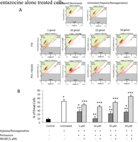
Our data suggest that 3 hours of 1% of hypoxia followed by 24 hours of reoxygenation resulted in a significant (p<0.05) decrease of cell viability compared to normoxic-cultured cells (Figure 1A and 1B). Treatment with (+)-pentazocine at 1, 10, 25 and 50 µM resulted in a significant (p<0.05) increase of cell viability when compared to untreated cells. In particular, the protective effect of (+)-pentazocine was more evident at a dose of 10 µM. In
order to determine whether the protective effect of (+)-pentazocine was dependent of σ 1R
experiments showed that NE-100 reversed the beneficial effect of (+)-pentazocine as demonstrated by a significant (p<0.05) reduction of cell viability when compared to (+)-pentazocine alone treated cells.
Figure 1. (A) Cytofluorimetric analysis of BV2 cell viability following various pharmacological treatments. Pictures are
representative of a single experiment. (B) Quantification of cell death percentage following various pharmacological treatments. Values represent the average of four separate experiments performed in triplicate.
(*p<0.01 when compared to normoxic untreated cells; **p<0.01 when compared to hypoxia/reoxygenation untreated cells; *** p<0.01 when compared to pentazocine alone treated cells)
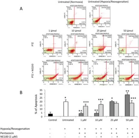
Apoptosis evaluation by cytofluorimetric Annexin V expression
Hypoxia/reoxygenation injury resulted in a significant (p<0.05) increase of apoptotic cell death compared to normoxic-cultured cells (Figure 2A and 2B). Treatment with (+)-pentazocine at 1 and 10 resulted in a significant (p<0.05) decrease of cell apoptosis when compared to untreated cells. Interestingly, 25 µM had no significant effect on cell apoptosis when compared to control (p>0.05) whereas 50 µM (+)-pentazocine resulted in a further increase of apoptotic cell death when compared to untreated cells (p<0.05). In order to determine whether the effect of (+)-pentazocine on apoptotic cell death was dependent
of σ1R stimulation, BV2 cells were also treated with NE-100. This set of experiments
showed that NE-100 reversed the beneficial effect of 1 and 10 µM (+)-pentazocine as demonstrated by a significant (p<0.05) increase of apoptosis when compared to (+)-pentazocine alone treated cells. Finally, NE-100 significantly (p<0.05) reduced 50 µM (+)-pentazocine induced apoptosis.
Figure 2. (A) Cytofluorimetric analysis of BV2 cell apoptosis following various pharmacological treatments. Pictures are
representative of a single experiment. (B) Quantification of cell apoptosis percentage following various pharmacological treatments. Values represent the average of four separate experiments performed in triplicate.
(*p<0.01 when compared to normoxic untreated cells; **p<0.01 when compared to hypoxia/reoxygenation untreated cells; *** p<0.01 when compared to pentazocine alone treated cells)
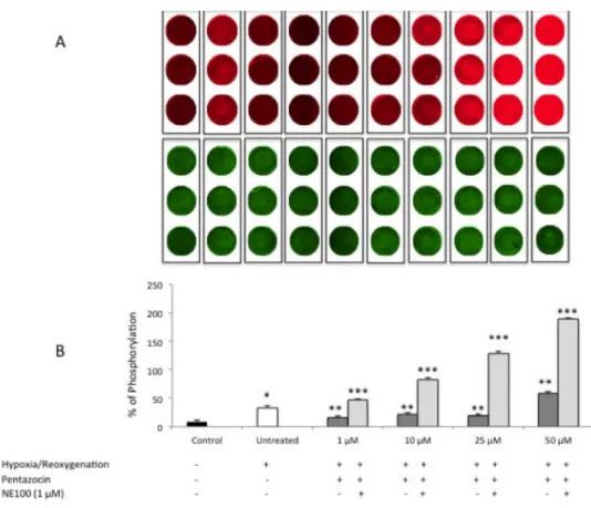
ERK1/2 phosphorylation assessment by in-cell western
In order to further evaluate the molecular mechanism underlying (+)-pentazocine treatment we evaluated the phosphorylation of extracellular-signal-regulated kinases (ERK-1/2), which is known to be activated also under hypoxic/reoxygenation conditions. This set of experiments showed a significant increase of ERK1/2 phosphorylation following
hypoxia/reoxygenation (Figure 3A and 3B). Interestingly, at 1, 10 and 25 µM (+)-pentazocine was able to prevent ERK1/2 activation. By contrast, 50 µM (+)-(+)-pentazocine increased phosphorylation of ERK-1/2 when compared to other (+)-pentazocine concentration or untreated group. Finally, NE-100 abolished (+)-pentazocine effect on ERK1/2 activation increasing its phosphorylation even to higher levels compared to untreated hypoxic/reoxygenated cell cultures.
Figure 3. (A) In cell western analysis of ERK1/2 phosphorylation following various pharmacological treatments.
Pictures are representative of a single experiment performed in triplicate. Red wells (IRdye 700) represent the phosphorylated form of ERK1/2 whereas the green wells (IRdye800) represent the total ERK1/2. (B) Quantification of ERK1/2 phosphorylation percentage following various pharmacological treatments. Values represent the average of four separate experiments performed in triplicate. (*p<0.01 when compared to normoxic untreated cells; **p<0.01 when compared to hypoxia/reoxygenation untreated cells; *** p<0.01 when compared to pentazocine alone treated cells)
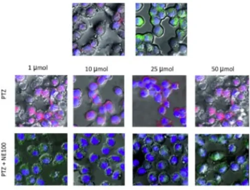
Confocal microscopy analysis of ΔΨm modification
ROS are known to stimulate activation of several signaling pathways leading to apoptosis including the mitochondrial death pathway. Mitochondrial function is highly susceptible to oxidative damage. We conducted a series of experiments to monitor the alterations of
mitochondrial functions. ΔΨm is known to be a critical factor determining the integrity of
mitochondria, and loss of ΔΨm can lead to cell apoptosis. ΔΨm was detected using JC-1
dye. Untreated control cells that were stained with the fluorescent dye JC-1 exhibited numerous, brightly stained mitochondria that emitted red-orange fluorescence (Figure 4), representing J aggregates that accumulate at normally hyperpolarized membrane potential. Cells after hypoxia/reoxygenation injury exhibited fewer orange J aggregates, indicating gradual dissipation of ΔΨm. All (+)-pentazocine concentrations resulted in a significant
restoration of ΔΨm and such effect was abolished by concomitant treatment with NE-100.
Figure 4. Confocal microscopy analysis of ΔΨ modification. JC-1 accumulates in mitochondria as a function of Δψ, is
excited at 490 nm, and emits at 527 nm when in monomeric form. At high Δψ, JC-1 is concentrated within mitochondria and forms J aggregates, resulting in a shift in emission to 585 nm.
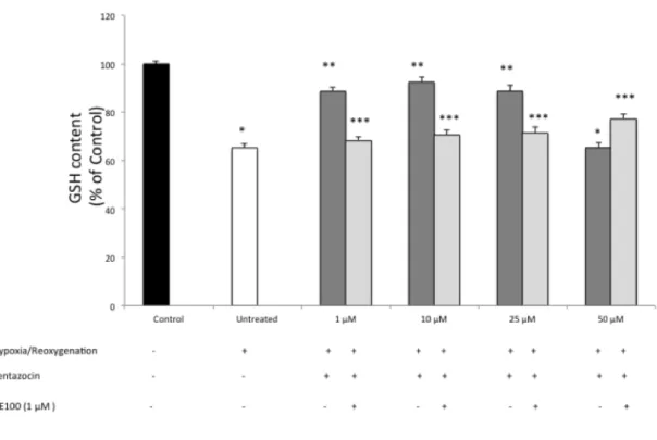
Reduced glutathione content measurement
In order to further demonstrate the antioxidant effect (+)-pentazocine we also determined the content of intracellular GSH. Our data suggest that 3 hours of 1% of hypoxia followed
by 24 hours of reoxygenation resulted in a significant (p<0.05) decrease of GSH content compared to normoxic-cultured cells (Figure 5). Treatment with (+)-pentazocine at 1, 10 and 25 µM resulted in a significant (p<0.05) restoration of intracellular GSH content when compared to untreated cells. Furthermore, 50 µM (+)-pentazocine had no significant effect on GSH restoration when compared to untreated cell cultures. In order to determine whether the protective effect of (+)-pentazocine was dependent of s1R stimulation, BV2
cells were also treated with NE-100. This set of experiments showed that NE-100 reversed the beneficial effect of (+)-pentazocine as demonstrated by a significant (p<0.05) reduction of GSH content when compared to (+)-pentazocine alone treated cells.
Figure 5. Evaluation of intracellular reduced glutathione content. Values represent the average of four different
experiments performed in triplicate. (*p<0.01 when compared to normoxic untreated cells; **p<0.01 when compared to hypoxia/reoxygenation untreated cells; *** p<0.01 when compared to pentazocine alone treated cells)
DISCUSSION
As one of the primary mechanisms of cerebral ischemia, the inflammatory response can cause secondary brain injury. Inflammatory cytokines, which are released from so-called inflammatory cells such as neutrophils, granulocytes, lymphocytes, and glia, can stimulate the adherence of leukocytes to the vascular endothelium, induce inflammatory responses, and cause tissue edema, destroying brain tissue [82]. Previous studies have demonstrated that, whether under the conditions of diseases or culture systems, the primary source of proinflammatory cytokines and oxidative stress mediators is microglia rather than astrocytes [82]. Based on the findings of our study, after hypoxia for 3 h followed by reoxygenation for 24 h, the levels of oxidative stress and apoptotic cell death increased and cell viability decreased. Our model is therefore consistent with previous data showing that hypoxia/reoxygenation results in a significant activation of microglia accompanied by a significant increase of various oxidative stress and inflammatory mediators. Various strategies have been investigated so far in the attempt to modulate and control microglial activation under various neurodegenerative disorders, however most of them did not translated into an improved outcome in a clinical setting. On the basis of the literature data and on the availability of clinical relevant class of molecules, we aimed at evaluating the effect of (+)-pentazocine on the redox balance in BV2 microglial cells following hypoxia/reoxygenation. Our results offer the first report, to our knowledge, of the effects of σR1 activation on microglia cells under this experimental condition. Previous reports have shown conflicting results with respect to the involvement of σ receptors in
inflammatory responses. Our data demonstrated that (+)-pentazocine treatment results in a significant decrease of oxidative stress as measured by increased GSH content, restoration of mitochondrial membrane potential and reduced apoptosis. Taken all together all these effects resulted in a significant increase of cell viability. Furthermore, our data strongly suggest that the beneficial effects of (+)-pentazocine are related to its binding to σ1R since
the effects were abolished by concomitant treatment of cell cultures with NE-100 (1 µM). Furthermore, pharmacologic analyses have demonstrated (+)-pentazocine to be a highly specific and potent ligand for σR1. In addition, when brain membranes derived from the s1R knockout mouse were analyzed in binding assays using [3H] (+)-pentazocine as the
radioligand, no binding activity was observed. Binding activity was reduced by half in heterozygous compared with wild-type animals [93]. As far as concern the antioxidant effect of (+)-pentazocine, very little is known in the scientific literature and no data are available related to our in vitro model. However, it should be noted that (+)-pentazocine has been shown to have opposite effects under various experimental conditions. In particular, previous data showed that following pharmacological treatment with DTG, a nonspecific σ1/2R ligand, results in a significant reduction of TNF-α, IL-10, and NO [87].
Similarly, treatment with σR ligands SR31747A and SSR125329A, decrease cytokine release in experimental models of rheumatoid arthritis and sepsis [94, 95]. However,
different results were obtained by Ruscher et al. [96] showing that σ1R ligand SA4503 had
no significant effect on the release of proinflammatory mediators following hypoxia/aglycemic conditions.
We also investigated the cell transduction mechanisms underlying (+)-pentazocine pharmacological effects. Our data showed a significant increase of ERK activation following hypoxia/reoxygenation whereas (+)-pentazocine was able to prevent ERK phosphorylation. Our data suggest that this effect is related to (+)-pentazocine selective
σR1 binding since this effect was abolished in the presence of NE-100. Interestingly, a higher dose of (+)-pentazocine (50 µM) further increase ERK activation when compared to untreated hypoxia/reoxygenated untreated cell thus suggesting that the effects of (+)-pentazocine are dose dependent. Our data are consistent with those reported by Tuerxun et al.[97] showing that the σ1R ligand SA4503 prevents cultured cortical neurons from
oxidative stress–induced cell death via suppression of MAPK/ERK pathway activation.
However, previous reports showed also that σ1R ligands resulted in a significant increase
in ERK activation, rather than a decrease, in the hippocampus and neuronal cultures [98]. Such apparent discrepancy may in part be related to the cell specific (i.e. neuronal type or glia) effects of σ1R ligands. Furthermore, previous data reported that σ1R exhibits multiple
discrete binding sites causing different responses. Finally, dose is an additional variable to take into due account when evaluating (+)-pentazocine effects on oxidative stress and inflammation.
CONCLUSIONS
Take all together, our results suggest that since (+)-pentazocine is a small molecule that can pass through the blood–brain barrier and shows beneficial effects, it could hold therapeutic potential for neuroprotection. Future studies are now warranted in order to
determine whether (+)-pentazocine or other s1R ligands show disease-relevant
neuroprotection.
ACKNOLEDGMENTS
This work was supported in part by “Associazione Mani Amiche onlus” (Catania, Italy) no-profit organization. We would like to thank also Edy Bonaccorsi (Leica Microsystems, Catania, Italy) for his extraordinary technical assistance.
(+)-pentazocine attenuates neuronal cell death, oxidative stress and microglial migration induced by conditioned medium from activated microglia
Kathrin Heiss1, MD, Marco Raffaele2, PharmD, Luca Vanella2, PhD, Paolo Murabito3,
MD, PhD, Orazio Prezzavento2, PhD, Agostino Marrazzo2, PharmD, Giuseppina Aricò,
PhD, Carlo Castruccio Castracani1, PharmD, Ignazio Barbagallo2, PhD, Agata Zappalà1,
PhD, Giovanni Li Volti1,4 MD, PhD
1Department of Biomedial and Biotecnological Sciences, University of Catania, Via S.
Sofia 64 95100 Catania (Italy); 2Department of Drug Sciences, University of Catania, V.le A. Doria 6, 95125 Catania (Italy); 3Azienda Ospedaliera Universitaria Policlinico “G. Rodolico”, University of Catania, (Italy), Via S. Sofia 78, 95125 Catania (Italy); 4
Euro-Mediterranean Institute of Science and Technology, Via Emerico Amari 131, 90100 Palermo (Italy)
Corresponding Author:
Prof. Giovanni Li Volti, MD, PhD
Department of Biomedical and Biotechnological Sciences University of Catania Via S. Sofia, 64 95100 Catania (Italy) tel: +39-0957384081 Fax: +39-0957384220 Email: [email protected]
ABSTRACT
Background: Sigma receptors (σ1R) are expressed both in neurons and microglia and
can be considered as a promising target for developing pharmacological strategies for neuroprotection in various experimental models. The aim of the present study was to test the effect of (+)-pentazocine, a putative σ 1R agonist, in an in vitro model of
neuron/microglia crosstalk following hypoxia/reoxygenation. Methods: Microglia (BV2 cells) was exposed (3 hours) to 1% oxygen and reoxygenation was allowed for 24 hours. Conditioned media obtained from this experimental condition was used to treat neuron (SH-SY5Y cells) in the presence or absence of (+)-pentazocine (25 μM). Cell viability was measured by cytofluorimetric analysis, whereas inflammation and oxidative stress were evaluated by the expression of Hsp70, GAD, SOD and p65. Microglial cell migration was also evaluated by Xcelligence technology. Results: Our results showed that (+)-pentazocine was able to increase neuronal cell viability following exposure to microglial-conditioned medium. Furthermore, (+)-pentazocine was also able to inhibit microglial cell toward neuron treated with hypoxic conditioned medium. Finally, pharmacological treatment reduced the expression of inflammatory and oxidative stress markers (GAD, SOD and p65). Interestingly, hypoxic medium was able to reduce the expression of Hsp70 and such effect was prevented by (+)-pentazocine treatment. Conclusions: (+)-pentazocine exhibits significant neuroprotective effects in our in vitro model of neuron/microglial crosstalk thus suggesting that σ1R may represent a possible strategy for neuroprotection.
Key Words: microglia, migration, neurons, (+)-pentazocine, oxidative stress, sigma
receptors
ABBREVIATIONS
1,3-Di-(2-tolyl) guanidine (DTG)
2,2-dithio-bis-nitrobenzoic acid (DTNB) central nervous system (CNS)
Heat shock Protein 70 (Hsp70) interleukin 10 (IL-10)
nitric oxide (NO)
lipopolysaccharide (LPS)
Phosphate Buffered Saline solution (PBS) reactive oxygen species (ROS)
Superoxide dismutase (SOD) tumor necrosis factor-α (TNF-α)
INTRODUCTION
Ischemic cerebrovascular diseases are among the most common disorders of the central nervous system. The cascade of events that underlie the mechanism of ischemia/reperfusion injury is very complex because it involves different cell types. Oxidative stress is considered to be one of the most important pathophysiological mechanisms of such damage since it would be at the base of the activation of the crosstalk between the different cell types of the brain such as astrocytes, neurons and microglia [81]. In particular, this last cell type plays an important role in sensing and in the modulation of neuronal function. Following an inflammatory stimulus, microglia takes on an amoeboid phenotype and upregulates numerous cell surface receptors involved in the innate immune response.
Besides regulating neuronal apoptosis, microglia secretes various cytokines and growth factors controlling synapse formation and plasticity. Unarguably, microglial cells have to be kept in check under normal conditions. Furthermore, since excessive inflammation causes significant damage to the CNS, the microglia activation must be limited by a series of counter-regulatory mechanisms for a proper maintenance of CNS homeostasis [85, 86]. Unfortunately, the mechanisms allowing the maintenance of a proper balance between these mechanisms remains to be fully elucidated. Various pharmacological strategies have been developed with the aim to regulate the crosstalk between neurons and microglia in order to obtain a significant neuroprotective effect. To this regard, σ 1R agonists are recently emerging as good
candidates for neuroprotective strategies under various experimental models [99]. σ1R are
highly expressed in neurons as well as microglia [87]. The activated σ1R can modulate the
been reported that σ1R are involved in the modulation of neuroinflammation. σ1R agonists,
such as (+)-pentazocine and 1,3-Di-(2-tolyl) guanidine (DTG), inhibit lipopolysaccharide (LPS)-stimulated activation of microglia, and decrease the production of reactive oxygen species (ROS), the release of pro-inflammatory molecules [tumor necrosis factor- α (TNF-α), nitric oxide (NO), interleukin 10 (IL-10), etc.] as well as the expression of monocyte chemoattractant protein-1 [87, 89-91]. Based on the findings described above, the present study aimed at evaluating the effect of (+)-pentazocine on the pathological crosstalk between neurons and microglia in an in vitro model using conditioned medium from hypoxia-activated microglial cells.
MATERIALS AND METHODS
Cell Culture and pharmacological treatments
Human neuronal SH-SY5Y and microglia BV2 cells were purchased from ATCC Company (Milan, Italy). Cells were suspended in culture medium (Dulbecco's Modified Eagle Medium (DMEM) containing 10% fetal bovine serum (FBS), 100 U/mL penicillin, and 100 U/mL streptomycin). At 80% confluency, cells were passaged using trypsin-EDTA solution (0.05% trypsin and 0.02% trypsin-EDTA). To obtain a conditioned media from activated microglia, BV2 cells were washed 3 times with Phosphate Buffered Saline solution (PBS), replaced with serum-free DMEM medium, and placed in a incubator at
37°C containing 1.0% O2 for 3 hours to initiate hypoxia, followed by 24h reoxygenation in
an incubator at 37°C containing 5% CO2. In order to test the neuroprotective effects of
(+)-pentazocine, neuronal cells were also treated with (+)-pentazocine (Sigma–Aldrich, Milan, Italy) at a final concentration of 25 µM. This concentration was used on the basis of our previous experiments in BV2 cells and by our preliminary data [100] assessing the toxicity of this compound under our experimental conditions (data not shown).
Cell viability evaluation cytofluorimetric analysis
Cell viability was assessed by Muse™ Count & Viability Kit (Catalog No. MCH100102, Millipore, Milan, Italy) according to the manufacture’s guidelines. Briefly, 50 µL of cell suspension containing 1 x 10⁶ cells/mL were mixed with 450 µL of Count & Viability Reagent. Cells were allowed to stain for 10 minutes at room temperature and samples were read by Muse™ Cell Analyzer (Millipore).
Microglia migration assay
Real‐time monitoring of BV2 cell migration was performed using the xCELLigence system with the CIM‐Plate 16 (Roche). The upper chamber was seeded with 50000 BV2. When BV2 cells migrated through the membrane into the bottom chamber seeded with neurons treated or untreated with pentazocine and cultured in the presence or absence of activated microglia conditioned medium, they contacted and adhered to the electronic sensors, resulting in an increase in impedance. The cell‐index values reflecting impedance changes were automatically and continuously recorded every 15 min.
Immunocytochemistry
Immunocytochemistry for oxidative and ER stress was performed following anti-Hsp70, SOD, p65 and GAD specific antibody (1:200) (R&D system, Milan, Italy), and then incubated with a species-specific FITC-conjugated secondary antibody (1:400) (Chemicon, Milan, Italy). Specimens were washed thoroughly in between incubations and counterstained with DAPI (4′,6-Diamidino-2-phenylindole dihydrochloride) (Sigma–Aldrich). The sections were mounted with polyvinyl alcohol mounting medium with DABCO (Sigma–Aldrich) and visualized under fluorescence microscope.
Statistical Analysis
The data were expressed as the means ± SD. Statistical analysis was performed via one-way analysis of variance (ANOVA) using SPSS11.0 software. P < 0.05 was considered to be significant.
RESULTS
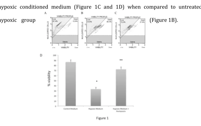
Our data showed that conditioned medium from hypoxia-activated microglia significantly reduced neuronal cell viability (Figure 1B and 1D) when compared to non-hypoxic conditioned medium (Figure 1A and 1D). Interestingly, (+)-pentazocine treatment resulted in a significant neuronal protection following treatment with hypoxic conditioned medium (Figure 1C and 1D) when compared to untreated
hypoxic group (Figure 1B).
Figure 1. Cytofluorimetric analysis of neurons viability cultured with non conditioned medium (A),
are representative of a single experiment. (C) Quantification of cell death percentage in the three experimental groups. Values represent the average of four separate experiments performed in triplicate. (* p<0.01 when compared to non conditioned medium; **p<0.01 when compared to microglial derived hypoxic medium).
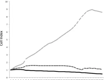
Furthermore, we showed that hypoxic medium results in a significant increase of microglia migration (Figure 2) and this effect is prevented by treatment with (+)-pentazocine.
Figure 2. Impedance-based technology migration assay. The CIM-Plate 16 provides a kinetic cell-response profile
throughout an experiment, detailing the onset and real time rate of BV2 migration.
To our knowledge, this is the first report showing the effect of (+)-pentazocine in microglia migration and thus suggesting a role in the crosstalk between neurons and microglia.
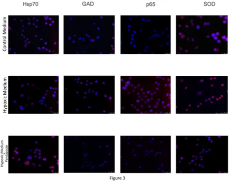
In order to further confirm the positive effects of (+)-pentazocine on the crosstalk between neuron and microglia we evaluated the expression of proteins involved in neuroinflammation and oxidative stress. In particular, this set of experiments showed that hypoxic medium resulted in a significant increase in the expression of proteins involved in inflammation and ER stress SOD, GAD and p65. Consistently with the above mentioned results, (+)-pentazocine resulted in a significant decrease in the
expression of such proteins when compared to untreated neurons cultured with hypoxic medium. Finally, hypoxic medium resulted in a significant decrease of Hsp70 and again this effect was prevented by treatment with (+)-pentazocine (Figure 3).
Figure 3. Immunocytochemical analysis of proteins involved in the oxidative and inflammatory response
following microglial activation. Nuclei were counterstained with DAPI.
DISCUSSION
The excessive microglial activation triggers a series of events that lead to a significant secondary brain damage. Microglia activation provides the triggering of a series of biochemical events consisting mainly in microglia phenotype modification, an increase of cell migration towards suffering neurons and the onset of a possible crosstalk between neurons and microglia leading to an irreversible neuronal damage. Given the importance of microglia both during development and in neurodegenerative diseases, many studies have focused on this cell type and their response in various experimental models of brain injury. Various strategies directing
at inhibiting microglial activation have been proposed as potential neuroprotective strategies. In particular, our recent data suggested that (+)-pentazocine treatment resulted in a significant reduction of oxidative stress in microglial cells via the ERK1/2
pathway. These results were consistent with previous evidence showing that σ 1R agonists
result in significant neuroprotective effects both in vitro and in vivo [99]. In the present work, we aimed at elucidating whether such beneficial effects could be exploited to confer neuroprotection in an in vivo pathological crosstalk between microglia and neurons. The first set of experiments suggested that hypoxic medium derived from microglial cells resulted in a significant reduction of neuronal cell viability. Interestingly, when neurons were treated with (+)-pentazocine, cell viability returned to the control levels. These data are consistent with previous data showing that activated microglia release a series of mediators with triggers a series of signaling cascade leading to neuronal cell loss and oxidative stress. Remarkably, our data also showed that neurons treated with hypoxic medium increase the recruitment of microglial cell by activating microglia migration. Consistently with above mentioned results, (+)-pentazocine abolished microglia migration. Such positive effect should be related to a protective effect of (+)-pentazocine on neurons, which may respond reducing the release of signals triggering microglia activation. This is to our knowledge the first in vitro demonstration that (+)-pentazocine inhibits microglial migration thus providing an additional neuroprotective effect of such compound. This set of experiments is consistently with a previous in vivo study [101] showing that PB190, a σ
1 agonist, blocks microglial migration toward injuries leaving cellular baseline motility and
the engulfment of apoptotic neurons unaffected.
In order to test the mechanisms underlying such modulation of neuron/microglia crosstalk, we examined the expression of various proteins involved in various aspect of neuronal damage. In particular, this set of experiments showed that hypoxic medium
resulted in a significant reduction of Hsp70 expression when compared to control. The influence of hypoxia on intracellular Hsp70 levels is still controversially discussed in the literature. Whereas Baek et al. observed an increase in Hsp70 expression after hypoxic exposure in RIF cells [102], another group showed differential effects on the Hsp70 expression in 18 different melanoma cell lines [103]. Our data are of particular interest in this setting since Hsp70 may play a major role in the immune response triggered by microglial cells. Future studies are now warranted in order to establish the molecular mechanisms underlying the expression of Hsp70 under our experimental conditions and whether Hsp70 secretion mechanisms are also involved in the crosstalk neurons/microglia. Consistently with previously published work our data showed that hypoxic medium triggers and inflammatory response and oxidative stress in neurons as measured by increased p65, GAD and SOD expression. Our results showed that (+)-pentazocine prevented up-regulation of such proteins thus suggesting this compound exhibits a significant neuroprotective effect also in our experimental conditions. These results are consistent with our recently published work showing that (+)-pentazocine reduces oxidative stress and apoptosis in microglial cells and that this effect was dependent on σ1R stimulation.
CONCLUSIONS
Taken all together, our results suggest that (+)-pentazocine may exert its beneficial effects by various mechanisms including a direct effect on neurons regulating the expression of important targets involved in oxidative stress and inflammation and an indirect effect involving recruitment of microglial cells and activation of a pathological inflammatory response. Given, the dimension of this molecule and its ability to cross the
blood–brain barrier, (+)-pentazocine may be considered a good candidate for pharmacological neuroprotective strategies.
ACKNOLEDGMENTS
This work was supported in part by “Associazione Mani Amiche onlus” (Catania, Italy) no-profit organization. We would like to thank also Edy Bonaccorsi (Leica Microsystems, Catania, Italy) for his extraordinary technical assistance.
References
1. P., D.R.-H., Microglia. Cytology and Cellular Pathology of the Nervous System, 1932: p. p. 482–1924 –534.
2. F, N., Ueber einige Beziehungen zwischen Nervenzeller- krankungen und gliiSsen
Erscheinungen bei verschiedenen Psy- chosen. Arch Psychiat, 1899.
3. Blinzinger, K. and G. Kreutzberg, Displacement of synaptic terminals from
regenerating motoneurons by microglial cells. Z Zellforsch Mikrosk Anat, 1968.
85(2): p. 145-57.
4. J., C., Estudie del compotamento de la microglia cultivade on vetro. Datos
concernientes a su histogenesis. Mem R Soc cep Hist nat, 1930.
5. Davalos, D., et al., ATP mediates rapid microglial response to local brain injury in
vivo. Nat Neurosci, 2005. 8(6): p. 752-8.
6. Nimmerjahn, A., F. Kirchhoff, and F. Helmchen, Resting microglial cells are
highly dynamic surveillants of brain parenchyma in vivo. Science, 2005.
308(5726): p. 1314-8.
7. Chan, W.Y., S. Kohsaka, and P. Rezaie, The origin and cell lineage of microglia:
new concepts. Brain Res Rev, 2007. 53(2): p. 344-54.
8. Brockhaus, J., et al., Membrane properties of ameboid microglial cells in the corpus callosum slice from early postnatal mice. J Neurosci, 1993. 13(10): p. 4412-21.
9. Haas, S., et al., ATP-induced membrane currents in ameboid microglia acutely
isolated from mouse brain slices. Neuroscience, 1996. 75(1): p. 257-61.
10. Mildner, A., et al., Microglia in the adult brain arise from Ly-6ChiCCR2+
monocytes only under defined host conditions. Nat Neurosci, 2007. 10(12): p. 1544-53.
11. Rezaie, P., et al., Microglia in the cerebral wall of the human telencephalon at second trimester. Cereb Cortex, 2005. 15(7): p. 938-49.
12. Sievers, J., R. Parwaresch, and H.U. Wottge, Blood monocytes and spleen macrophages differentiate into microglia-like cells on monolayers of astrocytes: morphology. Glia, 1994. 12(4): p. 245-58.
13. Wollmer, M.A., et al., ATP and adenosine induce ramification of microglia in vitro. J Neuroimmunol, 2001. 115(1-2): p. 19-27.
14. T., S., et al., Astrocyte-released cytokines induce ramification and outward K+ channel expression in microglia via distinct signalling pathways. Eur J Neurosci, 2001. 14: p. 463– 473.
15. Block, M.L., L. Zecca, and J.S. Hong, Microglia-mediated neurotoxicity:
uncovering the molecular mechanisms. Nat Rev Neurosci, 2007. 8(1): p. 57-69.
16. Colton, C. and D.M. Wilcock, Assessing activation states in microglia. CNS
Neurol Disord Drug Targets, 2010. 9(2): p. 174-91.
17. Davoust, N., et al., From bone marrow to microglia: barriers and avenues. Trends
Immunol, 2008. 29(5): p. 227-34.
18. Graeber, M.B. and W.J. Streit, Microglia: biology and pathology. Acta
Neuropathol, 2010. 119(1): p. 89-105.
19. Hanisch, U.K. and H. Kettenmann, Microglia: active sensor and versatile effector
cells in the normal and pathologic brain. Nat Neurosci, 2007. 10(11): p. 1387-94.
20. Kreutzberg, G.W., Microglia: a sensor for pathological events in the CNS. Trends
Neurosci, 1996. 19(8): p. 312-8.
21. Streit, W.J., et al., Role of microglia in the central nervous system's immune response. Neurol Res, 2005. 27(7): p. 685-91.
22. van Rossum, D. and U.K. Hanisch, Microglia. Metab Brain Dis, 2004. 19(3-4): p.
393-411.
23. Trapp, B.D., et al., Evidence for synaptic stripping by cortical microglia. Glia, 2007. 55(4): p. 360-8.
24. Wake, H., et al., Resting microglia directly monitor the functional state of synapses
in vivo and determine the fate of ischemic terminals. J Neurosci, 2009. 29(13): p. 3974-80.
25. Town, T., V. Nikolic, and J. Tan, The microglial "activation" continuum: from innate to adaptive responses. J Neuroinflammation, 2005. 2: p. 24.