UNIVERSITÀ DEGLI STUDI DELLA TUSCIA DI VITERBO
DIPARTIMENTO DI SCIENZE ECOLOGICHE E BIOLOGICHE
CORSO DI DOTTORATO DI RICERCA Scienze ambientali- XXIII Ciclo
CHARACTERIZATION OF BACTERIAL COMMUNITIES FROM SEAWATER SAMPLES: THE STUDY CASE OF KANDALAKSHA BAY, RUSSIA
(BIO/19)
Coordinatore: Prof. MAURIZIO PETRUCCIOLI
Tutor: Prof. MASSIMILIANO FENICE Co-Tutor: Dr. BELÉN JUÁREZ-JIMÉNEZ
During my Ph-D work we studied two different bacterial communities. The first part of the thesis was dedicated to investigate possible applications of bacterial isolated from samples (water, sediments etc.) obtained from the East sector of the Tyrrhenian sea. In this context, ca. 90 isolates have been plate screened for their extracellular enzyme activities (preliminary work carried out before the Ph-D course beginning, Fenice et al. 2007). Among them, few strains have been characterized for specific production of high level of enzymes and/or for some peculiar metabolic properties. In particular, we studied the “Production of chitinolytic enzymes by a strain (BM17) of
Paenibacillus pabuli isolated from crab shells samples collected in the East Sector of Central
Tyrrhenian Sea” (Juarez-Jimenez et al, 2008) and the “Metabolic characterisation of a strain (BM90) Delftia tsuruhatensis showing highly diversified capacity to degrade low molecular weight phenols” (Juarez-Jimenez et al. 2010).
These works had not been included in this thesis but only enclosed as published papers.
Due to the huge amount of data and for better coherence, only the second part of the thesis work, concerning the characterization of the bacterial community of Kadalaksha Bay, White Sea, Russia, has been reported here.
Index
Abstract
I
1. Introduction
The marine environment
11.1. Marine microrganisms
41.1.1.
Microorganisms and marine habitats 51.1.2.
Marine microorganisms and biotechnology 121.1.3.
Study of marine microorganisms by molecular methods 151.1.3.1. Genetic fingerprint techniques 17
1.1.3.2. PCR-DGGE/TGGE fingerprints analysis 19
1.2.
The White Sea
22
2. Materials and Methods
2.1. Microorganisms, media and culture conditions
29
2.1.1.
Chemicals 292.1.2.
Sampling, strain isolation and culture conditions 292.1.3.
Detection of growth temperatures profiles 302.1.4.
Detection of extracellular enzyme activities 322.2. Morphological physiological and biochemical tests
36
2.2.1.
Preliminary tests 362.2.2.
Metabolic characterization by BIOLOG 372.3.
Strain identification and phylogeny
43
2.3.2.
Polymerase Chain Reaction for amplification of the 16S rRNA gene 432.3.3.
Aligmennt and tree reconstruction 452.4.
Statistical analysis of data
45
2.5. Study of bacterial communities by temperature-gradient gel
electrophoresis (TGGE) fingerprinting analysis
462.5.1.
DNA extraction 462.5.2.
Study of total (eubacterial) community 472.5.2.1. 16 S rRNA gene amplification (First amplification) 47
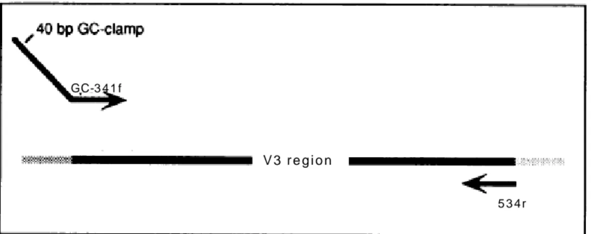
2.5.2.2. Hypervariable region V3 amplification (Second amplification) 48
2.5.2.3. Concentration and purification of PCR products 50
2.5.2.4. Fragments separation by TGGE 50
2.5.2.5. TGGE fingerprints analysis 52
2.5.2.6. TGGE bands amplification and sequencing 55
2.5.2.7. Sequences analysis and phylogenetic tree 56
2.5.3.
Study of Pseudomonas community 562.5.3.1. 16S rRNA gene amplification (First amplification) 56
2.5.3.2. Hypervariable region V3 amplification (Second amplification) 57
2.5.3.3. Fragments separation by TGGE 58
2.5.3.4. TGGE fingerprints analysis 58
3.
Results and discussion
3.1. Preliminary tests
60
3.2. Strain identification
64
3.3. Phylogeny and tree reconstruction
68
3.5. Metabolic characterization
87
3.5.1.
Screening for extracellular enzyme activity 873.5.2.
Metabolic profiles by BIOLOG 953.6.
Study of bacterial communities by PCR-TGGE fingerprinting
analysis
1123.6.1.
Study of total eubacterial community 1133.6.1.1. TGGE fingerprints analysis 113
3.6.1.2. Number of bands and statistical indexes in TGGE profiles 118 3.6.1.3. Taxonomical affiliation and phylogenetic study 126
3.6.2.
Study of Pseudomonas community 1333.6.2.1. Taxonomical affiliation and phylogenetic study 133
3.6.2.2. TGGE fingerprints analysis 138
3.6.2.3. Number of bands and statistical indexes in TGGE profiles 142
Conclusions
143
Aknowledgements
145
Appendix
146
In this work we studied the bacterial community obtained from water samples collected in Kandalaksha Bay (White Sea), Russia in order to obtain information both at the ecological level and for possible future application in biotechnology. The study has been carried out both on cultivable strains, isolated by traditional methods, and on the total bacterial (Eubacteria) community by molecular methods.
The isolates were preliminary investigated in order to understand their temperature preferences then their metabolic competences, including the production of extracellular enzyme activities, were tested for possible further biotechnological applications. Furthermore, a detailed taxonomical study was carried out both on cultivable and total bacterial population
Strain isolation and preliminary tests
Sea water was sampled in various areas of Kandalaksha Bay. The majority of samples were collected, at minimum tide level using sterile containers, in an intertidal zone pool and from the adjacent water surface. Others, from different offshore locations and depths (0.5, 2.5, 15, 70 m), were taken by scuba divers or boats using Niskin bottles. Water was filtered on membranes in order to obtain both bacteria pure cultures and DNA for molecular to studies. Pure cultures of isolates (ca. 500) were obtained by plate streak method. To discharge evident replicates of same isolates, preliminary tests were carried out considering strain morphological characteristics (shape, color and dimensions), Gram reaction and simple biochemical tests (catalase and oxidase production). This preliminary selection permitted to remove the majority of replicates and to keep 52 isolates.
Taxonomical identification of isolates by 16S rDNA
Bacterial genomic DNA was extracted from pure cultures and used for amplification of 16S rDNA, to allows taxonomical identification of each isolate.
Only 20 strains out of 52 (ca. 38%), showing high identity (99-100%) with a single known microorganism, were identified at species level. All other isolates were identified at the genus level only: for most of them, affiliation was not possible because 98-99% of identity was
recorded with various species of the same genus. Moreover 3 strains showed a very low percentage of identity (96-97%).
The majority of sequences were phylogenetically related to the genera Pseudomonas and
Serratia that are well worldwide distributed. However, only a typical bacteria of cold marine
environment Shewanella baltica (KB30) have been revealed.
When Blastn analyses supplied uncertain identification, sequences were aligned with highly similar 16S in the Genbank and phylogenetic analysis was performed. To get a confident branch-length, three different trees, Pseudomonas, Serratia and a group containing all the other genera, were inferred. Basing on the results some of the strains were identified with certainty at species level while some others, due to the branch length or the external position, probably belonged to new species. For all other strains identification was possible at genus level only. In many cases this could be due to the scarce informative power of the gene target used to discern the relations below the genus level.
Temperature growth profiles
The optimal temperatures for growth of the various strains were tested in the range 0-45°C on PCA plates (steps of 5 °C). Most of the strains (42% ca.) showed the optimum at 30°C. The lowest optimum was recorded at 15°C for one strain only (KB75), while the highest was at 40°C (KB22 and KB49). Most of the strains (56%) were able to growth at 0°C while only 25% grew at 45°C.In addition, the majority were able to grow in a rather broad range of temperature. The majority of the isolates (42%) seems to be psychrotrophics while no psychrophiles were detected. Almost all the strains could be considered as eurythermics, indicating adaptation to frequent and wide temperature variations such as those of Kandalaksha Bay. Moreover, it was evident that many KB strains had wider ranges of growth if compared with same species described in literature; optima were also different.
Extracellular enzyme activity
The strains where submitted to plate screening for the production of various extracellular enzyme activities (amylase, cellulase, chitinase, pectinase, phosphatase, protease, urease, lipases), at their optimal temperature, in order to obtain metabolic information and to find new microorganisms for possible applications.
Lipases, phosphatase and protease were common (ca. 54% and 44% of the isolates, respectively). Pectinase and amylase were present in about 32 % of the strains, while chitinase and urease were detected in a limited number of isolates (17% both); cellulase was not detected. Organisms producing large halos of activity were considered as possible high producers and could be further investigated for biotechnological application. However, none of the isolates produced all the tested activities and a rather large number of strains (17%) produced no activity at all. The results of the screening could be useful also at ecological level. In fact, the isolates producing a limited number of enzymes could be considered as specialized, while those with more diversified enzymatic competence, showing a higher eco-nutritional versatility, are probably advantaged in the harsh White Sea environment.
Metabolic competences by BIOLOG system
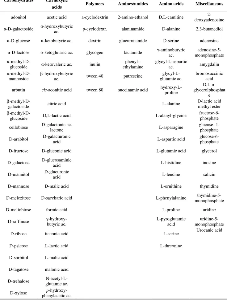
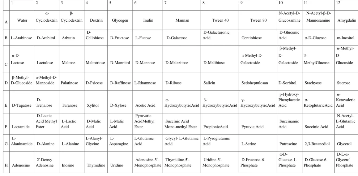
The BIOLOG system was used to test the metabolic competences of the strains by their ability to use different compounds as carbon sources. The system is able to detect the oxidation of 95 compounds (including sugars, fatty, organic amino and acids), used by microorganisms as sole carbon sources.In our case, amino acids were generally the preferred compounds: the most utilized carbon sources were L-Glutamic Acid and L-asparagine (96%
of strains). Also use of sugars was, as expected, rather common, being α-D-Glucose oxidized
by ca. 92%of strains. The information obtained by BIOLOG could be considered as an index regarding the strain metabolic complexity. A small number of strain apparently showed a rather simple metabolism being using a limited number of carbon sources and only few showed a very diversified metabolic competence. However, the majority of the bacteria tested
used about 30-50 compounds showing medium-high competence. In general, the strains able to use a wide array of carbon sources were also able to produce diversified extracellular enzyme patterns confirming their high eco-nutritional versatility.
Study on the total bacterial (Eubacteria) community
In order to have a complete overview of the bacterial community structure, by a cultivation-independent approach, total DNA was extracted from the filter-membranes. The bacteria biodiversity was studied by PCR-TGGE fingerprinting of partial 16S-rRNA gene amplicons. TGGE band patterns were normalized, compared and clustered. showed The community structure was revealed by the cluster analysis of the fingerprints. Samples collected both from the intertidal zone and the nearby sea surface, grouped together (80% similarity). Samples from open sea clearly clustered away. This was particularly evident for the sample collected at -70 m, which branched away at only 40% similarity.
TGGE gel images allow the analysis of band patterns generated from the environmental samples representing the various species present in the community. A single species is identified by a single band and the relative abundance of a single species is determined by the band intensity. Among the KB samples, a total of 70 different banding positions (band classes) were detected. The average number of bands per sample was 26 with a maximum of 30 in the samples from open sea. Simpson's Diversity Index calculated for all the samples showed very high level of diversity. Based on the total number of bands in each TGGE pattern and the relative temperature gradient, range-weighted richness indexes (Rr) were calculated and indicated a very high community biodiversity. Relative bands intensities were also calculated and expressed as percentages of the total band intensity in each TGGE lane. To render a graphic representation of the bacterial communities evenness, Pareto-Lorenz distribution curves were drawn based on the intensities. The functional community redundancy (response to perturbing environmental conditions) is evaluated by the Functional
medium Fo and a medium evenness. In other words, due to the elevated concentration of some species and the availability of many others, the community can potentially deal with environmental conditions changes thus preserving its functionality.
Prominent TGGE bands were excised from the gel, re-amplified and sequenced, to obtain the identities of the community predominant populations. Sequences phylogenetic analysis, showed a great presence of α-proteobacteria (16 sequences out of 27) with some γ-proteobacteria and some actinobacteria too. Some cyanobacteria were revealed also. Among α -proteobacteria strains can be affiliated mainly to the genus Roseobacter and one sequence was related to Ruegeria. All γ-proteobacteria showed highly similarity with the species of
Cobetia marina, while all the cyanobacteria were unknown.
Taxonomic results obtained by the total community study were quite different from those gained by the pure cultures: the pure culture strains were completely different (genera and species) from those by 16S rDNA analysis. This could be explained by the limits of TGGE technique. In fact, due to the bias introduced by the PCR reaction, this methodology can only detect bacteria representing at least 1% of the total community. It is possible that some species, even if present in a very low percentage, had prevailed due to favorable culture conditions of the isolation procedures. However, it is known that a large number of marine bacteria are uncultivable.
Another important information was that some bands showed very low sequence identity if compared with sequences present in the database, suggesting the possible presence of unknown bacteria.
Study on Pseudomonas species present in the community
In the TGGE gel relative to the total bacterial community, no Pseudomonas species were revealed. By contrast, the majority (ca. 45%) of the cultivable KB strains were affiliated to this genus. In order to find possible explications to this apparent incongruity, further TGGE analyses have been carried out focusing on Pseudomonas. Thus, total DNA was used for
PCR-TGGE fingerprinting of partial 16S-rRNA gene amplicons using specific primers for the amplification of this Genus and different temperature gradients.
Again, prominent TGGE bands, excised from the gel, were re-amplified and sequenced. The phylogenetic analysis showed that only 6 sequences out of 22 can be affiliated to
Pseudomonas. Although the primers used were specific for this genus, possible amplification
of other similar 16S rDNA (in particular γ-proteobacteria) could occur. In fact, a big cluster, comprising the majority of the sequences, showed very low similarity with Pseudomonas but high similarity with clones of marine invertebrates symbiotic γ-proteobacteria. It is possible that some new genera and/or species were present in our samples.
TGGE patterns were normalized, compared and clustered as reported for the total bacteria community. Cluster analysis of the fingerprints was similar to that obtained for total community. However, in this case sample collected at -70 m, was even more clearly separated from all the others samples: similarity was only 20%.
Analysis of bands number allowed to detect a total of 26 different banding positions
All the statistical indexes, discussed for total bacteria and related to community diversity and organization, in this case are meaningless because they would refer only to a limited portion of the entire population.
To the best of our knowledge, this work represents the first and extensive study carried out on Kandalaksha Bay bacterial community.
Chapter 1
Introduction
The marine environment
Seas and oceans represent the largest expanses of water on the planet. In total, they cover 3.61 x 10⁸ km2
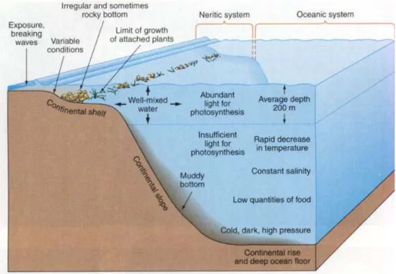
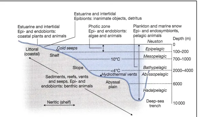
The different habitats, present in sea environment, can be divided into the benthic realm, comprising organisms living on seafloor, and the pelagic realm, including organisms living in the water column (Ross, 1995) (Fig 1.1). The benthic and pelagic environments are subdivided into zones based on depth. As for coastal habitats, in the benthic environment, the shallowest zones are called intertidal and subtidal zones, while for the pelagic environment the shallowest zone is the photic or surface zone, that extends from the
and contain 97% of all the water on earth (Austin, 1988).
A large proportion of all planet life exists in the oceans. Marine ecosystem is huge and it provides a lot of different habitats with a big variety of organisms adapted to its special conditions (Sumich and Morrissey, 2004).
shore line to the edge of the continental shelf. Most of marine life has been found in these coastal habitats, even though the shelf area occupies only 7% of the total ocean area. However it is recently recognized that life is abundant in the deep oceans also.
The physical and chemical structure of the oceans and the variations of factors, such as temperature, light, pressure, salinity and nutrient concentration, create distinct conditions to which organisms are adapted (Figure 1.2).
Temperature has an important role in the organisms distribution in the sea, since various biological processes depends on this factor. Solar radiation leads to thermal stratification: while on sea surface temperature can reach 25-30°C (tropical seas), at 100-150 m depth there is a marked thermocline where temperature strongly decreases (10°C or less) reaching almost 4°C in the benthic environment (Munn, 2011).
Light is another factor limiting organisms presence in the sea especially those responsible for primary productivity: photosynthesis is restricted in the upper 150-200 m of the sea surface
(photic zone) and various adaptation to different light regimes are needed by photosynthetic organisms (Sumich and Morrissey, 2004).
Pressure at sea surface is ca. 1 atm and increases of 1 atm every 10 m, thus only organisms that evolved special adaptation can inhabits deepest water. The majority of organisms living in these areas are bacteria and Archea even if presence of some fishes and invertebrates is reported (Gage and Tyler, 1992).
Seawater is a mixture of inorganic and organic compounds, including nutrients, and dissolved
gases (O2, CO2 etc.). Their concentration varies according to geographical and physical
factors and is responsible for organisms distribution in sea environments. As for chemical elements, they are responsible for salinity that is on average 35‰. Salinity increases from coastal to open sea and with depth: this gradient affects organisms presence depending on their tolerance (Austin, 1988). Dissolved oxygen plays an important role in the surviving of organisms, since it is present at high concentration only in the top 10-20 m of water, while it decreases with depth until it reaches a minimum between 200 and 1000 m due to its consumption by aerobic bacteria. Below this depth it increases again because oxygen consumption rates are low in relation with the supply trough cold, oxygen-rich deep waters from polar regions (Munn, 2011). In addition, the high abyssal hydrostatic pressure supports increased oxygen solubilization. Seawater presents very low nutrients concentration and ocean primary production is limited by their availability, affecting all the food chain. Essential nutrients such as nitrogen and phosphorus are normally at low concentration and are often the principal limiting factors even if recent studies demonstrates that also iron could limit primary production (Moore et al, 2001; Speight and Henderson, 2010).
Marine organisms are a vast resource, providing food, medicine, and raw materials. They contribute to important biogeochemical cycles (i.e. oxygen and carbon) and they show peculiar adaptation to the huge variability of sea conditions.
Even if the role of marine organisms and the knowledge of their ecology is rapidly increasing, large areas beneath the oceans still remain actually unexplored.
Figure 1.1 Divisions of the marine environment as reported by Ross (1995)
1.1
Marine microorganisms
Marine environment is a huge source of biological diversity and unexploited and/or unknown microorganisms. Nevertheless, investigation of seawater microorganisms for physiological, ecological and biotechnological studies is still scarce if compared with other environments (Pomponi,1999; Fenice et al., 2007).The main reasons for this lack of knowledge are probably the huge dimension of this environment, the difficulty of sampling in extreme habitats (i.e. ocean floor, hyper-thermophilic environments) and the problems to reproduce some microorganisms living conditions. Moreover, it is worth nothing that the majority of marine microorganisms, as occurs for other environments, cannot be isolated in pure cultures (Lewis, 2007). However, in the last decades, application of new technologies allowed a better knowledge of sea microorganisms. Powerful new tools in molecular biology, remote sensing and deep-sea explorations have led to important discoveries concerning the abundance and diversity of marine microbial life. Many distinct marine microbial ecosystems have been identified and studied. The most important microbial characteristic is the biodiversity and the ability to occupy habitats (i.e. polar or deep-seas and hydrothermal vents) generally inhospitable for any other organism. In fact, marine microorganisms growth occurs in all oceanic habitats from the seafloor to the top of ocean surface. Moreover, there is a growing awareness of the role of marine microorganisms in biogeochemical processes and their importance for the ocean food chain. For example, Cyanobacteria are major responsible of carbon organication and O2 production. It has been calculated that the picocyanobacteria
Prochlorococcus spp. and Synechoccus spp. contribute to ca. 50% of the photosynthetic
primary production in oceans (McIntyre, 2010) (Figure 1.3). Then, microorganisms can carry out metabolic processes such as biotransformation and degradation of organic matter (including a wide array of pollutants and recalcitrant compounds) and recycling of nutrients that have a central role for maintenance and support of all other life forms (Fowler et al.,
1987).Furthermore, they are responsible for planetary processes such as changes in the atmosphere, water and soil composition (Riebesell et al., 2007)
For all these reasons a deeper understanding of the distributions of marine microorganisms and their activities in maintaining ecosystem functions is needed.
Figure 1.3 Microscope image of Prochlorococcus marinus (Image copyright: Bob Andersen
and D. J. Patterson)
1.1.1 Microorganisms and marine habitats
As discussed, microorganisms are well represented in all the various marine habitats (Figures 1.4 and 1.5) as already reported by the early work of Sieburt (1979).
First of all, they constitute the major part of Plankton (small/microscopic organisms suspended in the water). Traditionally, plankton is divided in phyto- and zoo-plankton mainly taking into consideration animal and plants only. However, it is recognized that bacteria are an important part of plankton biomass. They have a basic role in planktonic marine microbial food webs and, being important agents of biogeochemical processes, their activity has a large impact on the entire ecosystem (Giovannoni et al., 1990; Gasol et al., 1999).
Microorganisms are also present on Neuston (the sea- air interface) that is rich in organic matter and, consequently, of microbial life. However, from some studies, carried out on coastal environments, there is no evidence of a stable an unique Neuston community, even
though it is possible that some very specialized bacteria, adapted to intense solar radiation, are prevalent (Maki, 2003, Agogue et al., 2005).
Surface of swimming animals(Nekton) as well as their digestive system (endobiotic environments)are widely colonized by microorganisms. Microbial biofilms occur on surfaces of all kind of animals and plants that, secreting organic compounds, provide nutrients. Many organisms seem to selectively enhance surface colonization by certain microorganisms and inhibit microbial growth or attachment of others trough the production of specific compounds (Jensen and Fenical 1994). Association among microorganisms and marine animals could be neutral or leading to mutual benefits (symbiosis) (Dubilier et al,. 2001, Goffredi et al., 2008). However, these microbial communities often strongly affect the health of marine animals and are important in fish spoilage (Banin et al., 2000; Gram and Dalgaard, 2002; Maloy et al., 2007).
The sea-sediment interface, including plants and animals, (Benthos) is widely colonized by microorganisms. The presence in the sediment of particulate organic debris and dissolved organic compounds lead to intense microbial activities. These activities are of paramount importance for the planetary carbon cycle but also for production/oxidation of methane and oxidation/reduction of sulfate (Iversen and Joergensen, 1985; Aller and Rude, 1988; Kirschner and Velimirov, 1999; Valentine, 2002). Concerning benthonic organisms, same consideration discussed for Nekton about biofilms formation and symbioses could be done.
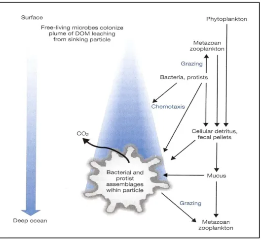
Microorganisms are also present in Seston (particulate matter).A continue flow of matter, which falls through the water column (marine snow, Figure 1.6), consists of aggregates of inorganic particles, plankton cells (including bacteria), debris from dead or dying plankton and fecal material glued together by a polymeric matrix produced by bacteria and phytoplankton (Alldredge and Silver, 1988).Marine snow is mainly produced in the upper part of the water column (100-200 m); large particles can sink up to various hundred meters per day reaching the ocean bottom in matter of days. This is the main mechanism by which a part
of the primary production is transported from the surface layers to deeper waters and see floor (Munn, 2011).During particle sinking organic material is degraded by extracellular enzymes and active microorganisms (Karner and Herndl, 1992). Microbial aerobic respiration creates anoxic conditions; therefore aerobic and anaerobic microorganisms coexist in different niches within the snow particle. The great rate of solubilization, due to microorganism activities, leads to formation of dissolved nutrients that can be utilized by other organisms and planktonic bacteria. Thus the majority of carbon rate is re-mineralized during its descent. However, the remaining material reaches the ocean floor where it is consumed by benthic organisms or contributes to sediment formation (Munn, 2011).
All solid materials in water, including plants, animals, rocks and anthropic structures, are colonized by microorganisms forming complexes called biofilms (epibiotic habitats). These structures are constituted by sessile communities that develop morphological and physiological characteristics differentiated from free-living forms. These communities are attached to solid surfaces by secretion of an extracellular polymeric matrix entrapping organic and inorganic components. In this matrix, complex physiochemical processes lead to formation of microenvironments colonized by mixed microbial communities. Biofilms are particularly important in shallow and intertidal waters and are interesting for their complex ecological interactions (Donlan, 2002; Munn, 2011).
Sea microorganisms are important also for their ability to live in extreme habitats were life for other organisms is difficult and/or impossible.
Deep-sea environments present conditions of low temperatures, oligotrophy and high pressure, thus various efficient adaptation strategies are needed. The majority of microorganisms living in these areas are Bacteria and Archea. They are found to be doubly extremophiles being both piezophilic and, generally, psychrophiles (Kato et al., 1995, Somero, 1992). Synergistic adaptation strategies are necessary. One of the most important
flexible and less subject to compression. Furthermore, piezophiles show control mechanisms of gene expression regulated by pressure. As for adaptation to nutrient low concentration, some bacteria (Photobacterium profundum and Shewanella sp.) produce high level of outer membrane proteins to increase transport providing a larger channel for easier nutrients assimilation (Bartlett, 1999, Munn, 2011). Another important adaptation, common both to pressures and to temperature, is the modified membrane composition. In fact, in all the microorganisms living in deep cold seas, the ratio of total unsaturated versus saturated fatty acids in the membrane lipids increased. This adaptation appear to be functional in maintaining membrane fluidity within a high range of pressures and at typical deep sea temperatures (Wirsen et al., 1986; Di Giulio, 2005).
In the ocean bottom a peculiar habitats is also constituted by hydrothermal systems (Figure 1.7). Hydrothermal vents are fissures in earth surface from which geothermally heated water springs. They are typical formations in the seafloor along the mid-ocean ridges, where two tectonic plates are diverging and new crust is being formed. The water emerging from hydrothermal vents consists mostly of sea water that permeated into the crust and had interacted with hot rocks or magma acquiring mineral elements. Two kind of vents has been discovered in the oceans up to ca. 5000 m of depth. The white smokers consist in warm water (20-23 °C) carrying low amount of minerals. Black smokers waters are very hot (350 °C) and transport lot of mineral elements such as metal sulfides, iron and manganese oxides and silicates that precipitate around the vent building a chimney structures. The gradient of temperatures and nutrients nearby the hydrothermal system create a wide array of water and sediments habitats colonized by microorganisms. Some of them are hyperthermophilic-chemosynthetic bacteria able to use sulfur compounds (i.e. hydrogen sulfide) to produce energyand obtain organic material by CO2 fixation. This metabolism supports a small food chain of this area that is hence independent from photosynthetic processes. For these microorganisms adaptation to high temperatures is needed also. Peculiar conformation of
protein permits a better stability of enzymes, while membranes with monolayer of isoprene-like units (in Archea, only) increase their stability (Aller and Rude, 1988; Jørgensen et al., 1992; Martin et al., 2008).
Microorganisms have also an important role for food chain in sea ice of polar regions. Sea ice is an ecosystem in which both physical phenomena and biological activity help to shape the overall habitat. Ecological and biological structures in sea ice are essentially driven by the prevailing temperature and salinity that are temporally and spatially highly variable. The structure of sea ice provides a wide array of different microhabitats that can be colonized by
mixed communities. Sea ice microbial populations have a major influence on various trophic
levels of the oceanic food web. They are particularly abundant in surface layers, within ice floes, and concentrated in ice-seawater interface. Bacterial populations are tightly coupled to primary productivity levels in sea ice. Microalgae photosynthesis contribute significantly to carbon fixation, while mineralization of dissolved organic matter is carried out primarily through the action of heterotrophic bacteria (Grossmann and Dieckmann, 1994; Arrigo and Thomas, 2004).
Although lot of information is available concerning various marine peculiar habitats, to date, a wide array of marine environments are still unknown under the microbiological point of view.
Figure 1.4 Schematic representation of the habitats for marine microorganisms based on
Sieburth (1979)
Figure 1.5 Schematic representation of the major ecological zones and habitats for marine
Figure 1.6 Schematic diagram of microbial processes during formation and fall of marine
snow as reported by Munn (2011)
1.1.2 Marine microorganisms and biotechnology
Biotechnology is an applied discipline that can be defined as “the integrated use of
biochemistry, microbiology and engineering sciences in order to achieve the technological (industrial) application of the capabilities of microorganisms, cultured tissue cells and parts thereof”. Biotechnology find applications in major industrial areas, including health care, food
production, agriculture, fine chemicals, biodegradable plastics, oils and biofuels, and for environmental uses.
Seas and oceans represent a pool of extremely interesting environments also for biotechnological studies. While much of the attention for this kind of investigations so far has been focused on others microbial ecosystems, marine environments remain largely unstudied and unexploited. Therefore, the enormous marine biodiversity still represents a source of microorganisms for a wide number of potential industrial and/or environmental applications (Fenice et al., 2007; Kennedy et al,. 2008).
Enzyme are among the most utilized compounds in biotechnology. The use of enzymes increased in the last 20 years, as well as their commercial value: total market for industrial enzymes reached $2.5 billion in the last few years reaching $6 billion in 2010 (Demain and Vaishnav, 2009; Munn, 2011). Strong increment of enzymes use was recorded not only in traditional fields (food and detergent industries), but in more emergent applications such as environmental depollution, pharmaceutical, bio-medical and chemical industries also (Fenice et al., 2007; Munn, 2011). Since various studies showed that marine bacteria are capable of producing unusual bioactive compounds, that are not observed in terrestrial sources, there is growing interest in obtaining higher value biochemicals from marine environments (Jha and Zi-rong, 2004, Debashish et al., 2005). The most commonly exploited enzymes are those that degrade polymers as proteins and carbohydrates. Marine bacteria can hydrolyze a wide array of polymers and particles using hydrolytic enzymes such as proteases, glucosidases, lipases, phosphatases, pectinase and chitinases (Hoppe, 2003;
Obayashi and Suzuki, 2008). Hence, oceans and seas can be valid alternatives to search microorganisms with high biodegradative properties. In particular, marine extreme environments represents a peculiar source of microorganisms showing activities of potential interest due to their adaptation to harsh conditions (Rothschild and Mancinelli, 2001; Bai et al., 2006; Srinivas et al., 2009). In fact, many successful biotechnological applications are due to enzymes from extremophiles (extremozymes). Many industrial processes often requires high temperatures, thus thermophilic enzymes can be useful in these processes due to their greater stability at these conditions. For example, thermo stable proteases and lipases are used in detergent formulation as stain removers. Their thermo-stability is often combined to resistance to the bleaching chemicals and surfactants present also in these products (Aaslyng et al., 1991, Munn, 2011). Enzymes from psychrophilic microorganisms, isolated from deep-sea sediments and/or polar regions, are useful in food-processing in which low temperatures are required to prevent spoilage and destruction of key ingredients as vitamins (van den Burg,, 2003).
Microbial bio-polymers found application in many industrial processes. For example, a wide variety of microorganisms are known to produce intracellular storage energy and carbon products in form of polyhydroxyalkanoates (PHA).The evaluation of PHA polyesters as natural, biodegradable, and biocompatible plastics for a wide range of possible applications, such as surgical sutures or packaging containers, was already reviewd by the early work of Brandl(1990).
Since global economy depends on oil for energy and chemicals, fossil sources of oil are subjected to overexploitation thus there is increasing interest in the use of renewable materials, such as land crops, for production of biofuels. However production costs and heavy ecological/social consequences make crops use unsustainable. Therefore, there is growing interest in use microalgae, including marine species, to produce biofuels. Microalgae are easy
and fatty acids stored in the cell as energy source can be converted into a variety of compounds with similar properties of petroleum products including biodiesel (Chisti, 2007; Munn 2011)
Microbial products provide many compounds for medicine and health care. The pharmaceutical industry is constantly searching for new compounds and many of the most successful drugs (i.e. antibiotics) are bacterial secondary metabolites. Even if, traditionally, investigation have been carried out mainly on terrestrial environments, the huge biodiversity of marine microorganisms led to a recent focus on sea organisms as source of novel compounds. Many new bioactive products are cyanobacterial peptides presenting huge diversity in structures and activity in different strains permitting to obtain a great variety of compounds with different pharmaceutical properties (Welker and Von Döhren, 2006; Gerwick et al., 2008).
Marine microorganisms can find application also in biodegradation and bioremediation of many chemical pollutant including oil spills. Oil tankers pollution produce immediate damaging on marine life but also long-term disruption of ecosystem communities. There are various strategies to deal with accidental spills. However, naturally occurring microorganisms, most of all bacteria, are able to degrade most of oil components especially hydrocarbons. The metabolic processes used by bacteria for biodegradation are mainly aerobic, being oxygenases (mono- and di-oxygenases) the key enzymes for degradation of paraffins and cleavage of aromatics. Since petroleum provide only the carbon source (hydrocarbons), these processes can be increased by supplementation of nitrogen, phosphorus and other elements (Kanaly and Harayama, 2000; McKew et al., 2007)
Many marine microorganisms are also able to degrade recalcitrant organic pollutant such as DDT, polychlorinated biphenyls (PCB), dioxine and furans. These substances are very persistent and resistant to biological and chemical degradation. However, aerobic and
anaerobic microbial degradation of various pollutants has been reported (Borja et al., 2005; Begonja Kolar et al., 2007).
1.1.3 Study of marine microorganisms by molecular methods
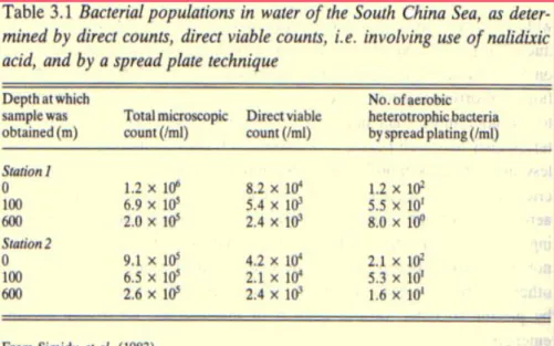
Already from the very early studies, it was clear that only a small fraction of marine bacteria can be isolated and cultivated in pure cultures. In fact, big differences were recorded between microscopic observations and counts (Figure 1.8; Simidu et al., 1983). This concept has been extended to other environments and recent studies confirm that the majority (99%) of microorganisms in nature are uncultivable or fail to grow in laboratory media (Wayne et al., 1987; Ward et al., 1992; Lewis, 2007). Possible reason could be the difficulty to reproduce natural growth conditions in laboratory due to lack of knowledge on strain physiology and adaptation strategies to peculiar environments (Amman et al., 1995; Muyzer 1999; Munn 2001). In addition, most bacteria are subjected to dormancy or inanition and could be very difficult to revitalize them (Lewis, 2007). Moreover, various symbiotic microorganism could require presence of their host to grow (Fisher, 1990). Thus, to obtain a complete overview of presence, diversity and role of microorganisms in the ecosystem, other approaches are needed to complement traditional culture-based microbiological methods. The application of molecular biology techniques have revolutionized the knowledge of environmental microbial diversity, offering new opportunities to analyze structure and species composition of microbial communities. In particular, nucleic acid based methods, allowed the detection and identification of organisms based on their genetic sequence. These culture-independent molecular techniques are based on direct extraction of genetic material from the environment, without culturing. The most widely used tool for microbial diversity investigation is the study of ribosomal RNA genes. In fact, the rRNA molecules(16S for bacteria) in the small ribosomal subunit (SSU) are universal in organisms, due to the role of ribosomes in protein synthesis. Furthermore, rRNA can be considered as phylogenetic maker
DNA extraction and amplification of specific region in the target gene by polymerase chain reaction (PCR). In rRNA molecules, some parts are highly conserved and can be amplified using the so called universal primers. Other fragments, present among these regions, show high degree of variability and are proved to be species specific. Thus, they can be used to study the different species present in a given environment and to determine structure and diversity in a microbial community.PCR products can be analyzed by cloning or genetic fingerprints. In cloning techniques, PCR products can be cloned in E. coli to create a gene library. Characterization of cloned sequences enables assessment of the genetic diversity of a community and can reveal the phylogenetic affiliation of the community members (Muyzer and Ramsing, 1995; Ranjard et al., 2000). Identification of particular populations is obtained by genetic fingerprints also. However, these techniques are particularly useful to get information on structure and dynamics of a community, based on the number and rank abundance of numerically common organisms (Bent and Forney, 2008).
Figure 1.8 Discrepancy between microscopic count and viable count observed by Simidu et
1.1.3.1 Genetic fingerprint techniques
These techniques are based on the principle of resolving the diversity, in size or composition, of amplified sequences by differential electrophoretic migration on agarose or polyacrylamide gels (Ranjard et al., 2000). Genetic fingerprints provide complex band profiles which are representative of the genetic community structure. Providing a relatively comprehensive description of a given community, they have been widely adopted. In particular, these techniques are extremely suitable to compare microbial community compositions in different environments (Justé et al., 2008). The most important fingerprints techniques utilized in microbial ecology are discussed below. However, since PCR-DGGE/TGGE has been used in this study it will be discussed in a separate paragraph.
Single-strand conformation polymorphism (SSCP)
This method allows separation of different DNA fragments of similar length based on conformational differences of folded single-stranded products. After denaturation, single stranded DNA fragments are loaded on a non-denaturing acrylamide gel that allows the formation of a stable secondary structure which is mainly determined by the intra-molecular interactions depending on the nucleotide sequence. Based on the migration of these secondary structures, products with similar molecular weight can be separated and visualized. Major limitation of SSCP is the formation of several different stable conformations of a single stranded DNA fragment, resulting in multiple bands on gel (Tiedje et al., 1999). Furthermore, sometime high rate of re-annealing of DNA strands during electrophoresis was observed regenerating double strand DNA (Selvakumar et al., 1997).
Terminal restriction fragment length polymorphism(T-FRLP)
Target genes are amplified with fluorescent primers and treated with specific restriction enzymes. Fragments are separated and detected on an automated sequencer (Liu et al., 1997). Only terminal labeled restriction fragments (TRFs) are detected and their length heterogeneity indicates community complexity. An internal size standard, labeled with a different
fluorescent dye, allows precise length assignment with single-base pair resolution. Because the use of a single restriction enzyme often does not provide sufficient resolution (Marsh, 2005), multiple restriction enzymes are typically used, increasing the specificity and the reliability of the assay (Osborne et al., 2006). Despite the high resolution and sensitivity, T-RFLP is highly dependent on 16S rRNA amplification which is affected by the DNA extraction method, PCR biases and the choice of universal primers (Kirk et al., 2004). Different enzymes will produce different community fingerprints and incomplete digestion by the restriction enzymes may lead to an overestimation of diversity (Dunbar et al., 1999; Osborn et al., 2000).
Restriction fragment length polymorphism (RFLP)
Restriction fragment length polymorphism, also known as amplified ribosomal DNA restriction analysis (ARDRA) is a relatively simple PCR-based fingerprinting technique centered on the digestion of amplified ribosomal community DNA followed by gel electrophoresis. RFLP can be used for microbial identification (Laguerre et al., 1994) or comparison of microbial communities and dynamics (Moyer et al.,1994). Differently from T-RFLP, all digested fragments are detected, increasing resolution. One single restriction enzyme generally does not provide sufficient resolution, and multiple restriction enzymes is needed. The main limitation of this method lies in the choice of restriction enzymes, which is crucial for obtaining optimal resolution (Ranjard et al., 2000).
Random amplified polymorphic DNA (RAPD)
The RAPD technique uses short random primers (about 10 bp) which anneal at different places on the genomic DNA, generating PCR products of various lengths further resolved on agarose or acrylamide gels. This technique is rapid and sensitive to reveal differences between similar complex prokaryotic genomes. Even if, some studies have evidenced possible lack of reproducibility due to its great sensitivity to PCR artifacts (Hadrys et al., 1992),sufficient reproducibility has been demonstrated to compare the genetic structure of complex bacterial
communities (Wikström et al., 1999). The main limitation is that it cannot provide phylogenetic information about the bacterial composition of the community (Ranjard, 2000).
Ribosomal intergenic spacer analysis (RISA)
RISA involves the analysis of the length polymorphism of the spacer between 16S and 23S genes. This intergenic spacer (IGS) region is species specific and successive sequencing of bands can allow taxonomic identification of definite populations within a community. The various amplified IGSs are directly separated on polyacrylamide gels by their size. To increase resolution, fluorescent primers can be used to further run RISA profiles on sequencing gel systems. Potential problems associated with RISA are the preferential amplification of shorter templates (Fisher and Triplett, 1999). Moreover, due to IGS length variation within a single genome, a single organism can contribute to more than one signal (Mateos and Markow, 2005).
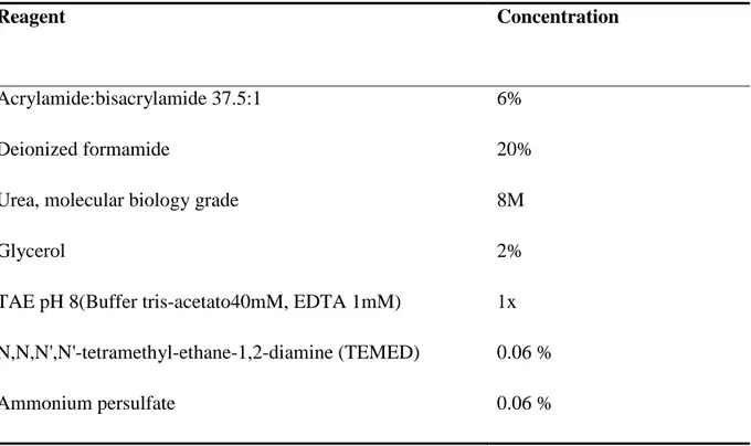
1.1.3.2 PCR-DGGE/TGGE fingerprints analysis
Both denaturing gradient gel electrophoresis (DGGE) andtemperature gradient gel electrophoresis (TGGE) examine microbial diversity of small PCR-amplified DNA fragments (200 – 700 bp) by poly-acrylamide gel electrophoresis (PAGE) under denaturing gradient (Muyzer et al., 1993; Muyzer and Smalla, 1998). DGGE uses denaturing chemicals such as formamide and urea, while a temperature gradient is applied in TGGE. Both techniques allows the separation of DNA fragments having the same length but different composition/sequences. Although gradient conditions need optimization to resolve specific samples (Ogier et al., 2002), these methods have the theoretical potential to detect differences of as little as a single or a few base pairs.
In TGGE, separation is based on the decreased electrophoretic mobility of partially melted double-stranded DNA molecules submitted to a linear temperature gradient. DNA fragments melting proceeds in discrete “melting domains”:stretches of base-pairs with identical melting
the temperature gradient gel, a transition from helical to a partially melted molecule occurs causing its migration stop. Sequence variations within such domains causes different melting temperatures, thus molecules with different sequences will stop at different positions. Generally, the melting behavior depends on the length of the product, its GC-content and nucleotide sequence.
By contrast, DGGE works at a fix temperature, thus, melting depends upon a chemical gradient.
For both these techniques, a sequence of guanines and cytosines (GC-clamp) is commonly added to the 5’end of one PCR primers, co-amplified and thus introduced into the amplified DNA fragments. The GC rich sequence is a high melting domain preventing the two DNA strands from complete dissociation into single strands (Sheffield et al., 1989).
Detection of correct size fragment of the 16S rRNA gene hypervariable regions usually requires a two-stage amplification (nested PCR). In fact, direct use of primers to amplify environmental DNA, often cause unspecific priming. Moreover, nested PCR improves amplification of scarcely represented species and yields richer band patterns (Nicolaisen and Ramsing, 2002; Ward and O’Mullan, 2002). For this reason, a first 16SrRNAgene PCR should be followed by a second PCR for the hypervariable regions. Furthermore, a PCR touch-down protocol during the second amplification is advisable (Watanabe et al., 2001). This technique consists in decreasing the annealing temperature, during PCR cycles, to avoid amplification of spurious DNA fragments (non-rRNA gene fragments and/or fragments with improper sizes).
Fragments separation generates samples profiles, representing the various species in the community being a single species revealed by a single band. Band intensities represent the relative abundance of each species. However, being PCR-based techniques, DGGE and TGGE detects only populations representing at least 1% of the total community targeted by
the primers (Muyzer, 1999). Therefore, the identified populations are only the most numerically predominant in the microbial community.
DGGE/TGGE are powerful tools to compare structural changes in microbial communities and for monitoring population dynamics (Sun et al., 2004; Camu et al., 2007). Several regions of the 16S rRNA gene have been used for DGGE/TGGE fingerprinting (Ercolini et al., 2003). However, length and species-specific heterogeneity of the V3 region within this gene make this fragment one of the better choices (Florez and Mayo, 2006). Furthermore, individual bands can be excised from the gel and identified by sequencing. However, reliable identification by sequencing may be hampered by the small fragment sizes of PCR products, which might not contain enough information for precise taxonomic classification (Øvreås, 2000). In addition, different sequences may have identical electrophoretic mobility, resulting in co-migration of different fragments (Sekiguchi et al., 2001). A major drawback often associated with DGGE/TGGE is lack of reproducibility. Actually, handling of big gels, primer–dimer formation and variable gel staining could affect reproducibility (Powell et al., 2005). However, these problems could be minimized by the inclusion of an internal standard, facilitating samples normalization within and between gels (Neufeld and Mohn, 2005).
1.2
The White Sea
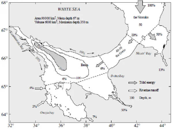
The White Sea (Figure 1.9) is located in the north-west of Russia, in Artic Sea basin. It can be considered a semi-enclosed sea which occupies a long gulf located south-east of the Kola Peninsula. It is joined to the Barents Sea by a strait between Cape Kanin and the Kola
peninsula. The White Sea covers approximately 95 000 km2 and a total volume of 6.000 km3
Kandalaksha Bay notably differs from the other basin its form being more elongated and having a limited width. The coasts of the bay present numerous inlets resembling fjords caused by glacial erosion. The longest inlet of Kandalaksha Bay, Chupa Inlet, is 37 km long, while its mean width is 1–2 km and the depths of the depressions are approximately 65 m. Concerning the bottom topography, Kandalaksha Bay is divided into two parts. The inner part (Zubov et al., 1956; Dobrosovol’skii et al., 1982).
The northern part, 40% of the total area, presents three regions (the Voronka, the Gorlo, and Mezen’ Bay) showing characteristic rather low depths (30–50 m). The southern part includes the Basin and three bays (Dvina, Onega, and Kandalaksha). A deep depression is located here with a maximum depth of 350 m; this occupies a small volume but plays a very important role in the White Sea ecosystem (Klenova, 1966).
The bays of the White Sea significantly differ in their morphometric properties. Mezen’ Bay has a very limited depth (ca. 10 m). The maximal tides of the White Sea are observed in this region, causing great cyclic variations in depths and horizontal displacements of the coastline. Dvina Bay is distinguished for having the most regular morphometric structure. The basin of the bay is open to the sea and here depths gradually increase reaching about 100 m.
In contrast, Onega Bay is characterized by a complex morphometric structure. The near-shore zone is shallow and has a typical skerry structure. Besides islands and “ludas” (small stony islands), there are many underwater and emerging rocks. The largest archipelago of the White Sea, the Solovets Archipelago, is located there.
is characterized by depth shallower than 50 m and an uneven relief, consisting of a complex alternation of depressions with depths gradually decreasing towards the apex of the bay (80 m, 50 m, and 30 m). The outer part of the bay is separated from the inner part by a sharp depth increase. The deepest point of the White Sea (335 m) is located there, off the Turii Peninsula (Pantyulin, 2003).
Temperature in the White Sea
The White Sea is located in the very north of the temperate climatic zone, but it shows subarctic climate characteristics. The climate is characterized by spatial variability and significant seasonal and inter-annual variations. However, Kandalaksha Bay, being located around the Arctic Polar Circle and above, could be considered as the most extreme area within White Sea.
Temperature is one of the crucial abiotic environmental factor determining the development and seasonal dynamic of many organisms: this has been widely demonstrated for White Sea also (Ushakova, 2003; Zubakha and Usov, 2004; Kuts and Ismailov, 2009). Temperature fluctuations (Figure 1.10) in White Sea and Kandalaksha Bay are quite broad and in the littoral zone they are even more evident (Savvichev et al., 2003). Winter is harsh and long (sea surface is covered with ice 6–7 months per year) and wheatear conditions could be very unpredictable. Temperature in winter may fall to -40°C, even if sometimes warm Atlantic air raises the temperature to ca. 5°C, while water temperature is about -1°C to -2°C. In summer, air could reach 30°C, albeit the average is in between 15-20°C, while sea water could reach 15°C on surface layers (Berger and Gorbushin, 2001; Shaporenko et al., 2005; Vershinin et al., 2006). In these extremely variable conditions, adaptation to wide temperature changes could be a winning surviving strategy as reported for microorganisms isolated in other extreme environments (Zucconi et al., 1996; Zhang et al., 2007; Srinivas et al., 2009).
Fig.1.10 Average annual temperature in White Sea
Freshwater contribution in the White Sea (Figure 1.11)
Freshwaters are transported to the sea mainly from river runoff and atmospheric precipitations. River runoff is the main input component of the freshwater budget in the White Sea (Pantyulin, 2003) and is largely due to three principal rivers Severnaya Dvina, Mezen’, and Onega.
The White Sea rivers are predominantly fed by snow melting, with a characteristic regime of spring floods and winter low-water periods. However, they differ in inter-annual runoff distribution. During the long winters (October–April) the White Sea bays freeze over (Klenova, 1966) and freshwater runoff to White Sea is restricted. The May/June melt brings a short period of extreme flood, which slowly decreases to the winter values (Gordeev&Sidorov, 1993).
Annually, between 500 and 600 mm of precipitation falls over White Sea surface. The precipitation is unevenly distributed throughout the year. It is most abundant from June through October (280-300 mm), accounting for about the 60% of the annual total. Evaporation and precipitation practically compensate each other, only a small part of freshwater contribution can be addressed to precipitation (Pantyulin 2003, Filatov et al.,
2005).Thus, in the White Sea water balance an essential role is played by the river run-off delivering about 95% of net fresh water entering the sea.
White Sea salinity is considerably low. This is due to the remarkable fresh water runoff and limited water exchange with the Barents Sea. The salinity of the surface waters in the Basin and open parts of bays varies from 24 to 27‰ with a typical range of 22–26‰ for the Kandalaksha Bay (Derjugin, 1928, Howland et al.,1999; Vershinin et al., 2006). However, in the estuaries of large rivers it falls down to 5-8 or less. The mixed layer depth is 25 m across the whole sea and, in general, surface waters are considerably fresher than the bottom waters. Sharp seasonal variations of surface water salinity are typical for the White Sea, which can be explained by the dynamics of freshwater inflow. Freshening of surface water begins in winter when the sea is covered with ice. The most prominent decrease in salinity of the upper layer is observed in April-May just before and during the period of ice-melting (Babkov, 1982).
Tides and Tidal Energy in the Sea (Figure 1.11)
Tides are the most important factor forming the hydrodynamic White Sea regime. Tides propagate into White Sea from the Barents Sea as regular semidiurnal waves, with the elevation of the tide and the velocity of the currents decreasing along the wave front from East to West. The maximum tidal currents velocities over the entire White Sea (up to 250 cm/s) are observed at the boundary of the Voronka with Mezen’ Bay, while the maximum tide height is located at the apex of Mezen’ Bay (9.8 m). Waves propagation into the Gorlo and the Basin occurs with their partial reflection and diffraction. The deviations are related to the influence of morphometry and are better reflected in the currents. In the Basin, the tide height decreases to 1.5 m and the velocities of the currents do not exceed 20 cm/s. Approximately identical tides are observed in Dvina Bay, but with higher velocities. In Kandalaksha Bay, the topographic effect leads to an increase in tide height up to 2.5 m, but it shows its major influence in the tidal currents: in numerous inlets the velocities of the currents can reach 80– 120 cm/s.
Kandalaksha Bay
Kandalaksha Bay is an estuarine system showing big sea level differences during tide cycles causing strong mixing of water (Melnikov et al., 2003; Savvichev et al., 2003). Estuaries are a dynamic ecosystem where seawater is diluted by the freshwater flowing from rivers and streams. Estuaries provide habitats for a large number of organisms and support very high productivity due mainly to phytoplankton (McLusky and Elliott, 2004). In Kandalaksha Bay, the seasonal extremes runoff of freshwater, due to the various rivers and intense precipitations, contributes to its very peculiar hydrodynamics (Howland et al., 1999; Dolotov et al., 2005).The coastal zone influenced by the tides (intertidal zone or littoral) is particularly interesting because all biogeochemical processes are most evident, especially if tidal phenomena are very pronounced as in Kandalaksha Bay. Organisms in this zone need adaptation to variable environmental conditions in which factors, such as water availability, temperature and salinity, change frequently (Savvichev et al., 2004).
Only a very limited number of studies is available on microorganisms from the White Sea and even less regards Kandalaksha Bay (Gorlenko et al., 1985; Dul’tseva et al., 1996; Rabold et al., 2006; Gorelova et al., 2009). In addition, most of these studies limit the investigation to the total number of bacteria in a determined environment (Savvichev et al., 2003; Savvichev et al., 2004; Kravchishina et al., 2008).
Fig. 1.11 Distribution of river runoff and tidal energy in the White Sea as reported by
Chapter 2
2 Materials and methods
2.1 Microorganisms, media and culture conditions
2.1.1 Chemicals
Chemical compounds used were as follows:
Plate Count Agar (PCA, Difco, USA), Nutrient Broth (NB, BD, USA), Soluble starch (Carlo Erba, Italy), Agar (BD, USA), Peptone (BD, USA) , Phenol red (Sigma-Aldrich, USA), Phenolphthalein diphosphate (Sigma-Aldrich, USA), Tween 20, 60 and 80 (Sigma-Aldrich,
USA), Yeast extract (BD, USA), Industrial pectin (Sigma-Aldrich, USA),
Hexadecyltrimethylammonium bromide (Sigma-Aldrich, USA), Carboxymethyl cellulose (Sigma-Aldrich, USA), Cetrimonium bromide (Sigma-Aldrich, USA), Skim milk (Fluka, CH), N,N,N,N tetrametil-p-phenylenediamine in water (Sigma-Aldrich, USA). All other chemicals were of analytical grade. Colloidal chitin was prepared by treating chitin from crab shells (Sigma-Aldrich, USA) with sulfuric acid as follows: 50 g of chitin were dissolved in 500 ml of 50% sulfuric acid (BDH Prolabo, Belgium) and then placed in a tank containing 15 volumes of distilled water. Chitin was washed various times until pH reached the value of 5.0.
After that, it was centrifuged at 11.000g for 15 minutes and autoclaved (121 ◦C, 20 min).
Chitin concentration (solid content of the sterile chitin suspension) by dry weight was evaluated by dehydration in oven at 105°C for 12 hours.
2.1.2 Sampling, strain isolation and culture conditions
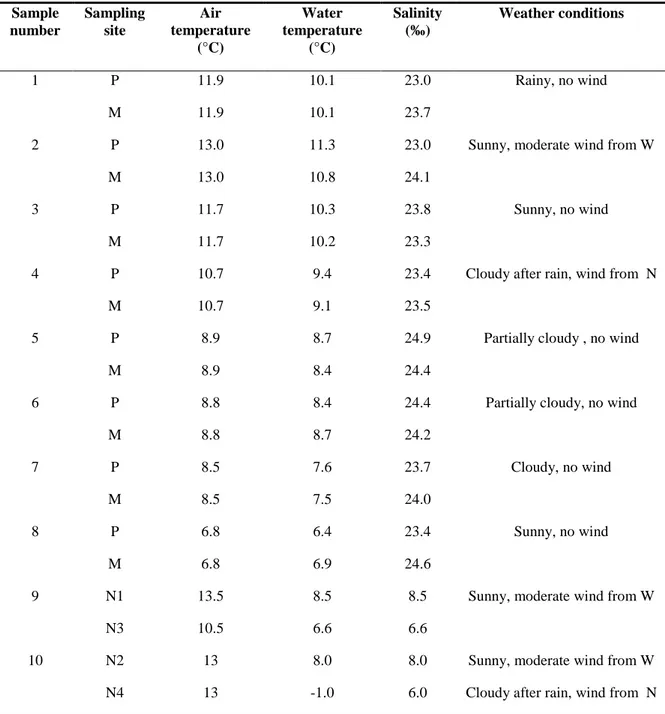
All the KB strains were obtained from water samples taken in various areas of Kandalaksha Bay, With Sea, Russia, during a 8 days sampling campaign in September 2008. The majority of samples were collected, at minimum tide level, in an intertidal zone pool and from the adjacent water surface using sterile containers. Others, from different offshore locations and depths (0.5, 2.5, 15, 70 m), were taken by scuba divers or boats using Niskin bottles sterilized by repeated washing with boiling sterile water and 70% (v/v) ethanol. Environmental
conditions, recorded during sampling, are reported in Table 2.1. Seawater was filtered on sterile membranes (0.22 µm, Millipore, USA) in order to obtain both pure cultures of bacteria and DNA to study the total bacterial community by molecular methods. Bacterial cultures were obtained placing the membranes on PCA plates and then incubating at 4, 15 and 25°C. Membranes to use for total DNA extraction, were maintained at 4°C in sterile tubes with silica-gel to avoid any contamination.
Pure cultures of isolates (ca. 500) were obtained on PCA by streak plate method. In order to discharge evident replicates of the same isolate, preliminary tests were carried out (see paragraph 2.2.1). Tests allowed to select 52 different isolates that were maintained on PCA slants at 4ºC and routinely sub-cultured.
2.1.3 Detection of growth temperature profiles.
Growth of the various isolates were tested in the range 0-45°C on PCA plates by steps of 5±0.5°C. Plates (diameter 90 mm) were inoculated in triplicate with inocula obtained pipetting 2 µl of bacterial suspension in sterile water (concentration was normalized spectrophotometrically at 600 nm). Plates were incubated at the different temperatures for 7 days and growth was monitored measuring the average increase of colony diameter. Plates were incubated for further 7 days to confirm the inability to grow at 0°C.
Table 2.1 Sampling description. Sample number Sampling site Air temperature (°C) Water temperature (°C) Salinity (‰) Weather conditions 1 P 11.9 10.1 23.0 Rainy, no wind M 11.9 10.1 23.7
2 P 13.0 11.3 23.0 Sunny, moderate wind from W
M 13.0 10.8 24.1
3 P 11.7 10.3 23.8 Sunny, no wind
M 11.7 10.2 23.3
4 P 10.7 9.4 23.4 Cloudy after rain, wind from N
M 10.7 9.1 23.5
5 P 8.9 8.7 24.9 Partially cloudy , no wind
M 8.9 8.4 24.4
6 P 8.8 8.4 24.4 Partially cloudy, no wind
M 8.8 8.7 24.2
7 P 8.5 7.6 23.7 Cloudy, no wind
M 8.5 7.5 24.0
8 P 6.8 6.4 23.4 Sunny, no wind
M 6.8 6.9 24.6
9 N1 13.5 8.5 8.5 Sunny, moderate wind from W
N3 10.5 6.6 6.6
10 N2 13 8.0 8.0 Sunny, moderate wind from W
N4 13 -1.0 6.0 Cloudy after rain, wind from N
Legend
Sampling sites:
P= intertidal zone pool, coordinates 66°33’15’’N; 33°05’47’’E; M= water surface nearby the intertidal zone pool, coordinates 66°33’15’’N; 33°05’47’’E; N1= water collected by scuba divers at – 2.5 m, coordinates: 66°32’29’’N, 33°14’94’’E; N3= water collected by scuba divers at – 15.5 m, coordinates: 66°32’30’’N, 33°14’94’’E; N2= water collected with boats at -0,5 m, coordinates: 66°32’21’’N, 33°14’77’’E; N4= water collected with boats at -70 m, coordinates: 66°32’21’’N, 33°14’77’’E
2.1.4 Detection of extracellular enzyme activities
The following activities were tested in triplicate: amylase (AMI), chitinase (CHI), cellulase (CEL), lipase on Tween 20, 60, 80 (T20, T60, T80), pectinase (PEC), phosphatase (PHO), protease (PRO), urease (URE). Plates for semi quantitative tests were prepared using previously reported techniques as follows: media for detection of chitinase, phosphatase, pectinase, lipase, cellulase and urease were prepared as previously reported Hankin and Anagnostakis (1975); media for detection of amylase and protease were prepared as reported by Paterson and Bridge (1994). Slight method modifications were carried out, when necessary, as described for each media. Where not specified, media were sterilized at 121°C for 20 minutes. Plates were inoculated with punctiform inocula using sterile needles from 1-2 day cultures grown on PCA plates. To avoid any interference from nearby colonies, only one isolate was inoculated onto each plate (60 mm diameter). The enzymatic activities were recorded measuring the diameter of the halo produced, after subtraction of colony diameter. For PHO, no activity halo is produced: positive colonies were recorded for their different color. The media for detection of extracellular enzyme activity were as follows:
Amilase
• Nutrient Broth 8.0 g/l
• Soluble starch 2.0 g/l
• Agar 20.0 g/l
Before sterilization, carried out at 0.75 atm for 30 minutes, pH was adjusted 7.0±0.2. After incubation plates were flooded with a iodine solution (Lugol) and amylolytic activity was indicated by the production of a yellow halo around the colony.
Urease
• Peptone 1.0 g/l